Cancer Association of South Africa (CANSA) - Cancer ...
←
→
Page content transcription
If your browser does not render page correctly, please read the page content below

Cancer Association of South Africa (CANSA)

                                                                             Fact Sheet
                                                                                 on
                                                                   Cancer of the Pharynx (Throat)

Introduction
In humans the pharynx (throat) is part of the digestive system and also of the conducting zone of the
respiratory system. The conducting zone also includes the nose, larynx, trachea, bronchi and
bronchioles. Their function is to filter, warm, and moisten air and conduct it into the lungs. It makes
up the part of the throat situated immediately posterior (behind) to the nasal cavity, posterior to the
mouth and superior (above) to the oesophagus and larynx. The human pharynx is conventionally
divided into three sections: the nasopharynx, the oropharynx and the hypopharynx. It is also
important in vocalisation.

Cancer of the Pharynx
Pharyngeal cancer (also referred to as pharyngeal carcinoma) is cancer that occurs in the pharynx. The
pharynx includes the space behind the nose and above the back of the throat.

It includes the following three parts:
• Nasopharynx
• Oropharynx
• Hypopharynx
                                       [Picture Credit: Pharynx]

The nasopharynx connects the back of the nose to the
back of the mouth. Cancer that develops in the
nasopharynx is called nasopharyngeal cancer. One
cannot see one’s own nasopharynx directly, but by
looking inside one’s mouth in the mirror, it lies above
the soft palate (the soft area at the back of the roof of
the mouth) and uvula (the dangly bit) at the back of the mouth.

The other two parts of the pharynx are:
• The oropharynx – the part of the throat at the back of the mouth. Cancers that start in this area
   are oropharyngeal cancers.
• The hypopharynx (sometimes called the laryngopharynx) – it sits behind and on either side of the
   larynx (voice box). Cancer can also start in the hypopharynx.

Researched and Authored by Prof Michael C Herbst
[D Litt et Phil (Health Studies); D N Ed; M Art et Scien; B A Cur; Dip Occupational Health; Dip Genetic Counselling; Dip
Audiometry and Noise Measurement; Diagnostic Radiographer; Medical Ethicist]
Approved by Ms Elize Joubert, Chief Executive Officer [BA Social Work (cum laude); MA Social Work]
January 2021                                                                                                     Page 1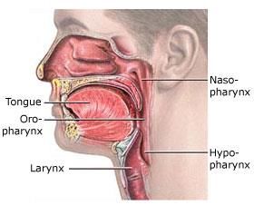
Incidence of Cancer of the Pharynx in South Africa
According to the outdated National Cancer Registry (2017), known for under reporting, the following
number of cases of cancer of the naso-oropharynx was histologically diagnosed in South Africa during
2017:

 Group - Males                         Actual                 Estimated           Percentage of
 2017                                No of Cases            Lifetime Risk          All Cancers
 All males                              338                     1:505                 0,85%
 Asian males                              6                    1:1 339                0,62%
 Black males                            175                     1:708                 1,34%
 Coloured males                          61                     1:326                 1,30%
 White males                             96                     1:305                 0,45%

 Group - Females                       Actual                 Estimated           Percentage of
 2017                                No of Cases            Lifetime Risk          All Cancers
 All females                            111                    1:2 133                0,27%
 Asian females                           11                     1:865                 0,78%
 Black females                           54                    1:3 559                0,29%
 Coloured females                        15                    1:1 405                0,33%
 White females                           31                    1:1 047                0,18%

The frequency of histologically diagnosed cases of cancer of the naso-oropharynx in South Africa for
2017 was as follows (National Cancer Registry, 2017):

 Group - Males                    0 – 19         20 – 29         30 – 39       40 – 49       50 – 59      60 – 69       70 – 79         80+
 2017                             Years           Years           Years         Years         Years        Years         Years         Years
 All males                           9             14              15            42            97           118           36             7
 Asian males                         0              0               0             0             3            2             0             1
 Black males                         9             12              13            28            49            49           14             1
 Coloured males                      0              0               1            10            21            22            4             3
 White males                         0              2               1             4            24            45           18             2

 Group - Females                  0 – 19         20 – 29         30 – 39       40 – 49       50 – 59      60 – 69       70 – 79         80+
 2017                             Years           Years           Years         Years         Years        Years         Years         Years
 All females                         4              6               5            16            26           34            15             5
 Asian females                       1              0               0             3             2            3             2             0
 Black females                       2              5               4            11             9           13             6             4
 Coloured females                    0              1               0             1             6            6             1             0
 White females                       1              0               1             1             9           12             6             1
N.B. In the event that the totals in any of the above tables do not tally, this may be the result of uncertainties as to the age, race or sex of
the individual. The totals for ‘all males’ and ‘all females’, however, always reflect the correct totals.

Ellington, T.D., Henley, S.J., Senkomago, V., O'Neil, M.E., Wilson, R.J., Singh, S., Thomas, C.C., Wu,
M. & Richardson, L.C. 2020.
“Cancers of the oral cavity and pharynx account for 3% of cancers diagnosed in the United States*
each year. Cancers at these sites can differ anatomically and histologically and might have different
causal factors, such as tobacco use, alcohol use, and infection with human papillomavirus (HPV) (1).
Incidence of combined oral cavity and pharyngeal cancers declined during the 1980s but began to
increase around 1999 (2,3). Because tobacco use has declined in the United States, accompanied by a
decrease in incidence of many tobacco-related cancers, researchers have suggested that the increase
in oral cavity and pharynx cancers might be attributed to anatomic sites with specific cell types in

Researched and Authored by Prof Michael C Herbst
[D Litt et Phil (Health Studies); D N Ed; M Art et Scien; B A Cur; Dip Occupational Health; Dip Genetic Counselling; Dip
Audiometry and Noise Measurement; Diagnostic Radiographer; Medical Ethicist]
Approved by Ms Elize Joubert, Chief Executive Officer [BA Social Work (cum laude); MA Social Work]
January 2021                                                                                                     Page 2
which HPV DNA is often found (4,5). U.S. Cancer Statistics† data were analyzed to examine trends in
incidence of cancers of the oral cavity and pharynx by anatomic site, sex, race/ethnicity, and age
group. During 2007-2016, incidence rates increased for cancers of the oral cavity and pharynx
combined, base of tongue, anterior tongue, gum, tonsil, oropharynx, and other oral cavity and
pharynx. Incidence rates declined for cancers of the lip, floor of mouth, soft palate and uvula, hard
palate, hypopharynx, and nasopharynx, and were stable for cancers of the cheek and other mouth
and salivary gland. Ongoing implementation of proven population-based strategies to prevent tobacco
use initiation, promote smoking cessation, reduce excessive alcohol use, and increase HPV vaccination
rates might help prevent cancers of the oral cavity and pharynx.

Risk Factors for Pharyngeal Cancer
Risk factors for pharyngeal cancer include:

General Risk Factors:
• Lack of fruits and vegetables: A diet low in fruits and vegetables can increase the likelihood of
   developing throat cancer

Lifestyle Factors:
• Tobacco use: The use of cigarettes, pipes and cigars all increase the likelihood of developing
    pharyngeal cancer.

Di Credico, G., Edefonti, V., Polesel, J., Pauli, F., Torelli, N., Serraino, D., Negri, E., Luce, D., Stucker,
I., Matsuo, K., Brennan, P., Vilensky, M., Fernandez, L., Curado, M.P., Menezes, A., Daudt, A.W.,
Koifman, R., Wunsch-Filho, V., Holcatova, I., Ahrens, W., Lagiou, P., Simonato, L., Richiardi, L., Healy,
C., Kjaerheim, K., Conway, D.I., Macfarlane, T.V., Thomson, P., Agudo, A., Znaor, A., Boaventura
Rios, L.F., Toporcov, T.N., Franceschi, S., Herrero, R., Muscat, J., Olshan, A.F., Zevallos, J.P., La
Vecchia, C., Winn, D.M., Sturgis, E.M., Li, G., Fabianova, E., Lissowska, J., Mates, D., Rudnai, P.,
Shangina, O., Swiatkowska, B., Moysich, K., Zhang, Z.F., Morgenstern, H., Levi, F., Smith, E., Lazarus,
P., Bosetti, C., Garavello, W., Kelsey, K., McClean, M., Ramroth, H., Chen, C., Schwartz, S.M.,
Vaughan, T.L., Zheng, T., Menvielle, G., Boccia, S., Cadoni, G., Hayes, R.B., Purdue, M., Gillison, M.,
Schantz, S., Yu, G.P., Brenner, H., D'Souza, G., Gross, N.D., Chuang, S.C., Boffetta, P., Hashibe, M.,
Lee, Y.A. & Dal Maso, L. 2019.
OBJECTIVES: This study aimed at re-evaluating the strength and shape of the dose-response
relationship between the combined (or joint) effect of intensity and duration of cigarette smoking
and the risk of head and neck cancer (HNC). We explored this issue considering bivariate spline
models, where smoking intensity and duration were treated as interacting continuous exposures.
MATERIALS AND METHODS: We pooled individual-level data from 33 case-control studies (18,260
HNC cases and 29,844 controls) participating in the International Head and Neck Cancer Epidemiology
(INHANCE) consortium. In bivariate regression spline models, exposures to cigarette smoking
intensity and duration (compared with never smokers) were modeled as a linear piecewise function
within a logistic regression also including potential confounders. We jointly estimated the optimal
knot locations and regression parameters within the Bayesian framework.
RESULTS: For oral-cavity/pharyngeal (OCP) cancers, an odds ratio (OR) >5 was reached after 30 years
in current smokers of ∼20 or more cigarettes/day. Patterns of OCP cancer risk in current smokers
differed across strata of alcohol intensity. For laryngeal cancer, ORs >20 were found for current
smokers of ≥20 cigarettes/day for ≥30 years. In former smokers who quit ≥10 years ago, the ORs

Researched and Authored by Prof Michael C Herbst
[D Litt et Phil (Health Studies); D N Ed; M Art et Scien; B A Cur; Dip Occupational Health; Dip Genetic Counselling; Dip
Audiometry and Noise Measurement; Diagnostic Radiographer; Medical Ethicist]
Approved by Ms Elize Joubert, Chief Executive Officer [BA Social Work (cum laude); MA Social Work]
January 2021                                                                                                     Page 3
were approximately halved for OCP cancers, and ∼1/3 for laryngeal cancer, as compared to the same
levels of intensity and duration in current smokers.
CONCLUSION: Referring to bivariate spline models, this study better quantified the joint effect of
intensity and duration of cigarette smoking on HNC risk, further stressing the need of smoking
cessation policies.
• Chewing tobacco or betel quid (gutkha)
• Alcohol use: Excessive use of alcohol can increase the risk for pharyngeal cancer
Other Factors:
• Human papillomavirus (HPV) Infection: New research has found that HPV infection is
responsible for rising rates of pharyngeal cancer, in particular oropharyngeal cancer
Tsang, C.M., Lui, V.W.Y., Bruce, J.P., Pugh, T.J. & Lo, K.W. 2020.
“Nasopharyngeal carcinoma (NPC), also named the Cantonese cancer, is a unique cancer with strong
etiological association with infection of the Epstein-Barr virus (EBV). With particularly high prevalence
in Southeast Asia, the involvement of EBV and genetic aberrations contributive to NPC tumorigenesis
have remained unclear for decades. Recently, genomic analysis of NPC has defined it as a genetically
homogeneous cancer, driven largely by NF-κB signaling caused by either somatic aberrations of NF-κB
negative regulators or by overexpression of the latent membrane protein 1 (LMP1), an EBV viral
oncoprotein. This represents a landmark finding of the NPC genome. Exome and RNA sequencing data
from new EBV-positive NPC models also highlight the importance of PI3K pathway aberrations in NPC.
We also realize for the first time that NPC mutational burden, mutational signatures, MAPK/PI3K
aberrations, and MHC Class I gene aberrations, are prognostic for patient outcome. Together, these
multiple genomic discoveries begin to shape the focus of NPC therapy development. Given the
challenge of NF-κB targeting in human cancers, more innovative drug discovery approaches should be
explored to target the unique atypical NF-κB activation feature of NPC. Our next decade of NPC
research should focus on further identification of the -omic landscapes of recurrent and metastatic
NPC, development of gene-based precision medicines, as well as large-scale drug screening with the
newly developed and well-characterized EBV-positive NPC models. Focused preclinical and clinical
investigations on these major directions may identify new and effective targeting strategies to further
improve survival of NPC patients.”
Tam, M. & Hu, K. 2019.
“Oropharyngeal carcinoma associated with the human papillomavirus is increasing in incidence and
represents a unique head and neck disease with favorable treatment outcomes. This review evaluates
the evolving role of radiotherapy in regional management with an overall goal of treatment de-
escalation in the appropriate patient. Determining the optimal approach and selection factors for
treatment de-escalation is under active investigation. Response to induction chemotherapy, refining
adverse pathologic factors after a primary surgical approach, decreasing radiation dose with or
without chemotherapy in the definitive or adjuvant settings as well as more selective nodal level
irradiation all are current strategies for treatment de-escalation. This review details the likely changes
in regional radiotherapy management for oropharyngeal carcinoma in the modern human
papillomavirus era and discusses future approaches to patient selection with the goal of reducing
toxicities while maintaining function preservation and quality of life in group of patients who are
younger and healthier than traditional head and neck cancer patients.”
Researched and Authored by Prof Michael C Herbst
[D Litt et Phil (Health Studies); D N Ed; M Art et Scien; B A Cur; Dip Occupational Health; Dip Genetic Counselling; Dip
Audiometry and Noise Measurement; Diagnostic Radiographer; Medical Ethicist]
Approved by Ms Elize Joubert, Chief Executive Officer [BA Social Work (cum laude); MA Social Work]
January 2021 Page 4• Gastro-oesophageal reflux disease (GERD): When acid leaks from the stomach into the
oesophagus (food tube), it causes acid reflux. Chronic acid reflux is called GERD, and increases
pharyngeal cancer risk depending on the frequency and severity of the acid reflux
• Contracting Epstein-barr virus (EBV): This common virus is transmitted via saliva. Contracting
EBV increases the likelihood of developing pharyngeal cancer
• Low immunity - Research has found that people have an increased risk of mouth and throat
cancer if they have a reduced immunity due to HIV or AIDS. Taking medicines to suppress
immunity after organ transplants also gives a higher risk of mouth and throat cancer than in
the general population.
• Previous cancer - People who have had mouth or oropharyngeal cancer have an increased risk
of getting a second one. Women have a higher risk of a second oral cancer than men.
• People who have had some other types of cancer also have an increased risk of mouth and throat
cancer. These include
▪ Cancer of the food pipe (oesophagus)
▪ Squamous cell skin cancer
▪ Cervical, anal or genital cancer in women
▪ Cancer of the back passage (rectum) in men
• Family history - People often worry that they are at a higher risk of cancer because someone
in their family has it. There does seem to be a slightly higher risk of getting mouth cancer if
one has a close relative (a parent, brother, sister or child) who has had mouth cancer. The
reason for this is unknown
• Mouth conditions - Sometimes changes can happen in the cells of the lining of the mouth and
they cause red or white patches to appear. These changes are called leukoplakia and
erythroplakia. In some people these changes may develop into cancer over some years.
Dentists can see these changes during dental checks so it is important to have regular dental
appointments to find these changes early.
• Genetic conditions - People with certain syndromes caused by inherited changes (mutations)
in particular genes have a high risk of mouth and throat cancer. These include:
[Picture Credit: Fanconi Anaemia]
• Fanconi anaemia – a genetic disorder that can affect
children and adults from any ethnic background. It is
also called Fanconi's syndrome. People with Fanconi
anaemia are short, have bone changes, and are at risk
of developing cancers, leukaemia, and bone
marrow failure (aplastic anaemia).
A common symptoms of Fanconi anaemia is abnormal hands.
Researched and Authored by Prof Michael C Herbst
[D Litt et Phil (Health Studies); D N Ed; M Art et Scien; B A Cur; Dip Occupational Health; Dip Genetic Counselling; Dip
Audiometry and Noise Measurement; Diagnostic Radiographer; Medical Ethicist]
Approved by Ms Elize Joubert, Chief Executive Officer [BA Social Work (cum laude); MA Social Work]
January 2021 Page 5• Dyskeratosis congenita – a genetic syndrome that can cause aplastic anaemia, skin rashes, and
abnormally shaped fingernails and toenails. People with this syndrome have a high risk of
developing cancer of the mouth and throat when they are young
Signs and Symptoms of Pharyngeal Cancer
Signs and symptoms of pharyngeal cancer may include:
• A persistent cough
• Changes in your voice, such as hoarseness
• Difficulty swallowing
• Ear pain
• A lump or sore that does not heal
• Difficulty chewing, swallowing, or moving the jaw or tongue
• Changes in speech
• A persistent sore throat or a feeling that something is caught in the throat
• Unexplained weight loss
• Chronic bad breath
• Fatigue
• Loss of appetite, especially when prolonged: this may happen later in the course of the illness
An appointment should be made to see a doctor if any of the above signs and symptoms persist or
any new signs and symptoms appear. Most pharyngeal cancer symptoms are not specific to cancer,
so the doctor will likely investigate other more common causes first.
(Mayo Clinic; Cancer.Net; Macmillan Cancer Support).
Types of Pharyngeal Cancer
More than 9 out of 10 mouth and oropharyngeal cancers (90%) are squamous cell carcinoma. Squamous
cells are the flat, skin like cells that cover the inside of the mouth, nose, larynx and throat. Carcinoma just
means cancer. So squamous cell carcinoma is cancer that starts in these cells.
There is an unusual type of squamous cell carcinoma called verrucous carcinoma. About 1 in 20 mouth
cancers (5%) are this type. Verrucous carcinoma rarely spreads to other parts of the body but can grow
very deeply into surrounding tissues.
Though most throat cancers involve the same types of cells, specific terms are used to differentiate
the part of the throat where cancer originated:
• Nasopharyngeal cancer begins in the nasopharynx - the part of the throat just behind the nose
• Oropharyngeal cancer begins in the oropharynx - the part of the throat right behind the mouth
that includes the tonsils
• Hypopharyngeal cancer (laryngopharyngeal cancer) begins in the hypopharynx (laryngopharynx) -
the lower part of the throat, just above the oesophagus and windpipe
• Glottic cancer begins in the vocal cords
• Supraglottic cancer begins in the upper portion of the larynx and includes cancer that affects the
epiglottis, which is a piece of cartilage that blocks food from going into the windpipe
Researched and Authored by Prof Michael C Herbst
[D Litt et Phil (Health Studies); D N Ed; M Art et Scien; B A Cur; Dip Occupational Health; Dip Genetic Counselling; Dip
Audiometry and Noise Measurement; Diagnostic Radiographer; Medical Ethicist]
Approved by Ms Elize Joubert, Chief Executive Officer [BA Social Work (cum laude); MA Social Work]
January 2021 Page 6• Subglottic cancer begins in the lower portion of the voice box, below the vocal cords
Diagnosis of Pharyngeal Cancer
The following tests may be used to diagnose pharyngeal cancer:
• Physical examination - Dentists and doctors often find lip and oral cavity cancers during
routine check-ups. If a person shows signs of oral or oropharyngeal cancer, the doctor will
take a complete medical history, asking about the patient’s symptoms and risk factors. The
doctor will feel for any lumps on the neck, lips, gums, and cheeks. Since patients with oral or
oropharyngeal cancer have a higher risk of other cancers elsewhere in the head and neck
region, the area behind the nose, the larynx (voice box), and the lymph nodes of the neck are
also examined.
• Endoscopy - This test allows the doctor to see inside the mouth and throat. Typically, an
endoscope (a thin, flexible tube with an attached light and view lens) is inserted through the
nose to examine the head and neck areas. Sometimes, a rigid endoscope (a hollow tube with
a light and view lens) is placed into the back of the mouth to see the back of the throat in
more detail. The examination has different names depending on the area of the body that is
examined, such as laryngoscopy (larynx), pharyngoscopy (pharynx), or a nasopharyngoscopy
(nasopharynx). To make the patient more comfortable, these examinations are performed
using an anaesthetic spray to numb the area. If tissue looks suspicious, the doctor will take a
biopsy. Tests are often done in the doctor’s office; however, sometimes, an endoscopy must
be performed in an operating room at a hospital using a general anaesthesia.
• Biopsy - A biopsy is the removal of a small amount of tissue for examination under a
microscope. Other tests can suggest that cancer is present, but only a biopsy can make a
definite diagnosis. The sample removed during the biopsy is analysed by a pathologist (a
doctor who specializes in interpreting laboratory tests and evaluating cells, tissues, and organs
to diagnose disease). The type of biopsy performed will depend on the location of the cancer.
In a fine needle aspiration biopsy, cells are withdrawn using a thin needle inserted directly
into the tumour. The cells are examined under a microscope for cancer cells (called cytologic
examination).
• Oral brush biopsy - During routine dental examinations, some dentists are using a newer,
simple technique to detect oral cancer in which a dentist uses a small brush to gather cell
samples of a suspicious area. The specimen is then sent to a laboratory for analysis. This oral
brush biopsy procedure is easy and can be done right in the dentist’s chair with very little or
no pain. If cancer is found using this method, it is recommended that a traditional biopsy (see
above) be done to confirm the results.
• X-ray - An x-ray is a way to create a picture of the structures inside of the body, using a small
amount of radiation. A dentist may take extensive x-rays of the mouth, including a panorex
(panoramic view; see below).
• Barium swallow - There are two types of these tests that are generally used to look at the
oropharynx and check a patient’s swallowing. The first is a traditional barium swallow. During
an x-ray exam, the patient is asked to swallow liquid barium so the doctor can look for any
Researched and Authored by Prof Michael C Herbst
[D Litt et Phil (Health Studies); D N Ed; M Art et Scien; B A Cur; Dip Occupational Health; Dip Genetic Counselling; Dip
Audiometry and Noise Measurement; Diagnostic Radiographer; Medical Ethicist]
Approved by Ms Elize Joubert, Chief Executive Officer [BA Social Work (cum laude); MA Social Work]
January 2021 Page 7changes in the structure of the oral cavity and throat and see whether the liquid passes easily
to the stomach. A modified barium swallow, or videofluoroscopy, is used to evaluate
swallowing. The patient is asked to swallow liquid barium, pudding, and a cracker coated with
barium.
• Panorex - This is a rotating, or panoramic, x-ray of the upper and lower jawbones to detect
bone destruction from cancer or to evaluate teeth before radiation therapy or chemotherapy.
• Computed tomography (CT or CAT) scan - A CT scan creates a three-dimensional picture of the
inside of the body with an x-ray machine. A computer then combines these images into a
detailed, cross-sectional view that shows any abnormalities or tumours. A CT scan can also be
used to measure the tumour’s size. Sometimes, a contrast medium (a special dye) is injected
into a patient’s vein or given orally (by mouth) to provide better detail. A CT scan can help a
doctor decide whether the tumour can be surgically removed and determine whether the
cancer has spread to lymph nodes in the neck or lower jawbone.
• Magnetic resonance imaging (MRI) - An MRI uses magnetic fields, not x-rays, to produce
detailed images of the body, especially images of soft tissue, such as the tonsils and base of
the tongue. A contrast medium may be injected into a patient’s vein or given orally to create
a clearer picture.
• Ultrasound - An ultrasound uses sound waves to create a picture of the internal organs. This
test can detect the spread of cancer to the lymph nodes in the neck (called the cervical lymph
nodes).
• Positron emission tomography (PET) scan - A PET scan is a way to create pictures of organs
and tissues inside the body. A small amount of a radioactive substance is injected into a
patient’s body. This substance is absorbed mainly by organs and tissues that use the most
energy. Because cancer tends to use energy actively, it absorbs more of the radioactive
substance. A scanner then detects this substance to produce images of the inside of the body.
• Panendoscopy - Is a diagnostic test used to examine the upper digestive system, including the
oesophagus, stomach and first part of the small intestine. In this exam, an individual is given
general anaesthesia in an operating room so that the entire region of the body can be closely
inspected for cancer. Endoscopes are used to look at the throat, larynx, oesophagus and
possibly the windpipe (trachea) and bronchi.
Other parts of the nose, mouth and throat, including the trachea (windpipe) and oesophagus,
are also examined during this procedure. The doctor performing the procedure will look for
any visible signs of a tumour. Doctors may use a special instrument through the scope to
biopsy pieces of tissue that look potentially cancerous.
Alves, C.G.B., Treister, N.S., Ribeiro, A.C.P., Brandão, T.B., Tonaki, J.O., Lopes, M.A., Rivera, C.
Santos-Silva, A.R. 2020.
Objective: This review aimed to explore the paradigms of disclosing a cancer diagnosis with a focus
on oral and oropharyngeal cancer and patient-related considerations.
Researched and Authored by Prof Michael C Herbst
[D Litt et Phil (Health Studies); D N Ed; M Art et Scien; B A Cur; Dip Occupational Health; Dip Genetic Counselling; Dip
Audiometry and Noise Measurement; Diagnostic Radiographer; Medical Ethicist]
Approved by Ms Elize Joubert, Chief Executive Officer [BA Social Work (cum laude); MA Social Work]
January 2021 Page 8Study design: A search of MEDLINE, Embase, and Scopus was conducted using the following keywords: oral cancer; mouth lesions; oncology; breaking bad news; truth disclosure; and communication skills training. English and Spanish language studies published through October 2019 were included. Results: The way bad news is conveyed to patients with cancer may affect their comprehension of information, emotional distress, treatment adherence, and health outcomes. Models of communication that are focused on patients' preferences may result in better treatment outcomes. Available protocols, such as SPIKES and ABCDE, have useful recommendations for health care professionals communicating an oral cancer diagnosis. However, it is important to be attentive to the particular information needs of patients. Conclusions: When communicating a cancer diagnosis, providers should employ validated methods of information delivery and support for oncology patients. Further studies are needed to evaluate the experiences and preferences of patients with oral cancer during these communications. Recurrent (relapsed) Cancer This is not an actual stage in the TNM system. Recurrent (relapsed) disease means that the cancer has come back (recurred) after treatment. Recurrent oral cavity or oropharyngeal cancer may return in the mouth or throat (local recurrence), in nearby lymph nodes (regional recurrence) or in another part of the body, such as the lungs (distant recurrence). Treatment of Pharyngeal Cancer Treatment is based on many factors, such as the location and stage of the cancer, the type of cells involved, overall health, and personal preferences. Benefits and risks of each of the options should be discussed with the treating doctor. Radiation therapy - Radiation therapy uses high-energy beams, such as X-rays, to deliver radiation to the cancer cells, causing them to die. Radiation therapy can come from a large machine outside your body (external beam radiation). Or radiation therapy can come from small radioactive seeds and wires that can be placed inside the body, near the cancer (brachytherapy). For early-stage pharyngeal (throat) cancers, radiation therapy may be the only treatment necessary. For more advanced throat cancers, radiation therapy may be combined with chemotherapy or surgery. In very advanced throat cancers, radiation therapy may be used to reduce signs and symptoms and make the patient more comfortable. Tam, M. & Hu, K. 2019. “Oropharyngeal carcinoma associated with the human papillomavirus is increasing in incidence and represents a unique head and neck disease with favorable treatment outcomes. This review evaluates the evolving role of radiotherapy in regional management with an overall goal of treatment de- escalation in the appropriate patient. Determining the optimal approach and selection factors for treatment de-escalation is under active investigation. Response to induction chemotherapy, refining adverse pathologic factors after a primary surgical approach, decreasing radiation dose with or without chemotherapy in the definitive or adjuvant settings as well as more selective nodal level irradiation all are current strategies for treatment de-escalation. This review details the likely changes in regional radiotherapy management for oropharyngeal carcinoma in the modern human Researched and Authored by Prof Michael C Herbst [D Litt et Phil (Health Studies); D N Ed; M Art et Scien; B A Cur; Dip Occupational Health; Dip Genetic Counselling; Dip Audiometry and Noise Measurement; Diagnostic Radiographer; Medical Ethicist] Approved by Ms Elize Joubert, Chief Executive Officer [BA Social Work (cum laude); MA Social Work] January 2021 Page 9
papillomavirus era and discusses future approaches to patient selection with the goal of reducing
toxicities while maintaining function preservation and quality of life in group of patients who are
younger and healthier than traditional head and neck cancer patients.”

Surgery - The types of surgical procedures that may be considered depend on the location and stage
of the cancer. Options may include:

•        Surgery for early-stage throat cancer. Throat cancer that is confined to the surface of the
         throat or the vocal cords may be treated surgically using endoscopy. The doctor may insert a
         hollow endoscope into the throat or voice box and then pass special surgical tools or a laser
         through the scope. Using these tools, the doctor can scrape off, cut out or, in the case of the
         laser, vaporise very superficial cancers.

•        Surgery to remove all or part of the voice box (laryngectomy) - For smaller tumours, the doctor
         may remove the part of the voice box that is affected by cancer, leaving as much of the voice
         box as possible. The doctor may be able to preserve the patient’s ability to speak and breathe
         normally. For larger, more-extensive tumours, it may be necessary to remove the entire voice
         box. The windpipe is then attached to a hole (stoma) in the throat to allow the person to
         breathe (tracheotomy). If the entire larynx is removed, the patient will have several options
         for restoring speech. He/she can work with a speech pathologist to learn to speak without a
         voice box.

•        Surgery to remove all or part of the throat (pharyngectomy) - Smaller throat cancers may
         require removing only part of your throat during surgery. Parts that are removed may be
         reconstructed in order to allow you to swallow food normally. Surgery to remove your entire
         throat usually includes removal of your voice box as well. Your doctor may be able to
         reconstruct your throat to allow you to swallow food.

•        Surgery to remove cancerous lymph nodes (neck dissection) - If throat cancer has spread deep
         within the neck, the doctor may recommend surgery to remove some or all of the lymph nodes
         to see if they contain cancer cells. Surgery carries a risk of bleeding and infection. Other
         possible complications, such as difficulty speaking or swallowing, will depend on the specific
         procedure the patient has to undergo.

Reconstructive surgery can help restore appearance and rehabilitate speech and swallowing function.
Prosthetic devices in the mouth may replace removed portions of teeth, gums and jaw. In more
advanced cases, a patient may need to use tubes for feeding and breathing and an artificial voice aid
for speaking.

Wei, W.I. & Chan, J.Y.W. 2019.
“It is not uncommon for patients with hypopharyngeal cancer to present at an advanced stage of
disease. Surgical treatment provides a cure for the tumour with immediate relief from obstruction to
the airway and the swallowing passage. Careful planning of surgery is important to ensure good
outcome of treatment and prevent complications, some of which may be fatal. The shape of the
hypopharynx resembles that of a funnel, with a wide circumference above in continuity with the
oropharynx, and a small circumference below where it joins with the cervical oesophagus. As a result,
while small tumours require the partial removal of the hypopharynx, large tumours, especially those
involving the post-cricoid region, warrant a complete, circumferential pharyngectomy. For tumours
Researched and Authored by Prof Michael C Herbst
[D Litt et Phil (Health Studies); D N Ed; M Art et Scien; B A Cur; Dip Occupational Health; Dip Genetic Counselling; Dip
Audiometry and Noise Measurement; Diagnostic Radiographer; Medical Ethicist]
Approved by Ms Elize Joubert, Chief Executive Officer [BA Social Work (cum laude); MA Social Work]
January 2021                                                                                                   Page 10
that invade the cervical esophagus, transcervical approach is still feasible, and this is facilitated by the
removal of the manubrium, allowing access to the tumour and resection with clear margins. In the
presence of synchronous tumours lower down in the esophagus, pharyngo-laryngo-esophagectomy is
indicated. Successful reconstruction of defects after tumour extirpation allows proper wound healing
and early delivery of adjuvant radiotherapy. It is also important to ensure quick recovery of the long-
term swallowing function. It ranges from the use of the soft tissue flap with skin island that is sutured
as a patch to the remnants of the pharyngeal mucosa, to the use of a visceral flap, such as the free
jejunal flap, to repair the circumferential pharyngectomy defects. The treatment protocol is
personalized according to the extent of the tumour and the characteristics of the patients.”
Chemotherapy - Chemotherapy uses chemicals to kill cancer cells. Chemotherapy is often used along
with radiation therapy in treating throat cancers. Certain chemotherapy drugs make cancer cells more
sensitive to radiation therapy. But combining chemotherapy and radiation therapy increases the side
effects of both treatments. Discuss with your doctor the side effects you're likely to experience and
whether combined treatments will offer benefits that outweigh those effects.
Targeted drug therapy - Targeted drugs treat throat cancer by taking advantage of specific defects in
cancer cells that fuel the cells' growth. Cetuximab (Erbitux) is one targeted therapy approved for
treating throat cancer in certain situations. Cetuximab stops the action of a protein that's found in
many types of healthy cells, but is more prevalent in certain types of throat cancer cells.
Other targeted drugs are being studied in clinical trials. Targeted drugs can be used in combination
with chemotherapy or radiation therapy.
About Clinical Trials
Clinical trials are research studies that involve people. They are conducted under controlled
conditions. Only about 10% of all drugs started in human clinical trials become an approved drug.
Clinical trials include:
• Trials to test effectiveness of new treatments
• Trials to test new ways of using current treatments
• Tests new interventions that may lower the risk of developing certain types of cancers
• Tests to find new ways of screening for cancer
The South African National Clinical Trials Register provides the public with updated information on
clinical trials on human participants being conducted in South Africa. The Register provides
information on the purpose of the clinical trial; who can participate, where the trial is located, and
contact details.
For additional information, please visit: www.sanctr.gov.za/
Researched and Authored by Prof Michael C Herbst
[D Litt et Phil (Health Studies); D N Ed; M Art et Scien; B A Cur; Dip Occupational Health; Dip Genetic Counselling; Dip
Audiometry and Noise Measurement; Diagnostic Radiographer; Medical Ethicist]
Approved by Ms Elize Joubert, Chief Executive Officer [BA Social Work (cum laude); MA Social Work]
January 2021 Page 11Medical Disclaimer This Fact Sheet is intended to provide general information only and, as such, should not be considered as a substitute for advice, medically or otherwise, covering any specific situation. Users should seek appropriate advice before taking or refraining from taking any action in reliance on any information contained in this Fact Sheet. So far as permissible by law, the Cancer Association of South Africa (CANSA) does not accept any liability to any person (or his/her dependants/estate/heirs) relating to the use of any information contained in this Fact Sheet. Whilst the Cancer Association of South Africa (CANSA) has taken every precaution in compiling this Fact Sheet, neither it, nor any contributor(s) to this Fact Sheet can be held responsible for any action (or the lack thereof) taken by any person or organisation wherever they shall be based, as a result, direct or otherwise, of information contained in, or accessed through, this Fact Sheet. Sources and References Consulted or Utilised Alves, C.G.B., Treister, N.S., Ribeiro, A.C.P., Brandão, T.B., Tonaki, J.O., Lopes, M.A., Rivera, C. Santos-Silva, A.R. 2020. Strategies for communicating oral and oropharyngeal cancer diagnosis: why talk about it? Oral Surg Oral Med Oral Pathol Oral Radiol. 2020 Apr;129(4):347-356. American Cancer Society http://www.cancer.org/cancer/oralcavityandoropharyngealcancer/detailedguide/oral-cavity-and-oropharyngeal-cancer- staging Cancer.Net http://www.cancer.net/cancer-types/oral-and-oropharyngeal-cancer/symptoms-and-signs http://www.cancer.net/cancer-types/oral-and-oropharyngeal-cancer/diagnosis Cancer Research UK http://www.cancerresearchuk.org/cancer-help/type/nasopharyngeal-cancer/about/what-is-the-nasopharynx http://www.cancerresearchuk.org/cancer-help/type/mouth-cancer/about/risks/definite-risks-for-mouth-and- oropharyngeal-cancer http://www.cancerresearchuk.org/cancer-help/type/mouth-cancer/about/types-of-mouth-and-oropharyngeal-cancer Cancer Treatment Centers of America http://www.cancercenter.com/throat-cancer/risk-factors/ http://www.cancercenter.com/throat-cancer/panendoscopy/ Di Credico, G., Edefonti, V., Polesel, J., Pauli, F., Torelli, N., Serraino, D., Negri, E., Luce, D., Stucker, I., Matsuo, K., Brennan, P., Vilensky, M., Fernandez, L., Curado, M.P., Menezes, A., Daudt, A.W., Koifman, R., Wunsch-Filho, V., Holcatova, I., Ahrens, W., Lagiou, P., Simonato, L., Richiardi, L., Healy, C., Kjaerheim, K., Conway, D.I., Macfarlane, T.V., Thomson, P., Agudo, A., Znaor, A., Boaventura Rios, L.F., Toporcov, T.N., Franceschi, S., Herrero, R., Muscat, J., Olshan, A.F., Zevallos, J.P., La Vecchia, C., Winn, D.M., Sturgis, E.M., Li, G., Fabianova, E., Lissowska, J., Mates, D., Rudnai, P., Shangina, O., Swiatkowska, B., Moysich, K., Zhang, Z.F., Morgenstern, H., Levi, F., Smith, E., Lazarus, P., Bosetti, C., Garavello, W., Kelsey, K., McClean, M., Ramroth, H., Chen, C., Schwartz, S.M., Vaughan, T.L., Zheng, T., Menvielle, G., Boccia, S., Cadoni, G., Hayes, R.B., Purdue, M., Gillison, M., Schantz, S., Yu, G.P., Brenner, H., D'Souza, G., Gross, N.D., Chuang, S.C., Boffetta, P., Hashibe, M., Lee, Y.A. & Dal Maso, L. 2019. Joint effects of intensity and duration of cigarette smoking on the risk of head and neck cancer: a bivariate spline model approach. Oral Oncol. 2019 Jul;94:47-57. doi: 10.1016/j.oraloncology.2019.05.006. Epub 2019 May 17. Ellington, T.D., Henley, S.J., Senkomago, V., O'Neil, M.E., Wilson, R.J., Singh, S., Thomas, C.C., Wu, M. & Richardson, L.C. 2020. Trends in Incidence of Cancers of the Oral Cavity and Pharynx - United States 2007-2016. MMWR Morb Mortal Wkly Rep. 2020 Apr 17;69(15):433-438. Researched and Authored by Prof Michael C Herbst [D Litt et Phil (Health Studies); D N Ed; M Art et Scien; B A Cur; Dip Occupational Health; Dip Genetic Counselling; Dip Audiometry and Noise Measurement; Diagnostic Radiographer; Medical Ethicist] Approved by Ms Elize Joubert, Chief Executive Officer [BA Social Work (cum laude); MA Social Work] January 2021 Page 12
Fanconi Anaemia
https://www.google.co.za/search?q=fanconi+anaemia&source=lnms&tbm=isch&sa=X&ei=YPJ9U6iNJoqs7QbtsoDwAQ&sqi
=2&ved=0CAYQ_AUoAQ&biw=1517&bih=714&dpr=0.9#facrc=_&imgdii=0hjY-
qEc6ghA9M%3A%3ByPI5Ha6fLgZwdM%3B0hjY-qEc6ghA9M%3A&imgrc=0hjY-
qEc6ghA9M%253A%3BEDAJL7y5QRXlsM%3Bhttp%253A%252F%252Fwww.pediatriconcall.com%252Fcgi_bin%252Fimg_gal
_anemia.jpg%3Bhttp%253A%252F%252Fwww.pediatriconcall.com%252FJournal%252FArticle%252FFullText.aspx%253Farti
d%253D482%2526type%253DI%2526tid%253D%2526imgid%253D194%2526reportid%253D%2526tbltype%253D%3B560%
3B446

MacMillan Cancer Support
http://www.macmillan.org.uk/Cancerinformation/Cancertypes/Headneck/Typesofheadneckcancers/Oropharynx.aspx

Mayo Clinic
http://www.mayoclinic.org/diseases-conditions/oral-and-throat-cancer/basics/symptoms/con-20042850
http://www.mayoclinic.org/diseases-conditions/oral-and-throat-cancer/basics/causes/con-20042850
http://www.mayoclinic.org/diseases-conditions/oral-and-throat-cancer/basics/treatment/con-20042850

National Cancer Institute
http://www.cancer.gov/cancertopics/pdq/treatment/oropharyngeal/Patient/page2#Keypoint9
http://www.cancer.gov/about-cancer/treatment/clinical-trials/what-are-trials

Pharynx
http://www.doctorcaruana.org/c_pharynx.php

Tam, M. & Hu, K. 2019. Regional radiation therapy for oropharyngeal cancer in the HPV era. Semin Radiat Oncol. 2019
Apr;29(2):126-136. doi: 10.1016/j.semradonc.2018.11.011.

Tsang, C.M., Lui, V.W.Y., Bruce, J.P., Pugh, T.J. & Lo, K.W. 2020. Translational genomics of nasopharyngeal cancer. Semin
Cancer Biol. 2020 Apr;61:84-100.

University of California San Francisco
http://www.ucsfhealth.org/conditions/throat_cancer/treatment.html

Wei, W.I. & Chan, J.Y.W. 2019. Surgical treatment of advanced staged hypopharyngeal cancer.
Adv Otorhinolaryngol. 2019;83:66-75. doi: 10.1159/000492312. Epub 2019 Feb 12.

Researched and Authored by Prof Michael C Herbst
[D Litt et Phil (Health Studies); D N Ed; M Art et Scien; B A Cur; Dip Occupational Health; Dip Genetic Counselling; Dip
Audiometry and Noise Measurement; Diagnostic Radiographer; Medical Ethicist]
Approved by Ms Elize Joubert, Chief Executive Officer [BA Social Work (cum laude); MA Social Work]
January 2021                                                                                                   Page 13
You can also read