Identification of ZG16B as a prognostic biomarker in breast cancer
←
→
Page content transcription
If your browser does not render page correctly, please read the page content below
Open Medicine 2021; 16: 1–13
Research Article
Haotian Lu#, Chunying Shi#, Xinyu Liu, Chen Liang, Chaochao Yang, Xueqi Wan, Ling Li*,
Ying Liu*
Identification of ZG16B as a prognostic biomarker
in breast cancer
https://doi.org/10.1515/med-2021-0004
received September 10, 2020; accepted October 14, 2020
1 Introduction
Abstract: Zymogen granule protein 16B (ZG16B) has been Breast cancer is the most commonly diagnosed cancer in
identified in various cancers, while so far the association women, which is one major cause of cancer death espe-
between ZG16B and breast cancer hasn’t been explored. cially in young women, second only to lung cancer [1–4].
Our aim is to confirm whether it can serve as a prognostic Based on the expression of estrogen receptor (ER), pro-
biomarker in breast cancer. In this study, Oncomine, Cancer gesterone receptor (PR), and human epidermal growth
Cell Line Encyclopedia (CCLE), Ualcan, and STRING data- factor receptor 2 (HER2), breast cancer is divided into
base analyses were conducted to detect the expression level Luminal A, Luminal B, Basal-like, and HER2-positive
of ZG16B in breast cancer with different types. Kaplan–Meier subtypes [5]. Many therapies have been developed and
plotter was used to analyze the prognosis of patients with used to detect and treat breast cancer [6–8]. However,
high or low expression of ZG16B. We found that ZG16B was due to the complex interactions between the environment
significantly upregulated in breast cancer. Moreover, ZG16B and hereditary factors, it’s still difficult to diagnose or
was closely associated with foregone biomarkers and crucial prevent breast cancer at initial stage [9]. It has been
factors in breast cancer. In the survival analysis, high reported that the abnormal increase of biomarkers in
expression of ZG16B represents a favorable prognosis in tumorigenesis can be detected in blood, urine, and tissue
patients. Our work demonstrates the latent capacity of and then help predict tumor’s grade malignancy, beha-
ZG16B to be a biomarker for prognosis of breast cancer. viors, and prognosis [10]. In previous studies, multiple
Keywords: ZG16B, breast cancer, database, biomarker, kinds of conventional biomarkers related to early diag-
prognosis nosis and prognosis for breast cancer have been devel-
oped, such as uPA [11], Rs/DJ-1 [12], and PAI-1 [13]. And
recently, circulating miRNAs [14], serum uPAR [15], KiSS1
[16], CD24 [17] etc. also have been recognized as strongly
associated with breast cancer development. In order to
improve the early detection, diagnosis, and prognosis or
# Two authors contributed equally to this work as co-first author. even discover therapeutic targets of breast cancer, more
specific biomarkers need to be identified.
Zymogen granule protein 16A (ZG16A), also known as
* Corresponding author: Ling Li, Department of Human Anatomy,
ZG16 or ZG16p, is a soluble lectin expressed in pancreatic
Histology and Embryology, School of Basic Medicine, College of
Medicine, Qingdao University, Qingdao, 266071, China, acinar cells and digestive tract, which mediates the con-
e-mail: liling743@126.com densation of pancreatic enzymes to the zymogen granule
* Corresponding author: Ying Liu, School of Basic Medicine, College membrane [18,19]. Zymogen granule protein 16B(ZG16B),
of Medicine, Qingdao University, Qingdao, 266071, China; Institute
also identified as Pancreatic adenocarcinoma upregulated
for Translational Medicine, The Affiliated Hospital of Qingdao
University, College of Medicine, Qingdao University, Qingdao,
factor, is a paralog of ZG16A which has a 65.5% of simi-
266071, China, e-mail: liuying_hero@163.com larity and 36% identity, first found to be overexpressed in
Haotian Lu, Xinyu Liu, Chen Liang, Chaochao Yang, Xueqi Wan: pancreatic ductal adenocarcinoma [19–21]. Both of these
School of Basic Medicine, College of Medicine, Qingdao University, two zymogen granule proteins exist in human alimentary
Qingdao, 266071, China
system and have the same structures such as β-prism
Chunying Shi: Department of Human Anatomy, Histology and
Embryology, School of Basic Medicine, College of Medicine, fold and glycosaminoglycan-binding site, suggesting their
Qingdao University, Qingdao, 266071, China potential functional similarity [19,21].
Open Access. © 2021 Haotian Lu et al., published by De Gruyter. This work is licensed under the Creative Commons Attribution 4.0
International License.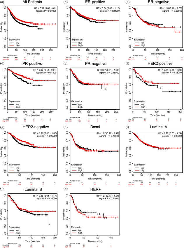
2 Haotian Lu et al.
ZG16A has been recognized as a mucus ingredient 2 Materials and methods
in colon fluid which blocks bacteria and upregulates
in colorectal cancer as a biomarker [22,23]. ZG16B was
2.1 Ethics statement
first discovered to act as a growth factor overexpressed
in pancreatic cancer, which enhances tumor prolifera-
Our work has been approved by the Ethics Committee
tion and helps escape from innate immune system
and Institutional Review Board of Qingdao University,
by intriguing TLR-mediated ERK signaling, inhibiting
China. Informed consent for publication was not required,
TLR-mediated NF-kappa B signaling and keeping β-catenin
as all patient data used in the study were obtained from
stable through phosphorylation [21,24,25]. Furthermore,
publicly available databases.
ZG16B enhances angiogenesis and vascular permeability
and then promotes tumor progression and metastasis of
pancreatic cancer by stimulating CXCR4 expression and
FAK activation [26–28]. Additionally, ZG16B helps pan- 2.2 Oncomine database analysis
creatic cancer cells to resist oncolytic parvovirus H-1
infection via IFNAR-mediated signaling [29]. As a special Oncomine database (https://www.oncomine.org), which pro-
factor in pancreatic cancer, ZG16B promotes activation vides 715 datasets and 86,733 samples, was referred to analyze
and maturation of DCs through TLR4 signaling pathway the expression pattern of ZG16B mRNA in different types of
to mediate immune system activation; meanwhile, it cancers. Pooled meta-analysis in different subtypes of breast
could also increase and activate MDSCs to benefit cancer and gene co-expression analysis of ZG16B were con-
tumor formation, showing a double-sided effect on ducted by Oncomine. The threshold of p-value was fixed to
the immunotherapy [30,31]. And in chemotherapy, 1 × 10⁻⁴. The threshold of fold change was fixed to 2.
ZG16B enhances the effect of gemcitabine and 5-FU
in pancreatic cancer [32,33].
In addition, ZG16B has been identified as a bio-
marker highly expressed in colorectal cancer and 2.3 CCLE analysis
enhancing the migration and invasion, leading to a
poor prognosis [20,34–36]. ZG16B is also confirmed to The expression level of ZG16B in different cell lines was ana-
exist in HeLa cells and upregulates in cervical cancer lyzed by Cancer Cell Line Encyclopedia (CCLE, https://portals.
[37,38]. Moreover, ZG16B can also have effect and be a broadinstitute.org/ccle), a database offering the expression
biomarker for early diagnosis and prognosis of pros- level sorting of 84,434 genes in 1,457 cancer cell lines.
tate cancer [39], oral squamous cell carcinoma [40],
and especially ovarian cancer [41–43]. Besides, ZG16B is
correlated with the prognosis of atherosclerosis and 2.4 Ualcan analysis
acute coronary syndrome [44], and it is demonstrated to
be abundant in the reflex tears as the key point in the Ualcan (http://ualcan.path.uab.edu/analysis.html) is a user-
ocular surface protection, maintaining the tear film stabi- friendly cancer database based on TCGA database. Data from
lity [45,46]. ZG16B has been detected as a biomarker in TCGA database were obtained through Ualcan to analyze the
various malignant tumors; however, the association be- expression discrepancy of ZG16B in diverse molecular sub-
tween ZG16B and breast cancer has not been noticed yet. types of breast cancer, as well as gender, age, cancer
In this report, we found that ZG16B expressed stages, and node metastasis status. The promoter methyla-
highly in breast cancer, and depth analysis was con- tion status of ZG16B was also analyzed using Ualcan.
ducted further to clarify the possible effect of ZG16B
in breast cancer through public medical databases.
Expression levels in different conditions, possible me- 2.5 STRING analysis
chanisms of action and the effect of prognosis of ZG16B
in breast cancer were presented, demonstrating its STRING (https://string-db.org) is a database that collects
potential value to be a biomarker for breast cancer in known and predicted protein–protein physical and func-
clinical practice. tional interaction information, the data of which originate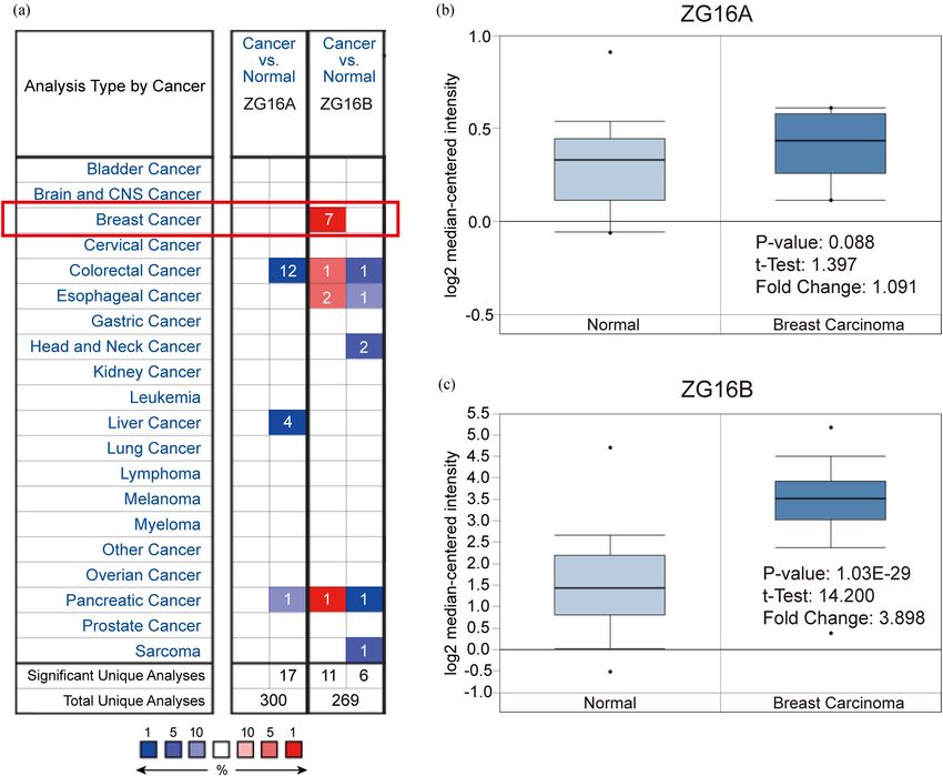
Identification of ZG16B as a prognostic biomarker in breast cancer 3
from computer prediction, knowledge sharing between high expression of ZG16A in breast cancer (Figure 1a).
organizations, and other databases. A protein network We analyzed the datasets uploaded by Ma [49] and Curtis
was drawn by STRING to find the interaction between [47]. No significant difference of ZG16A expression was
ZG16B and other proteins. observed between breast cancer tissue and normal tissue
(p = 0.088) (Figure 1b), while the fold change of ZG16B
expression was 3.898 (p = 1.03 × 10⁻²⁹) (Figure 1c), which
showed high significance.
2.6 Survival analysis Furthermore, in order to verify the high-level expres-
sion signal of ZG16B in breast cancer, we used CCLE
The Kaplan–Meier plotter (http://kmplot.com/analysis) analysis to detect the transcription level of ZG16B in
is an online graph plotter including the survival data of multiple cancer cell lines. The results demonstrated
patients with multiple types of cancers to draw survival that the transcription level of ZG16B was the highest
curves. The effect of ZG16B on prognosis in breast cancer among all types of cancer cell lines (Figure 2). These
was analyzed by Kaplan–Meier plotter on the scale of results indicated the special role of ZG16B in breast
relapse-free survival (RFS). ZG16B Affy ID: 228058_at. cancer.
The Probe set option is the user selected probe set. The In consideration that the evidences described above
cut-off value is determined by the median. indicate ZG16B expresses highly in breast cancer tissues
and cell lines, we have done a further pooled meta-
analysis including 2,780 samples from all the 18 researches
of breast carcinoma in Curtis’ [47], Finak’s [48], and TCGA
2.7 Statistical analysis databases to confirm the high expression in general situa-
tion of breast cancer. The pooled meta-analysis confirmed
Students t-tests were performed to detect the statistical that the mRNA upregulation of ZG16B was extremely
difference of ZG16B mRNA expression between breast significant in breast cancer (p = 5.97 × 10⁻⁴) (Figure 3).
cancer and normal tissues, as well as the mRNA expres- Persuasive testament was presented explaining the con-
sion level and promotor methylation level in different nection between the overexpression of ZG16B and breast
clinical indicators. Log-rank test and hazard ratio (HR) cancer.
analyses were conducted to examine the statistical differ-
ence of survival curves in different subtypes of breast
cancer with low or high expression of ZG16B. Data
were presented as the mean ± standard error of mean,
and P < 0.05 was considered to be statistically significant.
3.2 Analysis of ZG16B expression in
different clinical features of breast
cancer
3 Results Since it showed that ZG16B indeed upregulated in breast
cancer, in order to expound the expression pattern of
ZG16B in breast cancer, Ualcan analysis was conducted
3.1 ZG16B expression in breast cancer to compare the expression level of ZG16B in different clin-
ical indicators. The expression level of ZG16B increased in
Two kinds of zymogen granule proteins have been both female and rare male patients as shown in Figure 4a.
recognized in human body cells, including ZG16A and Although ZG16B expression level is higher in all patient
ZG16B. However, no report has announced their func- groups with different ages compared with normal groups,
tion and correlation in breast cancer yet. Oncomine no significance could be found between groups (Figure 4b);
database was used to analyze the different expression and the situations could be classified in similar way by in-
levels of ZG16A and ZG16B between multiple types of dividual cancer stages and nodal metastasis status as shown
cancer and the corresponding normal tissues. Among in Figure 4c and d. Intriguingly, ZG16B expression level is
all kinds of cancers, it was significant that the expres- significantly higher in luminal-like subtype (p = 1.624 ×
sion of ZG16B was upregulated in 7 analyses of 3 data- 10−12) and triple-negative subtype (p = 4.632 × 10⁻²) than in
sets from Curtis’ [47], Finak’s [48], and TCGA database normal tissues, whereas no significance was shown in HER2-
which met the threshold. And yet no dataset showed positive subtype (Figure 4e).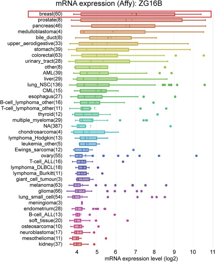
4 Haotian Lu et al.
Figure 1: Analysis of ZG16A and ZG16B mRNA expression in different types of cancer (Oncomine). (a) The mRNA expression pattern of ZG16A
and ZG16B in various cancers. The number in the grid represented the number of datasets that meet the fixed threshold (p < 1 × 10⁻⁴, fold
change >2). The color of the grid represented if the gene expressed higher (red) or lower (blue) (cancer vs. normal). The color depth of the
grid depended on the best gene rank percentiles in all genes detected in each analysis. Box plots derived from gene expression data in
Oncomine comparing the mRNA expression of ZG16A (b) and ZG16B (c) in normal and breast cancer tissues.
3.3 Hypomethylation of ZG16B promoter suggested that the overexpression regulatory mechanism
in breast cancer of ZG16B in breast cancer might be the consequence of
promoter demethylation.
It has been reported that abnormal promoter hypomethyla-
tion induces irregular gene upregulation and then affects
tumor progression [50–52]. Unsurprisingly, by the way of
Ualcan, we found that the promoter methylation level of 3.4 Protein interactions and gene
ZG16B significantly downregulated in primary breast tumor co-expression analysis
compared with that in normal tissues (Figure 5a). Patient
groups with different gender, age, and nodal metastasis STRING analysis was performed to extrapolate the pos-
status showed similar results, when compared with the sible mechanism of ZG16B action in breast cancer and
normal group; however, no significance was confirmed find the interaction between ZG16B and other proteins.
for within group comparison (Figure 5b–d). These results The protein network showed that UBAC1, LYZ, and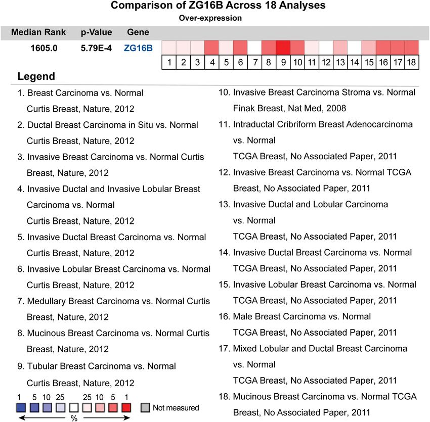
Identification of ZG16B as a prognostic biomarker in breast cancer 5
Figure 2: Analysis of ZG16B mRNA expression in cancer cells. ZG16B mRNA expression levels in 40 types of cancer cell lines were measured
by Affymetrix gene chips and arranged from the highest to the lowest (CCLE analysis). ZG16B mRNA expression level was the first highest in
breast cancer cells among all cancers.
CXCR4 are experimentally determined to have interac- SPINT1 (r = 0.828), TFAP2A (r = 0.826), and FOXA1 (r = 0.826)
tions with ZG16B, while ZBTB42 and LYZ co-expressed as shown in Figure 6b. These demonstrate that ZG16B has
with it. In addition, STRING computationally predicted strong interactions with various proteins involved in breast
ZG16B might have physical or functional relation with cancer formation, indicating the special role of ZG16B in
DUSP15, ZBTB42, S100PBP, PRR4, GTPBP10, FAM96B, breast cancer from another point of view.
and ANKEF1 (Figure 6a).
In order to obtain more detailed information at genetic
level, we have detected the co-expression genes of ZG16B
in breast cancer through Oncomine database. As reported 3.5 Overexpressed ZG16B represents
in Turashvili’s research [53], the expression of ZG16B is favorable prognosis of breast cancer
highly related to EPCAM (r = 0.946), SPDEF (r = 0.918), patients
KRT8 (r = 0.918), SCNN1A (r = 0.918), KRT19 (r = 0.918),
ERBB3 (r = 0.918), AP1M2 (r = 0.904), CLDN4 (r = 0.879), We further examined the impact of ZG16B on the prog-
AGR2 (r = 0.879), CA12 (r = 0.853), KIAA1324 (r = 0.839), nosis of distinct molecular types of breast cancer by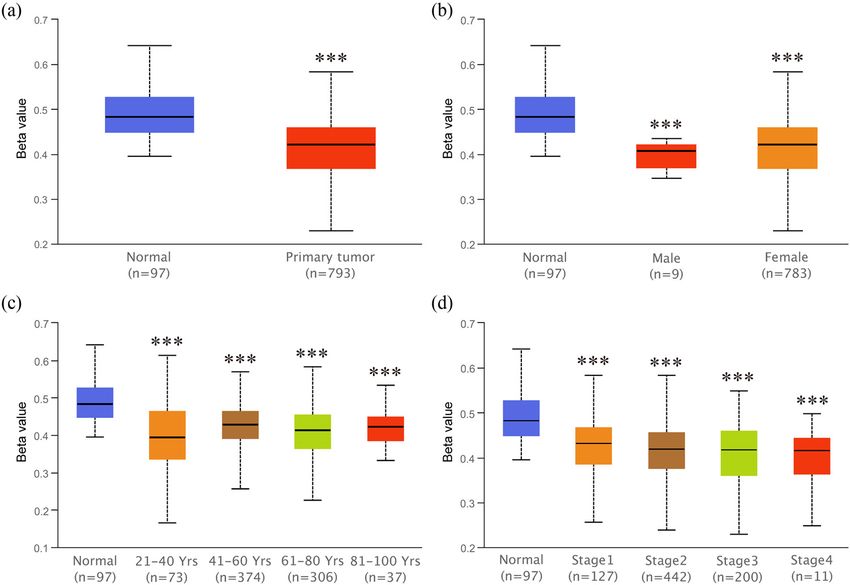
6 Haotian Lu et al.
Figure 3: The pooled meta-analysis of gene expression profiling for ZG16B gene across 18 analysis. The color of the grid represented if the
gene expressed higher (red) or lower (blue). The color depth of the grid depended on the median gene rank percentiles. The rank of ZG16B
was the median rank across each of the analyses. The p-value of ZG16B was for the median-ranked analysis.
Kaplan–Meier plotter using the RFS as an indicator. only to predict the invasiveness [57], recurrence, distant
Interestingly, higher expression of ZG16B represented metastases, and prognosis [58,59], but also to predict the
a longer RFS for all breast cancer patients (HR = 0.77, response to chemotherapy [60], monitor the use of medi-
p = 0.00095) (Figure 7a). The RFS of PR-positive patients cine [61], and being the target of therapy [62] to earn time
also had a longer RFS as ZG16B expressed higher (HR = and quality of life for patients. Fortunately, consensus has
0.62, p = 0.014) (Figure 7d), but there is no significance in been reached by biomedical researchers to establish bio-
other types of breast cancer (Figure 7b, c and e–k). These
informatics database such as Oncomine [63], TCGA data-
data indicated that ZG16B might be a general factor to
base [64], CCLE database [65], and so on to discover and
mark a relatively favorable prognosis in breast cancer.
predict potential biomarkers and therapeutic targets which
could be verified by experiments furthermore.
ZG16B as a biomarker of pancreatic cancer, ovarian
4 Discussion cancer, etc. has no research correlated to breast cancer
yet. In our work, we specially noticed in Oncomine data-
Currently, breast cancer threatens the health of women base that ZG16B was also upregulated in several reports.
around the world [3,54]. Various traditional early detec- In order to further verify the observation, CCLE analysis
tion methods including X-ray, CT, MRI, and ultrasound and pooled meta-analysis have been explored and con-
have been used to diagnose breast cancer [55,56]. In recent firmed that ZG16B indeed has high expression in tissues
years, biomarkers in breast cancer are found and used not and cell lines of breast cancer, as shown in Figures 1–3.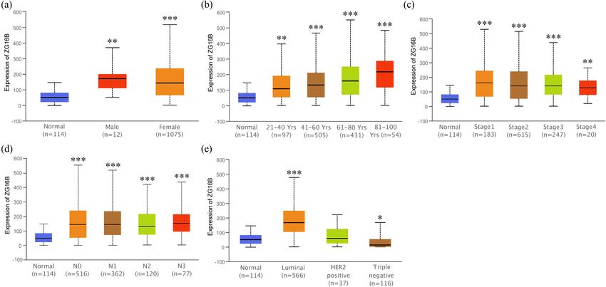
Identification of ZG16B as a prognostic biomarker in breast cancer 7 Figure 4: Analysis of ZG16B expression patterns in breast cancer patients with different clinical-pathologic features. Ualcan analysis showed the mRNA expression level of ZG16B in breast cancer patients with distinct gender (a), age (b), cancer stages (c), node metastasis status (d), and molecular subtypes (e). Asterisks were marked to show the significance of each breast cancer group compared with normal group (*p < 0.05, **p < 0.01, ***p < 0.001). Figure 5: Analysis of ZG16B promoter methylation status in breast cancer patients with different clinical-pathologic features. Ualcan analysis showed the methylation level of ZG16B promoter in primary tumor of breast cancer (a) and in breast cancer patients with distinct gender (b), age (c), and cancer stages (d). Asterisks were marked to show the significance of each group compared with normal group (*p < 0.05, **p < 0.01, ***p < 0.001).
8 Haotian Lu et al. Figure 6: Protein interactions and gene co-expression analysis of ZG16B. (a) The protein interaction network of ZG16B was drawn by STRING analysis. The colored nodes in the network represented ZG16B and its first stage of interactors, and the white nodes represented the second stage of interactors. The physical or functional relations between these proteins are interpreted in the conventional signs. (b) The gene co- expression analysis of ZG16B in Turashvili’s work was conducted by Oncomine. The co-expression genes of ZG16B in breast cancer and their coefficients were marked by red rectangles. To figure out the expression pattern of ZG16B in breast ZG16B has significantly high expression in luminal and triple- cancer, Ualcan database analysis confirms that ZG16B up- negative molecular subtypes of breast cancer, while this high regulates in different clinical classifications, including gender, expression is not found in HER2-positive subtype (Figure 4). In age, and nodal metastasis status; it also demonstrates that addition, the mechanism of demethylation epigenetic factor
Identification of ZG16B as a prognostic biomarker in breast cancer 9 Figure 7: Prognostic value of ZG16B expression in different subtypes of breast cancer. The RFS curves were drawn by Kaplan–Meier plotter in all breast cancer patients (a) and different subtypes of breast cancer, ER-positive (b), ER-negative (c), PR-positive (d), PR-negative (e), HER2-positive (f), HER2-negative (g), Basal (h), Luminal A (i), Luminal (j), and HER2+ (k).
10 Haotian Lu et al.
has been represented by Ualcan to explain the upregulation of PR-positive subtype ZG16B seemingly don’t show apparent
ZG16B in breast cancer (Figure 5). effect, ZG16B does represent a long RFS and good prog-
In order to explore the possible role of ZG16B in nosis for all kinds of patients. Patients with PR-positive
breast cancer, STRING analysis and Oncomine co-expres- breast cancer had the most favorable prognosis among
sion analysis have been performed successively and we breast cancer subtypes with ZG16B high expression, sug-
found that ZG16B had close relationships with UBCA1, gesting its special role in PR-positive breast cancer. All the
DUSP15, ZBTB42, S100PBP, PRR4, CXCR4, LYZ, GTPBP10, data discussed above confirm that ZG16B might be a poten-
FAM96B, ANKEF1, EPCAM, SPDEF, KRT8, SCNN1A, KRT19, tial biomarker of breast cancer which represents a favorable
ERBB3, AP1M2, CLDN4, AGR2, CA12, KIAA1324, SPINT1, prognosis.
TFAP2A, and FOXA1 (Figure 6). Among these proteins In conclusion, ZG16B upregulates in breast cancer and
related to ZG16B, LYZ [66] and PRR4 [67] have protective represents a favorable prognosis in patients. Furthermore,
function in tears and various body fluids, which are corres- ZG16B has correlations with various biomarkers and factors
ponding to Perumal’s research [45,46]. S100PBP [68], PRR4 of breast cancer, some of which have precisely inhibitory
[69], ANKEF1 [39,70], EPCAM [71], SPDEF [72], KRT8, effect on breast cancer. More work and experiments are
KRT19 [73], KIAA1324 [74,75], CXCR4 [76,77], AGR2 needed in order to further reveal more fundamental
[78–80], SCNN1A [81], AP1M2 [82], CLDN4 [83], and mechanism for its role in breast cancer.
ERBB3 [84,85] have been reported to have correlations
with breast cancer or have been identified as biomar- Acknowledgments: This study is supported by National Key
kers for diagnosis, metastasis, and prognosis of breast R&D Program of China (2016YFC1000808), the National
cancer and even as therapeutic targets. Interestingly, Natural Science Foundation of China (81470926, 81702785),
FAM96B is reported to inhibit VEGF receptor 2 pro- Natural Science Foundation of Shandong Province
moter to restrain endothelium activity through the (ZR2017PH013), Key R&D Program of Shandong Province
control of E2-2 expression [86]. SPINT1, which is one (2019GSF107037), the Science and Technology Project of
of the Kunitz-type serine protease inhibitors and also Qingdao, China (18-6-1-88-nsh), and Qingdao Postdoctoral
known as HAI-1, can inhibit hepatocyte growth factor func- Application Research Funded Project (2016067). In addition,
tion via regulation of HGFA, matriptase, and hepsin to in- we specially thank Dr. Rongtao Lu for comments on the
hibit the migration, proliferation, and invasion of breast manuscript.
cancer, indicating a good prognosis [87–89]. TFAP2A, also
known as AP-2-α, is a transcription factor regulating the Conflict of interest: The authors declare that there are no
differentiation and proliferation of breast, the upregulation conflicts of interest in this work.
of which inhibits cell cycle, promotes apoptosis, and sup-
presses invasion in breast cancer via the regulation of
various miRNAs [90–92]. FOXA1 high expression connects
with good prognosis in breast cancer by relieving the
epithelial-to-mesenchymal transition process and inhi-
References
biting migration, invasion, and metastasis [93,94]. These
four genes co-expressed with ZG16B are corresponding to
[1] DeSantis CE, Ma J, Sauer AG, Newman LA, Jemal A. Breast
our exploration, the interaction of which may interpret cancer statistics, 2017, racial disparity in mortality by state.
that ZG16B high expression represents a favorable prog- Ca-a Cancer J Clinic. 2017;67(6):439–48.
nosis in breast cancer. In addition, DUSP15 is recognized [2] Chen W, Zheng R, Baade PD, Zhang S, Zeng H, Bray F, et al.
as a special regulator gene for oligodendrocytes differen- Cancer Statistics in China, 2015. Ca-a Cancer J Clinic.
2016;66(2):115–32.
tiation [95]. GTPBP10 is a mitochondrial protein as a ribo-
[3] Siegel RL, Miller KD, Jemal A. Cancer Statistics, 2016. Ca-a
some biogenesis factor [96,97] correlated with multicentric Cancer J Clinic. 2016;66(1):7–30.
glioblastoma [98]. The two genes co-expressed with ZG16B [4] Liu Y, Ao X, Jia Z, Bai XY, Xu Z, Hu G, et al. FOXK2 transcription
may suggest a potential function of ZG16B in nervous system. factor suppresses ERalpha-positive breast cancer cell growth
Finally, to confirm whether ZG16B had clinical signifi- through down-regulating the stability of ERalpha via me-
chanism involving BRCA1/BARD1. Sci Rep. 2015;5:8796.
cance in breast cancer patients and investigate its effect on
[5] Perou CM, Sorlie T, Eisen MB, van de Rijn M, Jeffrey SS,
prognosis, we have done Kaplan–Meier plotter analysis to
Rees CA, et al. Molecular portraits of human breast tumours.
access the RFS survival curves in different molecular sub- Nature. 2000;406(6797):747–52.
types of breast cancer shown in Figure 7, and it has been [6] Van de Wiel M, Dockx Y, Van den Wyngaert T, Stroobants S,
observed that although most molecular subtypes except Tjalma WAA, Huizing MT. Neoadjuvant systemic therapy inIdentification of ZG16B as a prognostic biomarker in breast cancer 11
breast cancer: challenges and uncertainties. Eur J Obstet [22] Bergstrom JH, Birchenough GM, Katona G, Schroeder BO,
Gynecol Reprod Biol. 2017;210:144–56. Schutte A, Ermund A, et al. Gram-positive bacteria are held at a
[7] Piccart-Gebhart MJ, Procter M, Leyland-Jones B, Goldhirsch A, distance in the colon mucus by the lectin-like protein ZG16.
Untch M, Smith I, et al. Trastuzumab after adjuvant che- Proc Natl Acad Sci U S A. 2016;113(48):13833–8.
motherapy in HER2-positive breast cancer. N Engl J Med. [23] Meng H, Li W, Boardman LA, Wang L. Loss of ZG16 is asso-
2005;353(16):1659–72. ciated with molecular and clinicopathological phenotypes of
[8] Denkert C, Liedtke C, Tutt A, von Minckwitz G. Molecular colorectal cancer. BMC Cancer. 2018;18(1):433.
alterations in triple-negative breast cancer-the road to new [24] Park HD, Lee Y, Oh YK, Jung JG, Park YW, Myung K, et al.
treatment strategies. Lancet 2017;389(10087):2430–42. Pancreatic adenocarcinoma upregulated factor promotes
[9] Sun YS, Zhao Z, Yang ZN, Xu F, Lu HJ, Zhu ZY, et al. Risk factors metastasis by regulating TLR/CXCR4 activation. Oncogene.
and preventions of breast cancer. Int J Biol Sci. 2011;30(2):201–11.
2017;13(11):1387–97. [25] Cho IR, Koh SS, Min HJ, Kim SJ, Lee Y, Park EH, et al. Pancreatic
[10] Li G, Hu J, Hu G. Biomarker studies in early detection and adenocarcinoma up-regulated factor (PAUF) enhances the
prognosis of breast cancer. Adv Exp Med Biol. expression of beta-catenin, leading to a rapid proliferation of
2017;1026:27–39. pancreatic cells. Exp Mol Med. 2011;43(2):82–90.
[11] Janicke F, Schmitt M, Pache L, Ulm K, Harbeck N, Hofler H, et al. [26] Kim SJ, Lee Y, Kim NY, Hwang Y, Hwang B, Min JK, et al.
Urokinase (UPA) and its inhibitor PAI-1 are strong and inde- Pancreatic adenocarcinoma upregulated factor, a novel
pendent prognostic factors in node-negative breast-cancer. endothelial activator, promotes angiogenesis and vascular
Breast Cancer Res Treat. 1993;24(3):195–208. permeability. Oncogene. 2013;32(31):3638–47.
[12] Le Naour F, Misek DE, Krause MC, Deneux L, Giordano TJ, [27] Lee Y, Kim SJ, Park HD, Park EH, Huang SM, Jeon SB, et al. PAUF
Scholl S, et al. Proteomics-based identification of RS/DJ-1 as a functions in the metastasis of human pancreatic cancer cells
novel circulating tumor antigen in breast cancer. Clin Cancer and upregulates CXCR4 expression. Oncogene
Res. 2001;7(11):3328–35. 2010;29(1):56–67.
[13] Look MP, van Putten WLJ, Duffy MJ, Harbeck N, Christensen IJ, [28] Lee YS, Kim SJ, Min HJ, Jo JY, Park EH, Koh SS. PAUF promotes
Thomssen C, et al. Pooled analysis of prognostic impact of adhesiveness of pancreatic cancer cells by modulating focal
urokinase-type plasminogen activator and its inhibitor PAI-1 adhesion kinase. Exp Mol Med. 2011;43(5):291–7.
8377 breast cancer patients. J Natl Cancer Inst. [29] Kaowinn S, Cho IR, Moon J, Jun SW, Kim CS, Kang HY, et al.
2002;94(2):116–28. Pancreatic adenocarcinoma upregulated factor (PAUF) confers
[14] Zhao H, Shen J, Medico L, Wang D, Ambrosone CB, Liu S. A pilot resistance to pancreatic cancer cells against oncolytic parvo-
study of circulating miRNAs as potential biomarkers of early virus H-1 infection through IFNA receptor-mediated signaling.
stage breast cancer. PLoS One. 2010;5(10):e13735. Biochem Biophys Res Commun. 2015;459(2):313–8.
[15] Hao W, Friedman A. Serum uPAR as biomarker in breast cancer [30] Kang TH, Kim YS, Kim S, Yang B, Lee JJ, Lee HJ, et al. Pancreatic
recurrence: a mathematical model. PLoS One. adenocarcinoma upregulated factor serves as adjuvant by
2016;11(4):e0153508. activating dendritic cells through stimulation of TLR4.
[16] Singh R, Bhatt ML, Singh SP, Kumar V, Goel MM, Mishra DP, Oncotarget. 2015;6(29):27751–62.
et al. Evaluation of KiSS1 as a prognostic biomarker in North [31] Song J, Lee J, Kim J, Jo S, Kim YJ, Baek JE, et al. Pancreatic
Indian breast cancer cases. Asian Pac J Cancer Prev. adenocarcinoma up-regulated factor (PAUF) enhances the
2016;17(4):1789–95. accumulation and functional activity of myeloid-derived
[17] Jing X, Cui X, Liang H, Hao C, Yang Z, Li X, et al. CD24 is a suppressor cells (MDSCs) in pancreatic cancer. Oncotarget.
potential biomarker for prognosis in human breast carcinoma. 2016;7(32):51840–53.
Cell Physiol Biochem. 2018;48(1):111–9. [32] Gao CC, Xu XL, Li F, Gong BG, Liu S, Cui YQ, et al. Silencing
[18] Mito A, Nakano Y, Saitoh T, Gouraud SSS, Yamaguchi Y, Sato T, pancreatic adenocarcinoma upregulated factor (PAUF)
et al. Lectin ZG16p inhibits proliferation of human colorectal increases the sensitivity of pancreatic cancer cells to gemci-
cancer cells via its carbohydrate-binding sites. Glycobiology. tabine. Tumour Biol. 2016;37(6):7555–64.
2018;28(1):21–31. [33] Cho JH, Kim SA, Park SB, Kim HM, Song SY. Suppression of
[19] Kanagawa M, Satoh T, Ikeda A, Nakano Y, Yagi H, Kato K, et al. pancreatic adenocarcinoma upregulated factor (PAUF) in-
Crystal structures of human secretory proteins ZG16p and creases the sensitivity of pancreatic cancer to gemcitabine and
ZG16b reveal a Jacalin-related beta-prism fold. Biochem 5FU, and inhibits the formation of pancreatic cancer stem like
Biophys Res Commun. 2011;404(1):201–5. cells. Oncotarget. 2017;8(44):76398–407.
[20] Escudero-Paniagua B, Bartolome RA, Rodriguez S, de Los [34] Barderas R, Mendes M, Torres S, Bartolome RA, Lopez-
Rios V, Pintado L, Jaen M, et al. PAUF/ZG16B promotes color- Lucendo M, Villar-Vazquez R, et al. In-depth characterization
ectal cancer progression through alterations of the mitotic of the secretome of colorectal cancer metastatic cells identi-
functions and the Wnt/beta-catenin pathway. Carcinogenesis. fies key proteins in cell adhesion, migration, and invasion. Mol
2020;41(2):203–13. Cell Proteom. 2013;12(6):1602–20.
[21] Kim SA, Lee Y, Jung DE, Park KH, Park JY, Gang J, et al. [35] Liu PF, Wu YY, Hu Y, Wang L, He SB, Zhu YB, et al. Silencing of
Pancreatic adenocarcinoma up-regulated factor (PAUF), a pancreatic adenocarcinoma upregulated factor by RNA inter-
novel up-regulated secretory protein in pancreatic ductal ference inhibits the malignant phenotypes of human colorectal
adenocarcinoma. Cancer Sci. 2009;100(5):828–36. cancer cells. Oncol Rep. 2013;30(1):213–20.12 Haotian Lu et al.
[36] Kim JG, Chae YS, Lee SJ, Kang BW, Park JY, Lee EJ, et al. Genetic [51] Van Tongelen A, Loriot A, De Smet C. Oncogenic roles of DNA
variation in microRNA-binding site and prognosis of patients hypomethylation through the activation of cancer-germline
with colorectal cancer. J Cancer Res Clin Oncol. genes. Cancer Lett. 2017;396:130–7.
2015;141(1):35–41. [52] Lin IH, Chen DT, Chang YF, Lee YL, Su CH, Cheng C, et al.
[37] Wakana Y, van Galen J, Meissner F, Scarpa M, Polishchuk RS, Hierarchical clustering of breast cancer methylomes revealed
Mann M, et al. A new class of carriers that transport selective differentially methylated and expressed breast cancer genes.
cargo from the trans Golgi network to the cell surface. Embo J. PLoS One. 2015;10(2):e0118453.
2012;31(20):3976–90. [53] Turashvili G, Bouchal J, Baumforth K, Wei W, Dziechciarkova M,
[38] Choi CH, Chung JY, Park HS, Jun M, Lee YY, Kim BG, et al. Ehrmann J, et al. Novel markers for differentiation of lobular
Pancreatic adenocarcinoma up-regulated factor expression is and ductal invasive breast carcinomas by laser microdissec-
associated with disease-specific survival in cervical cancer tion and microarray analysis. BMC Cancer. 2007;7:55.
patients. Hum Pathol. 2015;46(6):884–93. [54] Torre LA, Bray F, Siegel RL, Ferlay J, Lortet-Tieulent J, Jemal A.
[39] Jin HJ, Jung S, DebRoy AR, Davuluri RV. Identification and va- Global Cancer Statistics, 2012. Ca-a Cancer J Clinic.
lidation of regulatory SNPs that modulate transcription factor 2015;65(2):87–108.
chromatin binding and gene expression in prostate cancer. [55] Roganovic D, Djilas D, Vujnovic S, Pavic D, Stojanov D. Breast
Oncotarget. 2016;7(34):54616–26. MRI, digital mammography and breast tomosynthesis: com-
[40] Sasahira T, Kurihara M, Nishiguchi Y, Nakashima C, Kirita T, parison of three methods for early detection of breast cancer.
Kuniyasu H. Pancreatic adenocarcinoma up-regulated factor Bosn J Basic Med Sci. 2015;15(4):64–8.
has oncogenic functions in oral squamous cell carcinoma. [56] Wang L. Early diagnosis of breast cancer. Sensors.
Histopathology. 2017;70(4):539–48. 2017;17(7):1572.
[41] Kim SK, Song SY, Kim S, Cho NH, Yim GW, Kim SW, et al. [57] Lim S, Janzer A, Becker A, Zimmer A, Schule R, Buettner R, et al.
Association of pancreatic adenocarcinoma up-regulated Lysine-specific demethylase 1 (LSD1) is highly expressed in
factor expression in ovarian mucinous adenocarcinoma with ER-negative breast cancers and a biomarker predicting ag-
poor prognosis. Int J Clin Exp Pathol. 2014;7(8): gressive biology. Carcinogenesis. 2010;31(3):512–20.
5103–10. [58] Millar EKA, Graham PH, O’Toole SA, McNeil CM, Browne L,
[42] Choi CH, Kang TH, Song JS, Kim YS, Chung EJ, Ylaya K, et al. Morey AL, et al. Prediction of local recurrence, distant metas-
Elevated expression of pancreatic adenocarcinoma upregu- tases, and death after breast-conserving therapy in early-
lated factor (PAUF) is associated with poor prognosis and stage invasive breast cancer using a five-biomarker panel.
chemoresistance in epithelial ovarian cancer. Sci Rep. J Clin Oncol. 2009;27(28):4701–8.
2018;8(1):12161. [59] Ao X, Ding W, Ge H, Zhang Y, Ding D, Liu Y. PBX1 is a valuable
[43] Zhang GH, Chen MM, Kai JY, Ma Q, Zhong AL, Xie SH, et al. prognostic biomarker for patients with breast cancer. Exp Ther
Molecular profiling of mucinous epithelial ovarian cancer by Med. 2020;20(1):385–94.
weighted gene co-expression network analysis. Gene. [60] Esserman LJ, Berry DA, Cheang MCU, Yau C, Perou CM, Carey L,
2019;709:56–64. et al. Chemotherapy response and recurrence-free survival in
[44] Martin-Lorenzo M, Zubiri I, Maroto AS, Gonzalez-Calero L, neoadjuvant breast cancer depends on biomarker profiles:
Posada-Ayala M, de la Cuesta F, et al. KLK1 and ZG16B proteins results from the I-SPY 1 TRIAL (CALGB 150007/150012; ACRIN
and arginine-proline metabolism identified as novel targets to 6657). Breast Cancer Res Treat. 2012;132(3):1049–62.
monitor atherosclerosis, acute coronary syndrome and [61] Dowsett M, Ebbs SR, Dixon JM, Skene A, Griffith C,
recovery. Metabolomics. 2015;11(5):1056–67. Boeddinghaus I, et al. Biomarker changes during neoadjuvant
[45] Perumal N, Funke S, Wolters D, Pfeiffer N, Grus FH. anastrozole, tamoxifen, or the combination: influence of hor-
Characterization of human reflex tear proteome reveals high monal status and HER-2 in breast cancer – a study from the
expression of lacrimal proline-rich protein 4 (PRR4). IMPACT trialists. J Clin Oncol. 2005;23(11):2477–92.
Proteomics. 2015;15(19):3370–81. [62] Ross JS, Fletcher JA, Linette GP, Stec J, Clark E, Ayers M, et al.
[46] Perumal N, Funke S, Pfeiffer N, Grus FH. Proteomics analysis of The HER-2/neu gene and protein in breast cancer 2003: bio-
human tears from aqueous-deficient and evaporative dry eye marker and target of therapy. Oncologist. 2003;8(4):307–25.
patients. Sci Rep. 2016;6:29629. [63] Rhodes DR, Yu JJ, Shanker K, Deshpande N, Varambally R,
[47] Curtis C, Shah SP, Chin SF, Turashvili G, Rueda OM, Ghosh D, et al. ONCOMINE: a cancer microarray database and
Dunning MJ, et al. The genomic and transcriptomic architec- integrated data-mining platform. Neoplasia. 2004;6(1):1–6.
ture of 2,000 breast tumours reveals novel subgroups. Nature. [64] Chin L, Meyerson M, Aldape K, Bigner D, Mikkelsen T,
2012;486(7403):346–52. VandenBerg S, et al. Comprehensive genomic characterization
[48] Finak G, Bertos N, Pepin F, Sadekova S, Souleimanova M, defines human glioblastoma genes and core pathways.
Zhao H, et al. Stromal gene expression predicts clinical Nature. 2008;455(7216):1061–8.
outcome in breast cancer. Nat Med. 2008;14(5): [65] Barretina J, Caponigro G, Stransky N, Venkatesan K,
518–27. Margolin AA, Kim S, et al. The cancer cell line encyclopedia
[49] Ma XJ, Dahiya S, Richardson E, Erlander M, Sgroi DC. Gene enables predictive modelling of anticancer drug sensitivity.
expression profiling of the tumor microenvironment during Nature. 2012;483(7391):603–7.
breast cancer progression. Breast Cancer Res. 2009;11(1): [66] McCanna DJ, Oh S, Seo J, Coles-Brennan C, Fadli Z,
R7. Subbaraman LN, et al. The effect of denatured lysozyme on
[50] Jones PA, Baylin SB. The fundamental role of epigenetic events human corneal epithelial cells. Invest Ophthalmol Vis Sci.
in cancer. Nat Rev Genet. 2002;3(6):415–28. 2018;59(5):2006–14.Identification of ZG16B as a prognostic biomarker in breast cancer 13
[67] Ekizoglu S, Ulutin T, Guliyev J, Buyru N. PRR4: a novel down- [82] Tang YC, Ho SC, Tan E, Ng AWT, McPherson JR, Goh GYL, et al.
regulated gene in laryngeal cancer. Oncol Lett. Functional genomics identifies specific vulnerabilities in PTEN-
2018;15(4):4669–75. deficient breast cancer. Breast Cancer Res. 2018;20(1):22.
[68] Savci-Heijink CD, Halfwerk H, Koster J, Horlings HM, van de [83] Wang F, Gao Y, Tang L, Ning K, Geng N, Zhang H, et al. A novel
Vijver MJ. A specific gene expression signature for visceral PAK4-CEBPB-CLDN4 axis involving in breast cancer cell
organ metastasis in breast cancer. BMC Cancer. migration and invasion. Biochem Biophys Res Commun.
2019;19(1):333. 2019;511(2):404–8.
[69] Rabet MM, Cabaud O, Josselin E, Finetti P, Castellano R, [84] Baselga J, Swain SM. Novel anticancer targets: revisiting
Farina A, et al. Nectin-4: a new prognostic biomarker for effi- ERBB2 and discovering ERBB3. Nat Rev Cancer.
cient therapeutic targeting of primary and metastatic triple- 2009;9(7):463–75.
negative breast cancer Ann Oncol. 2017;28(4):769–76. [85] Holbro T, Beerli RR, Maurer F, Koziczak M, Barbas CF,
[70] Nguyen NM, Andrade FD, Jin L, Zhang XY, Macon M, Cruz MI, Hynes NE. The ErbB2/ErbB3 heterodimer functions as an on-
et al. Maternal intake of high n-6 polyunsaturated fatty acid cogenic unit: ErbB2 requires ErbB3 to drive breast tumor cell
diet during pregnancy causes transgenerational increase in proliferation. Proc Natl Acad Sci U S A. 2003;100(15):8933–8.
mammary cancer risk in mice. Breast Cancer Res. 2017;19:77. [86] Yang W, Itoh F, Ohya H, Kishimoto F, Tanaka A, Nakano N, et al.
[71] Gao S, Sun Y, Liu X, Zhang D, Yang X. EpCAM and COX2 Interference of E2-2-mediated effect in endothelial cells by
expression are positively correlated in human breast cancer. FAM96B through its limited expression of E2-2. Cancer Sci.
Mol Med Rep. 2017;15(6):3755–60. 2011;102(10):1808–14.
[72] Doane AS, Danso M, Lal P, Donaton M, Zhang L, Hudis C, et al. [87] Oberst MD, Chen LY, Kiyomiya K, Williams CA, Lee MS,
An estrogen receptor-negative breast cancer subset charac- Johnson MD, et al. HAI-1 regulates activation and expression of
terized by a hormonally regulated transcriptional program and matriptase, a membrane-bound serine protease. Am J Physiol
response to androgen. Oncogene. 2006;25(28):3994–4008. Cell Physiol. 2005;289(2):C462–70.
[73] Qiu J, Du Z, Wang Y, Zhou Y, Zhang Y, Xie Y, et al. Weighted [88] Parr C, Jiang WG. Hepatocyte growth factor activation inhibi-
gene co-expression network analysis reveals modules and hub tors (HAI-1 and HAI-2) regulate HGF-induced invasion of
genes associated with the development of breast cancer. human breast cancer cells. Int J Cancer. 2006;119(5):1176–83.
Medicine. 2019;98(6):e14345. [89] Parr C, Watkins G, Mansel RE, Jiang WG. The hepatocyte growth
[74] Bolton KA, Holliday EG, Attia J, Bowden NA, Avery-Kiejda KA, factor regulatory factors in human breast cancer. Clin Cancer
Scott RJ. A novel polymorphic repeat in the upstream regula- Res. 2004;10(1 Pt 1):202-11.
tory region of the estrogen-induced gene EIG121 is not asso- [90] Berlato C, Chan KV, Price AM, Canosa M, Scibetta AG,
ciated with the risk of developing breast or endometrial Hurst HC. Alternative TFAP2A isoforms have distinct activities
cancer. BMC Res Notes. 2016;9:287. in breast cancer. Breast Cancer Res. 2011;13(2):R23.
[75] Schlumbrecht MP, Xie SS, Shipley GL, Urbauer DL, [91] Zhou B, Guo H, Tang J. Long non-coding RNA TFAP2A-AS1
Broaddus RR. Molecular clustering based on ERalpha and inhibits cell proliferation and invasion in breast cancer via
EIG121 predicts survival in high-grade serous carcinoma of the miR-933/SMAD2. Med Sci Monit. 2019;25:1242–53.
ovary/peritoneum. Mod Pathol. 2011;24(3):453–62. [92] Xu J, Zheng J, Wang J, Shao J. miR-876-5p suppresses breast
[76] Zou YR, Kottmann AH, Kuroda M, Taniuchi I, Littman DR. cancer progression through targeting TFAP2A. Exp Ther Med.
Function of the chemokine receptor CXCR4 in haematopoiesis 2019;18(2):1458–64.
and in cerebellar development. Nature 1998;393(6685):595–9. [93] Shou J, Lai Y, Xu J, Huang J. Prognostic value of FOXA1 in breast
[77] Mantovani A, Allavena P, Sica A, Balkwill F. Cancer-related cancer: a systematic review and meta-analysis. Breast.
inflammation. Nature. 2008;454(7203):436–44. 2016;27:35–43.
[78] Jung SY, Yun J, Kim SJ, Kang S, Kim DY, Kim YJ, et al. Basic [94] Xu Y, Qin L, Sun T, Wu H, He T, Yang Z, et al. Twist1 promotes
helix-loop-helix transcription factor Twist1 is a novel regulator breast cancer invasion and metastasis by silencing Foxa1
of anterior gradient protein 2 homolog (AGR2) in breast expression. Oncogene. 2017;36(8):1157–66.
cancer. Biochem Biophys Res Commun. 2019;516(1):149–56. [95] Tian Y, Wang L, Jia M, Lu T, Ruan Y, Wu Z, et al. Association of
[79] Ondrouskova E, Sommerova L, Nenutil R, Coufal O, Bouchal P, oligodendrocytes differentiation regulator gene DUSP15 with
Vojtesek B, et al. AGR2 associates with HER2 expression pre- autism. World J Biol Psychiatry. 2017;18(2):143–50.
dicting poor outcome in subset of estrogen receptor negative [96] Maiti P, Kim HJ, Tu YT, Barrientos A. Human GTPBP10 is
breast cancer patients. Exp Mol Pathol. 2017;102(2):280–3. required for mitoribosome maturation. Nucleic Acids Res.
[80] Salmans ML, Zhao F, Andersen B. The estrogen-regulated 2018;46(21):11423–37.
anterior gradient 2 (AGR2) protein in breast cancer: a potential [97] Lavdovskaia E, Kolander E, Steube E, Mai MM, Urlaub H,
drug target and biomarker. Breast Cancer Res. 2013;15(2): Richter-Dennerlein R. The human Obg protein GTPBP10 is
204. involved in mitoribosomal biogenesis. Nucleic Acids Res.
[81] Varley KE, Gertz J, Roberts BS, Davis NS, Bowling KM, 2018;46(16):8471–82.
Kirby MK, et al. Recurrent read-through fusion transcripts in [98] Kong DS, Kim J, Lee IH, Kim ST, Seol HJ, Lee JI, et al. Integrative
breast cancer. Breast Cancer Res Treat. 2014;146(2): radiogenomic analysis for multicentric radiophenotype in
287–97. glioblastoma. Oncotarget. 2016;7(10):11526–38.You can also read