Anti inflammatory effects and enhancing immune response of freshwater hybrid catfish oil in RAW264.7 cells
←
→
Page content transcription
If your browser does not render page correctly, please read the page content below
EXPERIMENTAL AND THERAPEUTIC MEDICINE 22: 1223, 2021

        Anti‑inflammatory effects and enhancing immune response
            of freshwater hybrid catfish oil in RAW264.7 cells
                   BUSSARIN TONGMEE1, ATCHARAPORN ONTAWONG2, NARISSARA LAILERD3,
                      KRIANGSAK MENGAMPHAN4 and DOUNGPORN AMORNLERDPISAN4,5

    1
     Agricultural Interdisciplinary Program, Faculty of Engineer and Agro‑Industry, Maejo University, Chiang Mai 50290;
   2
    Division of Physiology, School of Medical Sciences, University of Phayao, Phayao 56000; 3Department of Physiology,
Faculty of Medicine, Chiang Mai University, Chiang Mai 50200; 4Center of Excellence in Agricultural Innovation for Graduate
   Entrepreneur; 5Faculty of Fisheries Technology and Aquatic Resources, Maejo University, Chiang Mai 50290, Thailand

                                         Received February 8, 2021; Accepted July 14, 2021

                                                   DOI: 10.3892/etm.2021.10657

Abstract. The present study assessed the effect of fresh‑           driver of septic shock (3). LPS is able to activate macrophages
water hybrid catfish oil (FFO) on the inflammatory status of        to release several inflammatory cytokines (4). Activation of the
lipopolysaccharide (LPS)‑stimulated RAW264.7 cells and              inflammatory pathway may be induced by pro‑inflammatory
investigated the underlying mechanisms. RAW264.7 cells              mediators and cytokines being secreted, including nitric
were supplemented with various concentrations [0.125‑2%             oxide (NO), cyclooxygenase‑2 (COX‑2), tumor necrosis
in 0.5% propylene glycol (v/v)] of FFO with or without LPS          factor‑ α (TNF‑ α), interleukin‑1β (IL‑1β), IL‑6 and prosta‑
(1 µg/ml) for 24 h. Inflammatory cytokines and mediators were       glandin (PG)E2 (5). Inflammation is one cause of increased
quantified using ELISA and reverse transcription‑quantitative       morbidity and mortality in intensive care units, also resulting
PCR. The results revealed that FFO treatment inhibited the          in elevated hospital‑related costs (6,7). Nowadays, several
secretion and mRNA expression of the pro‑inflammatory cyto‑         anti‑inflammatory drugs are available, such as non‑steroidal
kines IL‑6, IL‑1β, TNF‑α. In line with this, FFO suppressed         anti‑inflammatory drugs (NSAIDs) (8). However, a previous
the expression and secretion of the inflammatory mediators          study suggested that NSAIDs may induce gastrointestinal tract
cyclooxygenase‑2 and prostaglandin E2. FFO also reduced             bleeding (9). Safe and effective strategies to prevent and treat
apoptotic body formation and DNA damage. Correspondingly,           inflammation and its associated diseases are thus urgently
FFO enhanced the immune response by modulating the cell             required.
cycle regulators p53, cyclin D2 and cyclin E2. Accordingly,             The freshwater hybrid catfish (Pangasius sp.) belongs to
FFO may be developed as a nutraceutical product to prevent          the freshwater catfish family. It has become one of the most
inflammation.                                                       popular freshwater fish species and has a high demand,
                                                                    particularly on the European and US markets. Fish contains
Introduction                                                        2‑30% fat and ~50% of its body weight is discarded as
                                                                    waste during the fish processing operation (10). One of the
Inflammation is one of the first lines of defense against           fish processing byproducts is fish oil (FO). FO is a source
harmful stimuli, such as pathogens, damaged cells, trauma,          of long‑chain polyunsaturated fatty acid (e.g. omega‑3 fatty
bacteria and irritants (1). Macrophages detect and react to         acids), particularly fish oil extracted from marine fish, which
certain pathogens and consequently regulate the inflamma‑           is mainly composed of cis‑5,8,11,14,17‑eicosapentaenoic
tory response (2). Lipopolysaccharide (LPS) is an endotoxin         acid (EPA) and cis‑4,7,10,13,16,19‑docosahexaenoic acid (11).
derived from the outer membrane of Gram‑negative bacteria           As a component in FO, omega‑3 fatty acids have several
and also a powerful mediator of systemic inflammation and a         benefits, including protection against atherosclerosis, arrhyth‑
                                                                    mias and chronic obstructive pulmonary diseases (12). They
                                                                    also reduce blood pressure, blood glucose and symptoms of
                                                                    asthma and cystic fibrosis (13‑15). However, a previous study
                                                                    by our group demonstrated that fish oil from freshwater hybrid
Correspondence to: Dr Doungporn Amornlerdpisan, Center of
                                                                    catfish contains a high level of monounsaturated omega‑9 fatty
Excellence in Agricultural Innovation for Graduate Entrepreneur,
Maejo University, 63 Sansai‑Phrao Road, Sansai, Chiang Mai 50290,
                                                                    acid (MUFA) (16). Furthermore, freshwater hybrid catfish oil
Thailand                                                            (FFO) was indicated to have anti‑diabetic effects by improving
E‑mail: doungpornfishtech@gmail.com                                 insulin resistance and adipokine imbalance in a rat model of
                                                                    type 2 diabetes and also suppress pro‑inflammatory cytokine
Key words: anti‑inflammatory, cytokine, DNA damage, freshwater      protein expressions in the skeletal muscle tissues of those
hybrid catfish oil, immune response                                 rats (17). The omega‑9 fatty acid increased of high‑density
                                                                    lipoprotein‑cholesterol and decreased low‑density lipopro‑
                                                                    tein‑cholesterol (17). However, the effect of FFO on the
2 TONGMEE et al: ANTI-INFLAMMATION AND IMMUNE ENHANCEMENT OF FRESHWATER HYBRID CATFISH OIL
inflammatory condition and the underlying mechanisms have medium containing 0.5 mg/ml of MTT (Thermo Fisher
remained elusive. In the present study, the anti‑inflammatory Scientific, Inc.) was added to each well, followed by incubation
effects of FFO on RAW264.7 macrophages stimulated by LPS at 37˚C for 4 h. The MTT solution was then aspirated and cells
were examined and the associated mechanism was investi‑ were washed once with ice‑cold PBS. The purple formazan
gated. crystals were dissolved in DMSO for 30 min and cell viability
was subsequently analyzed by measuring the absorption at a
Materials and methods wavelength of 570 nm using an M965 AccuReader microplate
reader (Metertech, Inc.). The lysed cells were detected at a
Chemicals. Dulbecco's modified Eagle's medium (DMEM) wavelength of 680 nm was used as a reference. Cell viability
and fetal bovine serum (FBS) were purchased from Gibco; was calculated as follows: Cell viability (%) = [(Absorbance
Thermo Fisher Scientific, Inc. β‑nicotinamide adenine dinucle‑ value‑reference value) x100]/[mean of (absorbance value‑refer‑
otide phosphate and LPS were purchased from Merck KGaA. ence value) in untreated cells].
All other chemicals with high purity were purchased from
commercial sources. ELISA. RAW264.7 cells were seeded into 12‑well plates at a
density of 1x10⁵ cells/ml and incubated for 24 h at 37˚C in
Preparation of FFO. FO of freshwater hybrid catfish a humidified atmosphere with 5% CO2. The culture medium
(Pangasius gigas x Pangasianodon hypophthalmus) was was removed and cells were treated with different concentra‑
purchased from a private company, Me Natural Co., Ltd., tions of FFO [0.125‑2% in 0.5% propylene glycol (v/v)] with
which cooperated and received the adipose tissue from the or without LPS (1 µg/ml) in fresh medium for 24 h at 37˚C
Center of Excellence in Giant Catfish and Buk Siam Catfish, in a humidified atmosphere with 5% CO2. Subsequently,
Faculty of Fisheries Technology and Aquatic Resources, the cells were homogenized and lysed cells were centri‑
Maejo University (Chiang Mai, Thailand). FO was extracted as fuged at 2,000 x g for 10 min at 4˚C. The supernatant was
previously described, which exhibited a high omega‑9 content collected and stored at ‑80˚C for quantification of IL‑6 (cat.
and biological activity (18). In brief, frozen adipose tissues no. BIOL‑431304), IL‑1β (cat. no. BIOL‑432604), TNF‑ α
were purified by cleaning and steaming at 90˚C for 30 min. (cat. no. BIOL‑430904), NO (cat. no. 780001) and PGE2 (cat.
The liquid oil was subsequently filtered through a filter sack no. ABBK‑KTE70765‑96T) concentrations using commercial
and squeezed using a screw compressor. The squeezed liquid kits (BioLegend, Inc.) according to the manufacturer's proto‑
was centrifuged at 2,268 x g for 10 min at 25˚C to separate cols.
the solid particles from the oil and the supernatant FFO was
separated. Solvent‑free extraction was used to obtain FFO. As NO assay. The nitrate/nitrite concentration was determined
previously, adipose tissue was extracted and partially purified using a colorimetric assay kit (Cayman Chemical Co.). In
as aforementioned, resulting in FFO at a yield of 300 ml per brief, cells were treated with different concentrations of FFO
1 kg of adipose tissue. [0.125‑2% in 0.5% propylene glycol (v/v)] with or without
LPS (1 µg/ml) for 24 h at 37˚C in a humidified atmosphere
Determination of fatty acids, fat‑soluble vitamins and heavy with 5% CO2. Treated cells were centrifuged at 10,000 x g for
metal levels of FFO. The chemical compounds, including the 20 min at 4˚C. The supernatant was subsequently collected to
fatty acids and fat‑soluble vitamins, were sent for analysis at a measure the NO concentration at a wavelength of 540 nm using
certified lab with international standardization in the field of an M965 AccuReader microplate reader (Metertech, Inc.).
information technology (ISO172025), the Central Laboratory
(Thailand) Co. Ltd., Chiang Mai Branch, following the TE‑CH Hoechst 33342 staining. To confirm the effect of FFO on
260 in‑house method of the Association of Official Analytical LPS‑induced DNA damage, RAW264.7 cells were seeded into
Chemists 996.06 (19). Heavy metal contamination of FFO was 8‑well cell culture slides and treated with different concen‑
also detected according to this in‑house method. trations of FFO [0.125‑2% in 0.5% propylene glycol (v/v)]
with or without LPS (1 µg/ml) for 24 h at 37˚C in a humidi‑
Cell culture. RAW264.7 cells were purchased from the fied atmosphere with 5% CO2. Treated cells were fixed with
American Type Culture Collection. Cells at passage 2‑22 4% paraformaldehyde for 10 min at room temperature and
were maintained in DMEM (Thermo Fisher Scientific, Inc) subsequently stained with Hoechst 33342 (5 µg/ml) for 10 min
containing 3.7 g/l NaHCO3 supplemented with 10% FBS at room temperature. Cells were washed twice with PBS and
(Thermo Fisher Scientific, Inc) and 1% penicillin/strepto‑ observed under a Nikon Eclipse Ni‑U fluorescent microscope
mycin in a humidified atmosphere at 37˚C with 5% CO2 and (original magnification, x40; Nikon Corporation).
sub‑cultured every 4‑5 days using 0.05% trypsin‑EDTA in PBS
(Thermo Fisher Scientific, Inc.). Cells were seeded at a density DNA damage assay. To further determine the protective effect
of 1x10⁵ cells/well and cultured in 6‑, 12‑ and 96‑well plates of FFO on LPS‑induced DNA damage, the effect of FFO on
for 3 days until subsequent experimentation. The medium was DNA impairment was investigated via ELISA. RAW264.7 cells
replaced every 2 days during culture. were seeded into 12‑well plates at a density of 1x10⁵ cells/ml
and incubated for 24 h at 37˚C in a humidified atmosphere
Determination of cell viability. The MTT assay was performed with 5% CO2. The culture medium was removed and cells
to assess the effect of FFO on cell viability. Cells were incubated were treated with different concentrations of FFO [0.125‑2%
with serum‑free medium with FFO at 0, 0.125, 0.25, 0.5, 1 or in 0.5% propylene glycol (v/v)] with or without LPS (1 µg/ml)
2% in 0.5% propylene glycol (v/v). Subsequently, serum‑free for 24h at 37˚C in a humidified atmosphere with 5% CO2.EXPERIMENTAL AND THERAPEUTIC MEDICINE 22: 1223, 2021 3
Table I. Primer sequences and expected amplicon sizes for gene amplification.
GenBank Amplicon
cDNA accession no. Forward primer Reverse primer size (bp)
TNF-α NM013693.3 5'-ACCTGGCCTCTCTACCTTGT-3' 5'-CCCGTAGGGCGATTACAGTC-3' 161
IL-1β NM008361.4 5'-GCCACCTTTTGACAGTGATGAG-3' 5'-AGTGATACTGCCTGCCTGAAG-3' 165
IL-6 NM031168.2 5'-CAACGATGATGCACTTGCAGA-3' 5'-TCTCTCTGAAGGACTCTGGCT-3' 201
COX-2 NM011198.4 5'-CCACTTCAAGGGAGTCTGGA-3' 5'-AGTCATCTGCTACGGGAGGA-3' 197
Cyclin D2 NM009829.3 5'-ACCTCCCGCAGTGTTCCTATT-3' 5'-CACAGACCTCTAGCATCCAGG-3' 93
Cyclin E2 NM001037134.2 5'-TCTGTGCATTCTAGCATCGACTC-3' 5'-AAGGCACCATCGTCTACACATTC-3' 149
p27 NM009875.4 5'-GCGGTGCCTTTAATTGGGTCT-3' 5'-GGCTTCTTGGGCGTCTGCT-3' 230
p53 NM011640.3 5'-ACCGCCGACCTATCCTTACC-3' 5'-TCTTCTGTACGGCGGTCTCTC-3' 118
GAPDH NM001289726.1 5'-TGTGTCCGTCGTGGATCTGA-3' 5'-TTGCTGTTGAAGTCGCAGGAG-3' 150
TNF-α, tumor necrosis factor‑α; IL, interleukin; COX-2, cyclooxygenase-2.
Treated cells were centrifuged at 10,000 x g for 20 min at Table II. Fatty acid composition and vitamin content of fresh‑
4˚C. The supernatant was collected and stored at ‑80˚C for water hybrid catfish oil.
quantification of 8‑hydroxy‑2'‑deoxyguanosine (8‑OHdG; cat.
no. AB‑EIADNAD), a DNA damage marker, using commer‑ Chemical component Amount
cial kit (Thermo Fisher Scientific, Inc.) according to the
Saturated fatty acids, g/100 g 40.38±2.94
manufacturer's protocol.
Unsaturated fatty acids, g/100 g 55.80±0.64
Reverse transcription‑quantitative (RT‑q)PCR. Total RNA Monounsaturated fatty acids, g/100 g 46.74±2.24
was extracted and purified from RAW264.7 cells using Oleic acid, g/100 g 42.07±1.79
TRIzol® reagent (Thermo Fisher Scientific, Inc.) according Omega-9 42.27±1.76
to the manufacturer's protocol and reverse transcribed into Polyunsaturated fatty acids, g/100 g 12.75±1.04
cDNA using the SensiFAST™ cDNA synthesis kit (Bioline). Omega-3 1.17±0.27
qPCR was subsequently performed using SYBR Real‑Time Omega-6 10.95±1.03
PCR Master Mix (Bioline) on a CFX Touch real‑time PCR
Vitamins
system (Bio‑Rad Laboratories, Inc.). PCR amplifications were
Vitamin A (retinol), µg/100 g 1.80±0.12
performed at a 20‑µl volume with the following thermocycling
conditions: A polymerase enzyme activation step at 95˚C Vitamin E (α-tocopherol), mg/100 g 0.69±0.06
for 2 min; followed by 40 cycles of denaturation at 95˚C for Values are expressed as means ± standard error of the mean (n=3).
5 sec, 10 sec of annealing at 60˚C depending on primers, and
10 sec of elongation at 72˚C. The primer sequences used for
qPCR were purchased from Macrogen, Inc. and used at a final
concentration of 0.4 µM. The primer sequences for mouse at 40.38, 55.80, 46.74 and 12.75 g/100 g of FFO, respectively.
TNF‑α, IL‑1β, IL‑6, COX2, p53, p27, cyclin D2, cyclin E2 Among the detected MUFAs, the predominant fatty acid was
and GAPDH are presented in Table I (20‑25). Gene expression omega‑9 (42.27±1.76 g/100 g of FFO). In addition, FFO also
was calculated using the 2‑ΔΔCq method (26) and normalized to contained PUFAs and the predominant fatty acids were omega‑3
GAPDH. Data were reported as the relative fold change. qPCR (1.17±0.39 g/100 g of FFO) and omega‑6 (10.95±1.46 g/100 g of
amplification was performed in duplicate for each synthesized FFO). In addition, vitamin A was present at 1.80±0.12 µg/100 g of
cDNA set. FFO and vitamin E was present at 0.69±0.06 mg/100 g of FFO.
Statistical analysis. Statistical analysis was performed using Heavy metal content profiles of FFO. The concentrations
SPSS version 23 software (IBM Corp.). Values are expressed of arsenic, copper, lead, mercury, tin and zinc in FFO are
as the mean ± standard error of the mean. One‑way ANOVA presented in Table III. The results demonstrated that FFO
followed by Dunnett's test was used to compare differences contained copper and lead at much lower concentrations, while
between multiple groups. P4 TONGMEE et al: ANTI-INFLAMMATION AND IMMUNE ENHANCEMENT OF FRESHWATER HYBRID CATFISH OIL Figure 1. Inflammatory response was inhibited by treatment with FFO in LPS‑stimulated RAW264.7 cells. RAW264.7 cells were pre‑treated with different concentrations of FFO in the presence or absence of LPS for 24 h. The production of (A) IL‑6, (B) IL‑1β and (C) TNF‑α was measured by ELISA. (D) Viability of RAW264.7 cells after exposure to different concentrations of FFO with or without LPS for 24 h. Values are expressed as the mean ± standard error of the mean (n=5). *P
EXPERIMENTAL AND THERAPEUTIC MEDICINE 22: 1223, 2021                                                             5

Figure 2. Gene expression of pro‑inflammatory cytokines in LPS‑stimulated RAW264.7 cells. RAW264.7 cells were pre‑treated with different concentrations
of FFO in the presence or absence of LPS for 24 h. The expression of (A) IL‑6, (B) IL‑1β and (C) TNF‑α was measured by reverse transcription‑quantitative
PCR. Values are expressed as the mean ± standard error of the mean (n=5). *P
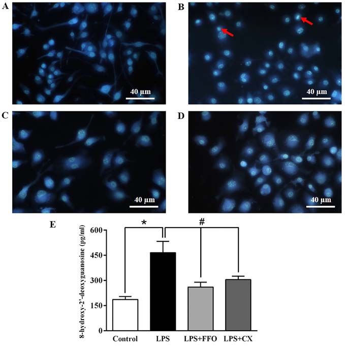
6            TONGMEE et al: ANTI-INFLAMMATION AND IMMUNE ENHANCEMENT OF FRESHWATER HYBRID CATFISH OIL

Figure 4. Cytoprotective effect of FFO in LPS‑stimulated RAW264.7 cells. Cells were seeded in 12‑well plates and treated with FFO or CX in the presence or
absence of LPS. After 24 h, cells were treated with Hoechst 33342 at 5 µg/ml for 10 min and then observed under an inverted fluorescence microscope (original
magnification, x40; total magnification, x400; scale bar, 40 µm). The cell nucleus changes of apoptotic cells are indicated by red arrows. (A) Control, (B) LPS
group, (C) LPS+FFO group and (D) LPS+CX group. (E) The amount of 8‑OHdG in the DNA was determined using an 8‑OHdG‑EIA kit. Values are expressed
as the mean ± standard error of the mean (n=5). *P
EXPERIMENTAL AND THERAPEUTIC MEDICINE 22: 1223, 2021                                             7

Discussion                                                              in vascular endothelial cells through upregulation of nuclear
                                                                        factor‑mediated antioxidant response and the decrease in intra‑
To the best of our knowledge, the present study was the first to        cellular reactive oxygen species (50). In addition, the present
demonstrate that FFO rich in omega‑9 exerts anti‑inflammatory           study demonstrated that the expression of cell cycle regula‑
effects in vitro by decreasing the expression and secretion of          tors, including cyclin D2 and cyclin E2, increased following
pro‑inflammatory cytokines and mediators, preventing DNA                treatment with FFO, while p53 expression was inhibited. A
damage via reduction of apoptotic body formation and 8‑OHdG,            previous study reported that cyclin D2 deficiency suppresses
and also promotes an immune response. A previous study demon‑           immune activity (51). On the other hand, hyperactive cyclin D2
strated that the activation of tissue macrophages releases various      expression promotes autoimmune disease or allograft rejec‑
pro‑inflammatory cytokines, including TNF‑α, IL‑1 and IL‑6,             tion (52). Other natural products merely promote immune
resulting in autoimmune and inflammatory diseases. In addition,         responses by regulating cell cycle regulators. For instance,
n‑3 polyunsaturated fatty acids (PUFAs) serve anti‑inflammatory         A. asphodeloides enhances the immune response of RAW264.7
effects by reducing the production of TNF‑α, IL‑1β, IL‑6 and            cells by extending the cell cycle S‑phase, suppressing p27 and
tissue factors by stimulated monocytes (36). Thus, inhibiting the       increasing cyclin D2 and cyclin E2 gene expression (53).
synthesis of these cytokines may prove useful for the treatment             In conclusion, the results of the present study demonstrated
of autoimmune and inflammatory diseases. The results of the             that FFO improved inflammation by suppressing the mRNA
present study demonstrated that FFO markedly decreased the              expression and secretion of pro‑inflammatory cytokines and
production of IL‑6, IL‑1β and TNF‑α and mRNA expression                 their mediators, and inhibiting apoptotic body formation and
levels in RAW264.7 cells, similar to NSAIDs. These results              DNA damage. FFO also enhanced the immune response by
suggested that FFO exerts an anti‑inflammatory effect by down‑          modulating cell cycle regulators. Thus, FFO may be used as
regulating pro‑inflammatory cytokines at both the transcriptional       a natural anti‑inflammatory supplement. Moreover, future
and translational levels, without any cell toxicity. Similarly, oleic   in vivo studies and clinical trials are required to elucidate
acid, one of the most representative monounsaturated omega‑9            whether FFO has an overall anti‑inflammatory effect in auto‑
fatty acids, was reported to mediate anti‑inflammatory effects by       immune or inflammatory diseases.
inhibiting reactive oxygen species, p38 MAPK and Akt signaling
pathways/IKK/NF‑κB in BV2 cells (37).                                   Acknowledgements
    Macrophages are associated with acute and chronic inflam‑
matory responses by stimulating NO generation, resulting in an          Not applicable.
increment of macrophage activity (38). NO and PGE2 produc‑
tion are critical immune‑regulatory biomarkers for chronic              Funding
inflammatory diseases, such as hepatic dysfunction and pulmo‑
nary disease (39). The results of the present study demonstrated        No funding was received.
that FFO decreased PGE2 and its synthase enzyme COX‑2,
but not the NO level, similar to the action of NSAIDs.                  Availability of data and materials
Previous studies have reported that natural products, including
coumarin, Indonesian cassia extract and Halocynthia auran‑              The datasets used and/or analyzed during the current study are
tium or docosahexaenoic acid‑omega‑3, decrease PGE2 and                 available from the corresponding author on reasonable request.
NO expression levels, which suggests that they have poten‑
tial as anti‑inflammatory agents (40‑42). Conversely, it has            Authors' contributions
been demonstrated that omega 3 increases the production of
PGE2 (43). The increment of the PGE2 concentration may be               BT performed the experiments, collected and analysed the
inhibited by the NF‑κ B signaling pathway and EP4 receptor,             data, and wrote the first draft of the manuscript. AO designed
resulting in anti‑inflammatory effects (44). There are contro‑          the experiments, collected and analysed the data and wrote the
versial data on the effect of PGE2 in inflammation. The results         manuscript. NL and KM provided, analyzed and interpreted the
of the present study demonstrated that FFO contains several             data. DA designed and verified the experiments, analysed the
fatty acids, including omega‑3, ‑6 and ‑9. Consistently, previous       data, and wrote and provided critical feedback for the manu‑
studies have demonstrated that omega‑3 fatty acids decrease             script. DA and AO confirm the authenticity of all the raw data.
PGE2 by decreasing the catalytic monomer of COX‑1 dimer by              All authors read and approved the final version of the study.
arachidonic acid and inhibiting COX‑1 oxygenation (45,46). In
addition, omega‑9 exerts anti‑inflammatory effects in inflam‑           Ethics approval and consent to participate
mation via a PPAR‑γ expression‑dependent mechanism (47).
    It is well‑known that there is a close association between          Not applicable.
inflammation and DNA damage (48). NO generated by
inflammatory cytokine stimulation is sufficient to induce               Patient consent for publication
oxidative DNA damage (49). The results of the present study
demonstrated that LPS induced DNA damage by nuclear                     Not applicable.
fragmentation, chromatin condensation and apoptotic body
formation, the effects of which were reversed following treat‑          Competing interests
ment with FFO and NSAIDs. Consistently, n‑3 polyunsaturated
fatty acids attenuate oxidative stress‑induced DNA damage               The authors declare that they have no competing interests.
8 TONGMEE et al: ANTI-INFLAMMATION AND IMMUNE ENHANCEMENT OF FRESHWATER HYBRID CATFISH OIL
References 21. Yen TL, Chang CC, Chung CL, Ko WC, Yang CH and Hsieh CY:
Neuroprotective effects of platonin, a therapeutic immuno‑
modulating medicine, on traumatic brain injury in mice after
1. Pereira DM, Correia‑da‑Silva G, Valentão P, Teixeira N and controlled corticalimpact. Int J Mol Sci 19: 1100, 2018.
Andrade PB: Anti‑inflammatory effect of unsaturated fatty 22. Teratake Y, Kuga C, Hasegawa Y, Sato Y, Kitahashi M,
acids and Ergosta‑7,22‑dien‑3‑ol from Marthasterias glacialis: Fujimura L, Watanabe‑Takano H, Sakamoto A, Arima M,
Prevention of CHOP‑mediated ER‑stress and NF‑κ B activation. Tokuhisa T, et al: Transcriptional repression of p27 is essential
PLoS One 9: e88341, 2014. for murine embryonic development. Sci Rep 6: 26244, 2016.
2. Lee HJ, Shin JS, Lee KG, Park SC, Jang YP, Nam JH and 23. Zhao H, Bauzon F, Fu H, Lu Z, Cui J, Nakayama K, Nakayama KI,
Lee KT: Ethanol extract of Potentilla supina Linne suppresses Locker J and Zhu L: Skp2 deletion unmasks a p27 safeguard
LPS‑induced inflammatory responses through NF‑κ B and AP‑1 that blocks tumorigenesis in the absence of pRb and p53 tumor
inactivation in macrophages and in endotoxic mice. Phytother suppressors. Cancer Cell 24: 645‑659, 2013.
Res 31: 475‑487, 2017. 24. Tokumoto M, Fujiwara Y, Shimada A, Hasegawa T, Seko Y, Nagase H
3. Bennett JE, Dolin R and Blaser MJ: Mandell, Douglas, and and Satoh M: Tokumoto1 M, Fujiwara Y, Shimada A, Hasegawa T,
Bennett's Principles and Practice of Infectious Diseases. 8th edition. Seko Y, Nagase H, Satoh M: Cadmium toxicity is caused by accumu‑
Bennett JE, Dolin R and Blaser MJ (eds). Elsevier/Saunders, lation of p53 through the down‑regulation of Ube2d family genes in
Philadelphia, PA, p27, 2015. vitro and in vivo. J Toxicol Sci 36: 191‑200, 2011.
4. Oh YC, Cho WK, Oh JH, Im GY, Jeong YH, Yang MC and 25. Chen YG, Zhang Y, Deng LQ, Chen H, Zhang YJ, Zhou NJ,
Ma JY: Fermentation by Lactobacillus enhances anti‑inflam‑ Yuan K, Yu LZ, Xiong ZH, Gui XM, et al: Control of meth‑
matory effect of Oyaksungisan on LPS‑stimulated RAW 264.7 icillin‑resistant Staphylococcus aureus pneumonia utilizing
mouse macrophage cells. BMC Complement Altern Med 12: 17, TLR2 agonist Pam3CSK4. PLoS One 11: e0149233, 2016.
2012. 26. Livak KJ and Schmittgen TD: Analysis of relative gene expression
5. Fard MT, Arulselvan P, Karthivashan G, Adam SK and data using real‑time quantitative PCR and the 2(‑Delta Delta
Fakurazi S: Bioactive extract from Moringa oleifera inhibits the C(T)) method. Methods 25: 402‑408, 2001.
pro‑inflammatory mediators in lipopolysaccharide stimulated 27. Institute of Medicine (US) Panel on Micronutrients: Dietary
macrophages. Pharmacogn Mag 11 (Suppl 4): S556‑S563, 2015. Reference Intakes for Vitamin A, Vitamin K, Arsenic, Boron,
6. Adrie C, Alberti C, Chaix‑Couturier C, Azoulay E, De Lassence A, Chromium, Copper, Iodine, Iron, Manganese, Molybdenum,
Cohen Y, Meshaka P, Cheval C, Thuong M, Troché G, et al: Nickel, Silicon, Vanadium, and Zinc. National Academies Press,
Epidemiology and economic evaluation of severe sepsis in France: Washington, DC, 2001.
Age, severity, infection site, and place of acquisition (community, 28. Shin S: Safety of celecoxib versus traditional nonsteroidal
hospital, or intensive care unit) as determinants of workload and anti‑inflammatory drugs in older patients with arthritis. J Pain
cost. J Crit Care 20: 46‑58, 2005. Res 11: 3211‑3219, 2018.
7. O'Brien DJ and Gould IM: Maximizing the impact of anti‑ 29. Park JY, Pillinger MH and Abramson SB: Prostaglandin E2 synthesis
microbial stewardship: The role of diagnostics, national and and secretion: The role of PGE2 synthases. Clin Immunol 119:
international efforts. Curr Opin Infect Dis 26: 352‑358, 2013. 229‑240, 2006.
8. Parvizi J and Kim GK: High Yield Orthopaedics. Saunders/Elsevier, 30. Valavanidis A, Vlachogianni T and Fiotakis C: 8‑Hydroxy‑2'-
Philadelphia, PA, pp325‑326, 2010. deoxyguanosine (8‑OHdG): A critical biomarker of oxidative
9. Dhikav V, Singh S, Pande S, Chawla A and Anand KS: stress and carcinogenesis. J Environ Sci Health Part C Environ
Non‑steroidal drug‑induced gastrointestinal toxicity: Mechanisms Carcinog Ecotoxicol Rev 27: 120‑139, 2009.
and management. JIACM 4: 315‑322, 2003. 31. Yuan L, Zhang Y, Xia J, Liu B, Zhang Q, Liu J, Luo L, Peng Z, Song Z
10. Wangcharoen W, Mengumphan K and Amornlerdpison D: Fatty acid and Zhu R: Resveratrol induces cell cycle arrest via a p53‑independent
composition, physical properties, acute oral toxicity and antioxidant pathway in A549 cells. Mol Med Rep 11: 2459‑2464, 2015.
activity of crude lipids from adipose tissue of some commercialized 32. Yuan L, Zhang Y, Xia J, Liu B, Zhang Q, Liu J, Luo L, Peng Z,
freshwater catfish. Warasan Khana Witthayasat Maha Witthayalai Song Z, Zhu R, Zhu R, et al: Resveratrol induces cell cycle arrest
Chiang Mai 42: 626‑636, 2015. via a p53‑independent pathway in A549 cells. Mol Med Rep 11:
11. Khoddami A, Ariffin A, Bakar J and Mohd Ghazali H: Fatty acid 2459‑2464, 2015
profile of the oil extracted from fish waste (head, intestine and liver) 33. Shaw PH: The role of p53 in cell cycle regulation. Pathol Res
(Sardinella lemuru). World Appl Sci J 7: 127-131, 2009. Pract 192: 669‑675, 1996.
12. Gammone MA, Riccioni G, Parrinello G and D'Orazio N: Omega‑3 34. Donehower LA: Phosphatases reverse p53‑mediated cell cycle
polyunsaturated fatty acids: Benefits and endpoints in sport. checkpoints. Proc Natl Acad Sci USA 111: 7172‑7173, 2014.
Nutrients 11: 46, 2018. 35. Møller MB: P27 in cell cycle control and cancer. Leuk Lymphoma 39:
13. Kim JS and Park JW: Mince from seafood processing by‑product 19‑27, 2000.
and surimi as food ingredients. In: Maximising the Value of Marine 36. Priante G, Bordin L, Musacchio E, Clari G and Baggio B: Fatty
By‑Products. Shahidi F (ed). Woodhead Publishing, Sawston, acids and cytokine mRNA expression in human osteoblastic
pp196‑228, 2007. cells: A specific effect of arachidonic acid. Clin Sci (Lond) 102:
14. Kim SK and Mendis E: Bioactive compounds from marine 403‑409, 2002.
processing byproducts - A review. Food Res Int 39: 383‑393, 2006. 37. Oh YT, Lee JY, Lee J, Kim H, Yoon KS, Choe W and Kang I: Oleic
15. Tawfik M: Proximate composition and fatty acids profiles in most acid reduces lipopolysaccharide‑induced expression of iNOS and
common available fish species in Saudi market. Asian J Clin Nutr 1: COX‑2 in BV2 murine microglial cells: Possible involvement
50-57, 2009. of reactive oxygen species, p38 MAPK, and IKK/NF‑kappaB
16. Bussarin T, Kriangsak M, Narissara L and Doungporn A: signaling pathways. Neurosci Lett 464: 93‑97, 2009.
Comparison of fatty acid profiles of freshwater hybrid catfish. In: 38. Olefsky JM and Glass CK: Macrophages, inflammation, and
Proceedings of MJU Annual Conference. The Office of Agricultural insulin resistance. Annu Rev Physiol 72: 219‑246, 2010.
Research and Extension Maejo, Maejo University. pp60‑61. 2018. 39. Ansar W and Ghosh S: Inflammation and inflammatory diseases,
17. Keapai W, Apichai S, Amornlerdpison D and Lailerd N: Evaluation markers, and mediators: Role of CRP in some inflammatory
of fish oil‑rich in MUFAs for anti‑diabetic and anti‑inflammation diseases. In: Biology of C Reactive Protein in Health and Disease.
potential in experimental type 2 diabetic rats. Korean J Physiol pp67‑107, 2016.
Pharmacol 20: 581‑593, 2016. 40. Kondreddy VK and Kamatham AN: Celecoxib, a COX‑2 inhibitor,
18. Amornlerdpison D, Rattanaphot T, Tongsiri S, Srimaroeng C and synergistically potentiates the anti‑inflammatory activity of doco‑
Mengumphan K: Effect of omega‑9‑rich fish oil on antioxidant sahexaenoic acid in macrophage cell line. Immunopharmacol
enzymes and relative immune gene expressions in Nile tilapia Immunotoxicol 38: 153‑161, 2016.
(Oreochromis niloticus). J Sci Technol 41: 1287‑1293, 2019. 41. Monmai C, Go SH, Shin IS, You SG, Lee H, Kang SB and
19. AOAC Official Method of Analysis: Official Method 996.06 Fat Park WJ: Immune‑enhancement and anti‑inflammatory activities
(Total, Saturated, and Unsaturated) in Foods. AOAC International of fatty acids extracted from Halocynthia aurantium tunic in
Chapter 41. Oils and Fats: 20-24. 2002. RAW264.7 cells. Mar Drugs 16: 16, 2018.
20. Li Y, Hao N, Zou S, Meng T, Tao H, Ming P, Li M, Ding H, Li J, 42. Sandhiutami NM, Moordiani M, Laksmitawati DR, Fauziah N,
Feng S, et al: Immune regulation of RAW264.7 cells in vitro Maesaroh M and Widowati W: In vitro assesment of anti‑inflam‑
by flavonoids from Astragalus complanatus via activating the matory activities of coumarin and Indonesian cassia extract
NF‑ κ B signalling pathway. J Immunol Res 2018: 7948068, in RAW264.7 murine macrophage cell line. Iran J Basic Med
2018. Sci 20: 99‑106, 2017.EXPERIMENTAL AND THERAPEUTIC MEDICINE 22: 1223, 2021 9
43. Denkins Y, Kempf D, Ferniz M, Nileshwar S and Marchetti D: 48. Kawanishi S, Ohnishi S, Ma N, Hiraku Y and Murata M:
Role of omega‑3 polyunsaturated fatty acids on cyclooxygenase‑2 Crosstalk between DNA damage and inflammation in the
metabolism in brain‑metastatic melanoma. J Lipid Res 46: multiple steps of carcinogenesis. Int J Mol Sci 18: 18, 2017.
1278‑1284, 2005. 49. Jaiswal M, LaRusso NF, Burgart LJ and Gores GJ: Inflammatory
44. Liu Y, Chen LY, Sokolowska M, Eberlein M, Alsaaty S, cytokines induce DNA damage and inhibit DNA repair in chol‑
Martinez‑Anton A, Logun C, Qi HY and Shelhamer JH: The angiocarcinoma cells by a nitric oxide‑dependent mechanism.
fish oil ingredient, docosahexaenoic acid, activates cytosolic Cancer Res 60: 184‑190, 2000.
phospholipase A2 via GPR120 receptor to produce prosta‑ 50. Sakai C, Ishida M, Ohba H, Yamashita H, Uchida H,
glandin E2 and plays an anti‑inflammatory role in macrophages. Yoshizumi M and Ishida T: Fish oil omega‑3 polyunsaturated
Immunology 143: 81‑95, 2014. fatty acids attenuate oxidative stress‑induced DNA damage in
45. Wada M, DeLong CJ, Hong YH, Rieke CJ, Song I, Sidhu RS, vascular endothelial cells. PLoS One 12: e0187934, 2017.
Yuan C, Warnock M, Schmaier AH, Yokoyama C, et al: Enzymes 51. Chunder N, Wang L, Chen C, Hancock WW and Wells AD:
and receptors of prostaglandin pathways with arachidonic Cyclin‑dependent kinase 2 controls peripheral immune tolerance.
acid‑derived versus eicosapentaenoic acid‑derived substrates and J Immunol 189: 5659‑5666, 2012.
products. J Biol Chem 282: 22254‑22266, 2007. 52. Laphanuwat P and Jirawatnotai S: Immunomodulatory roles of
46. Yuan C, Sidhu RS, Kuklev DV, Kado Y, Wada M, Song I and cell cycle regulators. Front Cell Dev Biol 7: 23, 2019.
Smith WL: Cyclooxygenase allosterism, fatty acid‑mediated 53. Ji KY, Kim KM, Kim YH, Im AR, Lee JY, Park B, Na M and
cross‑talk between monomers of cyclooxygenase homodimers. Chae S: The enhancing immune response and anti‑inflammatory
J Biol Chem 284: 10046‑10055, 2009. effects of Anemarrhena asphodeloides extract in RAW 264.7
47. Medeiros‑de‑Moraes IM, Gonçalves‑de‑Albuquerque CF, cells. Phytomedicine 59: 152789, 2019.
Kurz AR, Oliveira FM, de Abreu VH, Torres RC, Carvalho VF,
Estato V, Bozza PT, Sperandio M, et al: Omega‑9 oleic acid, This work is licensed under a Creative Commons
the main compound of olive oil, mitigates inflammation during Attribution-NonCommercial-NoDerivatives 4.0
experimental sepsis. Oxid Med Cell Longev 2018: 6053492, 2018. International (CC BY-NC-ND 4.0) License.You can also read