Omega 3 polyunsaturated fatty acids inhibit IL 11/STAT3 signaling in hepatocytes during acetaminophen hepatotoxicity
←
→
Page content transcription
If your browser does not render page correctly, please read the page content below
INTERNATIONAL JOURNAL OF MOLECULAR MEDICINE 48: 190, 2021
Omega‑3 polyunsaturated fatty acids inhibit IL‑11/STAT3
signaling in hepatocytes during acetaminophen hepatotoxicity
YUNZHI LIU1,2, JINGMIN LIN2, YU CHEN1,2, ZHUONAN LI3, JIA ZHOU2,
XIAO LU2, ZHENGLIANG CHEN2 and DAMING ZUO1,4
1
Department of Medical Laboratory, School of Laboratory Medicine and Biotechnology, Southern Medical University;
2
Guangdong Province Key Laboratory of Proteomics, Department of Immunology, School of Basic Medical Sciences,
Southern Medical University, Guangzhou, Guangdong 510515; 3College of Marine Life Sciences, Ocean University of China,
Qingdao, Shandong 266003; 4Department of Laboratory Medicine, Microbiome Medicine Center,
Zhujiang Hospital, Southern Medical University, Guangzhou, Guangdong 510282, P.R. China
Received March 31, 2021; Accepted July 2, 2021
DOI: 10.3892/ijmm.2021.5023
Abstract. Omega‑3 polyunsaturated fatty acids (n‑3 PUFAs) expression are responsible for the n‑3 PUFA‑mediated inhibi‑
exert a negative effect on IL‑6 production in several liver tory effect on IL‑11 production in APAP‑treated hepatocytes.
disorders, including cirrhosis, acute liver failure and fatty It was concluded that n‑3 PUFAs inhibit IL‑11 production and
liver disease. However, its effect on the production of IL‑11, further STAT3 activation in hepatocytes during APAP‑induced
another important IL‑6 family cytokine, remains unclear. liver injury. Therefore, ERK1/2‑mediated Fra‑1 expression is
IL‑11 was found to be significantly elevated in acetaminophen responsible for the effect of n‑3 PUFAs on IL‑11 expression.
(APAP)‑induced liver damage. The aim of the present study
was to investigate whether and how n‑3 PUFAs modulate IL‑11 Introduction
production during APAP‑induced liver injury. For that purpose,
wild‑type (WT) and fat‑1 transgenic mice were intraperitone‑ IL‑11 is a member of the IL‑6 cytokine family and has
ally injected with APAP to induce liver injury. Serum was been reported to exert important effects on various liver
collected for ELISA and alanine aminotransferase assay. The diseases (1). Lipid‑laden hepatocytes secrete IL‑11, which
hepatocytes of APAP‑injected mice were isolated for reverse acts via autocrine cis‑signaling and contributes to hepatocyte
transcription‑quantitative PCR and western blot analyses. For lipotoxicity in a reactive oxygen species (ROS)‑dependent
the in vitro study, primary hepatocytes isolated from WT or manner (2). Furthermore, IL‑11 receptor agonist enhances the
fat‑1 mice were stimulated with APAP. The results revealed proliferation of hepatocytes and ameliorates oxidative stress
that both endogenous and exogenous n‑3 PUFAs significantly during acetaminophen (APAP)‑induced liver injury. Among
aggravated APAP‑induced liver damage via the downregula‑ acute liver injury models, IL‑11 is markedly upregulated in
tion of STAT3 signaling. Notably, n‑3 PUFAs inhibited IL‑11 hepatocytes in response to APAP‑induced liver injury (3)
expression, but not IL‑6 expression in hepatocytes during and liver ischemia (4). APAP is a commonly used drug for
the APAP challenge. Furthermore, it was demonstrated that the relief of pain and fever. Although it is considered to be
limited phosphorylation of ERK1/2 and Fos‑like‑1 (Fra‑1) safe at therapeutic concentrations, its overdose can cause
acute liver damage (5). STAT3, a member of the STAT family,
can be activated by IL‑6 family cytokines, and has been
found to exert anti‑apoptotic and pro‑proliferative effects on
APAP hepatotoxicity (6). These functions mainly rely on the
Correspondence to: Professor Zhengliang Chen, Guangdong regulation of genes involved in cell fate, such as the apoptosis
Province Key Laboratory of Proteomics, Department of Immunology,
regulators Bcl‑2 and Bax (7,8).
School of Basic Medical Sciences, Southern Medical University,
1023 North Shatai Road, Guangzhou, Guangdong 510515, P.R. China Omega‑3 polyunsaturated fatty acids (n‑3 PUFAs), which
E‑mail: zhlchen@smu.edu.cn include eicosapentaenoic acid (EPA) and docosahexaenoic
acid (DHA), are important for human health. Several studies
Professor Daming Zuo, Department of Medical Laboratory, School
have reported the effect of n‑3 PUFAs on various liver
of Laboratory Medicine and Biotechnology, Southern Medical
diseases (9‑11). n‑3 PUFAs attenuate systemic inflamma‑
University, 1023 North Shatai Road, Guangzhou, Guangdong 510515,
P.R. China tion by reducing the circulating level and gene expression of
E‑mail: zdaming@smu.edu.cn IL‑6 (9,12). Dietary n‑3 PUFA intake is associated with lower
methylation at the IL‑6 promoter, leading to a decreased
Key words: acetaminophen, omega‑3 polyunsaturated fatty acids, plasma IL‑6 concentration (13). Reduced IL‑6 production
STAT3 signaling, IL‑11, ERK1/2‑Fos‑like‑1 signaling and limited STAT3 phosphorylation have been observed in
pancreatic acinar cells treated with n‑3 PUFAs during cerulein
exposure (14). Notably, previous studies have reported that
2                                         LIU et al: n-3 PUFAs INHIBIT IL-11 PRODUCTION

n‑3 PUFAs inhibited the phosphorylation of STAT3 in various        (rabbit anti‑mouse monoclonal; 1:1,000; cat. no. 12640),
disease models, thereby influencing cell differentiation,          p‑p44/42 MAPK (Erk1/2; rabbit anti‑mouse monoclonal;
promoting cell death, inhibiting cell migration and inducing       1:1,000; cat. no. 4376), p44/42 MAPK (Erk1/2) (rabbit
autophagy (15‑17). The complex of IL‑6 and IL‑6 receptor           anti‑mouse monoclonal; 1:1,000; cat. no. 4695) and GAPDH
(IL‑6R) binds to glycoprotein 130 (gp130), which dimerizes         (rabbit anti‑mouse monoclonal; 1:1,000; cat. no. 5174) were
and initiates intracellular STAT3 signaling (18). The presence     obtained from Cell Signaling Technology, Inc. Anti‑IL‑11 poly‑
of n‑3 PUFAs may reduce the surface expression of IL‑6R            clonal antibody (rabbit anti‑mouse; 1:1,000; cat. no. A1902) for
and its association with gp130 in lipid rafts, thereby leading     immunohistochemistry and immunoblotting was purchased
to decreased downstream STAT3 activation (16). In addition,        from ABclonal Biotech Co., Ltd. Anti‑Fra‑1 Ab (mouse
n‑3 PUFAs may reduce IL‑6‑induced gp130 dimerization               anti‑mouse monoclonal; 1:100; cat. no. sc‑271657) was obtained
and subsequent STAT3 phosphorylation. Notably, n‑3 PUFAs           from Santa Cruz Biotechnology, Inc. The antibody against
modulate STAT3 signaling by enhancing the expression of Src        Bcl‑2 (mouse anti‑mouse monoclonal; 1:100; cat. no. YM3041)
homology region 2 domain‑containing protein tyrosine phos‑         was from ImmunoWay Biotechnology Company. Anti‑Bax
phatase‑1, which is a well‑known negative regulator of STAT3       (rabbit anti‑mouse polyclonal; 1:1,000; cat. no. DB123) anti‑
signaling (19).                                                    body was purchased from DB Biotech, spol. s r.o.
    Fat‑1 mice, which exhibit increased levels of n‑3 PUFAs
in their organs and tissues compared with those of their           Isolation of mouse hepatocytes and hepatic mononuclear
wild‑type (WT) counterparts, have been reported to be a reli‑      cells. Hepatocytes were isolated as previously described (26).
able animal model to investigate n‑3 PUFAs (20). The present       Briefly, livers were perfused with calcium‑free salt solution and
study aimed to investigate whether n‑3 PUFAs modulate IL‑11        type IV collagenase through the portal vein in situ and then
expression and downstream STAT3 signaling during APAP              filtered with polyamide mesh. After centrifugation at 50 x g
hepatotoxicity.                                                    for 1 min at 4˚C, the precipitants were collected as hepatocytes
                                                                   for reverse transcription‑quantitative PCR (RT‑qPCR) and
Materials and methods                                              western blotting assays, or for primary hepatocyte culture. The
                                                                   supernatants were harvested for density gradient centrifugation
Mice. WT C57BL/6J mice were obtained from the Laboratory           using discontinuous 30/70% (v/v) Percoll gradients to obtain
Animal Center of Southern Medical University (Guangzhou,           mononuclear cells for RT‑qPCR. For primary mouse hepato‑
China). Fat‑1 transgenic mice, which has been reported to          cyte (PMH) culture, 1x10⁶ hepatocytes were seeded in 6‑well
carry the gene that encodes the enzyme that coverts n‑6 to         dishes coated with mouse tail collagen (cat. no. A1048301;
n‑3 PUFAs endogenously (20), were hybridized with WT               Gibco; Thermo Fisher Scientific, Inc.) in William's E medium
C57BL/6J mice, and the fat‑1 genotypes of each animal were         (cat. no. A1217601; Gibco; Thermo Fisher Scientific, Inc.)
recognized as we previously described (21). A total of 79 WT       containing 10% FBS (Gibco; Thermo Fisher Scientific,
C57BL/6J and 39 fat‑1 mice (male, 8 weeks old, 20‑25 g)            Inc.). After incubation for 4 h, the culture was replaced with
were included in this study. Mice included in the present          serum‑free RPMI‑1640 medium (cat. no. 11875101; Gibco;
study were housed under a 12 h light/dark cycle condition at       Thermo Fisher Scientific, Inc.). In some cases, cells were
a constant temperature (19‑23˚C) and (55±10%) humidity, fed        treated with 10 µM Sc144 (cat. no. S7124; Selleck Chemicals),
with commercial diet and had free access to food and water.        50 µM U0126 (cat. no. S1102; Selleck Chemicals) or 10 µM
All animal experiments in this study were approved by the          C188‑9 (cat. no. S8605; Selleck Chemicals) for 2 h before
Welfare and Ethical Committee for Experimental Animal              APAP stimulation at 37˚C.
Care of Southern Medical University (approval no. L2018234).
All mice were euthanized with 5% isoflurane. Mice were             Histology. Liver tissues were removed and collected in 1 ml
sacrificed at 0, 2, 6 and 24 h post‑APAP injection. Before being   4% paraformaldehyde for 24 h at room temperature. Sections
sacrificed, blood (50‑100 µl) was obtained from the tail vein.     were cut into 5 µm. Samples were stained with hematoxylin
After standing for 4 h, blood was centrifugated at 1,400 x g       and eosin (H&E) at room temperature for 3 min to observe
for 10 min at 4˚C, and the supernatant was collected as serum.     morphological changes (Nikon Intensilight DS‑Ri2; Nikon
                                                                   Corporation). The TUNEL experiment was performed using
APAP‑induced liver injury model. APAP (cat. no. sc‑203425;         a commercial kit (cat. no. C1091; Beyotime Institute of
Santa Cruz Biotechnology, Inc.) was intraperitoneally injected     Biotechnology) following the manufacturer's protocols. Harris
into 8‑week‑old male WT or fat‑1 mice at 400 mg/kg body            hematoxylin was used at room temperature for 3 min for nuclear
weight (overdose) (22,23) or at 600 mg/kg body weight (lethal      staining and sections were mounted with neutral resin. Three
dose) (24,25) as previously described. To demonstrate the          fields of view per section were observed (Nikon Intensilight
effect of exogenous n‑3 PUFAs, WT mice were fed with an n‑3        DS‑Ri2). For immunohistochemical analysis, specimens were
PUFA‑enriched diet for 3 weeks before APAP administration.         embedded in paraffin and cut into 5‑µm thick sections. The
n‑3 PUFA‑enriched diets were modified to contain 60 g/kg oils      sections were deparaffinized in xylene for 15 min at room
from DHA ethyl ester‑enriched fish oil (Ocean Nutrition Canada)    temperature and rehydrated with a graded series of alcohol,
containing 540 g/kg DHA and 50 g/kg EPA. Commercial mouse          following which a microwave antigen retrieval procedure
food was used for the normal diet (ND) control group.              was performed using 10 mM sodium citrate buffer at 105˚C
                                                                   for 10 min. Upon blocking endogenous peroxidase activity,
Reagents. Antibodies (Abs) against phosphorylated (p)STAT3         the samples were treated with 5% BSA (cat. no. SRE0096;
(rabbit anti‑mouse monoclonal; 1:2,000; cat. no. 9145), STAT3      Sigma‑Aldrich; Merck KGaA) at room temperature for 1 h to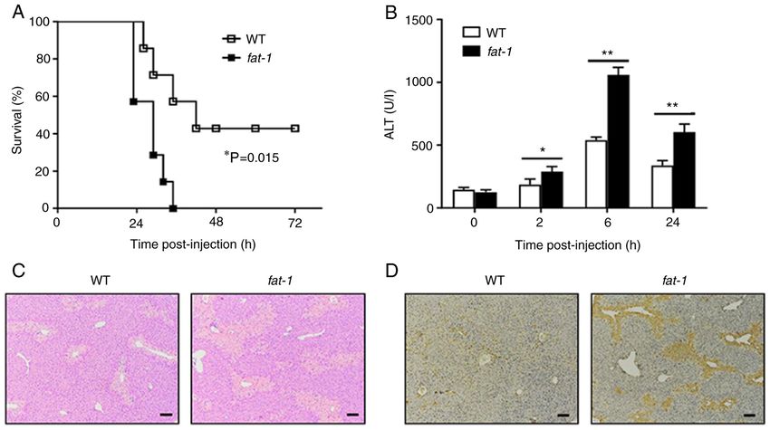
INTERNATIONAL JOURNAL OF MOLECULAR MEDICINE 48: 190, 2021                                          3

block non‑specific staining. The sections were then incubated       the corresponding HRP‑conjugated secondary antibody (goat
with an anti‑IL‑11 primary antibody (1:50; cat. no. A1902;          anti‑rabbit; 1:10,000; cat. no. S0001; Affinity Biosciences) at
ABclonal Biotech Co., Ltd.) at 4˚C overnight, followed by           room temperature for 1 h. The results were visualized with
incubation with the corresponding secondary antibody                ECL substrate (cat. no. 1705062; Bio‑Rad Laboratories, Inc.).
(HRP‑conjugated goat anti‑rabbit; 1:200; cat. no. S0001;            Each western blot analysis was performed three times.
Affinity Biosciences) at 37˚C for 1 h. Peroxidase activity was
detected (Nikon Intensilight DS‑Ri2) using DAB solution             RT‑qPCR analysis. Isolated hepatocytes were homogenized in
(Beyotime Institute of Biotechnology).                              1 ml TRIzol® (Thermo Fisher Scientific, Inc.), and total RNA
                                                                    was extracted based on the manufacturer's instruction. Then,
Serum alanine aminotransferase (ALT) assay and cytokine             1,000 ng RNA was synthesized into cDNA using TransScript®
assessment. The serum was collected at the indicated time           Fly First‑Stand cDNA Synthesis SuperMix (TransGen Biotech
points after APAP injection. Serum ALT activity was                 Co., Ltd.) with RNA removal reagent at 50˚C for 10 min
measured with a commercial kit (cat. no. C009‑2; Nanjing            and 85˚C for 5 sec. SYBR Green Real‑Time PCR Master
Jiancheng Bioengineering Institute), based on the manufac‑          Mix (cat. no. A46112; Applied Biosystems; Thermo Fisher
turer's instructions. Different cytokine levels in the serum        Scientific, Inc.) was applied for qPCR on an ABI Prism 7500
and cell culture supernatants were detected using commercial        Sequence Detection System (Applied Biosystems; Thermo
ELISA kits purchased from eBioscience (Thermo Fisher                Fisher Scientific, Inc.), according to the following thermo‑
Scientific, Inc.), including IL‑6 (cat. no. BMS603‑2), IL‑11        cycling conditions: Initial denaturation at 94˚C for 30 sec,
(cat. no. EMIL11), IL‑13 (cat. no. BMS6015) and IL‑22               followed by 40 cycles of denaturation at 94˚C for 5 sec, and
(cat. no. BMS6022).                                                 extension at 60˚C for 30 sec. The expression levels of the target
                                                                    genes were normalized to GAPDH gene expression and calcu‑
Flow cytometry. The Annexin V/PI apoptosis kit was obtained         lated using the 2‑∆∆Cq method (27). The primer sequences used
from Hangzhou Multi Sciences (Lianke) Biotech Co., Ltd., cells      in this experiment are shown in Table I.
were stained for 5 min in the dark at room temperature before
monitoring. Annexin V+/PI+ was identified as late apoptosis and     Statistical analysis. All experiments were independently
Annexin V+/PI‑ was identified as early apoptosis. Cells were        repeated in triplicate. Statistical analysis was performed
stained with JC‑1 dye (Nanjing KeyGen Biotech Co., Ltd.) for        using SPSS 12.0 (SPSS, Inc.). The data are presented as the
30 min at room temperature in the dark to evaluate mitochon‑        mean ± SD. An unpaired Student's t‑test was performed between
drial membrane potential. The cells were analyzed using the         WT and fat‑1 mice or ND control and n‑3 PUFA‑enriched diet
FACS LSRFortessa™ flow cytometer (BD Biosciences) with              mice at each time point. Comparison of the survival curves
BD FACSDiva™ software (version 8.0.1; BD Biosciences).              was analyzed with the Kaplan‑Meier method and log‑rank test.
                                                                    P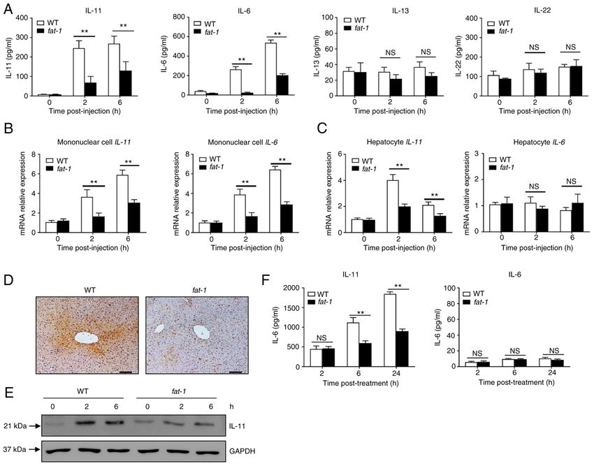
4                                                 LIU et al: n-3 PUFAs INHIBIT IL-11 PRODUCTION

Table I. Primers for the target genes.

Gene                                        Forward primer (5'→3')                                                   Reverse primer (5'→3')

Bcl‑2                               GGACTTGAAGTGCCATTGGT                                                    AGCCCCTCTGTGACA GCTTA
Bax                                 GGATGCGTCCACCAAGAAGC                                                    GGAGGAAGTCCAGTGTCCAGCC
IL‑11                               GTTTACAGCTCTTGATGTCTC                                                   GAGTCTTTAACAACAGCAGG
IL‑13                               CCTGGCTCTTGCTTGCCTT                                                     GGTCTTGTGTGATGTTGCTCA
Il‑22                               TGACGACCAGAACATCCAGA                                                    AATCGCCTTGATCTCTCCAC
IL‑6                                TACCACTTCACAAGTCGGAGGC                                                  CTGCAAGTGCATCATCGTTGTTC
Fra‑1                               TCATCTGGAGAGGTGGGTCC                                                    CTGCGGTTCTGACTCACTCG
GAPDH                               CGTCCCGTAGACAAAATGGT                                                    TTGATGGCAACAATCTCCAC

Fra‑1, Fos‑like 1.

Figure 1. N‑3 PUFAs exacerbate APAP‑induced liver damage. (A) APAP (600 mg/kg) was injected into WT and fat‑1 transgenic mice (n=7), and the survival
rate of mice was observed (P=0.015). (B‑D) WT and fat‑1 transgenic mice (n=5) were intraperitoneally injected with APAP (400 mg/kg). (B) Serum ALT
activities at different time points were measured. (C) Histological analysis of mouse livers was performed by hematoxylin and eosin staining 24 h post APAP
injection. Scale bar, 100 µm. (D) TUNEL staining was performed on paraffin‑embedded liver sections 24 h after APAP injection to mark apoptotic cells. Scale
bar, 100 µm. *P
INTERNATIONAL JOURNAL OF MOLECULAR MEDICINE 48: 190, 2021                                                        5

Figure 2. N‑3 PUFAs promote hepatocytes apoptosis through dephosphorylation of STAT3 signaling. (A‑D) Hepatocytes were isolated from WT and fat‑1
transgenic mice (n=5) at the indicated time after APAP (400 mg/kg) injection. (A) Protein levels of pSTAT3, STAT3 and GAPDH were determined by western
blotting analysis. (B) The mRNA levels of Bcl‑2 and Bax were measured by reverse transcription‑quantitative PCR and expressed as a ratio to GAPDH.
(C) The hepatic protein levels of Bcl‑2, Bax and GAPDH were evaluated at different time points by western blotting analysis. (D) Mitochondrial membrane
potential (ΔΨm) was detected in mouse hepatocytes by flow cytometry 6 h post‑APAP injection. (E‑G) Primary hepatocytes were isolated from WT and fat‑1
mice (n=5) and were stimulated with 20 mM APAP in vitro. In some cases, cells were pretreated with 10 µM Sc144 for 2 h before APAP stimulation. (E) The
levels of pSTAT3, STAT3 and GAPDH were detected by western blotting analysis. (F) Cellular apoptosis was measured by Annexin V‑PI staining 24 h
post‑APAP treatment. (G) The protein levels of Bcl‑2, Bax and GAPDH expression were determined by western blotting analysis 6 h post‑APAP treatment.
**
  P
6                                                  LIU et al: n-3 PUFAs INHIBIT IL-11 PRODUCTION

Figure 3. N‑3 PUFAs inhibit IL‑11 production in hepatocytes. (A‑D) WT and fat‑1 transgenic mice were injected with 400 mg/kg APAP. (A) The serum levels
of IL‑11, IL‑6, IL‑13 and IL‑22 were determined at the indicated time points. (B) Liver mononuclear cells and (C) hepatocytes were isolated from WT and fat‑1
transgenic mice (n=5) at the indicated time after APAP (400 mg/kg) injection. The levels of IL‑11 and IL‑6 expression were measured by reverse transcrip‑
tion‑quantitative PCR and expressed as a ratio to GAPDH. (D) The protein level of IL‑11 in liver tissues was detected by immunohistochemical staining 6 h
post‑APAP injection. Scale bar, 50 µm. (E) Protein levels of IL‑11 and GAPDH were determined by western blotting analysis. (F) Primary hepatocytes were
isolated from WT or fat‑1 transgenic mice and treated with 20 mM APAP for 24 h in vitro. The supernatants were collected, and the secreted levels of IL‑11
and IL‑6 were evaluated by ELISA. **P
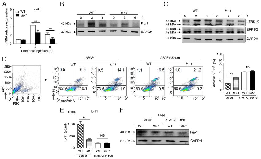
INTERNATIONAL JOURNAL OF MOLECULAR MEDICINE 48: 190, 2021                                                         7

Figure 4. N‑3 PUFAs inhibit IL‑11 production by downregulating ERK1/2 pathway. (A‑C) Hepatocytes were isolated from WT and fat‑1 transgenic mice (n=5)
at the indicated time after 400 mg/kg APAP administration. (A) The mRNA expression of Fra‑1 was evaluated by reverse transcription‑quantitative PCR and
expressed as a ratio to GAPDH. (B) The protein level of Fra‑1 was determined by western blotting analysis. (C) The protein levels of pERK, ERK and GAPDH
were detected by western blotting analysis. (D‑F) Primary hepatocytes were isolated from WT and fat‑1 mice (n=5) and stimulated with 20 mM APAP for 24 h
in vitro. In some cases, the cells were pretreated with 50 µM U0126 for 2 h before APAP stimulation. (D) Cellular apoptosis was measured by Annexin V‑PI
staining. (E) The culture supernatants were collected for evaluating IL‑11 production through ELISA. (F) The protein level of Fra‑1 in hepatocytes was
confirmed by immunoblotting analysis. **P
8                                                 LIU et al: n-3 PUFAs INHIBIT IL-11 PRODUCTION

Figure 5. Exogenous DHA aggravate APAP‑induced liver damage through dephosphorylated ERK‑mediated decreased IL‑11 production. (A‑D) An overdose
of APAP (400 mg/kg) was intraperitoneally injected into WT mice fed with ND or n‑3 PUFA‑enriched diet (n=5). (A) Histological analysis of mouse livers was
performed by hematoxylin & eosin staining. Scale bar, 100 µm. (B) Serum ALT activities at different time points were measured. (C) The serum level of IL‑11
was determined at the indicated time points post‑APAP injection. (D) The protein level of IL‑11 in the liver tissues was detected by immunohistochemical
staining 6 h post‑APAP injection. Scale bar, 50 µm. (E and F) Hepatocytes were isolated from WT mice fed with ND or n‑3 PUFA‑enriched diet (n=5) at
the indicated time post‑400 mg/kg APAP administration. (E) The levels of pERK, ERK, Fra‑1 and GAPDH were determined by western blotting analysis.
(F) The protein levels of pSTAT3, STAT3, Bcl‑2, BAX and GAPDH were detected by western blotting analysis. *P
INTERNATIONAL JOURNAL OF MOLECULAR MEDICINE 48: 190, 2021                                               9

of IL‑11 (3). Treatment with ERK inhibitors can efficiently         Authors' contributions
suppress TNF‑α‑induced IL‑11 induction in murine embry‑
onic fibroblasts (3). Fra‑1, a member of the activator protein 1    YL, ZC and DZ conceived and designed the present study.
family, was found to bind to the IL‑11 promoter before stimu‑       ZC and DZ provided administrative support. JZ and DZ were
lation, but recruitment of Fra‑1 was further enhanced after         responsible for the provision of study materials and patients.
oxidative stimulation (3,35). Gillies et al (36) found that Fra‑1   YL, JL and YC were responsible for the collection and
is expressed in proportion to the amplitude and duration of         assembly of data. YL, ZL, JZ, XL, ZC and DZ analyzed and
ERK1/2 activity. A previous study reported that the expres‑         interpreted the data. YL, ZC and DZ confirm the authenticity
sion of IL‑11 in hepatocytes is regulated by ERK1/2‑mediated        of all the raw data. All authors have read and approved the
Fra‑1 expression (37). n‑3 PUFAs appear to act via receptors        final manuscript.
or sensors, thereby controlling cellular signaling processes
that influence gene expression patterns (38). Several studies       Ethics approval and consent to participate
have reported that the anticancer properties of n‑3 PUFAs
involve the altered phosphorylation of ERK1/2 (39,40). It has       The authors are accountable for all aspects of the work in
been reported that n‑3 PUFAs induce apoptosis in human              ensuring that questions related to the accuracy or integrity
breast cancer cells through inhibition of ERK1/2 activa‑            of any part of the work are appropriately investigated and
tion (40). n‑3 PUFAs are able to suppress VEGF expression           resolved. All animal protocols in this study were approved by
in colon cancer cells by limiting ERK1/2 phosphorylation and        the Welfare and Ethical Committee for Experimental Animal
hypoxia‑inducible factor 1α overexpression (41). In addition,       Care of Southern Medical University (approval no. L2018234;
the anti‑inflammatory effect of n‑3 PUFAs on the endothelium        Guangzhou, China).
depends on the dephosphorylation of ERK1/2 (42). The present
study demonstrated that n‑3 PUFAs inhibited ERK1/2 phos‑            Patient consent for publication
phorylation and Fra‑1 expression in hepatocytes in response to
the APAP treatment. Notably, inhibition of ERK1/2 activation        Not applicable.
attenuated n‑3 PUFA‑induced hepatic IL‑11 production and
subsequent liver injury in APAP‑treated mice, suggesting that       Competing interests
n‑3 PUFAs affect IL‑11 production and APAP hepatotoxicity
via regulation of the ERK1/2 signaling pathway. However, the        The authors declare that they have no competing interests.
specific underlying mechanism by which n‑3 PUFAs regulate
ERK1/2 signaling needs further investigation. Fluorescence          References
labeled n‑3 PUFAs could be used to evaluate whether there
is a physical interaction between n‑3 PUFAs and ERK1/2               1. Cook SA and Schafer S: Hiding in plain sight: Interleukin‑11
                                                                        emerges as a master regulator of fibrosis, tissue integrity, and
protein in vitro, while IL‑11 receptor antagonist or IL‑11/IL‑11        stromal inflammation. Annu Rev Med 71: 263‑276, 2020.
receptor knockout by CRISPR/CRISPR‑associated protein                2. Dong J, Adami E, Chothani SP, Viswanathan S, Ng B, Lim WW,
9 could be used to further identify whether the effect of n‑3           Sing BK, Zhou J, Ko NSJ, Shekeran SG, et al: Autocrine IL11
                                                                        cis‑signaling in hepatocytes is an initiating nexus between lipo‑
PUFAs on APAP‑induced liver injury depends on the produc‑               toxicity and non‑alcoholic steatohepatitis. BioRxiv, 2020.
tion of IL‑11.                                                       3. Nishina T, Komazawa‑Sakon S, Yanaka S, Piao X, Zheng DM,
    In summary, the present results revealed that n‑3 PUFAs             Piao JH, Kojima Y, Yamashina S, Sano E, Putoczki T, et al:
                                                                        Interleukin‑11 links oxidative stress and compensatory
inhibit IL‑11 production and downstream STAT3 phosphory‑                proliferation. Sci Signal 5: ra5, 2012.
lation in hepatocytes, thereby aggravating APAP‑induced liver        4. Zhu M, Lu B, Cao Q, Wu Z, Xu Z, Li W, Yao X and Liu F: IL‑11
injury. The present study demonstrated that the ERK1/2‑Fra‑1            attenuates liver ischemia/reperfusion injury (IRI) through STAT3
                                                                        signaling pathway in mice. PLoS One 10: e0126296, 2015.
axis is important for the regulatory function of n‑3 PUFAs on        5. Bernal W, Auzinger G, Dhawan A and Wendon J: Acute liver
IL‑11 expression.                                                       failure. Lancet 376: 190‑201, 2010.
                                                                     6. Mühl H: STAT3, a key parameter of cytokine‑driven tissue
                                                                        protection during sterile inflammation‑the case of experimental
Acknowledgements                                                        acetaminophen (paracetamol)‑induced liver damage. Front
                                                                        Immunol 7: 163, 2016.
                                                                     7. Harrison DA: The Jak/STAT pathway. Cold Spring Harb Perspect
Not applicable.                                                         Biol 4: a011205, 2012.
                                                                     8. Nielsen M, Kaestel CG, Eriksen KW, Woetmann A, Stokkedal T,
Funding                                                                 Kaltoft K, Geisler C, Röpke C and Odum N: Inhibition of
                                                                        constitutively activated Stat3 correlates with altered Bcl‑2/Bax
                                                                        expression and induction of apoptosis in mycosis fungoides
This work was supported in part by the National Natural                 tumor cells. Leukemia 13: 735‑738, 1999.
Science Foundation of China (grant nos. 82071781, 81873872           9. Schmöcker C, Weylandt KH, Kahlke L, Wang J, Lobeck H,
                                                                        Tiegs G, Berg T and Kang JX: Omega‑3 fatty acids alleviate
and 81771771), and the Innovation Team of Chronic Kidney                chemically induced acute hepatitis by suppression of cytokines.
Disease with Integrated Traditional Chinese and Western                 Hepatology 45: 864‑869, 2007.
Medicine (grant no. 2019KCXTD014).                                  10. Li Y, Tang Y, Wang S, Zhou J, Zhou J, Lu X, Bai X, Wang XY,
                                                                        Chen Z and Zuo D: Endogenous n‑3 polyunsaturated fatty acids
                                                                        attenuate T cell‑mediated hepatitis via autophagy activation.
Availability of data and materials                                      Front Immunol 7: 350, 2016.
                                                                    11. Yang J, Fernández‑Galilea M, Martínez‑Fernández L, González-
                                                                        Muniesa P, Pérez‑Chávez A, Martínez JA and Moreno‑Aliaga MJ:
All data generated or analyzed during this study are included           Oxidative stress and non‑alcoholic fatty liver disease: Effects of
in this published article.                                              omega‑3 fatty acid supplementation. Nutrients 11: 872, 2019.
10 LIU et al: n-3 PUFAs INHIBIT IL-11 PRODUCTION
12. Kelley DS, Siegel D, Fedor DM, Adkins Y and Mackey BE: 29. Kroy DC, Beraza N, Tschaharganeh DF, Sander LE, Erschfeld S,
DHA supplementation decreases serum C‑reactive protein and Giebeler A, Liedtke C, Wasmuth HE, Trautwein C and
other markers of inflammation in hypertriglyceridemic men. Streetz KL: Lack of interleukin‑6/glycoprotein 130/signal trans‑
J Nutr 139: 495‑501, 2009. ducers and activators of transcription‑3 signaling in hepatocytes
13. Ma Y, Smith CE, Lai CQ, Irvin MR, Parnell LD, Lee YC, predisposes to liver steatosis and injury in mice. Hepatology 51:
Pham LD, Aslibekyan S, Claas SA, Tsai MY, et al: The effects 463‑473, 2010.
of omega‑3 polyunsaturated fatty acids and genetic variants on 30. Streetz KL, Wüstefeld T, Klein C, Kallen KJ, Tronche F,
methylation levels of the interleukin‑6 gene promoter. Mol Nutr Betz UA, Schütz G, Manns MP, Müller W and Trautwein C:
Food Res 60: 410‑419, 2016. Lack of gp130 expression in hepatocytes promotes liver injury.
14. Song EA, Lim JW and Kim H: Docosahexaenoic acid inhibits Gastroenterology 125: 532‑543, 2003.
IL‑6 expression via PPARγ‑mediated expression of catalase in 31. Garbers C and Scheller J: Interleukin‑6 and interleukin‑11: Same
cerulein‑stimulated pancreatic acinar cells. Int J Biochem Cell same but different. Biol Chem 394: 1145‑1161, 2013.
Biol 88: 60‑68, 2017. 32. Wang J, Homer RJ, Hong L, Cohn L, Lee CG, Jung S and
15. D'Eliseo D, Di Renzo L, Santoni A and Velotti F: Docosahexaenoic Elias JA: IL‑11 selectively inhibits aeroallergen‑induced pulmo‑
acid (DHA) promotes immunogenic apoptosis in human multiple nary eosinophilia and Th2 cytokine production. J Immunol 165:
myeloma cells, induces autophagy and inhibits STAT3 in both 2222‑2231, 2000.
tumor and dendritic cells. Genes Cancer 8: 426‑437, 2017. 33. Benigni F, Fantuzzi G, Sacco S, Sironi M, Pozzi P, Dinarello CA,
16. Allen MJ, Fan YY, Monk JM, Hou TY, Barhoumi R, McMurray DN Sipe JD, Poli V, Cappelletti M, Paonessa G, et al: Six different
and Chapkin RS: n‑3 PUFAs reduce T‑helper 17 cell differentiation cytokines that share GP130 as a receptor subunit, induce serum
by decreasing responsiveness to interleukin‑6 in isolated mouse amyloid A and potentiate the induction of interleukin‑6 and
splenic CD4+ T cells. J Nutr 144: 1306‑1313, 2014. the activation of the hypothalamus‑pituitary‑adrenal axis by
17. Tasaki S, Horiguchi A, Asano T, Ito K, Asano T and Asakura H: interleukin‑1. Blood 87: 1851‑1854, 1996.
Docosahexaenoic acid inhibits the phosphorylation of STAT3 34. Widjaja AA, Singh BK, Adami E, Viswanathan S, Dong JR,
and the growth and invasion of renal cancer cells. Exp Ther D'Agostino GA, Ng B, Lim WW, Tan J, Paleja BS, et al: Inhibiting
Med 14: 1146‑1152, 2017. interleukin 11 signaling reduces hepatocyte death and liver
18. O'Shea JJ, Schwartz DM, Villarino AV, Gadina M, McInnes IB and fibrosis, inflammation, and steatosis in mouse models of nonal‑
Laurence A: The JAK‑STAT pathway: Impact on human disease coholic steatohepatitis. Gastroenterology 157: 777‑792.e14, 2019.
and therapeutic intervention. Annu Rev Med 66: 311‑328, 2015. 35. Shin SY, Choi C, Lee HG, Lim Y and Lee YH: Transcriptional
19. Xiong A, Yu W, Liu Y, Sanders BG and Kline K: Elimination regulation of the interleukin‑11 gene by oncogenic Ras.
of ALDH+ breast tumor initiating cells by docosahexanoic acid Carcinogenesis 33: 2467‑2476, 2012.
and/or gamma tocotrienol through SHP‑1 inhibition of Stat3 36. Gillies TE, Pargett M, Minguet M, Davies AE and Albeck JG:
signaling. Mol Carcinog 55: 420‑430, 2016. Linear integration of ERK activity predominates over persis‑
20. Kang JX, Wang J, Wu L and Kang ZB: Transgenic mice: Fat‑1 tence detection in Fra‑1 regulation. Cell Syst 5: 549‑563.e5, 2017.
mice convert n‑6 to n‑3 fatty acids. Nature 427: 504, 2004. 37. Nishina T, Deguchi Y, Miura R, Yamazaki S, Shinkai Y,
21. Liu Y, Chen Y, Xie X, Yin A, Yin Y, Liu Y, Dong L, Zhu Z, Kojima Y, Okumura K, Kumagai Y and Nakano H: Critical
Zhou J, Zeng Q, et al: Gender difference on the effect of omega‑3 contribution of nuclear factor erythroid 2‑related factor 2
polyunsaturated fatty acids on acetaminophen‑induced acute (NRF2) to electrophile‑induced interleukin‑11 production. J Biol
liver failure. Oxid Med Cell Longev 2020: 8096847, 2020. Chem 292: 205‑216, 2017.
22. Henderson MW, Sparkenbaugh EM, Wang S, Ilich A, 38. Calder PC: Mechanisms of action of (n‑3) fatty acids. J Nutr 142:
Noubouossie DF, Mailer RK, Renné T, Flick MJ, Luyendyk JP, 592S‑599S, 2012.
Chen ZL, et al: Plasmin‑mediated cleavage of high molecular 39. Serini S and Calviello G: Modulation of Ras/ERK and phos‑
weight kininogen contributes to acetaminophen‑induced acute phoinositide signaling by long‑chain n‑3 PUFA in breast cancer
liver failure. Blood: Apr 7, 2021 (Epub ahead of print). and their potential complementary role in combination with
23. Saha B and Nandi D: Farnesyltransferase inhibitors reduce Ras targeted drugs. Nutrients 9: 185, 2017.
activation and ameliorate acetaminophen‑induced liver injury in 40. Sun H, Hu Y, Gu Z, Owens RT, Chen YQ and Edwards IJ:
mice. Hepatology 50: 1547‑1557, 2009. Omega‑3 fatty acids induce apoptosis in human breast cancer
24. Zhang C, Feng J, Du J, Zhuo Z, Yang S, Zhang W, Wang W, cells and mouse mammary tissue through syndecan‑1 inhibition
Zhang S, Iwakura Y, Meng G, et al: Macrophage‑derived IL‑1α of the MEK‑Erk pathway. Carcinogenesis 32: 1518‑1524, 2011.
promotes sterile inflammation in a mouse model of acetamino‑ 41. Calviello G, Di Nicuolo F, Gragnoli S, Piccioni E, Serini S,
phen hepatotoxicity. Cell Mol Immunol 15: 973‑982, 2018. Maggiano N, Tringali G, Navarra P, Ranelletti FO and Palozza P:
25. Torres S, Baulies A, Insausti‑Urkia N, Alarcón‑Vila C, n‑3 PUFAs reduce VEGF expression in human colon cancer
Fucho R, Solsona‑Vilarrasa E, Núñez S, Robles D, Ribas V, cells modulating the COX‑2/PGE2 induced ERK‑1 and ‑2 and
Wakefield L, et al: Endoplasmic reticulum stress‑induced upreg‑ HIF‑1alpha induction pathway. Carcinogenesis 25: 2303‑2310,
ulation of STARD1 promotes acetaminophen‑induced acute liver 2004.
failure. Gastroenterology 157: 552‑568, 2019. 42. Liu KL, Yang YC, Yao HT, Chia TW, Lu CY, Li CC, Tsai HJ,
26. Osawa Y, Uchinami H, Bielawski J, Schwabe RF, Hannun YA Lii CK and Chen HW: Docosahexaenoic acid inhibits inflam‑
and Brenner DA: Roles for C16‑ceramide and sphingosine mation via free fatty acid receptor FFA4, disruption of
1‑phosphate in regulating hepatocyte apoptosis in response to TAB2 interaction with TAK1/TAB1 and downregulation of
tumor necrosis factor‑alpha. J Biol Chem 280: 27879‑27887, ERK‑dependent Egr‑1 expression in EA.hy926 cells. Mol Nutr
2005. Food Res 60: 430‑443, 2016.
27. Pang Y, Liu Z, Han H, Wang B, Li W, Mao C and Liu S:
Peptide SMIM30 promotes HCC development by inducing
SRC/YES1 membrane anchoring and MAPK pathway activation.
This work is licensed under a Creative Commons
J Hepatol 73: 1155‑1169, 2020.
28. Lafdil F, Wang H, Park O, Zhang W, Moritoki Y, Yin S, Attribution-NonCommercial-NoDerivatives 4.0
Fu XY, Gershwin ME, Lian ZX and Gao B: Myeloid STAT3 International (CC BY-NC-ND 4.0) License.
inhibits T cell‑mediated hepatitis by regulating T helper 1
cytokine and interleukin‑17 production. Gastroenterology 137:
2125‑2135.e1‑e2, 2009.You can also read