Interphotoreceptor Retinol-Binding Protein Ameliorates Diabetes-Induced Retinal Dysfunction and Neurodegeneration Through Rhodopsin
←
→
Page content transcription
If your browser does not render page correctly, please read the page content below
788                                                                                                                         Diabetes Volume 70, March 2021

                Interphotoreceptor Retinol-Binding Protein Ameliorates
                Diabetes-Induced Retinal Dysfunction and
                Neurodegeneration Through Rhodopsin
                Jianglei Chen,1 Yan Shao,1,2 Temmy Sasore,1 Gennadiy Moiseyev,1 Kelu Zhou,1 Xiang Ma,1 Yanhong Du,1 and
                Jian-xing Ma1,3
                Diabetes 2021;70:788–799 | https://doi.org/10.2337/db20-0609

                Patients with diabetes often experience visual defects                              to a 2017 report by the International Diabetic Federation,
                before any retinal pathologies are detected. The molec-                             the worldwide prevalence of diabetes mellitus (DM) is 1 in
                ular mechanism for the visual defects in early diabetes                             11 adults (425 million), and the number is skyrocketing
                has not been elucidated. Our previous study reported                                accompanying the economic growth and increasing life
                that in early diabetic retinopathy (DR), rhodopsin levels                           span in developing countries.
                were reduced due to impaired 11-cis-retinal regenera-                                   DR was traditionally considered a microvascular com-
                tion. Interphotoreceptor retinol-binding protein (IRBP) is                          plication of diabetes, and pathological angiogenesis and
COMPLICATIONS

                a visual cycle protein and important for 11-cis-retinal                             vascular dysfunction were regarded as the priority in
                generation. IRBP levels are decreased in the vitreous and                           clinical treatment (2). Emerging evidence suggests that
                retina of DR patients and animal models. To determine the
                                                                                                    dysfunction of the retinal neurons and retinal neurode-
                role of IRBP downregulation in the visual defects in early DR,
                                                                                                    generation play important roles in the pathogenesis of DR
                we induced diabetes in transgenic mice overexpressing
                                                                                                    (3,4). Extensive reports have shown that oxidative stress,
                IRBP in the retina. IRBP overexpression prevented diabetes-
                                                                                                    characterized by overproduction of reactive oxygen species
                induced decline of retinal function. Furthermore, IRBP over-
                expression also prevented decreases of rhodopsin levels                             (ROS), plays an important role in DR (5). ROS-induced
                and 11-cis-retinal generation in diabetic mice. Diabetic                            chronic inflammation can lead to retinal degeneration and
                IRBP transgenic mice also showed ameliorated retinal                                vascular injury in patients with diabetes (6). Anti-vascular
                oxidative stress, inflammation, apoptosis, and retinal de-                           endothelial growth factor drugs and laser photocoagula-
                generation compared with diabetic wild-type mice. These                             tion have been used to ameliorate retinal vascular pathol-
                findings suggest that diabetes-induced IRBP downregula-                              ogies in DR, especially in proliferative DR (7). Nevertheless,
                tion impairs the regeneration of 11-cis-retinal and rhodop-                         there is no effective treatment for retinal degeneration in
                sin, leading to retinal dysfunction in early DR. Furthermore,                       DR.
                increased 11-cis-retinal–free opsin constitutively activates                            Photoreceptors are the most abundant cells in the retina
                the phototransduction pathway, leading to increased oxi-                            and the most metabolically active neurons in the central
                dative stress and retinal neurodegeneration. Therefore,                             nervous system (8). Under diabetic conditions, increased
                restored IRBP expression in the diabetic retina may confer                          oxidative stress caused by mitochondrial dysfunction in
                a protective effect against retinal degeneration in DR.                             photoreceptor cells subsequently results in retinal inflam-
                                                                                                    mation, leading to vascular dysfunction (9).
                                                                                                        Interphotoreceptor retinol-binding protein (IRBP) was
                Diabetic retinopathy (DR) is the leading cause of vision loss                       first identified as a visual cycle protein, which is a retinol-
                and disability in the working-age population (1). According                         binding protein secreted from the photoreceptors and is

                1Department of Physiology, University of Oklahoma Health Sciences Center,           This article contains supplementary material online at https://doi.org/10.2337/
                Oklahoma City, OK                                                                   figshare.13363265.
                2Tianjin Medical University Eye Hospital, Eye Institute & School of Optometry and
                                                                                                    © 2020 by the American Diabetes Association. Readers may use this article as
                Ophthalmology, Tianjin, China                                                       long as the work is properly cited, the use is educational and not for profit, and the
                3Harold Hamm Diabetes Center, University of Oklahoma Health Sciences Center,
                                                                                                    work is not altered. More information is available at https://www.diabetesjournals
                Oklahoma City, OK                                                                   .org/content/license.
                Corresponding author: Jian-xing Ma, jian-xing-ma@ouhsc.edu
                Received 9 June 2020 and accepted 10 December 2020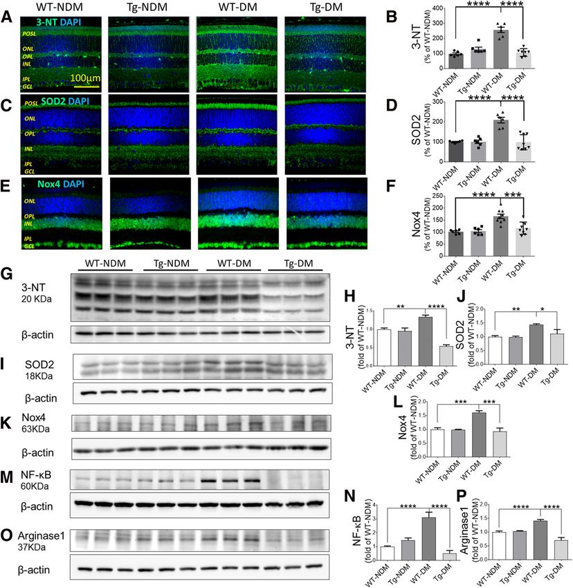
diabetes.diabetesjournals.org                                                                   Chen and Associates   789

present in the interphotoreceptor matrix of the retina         For cone function, a series of flashes with 600 cd $ s/m2
(10). IRBP transports retinoids between the photorecep-        intensity were applied to record the cone photoreceptor
tors and the retinal pigment epithelium (RPE) (10,11) and      response after 10-min light adaptation under a back-
plays an important role in the regeneration of 11-cis-         ground light of 50 cd/m2. ERG responses of both eyes
retinal, the chromophore of visual pigments, and is thus       were simultaneously recorded and analyzed.
essential for maintaining normal visual function. IRBP also
plays a role in retinal development (12,13) and shows          Optical Coherence Tomography
antioxidant activities via binding all-trans-retinal (14).     The retinal thickness was measured using a spectral do-
Recently, IRBP levels were reported to be decreased in the     main optical coherence tomography (SD-OCT) device
vitreous of DR patients, and IRBP conferred protective         (Bioptigen, Durham, NC). Images were captured with the
effects against DR in rodent models (15,16).                   rectangular scan at 1,000 A-scans per B scan, and 100 B-
   Rhodopsin regeneration and homeostasis are critical for     scans per frame. Total retinal thickness was recorded and
retinal function and health. The visual chromophore            averaged automatically using the InVivoVue diver software
11-cis-retinal binds to opsin to form rhodopsin in the dark,   (Bioptigen) by researchers blinded to the animal group
which locks the opsin in an inactive state. Unbound free       information.
opsin is known to be constitutively active, which can
exhaust photoreceptor cells and promote retinal degener-       Immunohistochemistry and TUNEL of Eyecup Sections
ation (17). In early diabetes, increased chromophore-free      Mouse eyes were carefully enucleated and fixed in David-
opsin due to insufficient 11-cis-retinal generation has been    son’s fixation solution for 48 h The eyecups were paraffin
suggested to have deleterious effects on photoreceptor         embedded, and 5-mm sections were collected. The sections
cells and accelerate DR progress (18).                         were immunostained with antibodies for 3-nitrotyrosine
   To identify the effect of diabetes-induced reduction of     (3-NT) (ab61392; Abcam), glial fibrillary acidic protein
IRBP on rhodopsin deficiency and retinal neurodegenera-         (GFAP) (G-3893; Sigma-Aldrich), superoxide dismutase
tion, we induced diabetes in transgenic mice overexpress-      2 (SOD2) (06-984; Millipore), NADPH oxidase 4 (NOX4)
ing IRBP in photoreceptors. We assessed the role of IRBP       (ab133303; Abcam), ionized calcium-binding adaptor mol-
expression in diabetes-induced retinal function decline,       ecule 1 (Iba1) (019-19741; Wako), arginase I (NBP1-
retinal degeneration, and decreased 11-cis-retinal and         32731; Novus), and inducible nitric oxide synthase (iNOS)
rhodopsin levels. The potential antioxidation and anti-        (NBP1-97471; Novus) following the manufacturers’ pro-
inflammation effects of IRBP on the diabetic retina were        tocols. TUNEL staining was performed using the in situ
further investigated.                                          cell-death detection kit, TMRed (Roche Diagnostics, Indi-
                                                               anapolis, IN) according to the manufacturer’s instruction.
RESEARCH DESIGN AND METHODS                                    Histological staining was performed on three sections per
Animals                                                        animal, with five to six animals in each group of the same
The animal experiments were conducted in compliance            condition. Observation and imaging were performed under
with the Association for Research in Vision and Ophthal-       the same setting for each experiment using Olympus
mology Statement for the Use of Animals in Ophthalmic          FluoView (Version 2.1a) (Olympus, Tokyo, Japan). The
and Vision Research. The protocol was approved by the          images were analyzed and semiquantified with ImageJ
University of Oklahoma Health Sciences Center Institu-         (National Institutes of Health) by researchers blinded to
tional Animal Care and Use Committee (Oklahoma City,           the specimen identifications.
OK).
   To induce diabetes, IRBP transgenic (IRBP-Tg) mice and      Retinoid Profile Analysis
wild-type (WT) littermates (12 weeks of age) received daily    The retinoid profile was analyzed using high-performance
intraperitoneal injections of streptozotocin (STZ; Sigma-      liquid chromatography (HPLC), as described previously
Aldrich) (55 mg/kg in 10 mmol/L of citrate buffer, pH 4.5)     (18). Briefly, mice were sacrificed under dim red light after
for five consecutive days. Blood glucose levels were mea-       16-h dark adaptation. The eyecup was homogenized in-
sured 1 week after the last STZ injection and monthly          dividually in lysis buffer (10 mmol/L NH2OH, 50% etha-
thereafter. Animals with blood glucose levels .350 mg/dL       nol, 50% 2-[N-morpholino] ethanesulfonic acid, pH 6.5),
were defined as diabetic animals.                               and retinoids were extracted with hexane. The retinoids
                                                               were dried under argon gas, resuspended in 200 mL of
Electroretinography Recording                                  mobile phase (11.2% ethyl acetate, 2.0% dioxane, 1.4%
Scotopic electroretinography (ERG) and photopic ERG            octanol, 85.4% hexane), and injected into HPLC (515 HPLC
were both recorded monthly using the Diagnosys Espion          pump; Waters, Milford, MA) for separation using a normal-
Visual Electrophysiology System (Lowell, MA), as described     phase column (LiChrosphere Si-60; 4.6 3 250 mm; 5 mm)
previously (18). Briefly, after dark adaptation for 16 h,       (Alltech, Deerfield, IL) with isocratic elution (1 mL/min).
mice were anesthetized with their pupils dilated. A series     Each retinoid isomer was quantified from the area of its
of flashes with intensities 0.002–600 candela (cd) $ s/m2       corresponding absorption peak based on synthetic retinoid
were applied to induce rod response under dark adaptation.     standards for calibration.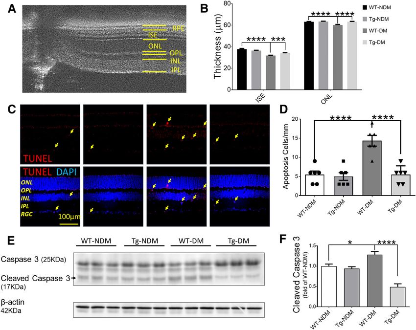
790    IRBP in Diabetic Retinopathy                                                                   Diabetes Volume 70, March 2021

Rhodopsin Content Measurement                                         Protein bands were visualized, examined, and quantified
After 16-h dark adaptation, each retina was isolated in-              by densitometry using ChemiDoc Imaging System and
dividually and homogenized in 150 mL of 13 PBS contain-               Image Lab software (Bio-Rad, Hercules, CA).
ing 1% (w/v) dodecyl maltoside. The retinal homogenates
were centrifuged at 70,000g for 1 h. The supernatant was              Statistical Analysis
transferred to clean spectrophotometer cuvettes and scanned           Data were entered and extracted from PRISM 7 (GraphPad
from 250 to 700 nm of wavelength using a DU800 spec-                  Software, San Diego, CA). All of the experiments were
trophotometer (Beckman Coulter, Brea, CA). The difference             performed at least three times. Quantitative data are
of absorbance spectra between prebleached and postbleached            presented as mean 6 SEM and were analyzed by ANOVA.
samples at 500 nm was used to calculate rhodopsin content             A P value of ,0.05 was considered statistically significant.
using a molar extinction coefficient of 42,000 mol/L21 $ cm21.
The data were normalized to the total volume of superna-              Data and Resource Availability
tant and presented as rhodopsin content per retina.                   The data sets and source data generated during and/or
                                                                      analyzed during the current study are available from the
Western Blot Analysis                                                 corresponding author upon reasonable request.
Eyecups were homogenized in the radioimmunoprecipita-
tion assay buffer containing a protease inhibitor cocktail            RESULTS
(Thermo Fisher, Waltham, MA). Total protein concentra-                IRBP-Tg mice on a C57BL/6J background overexpressing
tions were measured by bicinchoninic acid protein assay               IRBP under the rhodopsin promoter were generated through
(Thermo Fisher). Western blotting used antibodies for                 a contracted service with Cyagen Biosciences (Santa Clara,
IRBP (14352; Proteintech), caspase 3 (9661; Cell Signaling),          CA) (Supplementary Fig. 1A).
3-NT (10006966; Cayman), SOD2 (06-984; Millipore),                        Diabetes was induced in the IRBP-Tg mice and their WT
NOX4 (ab133303; Abcam), nuclear factor (NF)-kB (P65)                  littermates using injections of STZ. Blood glucose concen-
(ab16502; Abcam), and arginase I (NBP1-32731; Novus)                  trations and body weights of the mice were monitored and
and followed the conditions suggested by the manufacturers.           recorded, before and after the onset of STZ-induced

Figure 1—Overexpression of IRBP prevented retinal function decline in diabetic mice. Retinal function was evaluated using ERG in diabetic
WT (WT-DM), diabetic IRBP-Tg (IRBP-Tg-DM), and their nondiabetic controls (NDM). Amplitudes of scotopic a-wave (A), scotopic b-wave
(B), photopic a-wave (C), and photopic b-wave (D) at 2, 3, and 4 months after diabetes onset. *WT-NDM vs. WT-DM, 1IRBP-Tg-NDM vs.
IRBP-Tg-DM, ^WT-DM vs. IRBP-Tg-DM. Data are expressed as mean 6 SEM (N $ 8). */1/^P , 0.05, **/11/^^P , 0.01, ***/111/^^^P ,
0.001, and ****/1111/^^^^P , 0.0001.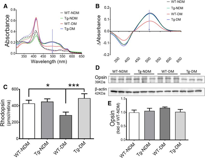
diabetes.diabetesjournals.org Chen and Associates 791
diabetes, together with nondiabetic controls (NDM). Di- To determine whether IRBP downregulation in diabetes is
abetic groups (DM) showed lower body weights and higher responsible for the deficiency in 11-cis-retinal regeneration
blood glucose levels compared with age-matched NDM and decrease in rhodopsin, we measured rhodopsin levels at
mice, with no significant difference between the IRBP-Tg 3 months after diabetes onset in IRBP-Tg and WT mice.
and WT mice in both DM and NDM subgroups (Supple- Rhodopsin was extracted after dark adaptation, and rho-
mentary Fig. 2). Western blotting using an IRBP antibody dopsin absorbance spectra were recorded before and after
showed that in the retinas of heterozygous IRBP-Tg mice, visual pigment bleach by light. The differential absorption
IRBP levels increased ;20% compared with that of WT spectra were used to quantify rhodopsin levels. As expected,
littermates. As expected, IRBP levels were decreased by rhodopsin levels were decreased in the retinas of WT-DM
;25% in the retinas of WT-DM mice compared with those mice compared with those of WT-NDM mice. Overexpres-
of WT-NDM mice. Retinal IRBP levels of IRBP-Tg-DM mice sion of IRBP prevented the decline of rhodopsin content
were similar to those of the IRBP-Tg-NDM mice. As a re- in IRBP-Tg-DM mice (Fig. 3A–C). Western blot analysis
sult, the retinal IRBP level in IRBP-Tg-DM mice was ;40% showed similar opsin levels in all of the groups (Fig. 3D and
higher than that of WT-DM mice (Supplementary Fig. 1B E), confirming the previous observation (18) that diabetes
and C). This difference of IRBP levels between IRBP-Tg and did not affect opsin expression or stability and suggesting
WT mice under diabetic conditions were comparable to that the decreased rhodopsin was likely caused by deficient
those between patients with DR and without DR (16). chromophore regeneration. Taken together, these results
suggest that overexpression of IRBP alleviates diabetes-
IRBP Overexpression Prevents Diabetes-Induced induced deficiency of visual pigment formation.
Retinal Dysfunction
Electrophysiological analysis was performed before diabe- IRBP Overexpression Normalizes 11-cis-Retinal Levels
tes onset and at the 2-, 3-, and 4-month time points after in Diabetic Mice
diabetes onset. WT-DM mice showed continuous decline It has been well established that IRBP is important for
of scotopic a-wave and b-wave amplitudes at 2, 3, and 11-cis-retinal transport and subsequent rhodopsin regen-
4 months compared with age-matched WT-NDM mice. eration (12,19). We also reported previously that 11-cis-
Under the same diabetic conditions, the decline of ERG retinal levels were decreased in diabetic rats compared
amplitudes was prevented completely (a-wave, Fig. 1A) or with nondiabetic controls (18). In this study, 11-cis-retinal
partially (b-wave, Fig. 1B) at 2 months after diabetic onset concentrations in the dark-adapted eyes of diabetic and
in IRBP-Tg-DM mice. Similarly, the decline of photopic nondiabetic IRBP-Tg and WT mice were measured by HPLC.
a-wave and b-wave was observed after 3 months of di- The level of 11-cis-retinal in the eye of WT-DM mice was
abetes in WT-DM mice, whereas the declines were partially significantly lower than that of WT-NDM, consistent with
prevented in IRBP-Tg-DM mice (Fig. 1C and D). our previous finding in diabetic rats (18). Overexpression of
IRBP prevented the diabetes-induced decline of 11-cis-
IRBP Overexpression Delays Neurodegeneration in the retinal levels in IRBP-Tg-DM mice (Fig. 4A–E), and as
Retina of Diabetic Mice a result, 11-cis-retinal levels in IRBP-Tg-DM mice were
OCT measurement demonstrated that the thickness of the similar to those of WT-NDM mice, supporting the notion
photoreceptor layers (inner segment ellipsoid and outer that the increased rhodopsin levels are a result of restored
nuclear layer) was significantly decreased in WT-DM mice 11-cis-retinal regeneration by IRBP overexpression in the
compared with that of WT-NDM mice. In IRBP-Tg-DM retina of diabetic IRBP-Tg mice.
mice, however, IRBP overexpression in the retina pre-
vented the decrease of photoreceptor layer thickness (Fig. IRBP Protects the Retina From Oxidative Stress in
2A and B). Consistently, TUNEL showed increased apo- Diabetic Mice
ptosis in all of the retinal nuclear layers in WT-DM mice, Because chromophore-free opsin is known to constitutively
which was attenuated in IRBP-Tg-DM mice (Fig. 2C and activate the phototransduction pathway in photoreceptor
D). Moreover, Western blotting showed cleaved caspase cells, resulting in excess oxidative stress, we measured 3-NT
3 levels were increased in the retina of WT-DM mice while as a marker of oxidative stress in the retinas of diabetic
significantly decreased in the retina of IRBP-Tg-DM mice mice. As shown by immunostaining, the 3-NT signal was
(Fig. 2E and F). Taken together, these observations significantly more intense in the WT-DM retina compared
suggested a critical role of IRBP in the prevention of with that in WT-NDM mice. Diabetic IRBP-Tg mice showed
diabetes-induced retinal neurodegeneration and retinal lower 3-NT staining compared with diabetic WT mice with
cell apoptosis. the same diabetic duration and similar glucose levels (Fig.
5A and B). Consistently, SOD2 was upregulated in the
IRBP Overexpression Improves Rhodopsin retinas of WT-DM mice, especially in the photoreceptor
Regeneration in Diabetic Mice outer segment region, but not in the retinas of IRBP-
Our previous study demonstrated that deficient regener- Tg-DM mice, supporting that IRBP expression alleviated
ation of the visual pigment rhodopsin in diabetic rats oxidative stress in the retina (Fig. 5C and D). Moreover, the
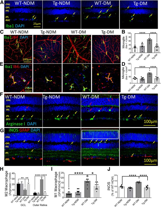
contributed to the decreased ERG response in early DR (18). increased immunostaining signal of NOX4, a major source792 IRBP in Diabetic Retinopathy Diabetes Volume 70, March 2021 Figure 2—Overexpression of IRBP-attenuated retinal neurodegeneration and apoptosis in diabetic mice. A: A representative mouse retina OCT scan. B: The outer nuclear layer (ONL) and inner segment ellipsoid thicknesses were measured using OCT and compared among diabetic WT (WT-DM) and diabetic IRBP-Tg (IRBP-Tg-DM) mice at 3 months after diabetes onset, as well as their nondiabetic controls (NDM). C: Retinal sections were collected 3 months after diabetes onset for TUNEL staining (red), with nuclei counterstained by DAPI (blue). The apoptotic cells are indicated by arrows. D: Apoptotic cells were quantified and expressed as cells per mm of retinal length and compared. A representative Western blot of caspase 3 and cleaved caspase 3 in the retinas (E), and analysis by densitometry and normalization by b-actin (F). Data are expressed as mean 6 SEM (N $ 6). **P , 0.01, ***P , 0.001, and ****P , 0.0001. of oxidative stress, was observed in the retinas of WT-DM IRBP Protects the Retina Against Diabetes-Induced mice but not in those of IRBP-Tg-DM mice (Fig. 5E and F). Retinal Inflammation Western blot analysis further confirmed that IRBP ex- DM is known to induce chronic inflammation in the retina, pression attenuated the upregulation of 3-NT, NOX4, leading to activation of microglia and leukocyte infiltration and SOD2 in the retinas of diabetic mice, suggesting an in the retina. Indeed, in the retina of diabetic WT mice, antioxidant effect of IRBP in diabetic retinas (Fig. 5G–P). activated microglial cells, labeled by Iba1, were increased More interestingly, the activation of redox-sensitive and migrated from the retinal ganglion cell (RGC) layer NF-kB, a bridge connecting oxidative stress and inflam- toward the photoreceptor layer. This increase and migra- mation in DR, was also ameliorated by overexpression of tion were not observed in the retinas of diabetic IRBP-Tg IRBP (Fig. 5M and N). Arginase I, a macrophage marker mice (Fig. 6A and B). Immunostaining of Iba1 on the flat- and mediator of DR (20), was increased in the retina of mounted retina showed increased numbers of activated WT-DM mice and was normalized by IRBP overexpres- and deformed microglia in the WT-DM mice, which was sion in IRBP-Tg-DM mice (Fig. 5O and P). These obser- prevented in IRBP-Tg-DM mice (Fig. 6C–E). Immunostain- vations indicated that IRBP overexpression protected the ing of arginase I, a marker of M2 macrophages, showed retina from diabetes-induced oxidative stress and resul- a decreased number of innate M2 in the RGC layer and an tant inflammation. increased number of activated M2 in the outer retinal
diabetes.diabetesjournals.org                                                                                Chen and Associates       793

Figure 3—Overexpression of IRBP prevented the decline of rhodopsin levels in diabetic mice. After 3 months of STZ-induced diabetes, the
eyeballs of IRBP-Tg mice and WT littermates were collected after 16-h dark adaptation. Rhodopsin levels were measured by absorption
spectrophotometry before and after photobleaching. A: Representative spectra of rhodopsin absorbance recorded by spectrophotometry.
An absorption peak with maximum absorption at ;500 nm is indicated by an arrow. The spectra before rhodopsin bleaching are shown in
solid lines and after bleaching in dashed lines. B: Photobleaching difference (DAbsorbance) spectra acquired using the difference between
the spectra before photobleaching and the spectra after photobleaching. C: Rhodopsin levels were calculated as pmol/eye and averaged
within each group. D: Rod opsin in the retina was measured by Western blot analysis using an anti-rhodopsin (1D4) antibody. E: Opsin levels
were analyzed by densitometry and normalized by b-actin levels. Data are expressed as mean 6 SEM (N 5 6). *P , 0.05 and ***P , 0.001.

layers of WT-DM mice compared with WT-NDM mice.                        neurodegeneration also play important roles in DR path-
Overexpression of IRBP attenuated the migration of M2                  ogenesis (22). It is well known that patients with diabetes
macrophages induced by diabetes (Fig. 6F and H). On the                often experience visual defects, such as delayed dark
other hand, immunostaining of M1 macrophages with                      adaptation in the early stages of DR, before any structural
marker iNOS (Fig. 6G) showed augmented iNOS expres-                    changes in the retina can be detected (23). Consistently,
sion in the photoreceptor layer (Fig. 6G and J, white box)             retinal function can be impaired before any sign of vascular
and an increase of M1 macrophages that had colocalized                 changes (24). However, the exact underlying molecular
with GFAP (21) in the retinas of WT-DM mice (Fig. 6G and               mechanisms are not yet fully understood. Our previous
I, arrows), which was prevented in the retinas of IRBP-                study reported that visual pigment formation was deficient
Tg-DM mice. All of these results suggested that IRBP                   in a diabetic model due to the impaired regeneration of
overexpression alleviated diabetes-induced chronic inflam-              11-cis-retinal, the chromophore for visual pigments (18),
mation in the retina.                                                  suggesting that the disturbance of the visual cycle or
                                                                       retinoid metabolism may contribute to DR. To identify the
DISCUSSION                                                             molecule that is responsible for this visual cycle defect in
DR was first defined as a microvascular disease, and later               diabetes, the current study investigated the role of IRBP,
studies demonstrated that retinal inflammation and                      which is decreased in the diabetic retina. Our results
794 IRBP in Diabetic Retinopathy Diabetes Volume 70, March 2021 Figure 4—Overexpression of IRBP recovered 11-cis-retinal levels in diabetic mice. At 3 months after diabetes onset, eyeballs were collected in the dark after 16-h dark adaption, and retinoids were extracted and analyzed by HPLC. Representative HPLC chromatographs at absorbance of 360 nm are shown for WT-NDM (A), IRBP-Tg-NDM (B), WT-DM (C), and IRBP-Tg-DM (D). Peaks were identified according to retinoid standards as: 1) retinyl esters; 2) syn-11-cis-retinal oxime (marked by *); 3) syn-all-trans-retinal oxime; 4) anti–all-trans-retinal oxime. E: We quantified 11-cis-retinal by measuring the peak areas of the corresponding 11-cis-retinal oximes (mean 6 SEM; N 5 5 to 6). **P , 0.01. demonstrate that diabetes-induced IRBP downregulation phototransduction pathway and exhausts photoreceptors. may be responsible, at least in part, for the impaired The deficient visual pigment formation and exhausted regeneration of 11-cis-retinal, leading to reduced forma- photoreceptors can contribute to the declined ERG re- tion of visual pigments. As a result, the increased free sponse and retinal degeneration in diabetes. These find- opsin (without 11-cis-retinal) constitutively activates the ings revealed the molecular basis for the functional defects
diabetes.diabetesjournals.org                                                                                   Chen and Associates        795

Figure 5—Overexpression of IRBP alleviated diabetes-induced oxidative stress in the retina. At 3 months after diabetes onset, 5-mm cross-
sections of eyeballs were collected from diabetic WT (WT-DM) and diabetic IRBP-Tg (Tg-DM), with age-matched nondiabetic WT (WT-NDM)
and nondiabetic IRBP-Tg (IRBP-Tg-NDM) mice as controls. A–F: Immunostaining of 3-NT (A), SOD2 (C), and Nox4 (E) (green), with the nuclei
counterstained by DAPI (blue). GCL, ganglion cell layer; INL, inner nuclear layer; IPL, inner plexiform layer; OPL, outer plexiform layer; ONL,
outer nuclear layer; POSL photoreceptor outer segment layer. The expression levels of 3-NT (B), SOD2 (D), and Nox4 (F) were semiquantified
by the fluorescence intensity. G–P: Representative Western blots of 3-NT, SOD2, Nox4, NF-kB, and arginase I in the retinas, and protein
levels analyzed by densitometry and normalized by b-actin levels (mean 6 SEM, N $ 6). *P , 0.05, **P , 0.01, ***P , 0.001, and ****P ,
0.0001.

in early DR and shed light on the development of a new                   results from the current study further confirmed that
therapeutic strategy for treating early DR and preventing                11-cis-retinal and rhodopsin levels were decreased in di-
its progression (25).                                                    abetic WT mice, supporting the notion that an impaired
    Our previous study reported that rhodopsin levels were               visual cycle or retinoid metabolism is associated with the
decreased in the retinas of diabetic rats, which was attrib-             functional decline of the retina in diabetes. To identify the
uted to the deficiency of 11-cis-retinal generation (18). The             molecular mechanism responsible for the visual cycle
796 IRBP in Diabetic Retinopathy Diabetes Volume 70, March 2021 Figure 6—Overexpression of IRBP prevented diabetes-induced microglia activation in the retinas. After 3 months of diabetes induced by STZ, eyeball sections from diabetic WT (WT-DM), diabetic IRBP-Tg (IRBP-Tg-DM), and their nondiabetic controls (NDM) were collected for immunohistochemistry. A: Immunostaining of Iba-1 (green) with the nuclei counterstained by DAPI (blue). B: The activated microglia (Iba-11, indicated by arrows) were counted and compared. C: Coimmunostaining of Iba-1 (green) and isolectin B4 (IB4, red) on the flat-mounted retinas with the nuclei counterstained with DAPI (blue). D: Activated microglial cells (Iba-11, indicated by arrows) were counted in the retinal flat-mounts and compared. E: The morphology and number changes of activated microglia in the flat-mounted retinas of diabetic IRBP-Tg and WT mice (green). F: Immunostaining of arginase-I (green) with the nuclei counterstained with DAPI (blue). The microglial cells in the RGC layer are indicated by yellow arrows, and the migrating microglial cells are indicated by red arrows. GCL, ganglion cell layer; INL inner nuclear layer; IPL, inner plexiform layer; ONL, outer nuclear layer; OPL, outer plexiform layer. G: Immunostaining of iNOS (green) and GFAP (red) with the nuclei counterstained with DAPI (blue). The activated M1 macrophages (GFAP/iNOS1) are indicated by yellow arrows. The augmented
diabetes.diabetesjournals.org                                                                              Chen and Associates       797

deficiency in diabetes, the current study investigated the             the photoreceptor cells from oxidative stress (14) and light-
role of IRBP. IRBP is a 135-kDa secreted protein that is              induced injury (35), also pointing to the protective role of
almost exclusively produced by photoreceptor cells and is             IRBP against oxidative stress.
a major soluble protein in the interphotoreceptor matrix                  Western blot analysis and immunostaining both showed
(26). IRBP binds, stabilizes, and transports retinoids (27)           SOD2 accumulation in the photoreceptor outer segment
and facilitates the regeneration of 11-cis-retinal through            region of WT-DM mice, indicating the activation of retinal
the visual cycle, and thus, rhodopsin formation (27–29).              intrinsic antioxidant defensive machinery and implying
To investigate the direct role of IRBP in visual cycle                a local elevation of oxidative stress. As a scavenging en-
homeostasis in DR, we induced diabetes in IRBP-Tg mice in             zyme of superoxide radicals, the activity of SOD2 has been
which the IRBP expression is under the control of a rho-              reported to be decreased in the retinal vasculature in DR
dopsin promoter and is thus not downregulated by di-                  (36). The upregulated levels of SOD2 in photoreceptors
abetes. Our data showed that restored IRBP expression in              may represent a compensatory response to counter the
the retina prevented the diabetes-induced decline of ERG              increased oxidative stress in DR.
response and partially restored both rhodopsin formation                  DR is also a chronic inflammatory complication of
and 11-cis-retinal regeneration. These findings suggest                diabetes (37,38) known to be driven by oxidative stress (5).
that diabetes-induced IRBP downregulation is responsible,             In the retina, oxidative stress activates redox-sensitive
at least in part, for the deficient rhodopsin formation and            NF-kB (39), which is a key regulator of inflammatory
declined ERG a-wave in early DR. This notion is supported             response (40). However, the increased levels of NF-kB and
by the observation that IRBP transgenic expression showed             arginase I in diabetic retinas were attenuated by IRBP
a more prominent protection against diabetes-induced                  overexpression in diabetic IRBP-Tg mice (Fig. 5), further
a-wave decline, because a-wave is primarily from the photo-           demonstrating the potential protective role of IRBP
receptor response. These findings suggest that diabetes-               against oxidative stress and inflammation in the diabetic
induced IRBP downregulation prominently impairs                       retina.
photoreceptor function through reduced regeneration of                    Through NF-kB activation, oxidative stress activated
chromophore and rhodopsin.                                            retinal microglia, the resident monocytes in the retina,
    On the other hand, insufficient 11-cis-retinal regener-            resulting in neurotoxicity, tissue damage, and retinal
ation in DR results in an increased level of chromophore-free         angiogenesis (41,42). Activation of retinal microglia was
opsin, which constitutively activates the phototransduction           characterized by microglial cell proliferation, migration
pathway in the photoreceptor, exhausts photoreceptor                  (from ganglion cell layer, inner nuclear layer, and inner
cells, and increases ROS production, further leading to               plexiform layer to inner nuclear layer and outer nuclear
retinal degeneration (17). However, the precise mecha-                layer), and morphological changes (from ramified to
nism by which the visual cycle and its components                     amoeboid) in the diabetic retina (Fig. 6), all of which
modulate retinal function and integrity remains controver-            were inhibited by overexpression of IRBP in IRBP-Tg-DM
sial (30,31). It is well known that DR development is closely         mice. The microglia under diabetic inflammatory stress in
associated with ROS overproduction and oxidative stress               the retina developed not only classic proinflammatory
(5,32). The eye is one of the most vulnerable targets of ROS          M1 microglia/macrophages but also anti-inflammatory
attack, especially under diabetic conditions (6,32). Increased        M2 microglia/macrophages. The M2 microglia were acti-
oxidative stress in the diabetic retina can result in degen-          vated and migrated from the RGC layer toward the outer
eration of photoreceptors (33), Müller cells (34), and other          retina region to address anti-inflammatory requirements
retinal neurons (5,6). Our results of apoptosis and OCT               at the early stage of DR; however, the M2 microglia de-
analyses confirmed the increased retinal cell death in                 creased as the disease progressed (43).
diabetic mice, suggesting that the increased oxidative                    In addition to the well-established function of binding
stress resulted in retinal degeneration, leading to ERG               and transporting 11-cis/trans retinols between photore-
b-wave decline. Our data also demonstrated that IRBP                  ceptors and RPE, IRBP is also important in retinal de-
overexpression prevented decline of the b-wave ampli-                 velopment (13,44), and loss of IRBP leads to retinal
tudes in diabetic mice, reflecting the protective effects of           degeneration (45–48). In addition, it has been shown that
IRBP on the functional integrity of the inner retina, which           purified IRBP possesses thiol-dependent antioxidant ac-
encouraged us to further investigate the other potential              tivity (49). Recent reports demonstrated that decreased
roles of IRBP. Indeed, our data showed that the diabetes-             IRBP levels in the vitreous of DR patients were associated
induced oxidative stress was ameliorated in diabetic                  with the severity of DR (15,16). All of these reports
IRBP-Tg mice (Fig. 5). Studies from other groups using                demonstrated that IRBP, a photoreceptor protein, exerts
different rodent models demonstrated that IRBP protected              its protective role spanning the entire retina.

expression of iNOS in the outer nuclear layer of WT-DM retinas is highlighted by the box. Numbers of residing and migrating microglia (H)
and activated M1 macrophages (I) in the retinas of WT-DM and IRBP-Tg-DM mice. J: The expression level of iNOS was semiquantified by
the fluorescence intensity. Data are expressed as mean 6 SEM (N $ 6). **P , 0.01 and ****P , 0.0001.
798 IRBP in Diabetic Retinopathy Diabetes Volume 70, March 2021
The mechanism by which IRBP protects the retina from 2. Barrett EJ, Liu Z, Khamaisi M, et al. Diabetic microvascular disease: an En-
diabetes-induced oxidative stresses and inflammatory re- docrine Society scientific statement. J Clin Endocrinol Metab 2017;102:4343–410
sponse warrants further investigation. Nevertheless, the 3. Rossino MG, Dal Monte M, Casini G. Relationships between neuro-
degeneration and vascular damage in diabetic retinopathy. Front Neurosci
current study provided clear evidence in support of the
2019;13:1172
notion that IRBP attenuates diabetes-induced oxidative
4. Lynch SK, Abràmoff MD. Diabetic retinopathy is a neurodegenerative dis-
and inflammatory stresses, at least partially, via mainte- order. Vision Res 2017;139:101–107
nance of homeostasis in visual pigment regeneration. 5. Kowluru RA, Chan PS. Oxidative stress and diabetic retinopathy. Exp Diabetes
As one of the major soluble proteins of the interpho- Res 2007;2007:43603
toreceptor matrix, IRBP plays an important role in main- 6. Calderon GD, Juarez OH, Hernandez GE, Punzo SM, De la Cruz ZD. Oxidative
taining a healthy environment for the photoreceptors, stress and diabetic retinopathy: development and treatment. Eye (Lond) 2017;31:
retina, and RPE (28,50). In addition to its retinoids-binding 1122–1130
capacity, IRBP also contains a potential fatty acid-binding 7. Stewart MW. Treatment of diabetic retinopathy: recent advances and un-
pocket (51), suggesting a probable role in lipid transport and resolved challenges. World J Diabetes 2016;7:333–341
metabolism. Recently, it was reported that IRBP inhibited 8. Kern TS. Do photoreceptor cells cause the development of retinal vascular
disease? Vision Res 2017;139:65–71
glucose transport in retinal cells through binding GLUT1
9. Liu H, Tang J, Du Y, et al. Photoreceptor cells influence retinal vascular
and downregulating the vascular endothelial growth factor
degeneration in mouse models of retinal degeneration and diabetes. Invest
pathway (16). These previous studies suggest that IRBP may Ophthalmol Vis Sci 2016;57:4272–4281
confer protective effects through multiple mechanisms, 10. Pfeffer B, Wiggert B, Lee L, Zonnenberg B, Newsome D, Chader G. The
such as lipid oxidation, glucose metabolism, and retinoid presence of a soluble interphotoreceptor retinol-binding protein (IRBP) in the retinal
metabolism. Considering that the retina is a complex tissue interphotoreceptor space. J Cell Physiol 1983;117:333–341
with intertwined cross talk among its many different 11. Okajima TI, Pepperberg DR, Ripps H, Wiggert B, Chader GJ. Interphotor-
resident cell types, in concordance with the multifactorial eceptor retinoid-binding protein: role in delivery of retinol to the pigment epi-
characteristic of DR pathogenesis, the multiple functions thelium. Exp Eye Res 1989;49:629–644
of IRBP warrant further investigation. 12. Okajima TI, Pepperberg DR, Ripps H, Wiggert B, Chader GJ. Interphotor-
Taken together, our findings suggest that IRBP protects eceptor retinoid-binding protein promotes rhodopsin regeneration in toad pho-
toreceptors. Proc Natl Acad Sci U S A 1990;87:6907–6911
the retina from diabetes-induced oxidative stress, inflam-
13. Gonzalez-Fernandez F, Healy JI. Early expression of the gene for inter-
mation, and neurodegeneration at least partially through
photoreceptor retinol-binding protein during photoreceptor differentiation suggests
maintaining the homeostasis of 11-cis-retinal and rhodop- a critical role for the interphotoreceptor matrix in retinal development. J Cell Biol
sin regeneration, especially at the early stages of DR. These 1990;111:2775–2784
findings suggest that IRBP has therapeutic potential for 14. Lee M, Li S, Sato K, Jin M. Interphotoreceptor retinoid-binding protein
early intervention in DR and could aid in slowing or mitigates cellular oxidative stress and mitochondrial dysfunction induced by all-
preventing further disease progression. trans-retinal. Invest Ophthalmol Vis Sci 2016;57:1553–1562
15. Garcia-Ramírez M, Hernández C, Villarroel M, et al. Interphotoreceptor
retinoid-binding protein (IRBP) is downregulated at early stages of diabetic
Acknowledgment. The authors thank the Diabetic Animal Core and retinopathy. Diabetologia 2009;52:2633–2641
Histology and Imaging Core supported by the Diabetes Center of Biomedical 16. Yokomizo H, Maeda Y, Park K, et al. Retinol binding protein 3 is increased in
Research Excellence (CoBRE) at the University of Oklahoma Health Sciences Center the retina of patients with diabetes resistant to diabetic retinopathy. Sci Transl Med
for their assistance. The authors thank Amy Whelchel (Department of Physiology, 2019;11:eaau6627
University of Oklahoma Health Sciences Center) for critical review of this manuscript. 17. Park PS. Constitutively active rhodopsin and retinal disease. Adv Pharmacol
Funding. This study was funded by National Institutes of Health, National Eye 2014;70:1–36
Institute grants (EY018659, EY019309, EY012231, EY028949), a National Institute 18. Malechka VV, Moiseyev G, Takahashi Y, Shin Y, Ma JX. Impaired rhodopsin
of General Medical Sciences grant (GM122744), a JDRF grant (SRA-2019-711-S- generation in the rat model of diabetic retinopathy. Am J Pathol 2017;187:2222–2231
B), and an Oklahoma Center for the Advancement of Science and Technology 19. Carlson A, Bok D. Promotion of the release of 11-cis-retinal from cultured
(OCAST) grant (HR16-041). retinal pigment epithelium by interphotoreceptor retinoid-binding protein. Bio-
Duality of Interest. No potential conflicts of interest relevant to this article chemistry 1992;31:9056–9062
were reported. 20. Patel C, Rojas M, Narayanan SP, et al. Arginase as a mediator of diabetic
Author Contributions. J.C. contributed to the concept, designed and retinopathy. Front Immunol 2013;4:173
performed the experiments, acquired, analyzed, and interpreted data, and wrote 21. O’Callaghan JP, Sriram K. Glial fibrillary acidic protein and related glial
the manuscript. Y.S. contributed to the concept and analyzed the data. T.S., G.M., proteins as biomarkers of neurotoxicity. Expert Opin Drug Saf 2005;4:433–442
and X.M. performed experiments and acquired data. K.Z. and Y.D. assisted in 22. Tavares Ferreira J, Alves M, Dias-Santos A, et al. Retinal neurodegeneration
animal studies. J.-x.M. designed and directed the study and contributed to writing in diabetic patients without diabetic retinopathy. Invest Ophthalmol Vis Sci 2016;
and editing the manuscript. All authors approved the final version of the 57:6455–6460
manuscript. J.-x.M. is the guarantor of this work and, as such, had full access 23. Greenstein VC, Thomas SR, Blaustein H, Koenig K, Carr RE. Effects of early
to all the data in the study and takes responsibility for the integrity of the data and diabetic retinopathy on rod system sensitivity. Optom Vis Sci 1993;70:18–23
the accuracy of the data analysis. 24. Sohn EH, van Dijk HW, Jiao C, et al. Retinal neurodegeneration may precede
microvascular changes characteristic of diabetic retinopathy in diabetes mellitus.
References Proc Natl Acad Sci U S A 2016;113:E2655–E2664
1. Cheung N, Mitchell P, Wong TY. Diabetic retinopathy. Lancet 2010;376:124– 25. Simó R, Stitt AW, Gardner TW. Neurodegeneration in diabetic retinopathy:
136 does it really matter? Diabetologia 2018;61:1902–1912diabetes.diabetesjournals.org Chen and Associates 799 26. Carter-Dawson L, Alvarez RA, Fong SL, Liou GI, Sperling HG, Bridges CD. 39. Zeng HY, Tso MO, Lai S, Lai H. Activation of nuclear factor-kappaB during Rhodopsin, 11-cis vitamin A, and interstitial retinol-binding protein (IRBP) during retinal degeneration in rd mice. Mol Vis 2008;14:1075–1080 retinal development in normal and rd mutant mice. Dev Biol 1986;116:431–438 40. Kowluru RA, Koppolu P, Chakrabarti S, Chen S. Diabetes-induced activation 27. Gonzalez-Fernandez F, Betts-Obregon B, Yust B, et al. Interphotoreceptor of nuclear transcriptional factor in the retina, and its inhibition by antioxidants. Free retinoid-binding protein protects retinoids from photodegradation. Photochem Radic Res 2003;37:1169–1180 Photobiol 2015;91:371–378 41. Altmann C, Schmidt MHH. The role of microglia in diabetic retinopathy: 28. Gonzalez-Fernandez F. Interphotoreceptor retinoid binding protein; myths inflammation, microvasculature defects and neurodegeneration. Int J Mol Sci and mysteries. J Ophthalmic Vis Res 2012;7:100–104 2018;19:110 29. Jin M, Li S, Nusinowitz S, et al. The role of interphotoreceptor retinoid- 42. Block ML, Zecca L, Hong JS. Microglia-mediated neurotoxicity: uncovering binding protein on the translocation of visual retinoids and function of cone the molecular mechanisms. Nat Rev Neurosci 2007;8:57–69 photoreceptors. J Neurosci 2009;29:1486–1495 43. Arroba AI, Valverde AM. Modulation of microglia in the retina: new insights 30. Bavik C, Henry SH, Zhang Y, et al. Visual cycle modulation as an approach into diabetic retinopathy. Acta Diabetol 2017;54:527–533 toward preservation of retinal integrity. PLoS One 2015;10:e0124940 44. Wisard J, Faulkner A, Chrenek MA, et al. Exaggerated eye growth in IRBP- 31. Chen Y, Okano K, Maeda T, et al. Mechanism of all-trans-retinal toxicity with deficient mice in early development. Invest Ophthalmol Vis Sci 2011;52:5804– implications for Stargardt disease and age-related macular degeneration. J Biol 5811 Chem 2012;287:5059–5069 45. den Hollander AI, McGee TL, Ziviello C, et al. A homozygous missense 32. Rohowetz LJ, Kraus JG, Koulen P. Reactive oxygen species-mediated mutation in the IRBP gene (RBP3) associated with autosomal recessive retinitis damage of retinal neurons: drug development targets for therapies of chronic pigmentosa. Invest Ophthalmol Vis Sci 2009;50:1864–1872 neurodegeneration of the retina. Int J Mol Sci 2018;19:3362 46. van Veen T, Ekstrom P, Wiggert B, et al. A developmental study of inter- 33. Kunchithapautham K, Rohrer B. Apoptosis and autophagy in photoreceptors photoreceptor retinoid-binding protein (IRBP) in single and double homozygous rd exposed to oxidative stress. Autophagy 2007;3:433–441 and rds mutant mouse retinae. Exp Eye Res 1988;47:291–305 34. Toft-Kehler AK, Gurubaran IS, Desler C, Rasmussen LJ, Skytt DM, Kolko M. 47. Narfström K, Nilsson SE, Wiggert B, Lee L, Chader GJ, van Veen T. Oxidative stress-induced dysfunction of Müller cells during starvation. Invest Reduced level of interphotoreceptor retinoid-binding protein (IRBP), a possible Ophthalmol Vis Sci 2016;57:2721–2728 cause for retinal degeneration in the Abyssinian cat. Cell Tissue Res 1989;257: 35. Sun Z, Zhang M, Liu W, Tian J, Xu G. Photoreceptor IRBP prevents light 631–639 induced injury. Front Biosci (Landmark Ed) 2016;21:958–972 48. Wiggert B, Kutty G, Long KO, et al. Interphotoreceptor retinoid-binding protein 36. Zhong Q, Kowluru RA. Epigenetic changes in mitochondrial superoxide (IRBP) in progressive rod-cone degeneration (prcd)–biochemical, immunocyto- dismutase in the retina and the development of diabetic retinopathy. Diabetes chemical and immunologic studies. Exp Eye Res 1991;53:389–398 2011;60:1304–1313 49. Gonzalez-Fernandez F, Sung D, Haswell KM, Tsin A, Ghosh D. Thiol-de- 37. Semeraro F, Cancarini A, dell’Omo R, Rezzola S, Romano MR, Costagliola C. pendent antioxidant activity of interphotoreceptor retinoid-binding protein. Exp Eye Diabetic retinopathy: vascular and inflammatory disease. J Diabetes Res 2015; Res 2014;120:167–174 2015:582060 50. Gonzalez-Fernandez F. Interphotoreceptor retinoid-binding protein–an old 38. Zhang W, Liu H, Al-Shabrawey M, Caldwell RW, Caldwell RB. Inflammation gene for new eyes. Vision Res 2003;43:3021–3036 and diabetic retinal microvascular complications. J Cardiovasc Dis Res 2011;2: 51. Ghosh D, Haswell KM, Sprada M, Gonzalez-Fernandez F. Structure of ze- 96–103 brafish IRBP reveals fatty acid binding. Exp Eye Res 2015;140:149–158
You can also read