MicroMAX: a brilliant beamline for microfocus crystallography
←
→
Page content transcription
If your browser does not render page correctly, please read the page content below
MicroMAX: a brilliant beamline for microfocus crystallography

Scientific vision
Structural biology aims to elucidate the three dimensional structures of biological
macromolecules (proteins, nucleic acids and their complexes) at atomic resolution, providing
a rational basis for elucidating biological function. X-ray structures of macromolecules have
made a pivotal contribution in understanding enzyme catalysis, substrate selectivity,
transcription and translation, energy transduction, protein-protein interactions, and signal
propagation. Protein structures are also of the utmost importance for rational drug design.
During the last two decades high resolution X-ray structures of proteins, DNA, RNA, and
their complexes have revolutionised our understanding of almost all fundamental processes in
molecular biology and biochemistry.
A key factor driving the explosion of structural biology is the use of X-rays derived from
synchrotron radiation. The first phase of beamlines at MAX IV will see the construction of a
high-throughput, tuneable wavelength macromolecular crystallography beamline (BioMAX),
funded from the KAW First Beamlines Grant and supported by all major Swedish
Universities. BioMAX will deliver a top performing beamline that is excellent in terms of
reliability, ease of use and user support, but which is relatively conservative in terms of
technical specifications given the exceptionally low emittance of the MAX IV laboratory
ring. The vision of BioMAX is to serve the large Swedish and Scandinavian structural
biology communities, making MAX IV their synchrotron of choice for data collection.
Here we propose a revolutionary beamline: MicroMAX, that is designed explicitly to exploit
the uniquely low emittance properties of MAX IV. MicroMAX will both extend the capacity
of BioMAX by allowing diffraction data to be collected from microcrystals using tried-and-
trusted approaches to structural biology but pushing these to a new state-of-the-art; while also
providing a world leading platform on which to develop completely new ways of collecting
diffraction data from microcrystals using synchrotron radiation.
MicroMAX will be a world leading microfocus beamline dedicated to macromolecular
crystallography. This instrument will simultaneously deliver exceptional microfocus (0.7 μm2
focal spot) and X-ray flux (>1013 photons/second of monochromatic X-rays; approaching to
1015 photons/second polychromatic X-rays). In monochromatic mode, MicroMAX will
deliver a 100-fold gain in X-ray flux density over BioMAX; and a 10 000 fold gain using
polychromatic mode. MicroMAX will truly be in a class of its own, will create revolutionary
possibilities for rapid collection of X-ray diffraction data from tiny crystals using a serial
approach. MicroMAX willbecome a hotbed for innovation at the very cutting edge of
structural biology, inspiring ambitious Scandinavian scientists to take on the most
challenging biological targets. The mandate of MicroMAX will be to push the structural
information that can be extracted from tiny crystals of biomolecules to new limits.

                                               1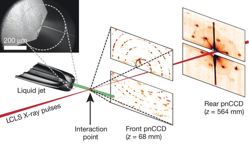
Scientific impact
Background
Structural biology is currently going through a golden age, as highlighted by the growth of
structural information over the last two decades: the structural biology databank (PDB) will
soon contain 100 000 structures! These successes have allowed scientists to obtain detailed
chemical information, from the mechanisms of chemical reactions catalysed by enzymes to
the intricate machinery and regulation of enormous macromolecular complexes like the
ribosome.
Not all areas of biology are covered equally well. Although the PDB contains over 91000
structures, only 379 of these (or 0.4%) are of unique membrane protein structures whereas
membrane proteins make up 20–30% of all expressed proteins (Wallin & von Heijne, 1998).
Information for multicomponent systems and protein-protein complexes is also relatively
scarce. Membrane proteins represent some of the largest classes of drug-targets (e.g. G
protein-coupled receptors and ion channels), while interactions between proteins are
important in many cellular processes such as immune response, signalling and regulatory
pathways. These discrepancies stem from the fact that such targets are more difficult to
isolate as functional proteins or complexes (see e.g. Nie et al., 2009). Crystallisation is also
more difficult, often yielding very small, inhomogeneous crystals with large cell dimensions.
To obtain structural information on these systems, advanced data collections strategies using
cutting-edge microfocus beamlines often provides a critical advantage (Moraes et al., 2013).

The scientific impact of microfocus macromolecular crystallography
                                        In 2012, the Nobel Prize in Chemistry was awarded
                                        to Brian Kobilka for his work on the structural
                                        biology of GPCRs (Figure 1). Since crystals of
                                        GPCRs are typically small and fragile, this work
                                        relied heavily on the use of microfocus data
                                        collection at Argonne National Laboratory. The
                                        foundations of the field were laid in Europe by
                                        Christian Riekel, Gebhard Schertler and coworkers at
                                        ID13 at the ESRF (Figure 2), who demonstrated the
                                        advantages of using a highly focused beam for
                                        protein crystallography (Riekel et al., 2005). Put
                                        simply: small crystals require small X-ray beams in
                                        order to minimize the background scatter and thereby
                                        maximize the signal to noise. Although ID13 was a
                                        multi-functional beamline, more recent protein
                                        crystallography dedicated microfocus beamlines such
                                        as ID23-2 and ID29 at the ESRF, the microfocus
                                        beamlines I24 at Diamond, 24-ID-C (23-ID-B/D) at
 Figure 1: Issue of Nature in which     the APS and X06SA at the SLS, have had large
 the first structure of a GPCR:G-       impact on the field of protein crystallography. There
 protein complex was presented by the   is universal agreement that there are dramatic
 Kobilka laboratory.                    benefits     in    pushing      protein    microfocus
                                        crystallography to its physical limits.

                                               2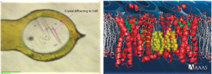
Figure 2: Microcrystals of a
human 2 adrenergic G-protein-
coupled receptor mounted at
ID13 of the ESRF, and the
resulting structure (Rasmussen et
al, 2007). Figure courtesy of
Riekel & Schertler.
The physical limits of macromolecular crystallography arise from protein, DNA and RNA
crystals being susceptible to radiation damage, with the concomitant degradation of structural
and chemical information. Advances in cryo-cooling technologies have meant that single
crystals frequently yield multiple complete datasets, which has been the basis for advanced
phasing methods such as MAD and SAD. Cryo-cooled crystals can withstand approximately
70 times higher radiation dose than non-cooled crystals (Nave & Garman, 2005; Nave &
Garman, 2009), but radiation damage often remains the major experimental challenge.
At a modern microfocus beamline (eg. 8 × 8 μm2 with 1012 ph/s flux) the Henderson radiation
dose limit of 20 MGy (Henderson, 1990) is reached within a few seconds (Evans, 2011a). At
MicroMAX, with tighter focus and higher flux, these limits could be reached within
milliseconds (Schneider et al, 2013). The only answer to radiation damage is to collect
diffraction data from hundreds or thousands of microcrystals, and to merge diffraction data
together to recover complete data-sets. Dramatic advances have recently been made on this
front due to the availability of X-ray free electron laser radiation.
Serial Crystallography at X‐ray free electron lasers
Arguably the most exciting recent technical developments in the field relate to the application
of X-ray free electron lasers (XFELs) to macromolecular crystallography. XFELs provide
extremely intense X-ray pulses of ~ 1012 photons/pulse in pulses of ~ 40 fs in duration
focused to a spot of 0.1 to 1 m. Since the Henderson radiation damage limit is exceeded
within a single pulse, the sample rapidly explodes (Neutze et al., 2000). Despite this damage
process, diffraction data is collected before the destruction of the sample (Chapman et al.,
2011; Barty et al., 2012) and high-resolution crystallographic data sets can be recovered by
merging diffraction data from thousands of microcrystals (Boutet et al., 2012; Redecke et at.
2013, Johansson et al., 2012, Johansson et al., 2013). This approach was highlighted by
Science who named the first new protein structure solved at XFEL (Redecke et al. 2013) as
one of the key scientific breakthroughs of 2012. Since every crystal exposed to the XFEL
beam yields only a single diffraction image, data is collected from a continuous flow of
microcrystals and the approach has been coined serial crystallography. As well as facilitating
the collection of diffraction data from nanocrystals, serial crystallography is a room-
temperature approach and allows time-resolved studies to be pursued.
Figure 3: Experimental setup used in
XFEL based serial femtosecond
crystallography studies at the LCLS. A
liquid microjet delivers a constant stream
of microcrystals across the focused XFEL
beam. The X-ray detector is read every
exposure and complete data is built up by
merging diffraction data from thousands
of microcrystals (Chapman et al., 2011).
3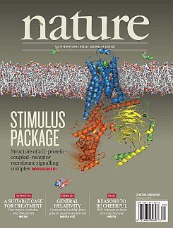
Vision for MicroMAX
Our vision for MicroMAX is to bridge this gap between what is possible with conventional
synchrotron based crystallography at synchrotron sources, and what is possible using serial
femtosecond crystallography at an XFEL. The concept is to apply the proven approach of
synchrotron-based microcrystallography with sorting and merging of diffraction data from
a very large number of microcrystals, in combination with a variety of sample delivery
technologies adapted and developed for application at a synchrotron based beamline. This
philosophy will push the physical limits of data collection at a storage ring.
There are several compelling scientific reasons for bridging this gap:
Macromolecular cryo-crystallography is an extremely successful method that has had
dramatic impact on life-science. Unlike XFEL radiation, cryo-crystallography allows
oscillation data to be collected from single-crystals but using a similar X-ray fluence
to XFEL studies. Adapting cryo-microcrystallography to allow very high-throughput
serial data collection creates a win-win situation that combines the benefits of XFEL
based serial crystallography with proven synchrotron based approaches.
Room-temperature serial crystallography at synchrotron sources will rely upon a
lower X-ray fluence than XFEL studies, yet there exists a domain and time-scale
where room temperature data can be collected from microcrystals (~ 1 to 5 m)
facilitating micro-crystal screening, micro-crystal optimization, data-collection free of
freezing artefacts (Fraser et al, 2011) and time-resolved diffraction studies.
Synchrotron based experiments using short X-ray exposures have shown a dose-rate
effect at room temperature (Owen et al, 2012; Warkentin at al, 2012) that allows
radiation damage to be partially outrun when collecting diffraction data on the single
millisecond timescale, which is ideal for MicroMAX.
XFEL beamtime will continue to be very limited and synchrotron based experiments
will always have an enormous cost-per-experiment advantage over XFEL sources.
There is therefore a compelling case for developing and optimizing micro-
crystallisation conditions using synchrotron radiation as a complement to certain
XFEL base studies.
Sample delivery at MicroMax
Inspired by the development of XFEL serial femtosecond crystallography (Chapman et al.,
2011), there is a large international effort in developing sample delivery technologies suitable
for serial crystallography experiments at both XFELs and storage rings. Proof-of-principle
demonstrations used the Spence liquid microjet (Doak et al., 2012). This delivers
microcrystals as a continuous stream in a jet approximately 3 m in diameter moving at a
velocity of about 10 m/s. This works beautifully at the XFEL, but this sample delivery
technology needs to be adapted for MicroMax due to the time-of-transit (~100 ns) across a
1m focus being too short for useful diffraction data to be collected.
The simplest extension of this approach to synchrotron radiation is to flow microcrystals
through a quartz capillary and collect serial diffraction data. This concept was recently
demonstrated at the beamlines P11 of PetraIII (White et al., unpublished) using a solution of
lysozyme microcrystals. Each micro-crystal was exposed to a radiation dose of 0.2 MGy (7 ×
15 m2 focus; 2×10¹² photons/sec; 10 msec exposure); diffraction data were recorded from
50,000 microcrystals; and an electron density map to 2.2 Å resolution was recovered.
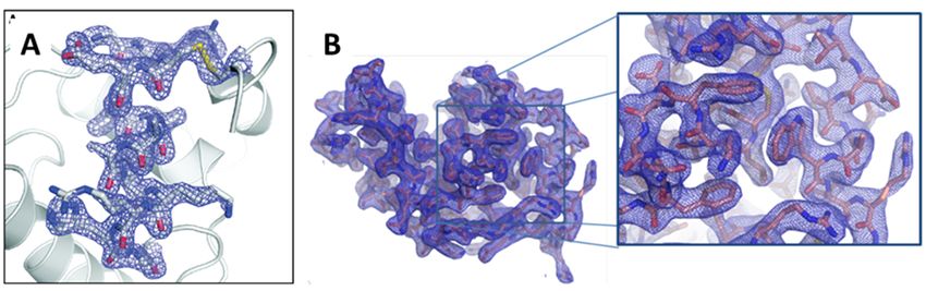
Comparison of the LCLS and PetraIII electron density maps of lysozyme shows that the
quality of the electron density is similar for both experiments (Figure 4).
4Figure 4: Electron density recovered from room-temperature microcrystals of lysozyme. A, From
serial femtosecond crystallography data recorded at the LCLS (dose 33 MGy) (Boutet et al.,
2012). B, From serial millisecond crystallography data recorded at PetraIII (dose 0.2 MGy). This
result demonstrates that serial crystallography can be successfully applied at a storage ring.
The Spence group has also developed a slower-moving lipidic cubic phase (LCP) microjet
which delivers highly viscous samples containing microcrystals. The LCP microjet moves at
approximately 1 mm/s, which equates to a transit time of 1ms for a 1m X-ray focus, ideal
for MicroMax. The LCP microjet has been tested using microcrystals of G protein-coupled
receptors at the LCLS (Cherezov et al, Science in press) and tested at the Swiss Light Source
(SLS) using microcrystals of lysozyme suspended in the LCP (Schlichting et al.,
unpublished). Diffraction from lysozyme microcrystals injected into a synchrotron beam
using the LCP microjet extended to approximately 2.1 Å resolution.
An alternative strategy was used in studies of frozen microcrystals of cathepsin B at P14 of
PetraIII (Redecke et al., unpublished). In this case solutions of cathepsin B microcrystals
were frozen in a litholoop and the entire loop was scanned using a series of continuous
helical scans, with a dose per crystal up to 50 MGy. Interpretable electron density maps were
again recovered to 2 Å resolution. Closely related studies at the LCLS used a micro-grid
mounting systems for which cryo-diffraction data from 932 microcrystals of myoglobin
(from 32 grids) were merged to recover diffraction data to 1.4 Å resolution (Cohen et al.,
unpublished). This strategy, of raster-screening cryo-cooled loops or micro-grids containing
dozens to hundreds of microcrystals, will be standardized at MicroMAX. This will provide a
valuable complement to data-collection studies from larger crystals at BioMAX, but will
require some development to optimize the sample mounting and data collection.
More technically challenging approaches to sample delivery are also being developed by the
Soares group at NSLS-II (Brookhaven) (Roesler et al., 2013) and the Cohen group at SSRL.
These emerging technologies including the use of a rullator: whereby microcrystals are
delivered onto a low-background thin-film and rolled across the X-ray beam at a controlled
pace and oscillation angle; Drop on demand delivery: whereby microdrops containing
microcrystals are delivered using acoustic sample delivery technologies; adaption of carbon
cryo-grid technologies from electron microscopy applications: whereby hundreds of
microcrystals are mounted on mature support structures that have been developed for
diffraction applications using electron microscopes (Zarrine-Afsar et al., 2012).
From this sampling of emerging solutions for sample mounting and manipulation designed
for microfocus data collection at synchrotron and XFEL sources, there can be no doubt that
many technologies will be mature when MicroMax comes online in 2018. It is our
consideration that this facet of MicroMAX, which was considered radical untested
technology only two years ago, is now within appropriate bounds of even the most
conservative risk assessment concerning future beamlines at MAX IV laboratory.
5Millisecond & microsecond X‐ray choppers and X‐ray detectors
MicroMAX will deliver a remarkably hot synchrotron beam, focussing 1013 photons/sec into
a spot ~0.5 m2, and 1015 photons/sec in polychromatic mode. This means that, even at
cryogenic temperatures, micron sized crystals will lose diffraction power within a few
milliseconds. This creates new challenges in terms of X-ray optics and detector technology.
X-ray choppers have been developed for time-resolved diffraction studies at polychromatic
beamlines (Cammarata et al., 2009; Husheer et al., 2010) and enable X-ray pulse trains down
~1 sec in duration to be isolated (or individual X-ray pulses in certain fillings of the storage
ring). When operating in polychromatic mode, it will be appropriate to isolate pulse-trains of
10 to 100 s in duration at MicroMAX. These specifications are easily attained with existing
technologies, such as the Julich X-ray chooper used in combination with Eiger detectors
developed at the Paul Scherrer Institute (Johnson et al., 2012). Adaptive Gain Integrating
Pixel Detectors (AGIPD) are now being developed at DESY for application at the European-
XFEL and they provide an even more attractive option for MicroMAX. These integrating
pixel detectors are designed to adjust the gain in each pixel according to the count rate of the
pixel, and have a frame readout rate of 3 kHz.
These combined technologies: a high-flux dedicated protein crystallography microfocus
beamline with a polychromatic option, equipped with a sec chopper and an Eiger or
AGIPD X-ray detector, is going to create a world-leading experimental protein
crystallography station for MaxIV laboratory. This is not business as usual, but is an
experimental station dedicated to pushing back the physical limits of what is possible at a
storage ring and taking an international lead, while at the same time bridging the gap
between conventional cryo-crystallography at more standard macromolecular beamlines and
serial femtosecond crystallography at XFELs.
User base and demand
Macromolecular crystallography is the most established of all Swedish user communities of
synchrotron radiation. Within the Swedish academia there are currently 13 professors, 26
academic scientists at associate professor, lecturer or assistant professor level, approximately
110 postdocs and PhD students working in the field of macromolecular crystallography.
Another 30 scientists are dedicated full time to the field of macromolecular X-ray
crystallography within Swedish industry. There are active research groups at all major
Swedish universities (Umeå University; Uppsala University; Swedish Agricultural
University, Royal Institute of Technology, Karolinska Institutet, University of Gothenburg,
Stockholm University, Lund University) and in both large pharmaceutical (AstraZeneca) and
small biotech (KaroBio, Medivir, Sprint Bioscience, SARomics Biostructures) companies.
There are very active macromolecular crystallography communities within Denmark,
Norway and Finland, also with structural biology groups established at most major
universities. Interest for protein crystallography is growing within the Baltic states and in
Poland. Specific scientific research centres dedicated to structural biology are funded in
Norway (Norstruct, Tromsø), Denmark (PUMPkin, Århus), Finland (Biocenter Structural
Biology, Helsinki/Oulu) and Poland (CBB, Poznan and IIMCB, Warsaw, Poland). Industrial
interest in MX is particularly strong in Denmark, where Novo Nordisk has a dedicated MX
group and many other companies collaborate intensively with academia.
In addition to Swedish, Scandinavian and Baltic interest, we foresee that MicroMAX will
become one of the flagship beamlines of MaxIV Laboratory and attract a large international
user community from within Europe, the USA and Asia.
6Instrument performance
Beamline Specifications
MicroMAX is a dedicated macromolecular crystallography beamline with the capability to
focus a high-intensity X-ray beam down to a spot size of 0.7 x 1 μm2: in monochromatic
mode >1013 photons/sE/E~10-4; in polychromatic mode ~1015 photons/sE/E~3×10-2. The
combination of a stable, highly focused, brilliant X-ray beam exploits the uniquely low
emittance of MAX IV while respecting the demands of the life-science user community.
MicroMAX will be constructed with both a "Standard MX setup" and the "Serial
crystallography setup".
For the Standard MX setup: Automated sample changers will be used to both enable rapid
and reliable sample changing and to minimize temperature changes as users enter and leave
the hutch. For the Serial crystallography setup: A variety of sample technologies are
discussed in detail in the previous section and include the use of the LCP microjet, rullators,
acoustic microdrop technology.
MicroMAX will be energy-tuneable over a wide range, with energy resolution allowing the
exploitation of anomalous scattering. The energy domain should be sufficient to explore
potential benefit of long wavelength radiation due to the low X-ray absorption in micro-
crystals (Evans et al, 2011).
MicroMAX requires a large pixel based detector (Eiger detector or AGIPD detector) with
rapid readout, that optimizes the signal to noise ratio for micron or sub-micron sized crystals.
A rapid X-ray detector will make it possible to follow the decay of diffraction quality
retrospectively, and thereby accept or reject data with sub-millisecond temporal resolution.
For polychromatic mode, MicroMAX will require a sec X-ray chopper, as has been
developed and applied at time-resolved diffraction beamlines at the ESRF and APS.
Table 1: MicroMAX beamline characteristics.
Photon source In vacuum or cryo-cooled undulator using the full straight
section
Monochromator Si(111), liquid nitrogen cooled
Focusing elements Two horizontally focusing mirrors, one vertically focusing
mirror.
Energy (wavelength) range 5 – 30 keV (0.5 – 2.5 Å)
Energy resolution E/E ~ 2 x 10⁴ with Si (111)
Photon flux at sample > 1013 ph/s with Si (111) at 1 Å
Beam size (horizontal × vertical) 1 × 0.7 μm2 with 0.7 × 0.5 mrad2 divergence, beam size
adjustable by working out of focus, in the horizontal the
secondary source can also be used to adjust the focus size
Beam stability Specified to 10% of the beam size
Experimental set-up High precision goniostat with kappa possibility, large area
detector, sample changer, cryostat, fluorescence detector
7Technical challenges
Beamline optics
The low emittance of the storage ring provides the basis for the design of MicroMAX. To
achieve the design goals it will be necessary to take extreme care with mechanical stability,
suppression of vibrations and handling of heat load effects.
The goal of achieving the maximum flux density at the sample leads to the choice of a long
undulator with short period and high magnetic field. It should have similar performance to
the BioMAX in-vacuum undulator (18 mm period, 1.5 m magnetic length) but a longer total
magnetic length and potentially cryogenically cooled to increase the brilliance.
Several optics designs are possible (see e.g. Evans et al., 2011b, Smith et al., 2012). We
propose a double crystal monochromator that is horizontally deflecting the beam, as
suggested for the BioMAX beamline. The heat load density on the first crystal will be high
(25 W/mm2 at 12 keV assuming twice the power from the BioMAX undulator, and 50
W/mm2 at 5 keV). This needs detailed studies and could limit the lower energy limit of the
beamline. For a larger energy bandwidth a multilayer monochromator is needed. The energy
bandwidth will depend on the undulator specifications and acceptable beam divergence.
Space should be reserved to be able to add a multilayer monochromator later.
The requirement of rapid energy-tuneability leads us to propose focusing mirrors rather than
e.g. compound refractive lenses or kinoform lenses for focusing. The requirements on the
mirror quality will be very high with slope error and surface error specifications at the upper
limit on what is available. We propose to use a two-stage focusing in the horizontal direction.
The first mirror focuses the beam to a secondary source that is then refocused at the sample
position by the second mirror. The two-stage focusing will give more space around the
sample position. The requirement to focus the undulator source down to 1 μm with a one-
stage focusing would either give very limited space around the sample or substantially
reduced flux by creating a secondary source without primary focusing. By using a slit at the
secondary source position, the sensitivity to movements of the experimental setup, including
the slit and second focusing stage, relative to everything upstream of the secondary source is
reduced, at the expense of some flux. The secondary source slit can also be used to rapidly
modify the beam size at the sample position. Both horizontally focusing mirrors should have
adjustable radii to be able to modify the beam size at the position of the secondary source slit
and at the sample.
In the vertical direction, the source size is so small that only a minor demagnification is
needed, and the space around the sample is not limited by the optical focusing element. With
a one-stage focusing, a second mirror is avoided which means higher beam quality and lower
cost. The vertically focusing mirror should also be adjustable to allow the beam size at the
sample to be adjustable.
Pink beam & chopper
In order to obtain a pink beam (E/E ≈ 0.01) at the MAX IV 3 GeV ring it will be needed to
install a multilayer monochromator. A high-heat-load chopper will be required to reduce the
average power load on downstream components and the sample, and a microsecond chopper
to provide an adequate time structure of the X-ray pulse trains.
Experimental setup
We propose to combine a standard MX setup with a serial crystallography setup at the same
position, i.e. the optics are not modified when changing from one mode to the other. The
8standard MX setup will consist of a rotation axis for data collection, a cold gas device for
sample cooling and an automatic sample changer. The setup will not be standard in the sense
that the stability and precision needs are higher than for today's standard beamlines. The
rotation axis will be vertical since this makes it easier to achieve a small sphere of confusion.
The rotation axis should accept standard sample holders (SPINE as used today or a modified
version as being developed). Several details will have to be worked out during the design
stage, in particular how to optimally use the space around the sample position.
For serial crystallography an additional device such as a LCP microjet or a rullator, can be
positioned in the horizontal plane orthogonally to the X-ray beam.
For optimal data quality the development of a vacuum environment from the optics up to the
detector will be needed. In the first phase this is envisioned as a vacuum cone beginning just
behind the mounted crystal and extending to the surface of the X-ray detector, with the back-
stop in vacuum. A silicon nitride X-ray window can be used to seal the vacuum immediately
after the sample, allowing microcrystals to be manipulated and cryo-cooled in air. For some
setups, such as the LCP microjet, it may be appropriate to develop these in vacuum
technology from the outset.
Computing needs
MicroMAX should not only have the hardware to perform challenging diffraction
experiments, but the computing environment that allows us to manage the experimental
hardware, the large number of samples, the large volume of data, the tools to perform the data
analysis, need also to be provided in a way easily applied by non-expert users. Here all
developments happening at BioMAX will be implemented and it will be a shared effort
between the two beamlines.
Comparison of BioMAX with other microfocus beamlines
All other microfocus beamlines available today worldwide have considerably lower brilliance
than MicroMAX. ID13 of the ESRF is currently the only microfocus beamline in Europe that
regularly achieves a spot focus of 1 x 1 μm2. ID13, however, is only available to the
macromolecular crystallography community for approximately 10 % of its operational time
and the X-ray flux is approximately 1% of what will be available at MicroMAX.
BL32XU at SPring-8 is currently the world’s only operational 1 x 1 μm2 microfocus beamline
dedicated to protein crystallography, and its flux is comparable with that of ID13. It is also
tuneable, with an energy range between 8.5 and 20 keV.
ID23-2 of the ESRF has a spot size of 3.5 x 7 μm2 but is fixed energy. ID23-2 is regarded
within the European membrane protein crystallography community as a superb data-
collection station.
I24 at Diamond Light Source is a tuneable beamline with a 5 x 5 μm2 focus and is proven to
fill an important need, not least in screening and data collection of micro-crystals at room
temperature. I24, however, lacks the brilliance to optimize data collection for the smallest
crystals, a need that MicroMAX could satisfy.
Several microfocus beamlines are under development or are being commissioned, some of
which will approach the intensity of MicroMAX. A further advantage is the low divergence
that MicroMAX will achieve in comparison to these other facilities due to the characteristics
of the source itself.
9Table 2: Synchrotron based beamlines with focus in at least one direction ≤ 5 micron.
Beamline beam size Flux [ph/s]
h x v [µm x µm]
ESRF ID13 not dedicated to MX 1x1 8 x 10¹⁰
ESRF ID23-2, fixed λ 7 x 4 (eventually 1x1) 4 x 1011
SLS X06SA 15 x 5 (tunable 5.7 – 17.5 keV) 2 x 10¹²
APS 23ID-B and 23ID-D (GM/CA- 65 x 20 (standard) 2 x 10¹³
CAT) 5 x 5 (mini) 5 x 10¹⁰
1 x 1 (micro) 3 x 10⁹
tunable (5 – 20 keV)
APS 19ID [SBC-CAT) 5 x 20 (6-19.5 keV) 1.3 x 10¹³
APS 24ID-E (NE-CAT) fixed λ 5 – 100 (apertures) 3 x 10¹²
Diamond I24 5 x 5 (tunable 5 – 17 keV) 1.1 x 10¹²
SPring-8 BL32XU 1x1 6 x 10¹⁰
Soleil PROXIMA 2A 5 x 4 (tunable 5 – 15 keV) 1.1 x 10¹²
PETRA III P11€ X x X (tunable 7 – 20 keV) 1 x 10¹³ to 1015
PETRA III P14 1 x 5 (tunable 7 – 20 keV) 1 x 10¹³ to 1015
APS 23ID-D (under construction 1- 20 (tunable 6 - 35 keV) 1 x 10¹³
NSRRC TPS-05A1 (under 1 - 50 (tunable 5.7- 20 keV) 1 x 10¹²
construction
SSRF NFPS (under construction) 10 x 5 (tunable 5.7- 20 keV) -
NSLS-II FMX under development 1 x 1 (tunable 5 – 23 keV) 1 x 10¹³
NSLS-II NYX 5 – 50 (tunable 3.5 – 17.5 keV) 1 x 10¹³ (?)
ALBA MicroFocus proposed 3x1 3 x 10¹²
BioMAX under development 20 x 5 (tuneable ) 1 x 10¹³
Micromax proposed 0.7 x 1 (tuneable ) 1 x 10¹³ to 1015 *
* = option for pink beam
€ = shared beamline with bio-imaging
Conclusions
MicroMAX, a microfocus macromolecular crystallography beamline, is proposed as a state
of-the-art exploratory beam line at MAXIV. MicroMAX will deliver a 0.7 μm2 focal spot
beam diameter with an X-ray flux >1013 photons/sec in monochromatic mode, and
approaching 1015 photons/sec in polychromatic mode. Because of these state-of-the-art
specifications, MicroMAX will allow very large data sets to be rapidly collected from
thousands of micron sized macromolecular crystals cooled to cryogenic temperatures using
sample mounting and freezing technologies familiar to any protein crystallographer. In
parallel, MicroMAX will develop novel sample delivery environments that will allow the
pursuit of serial data collection strategies both at room temperature and using cryo-
technologies. This beamline will allow new structures to be solved from very small crystals;
and will accelerate the rate of progress towards structures of extremely challenging
crystallization targets. The ease of access to MicroMAX for the entire Nordic Structural
Biology community will stoke ambition and drive innovative science, providing a flagship
example of how MAXIV Laboratory and its user community can work together to achieve
scientific goals unreachable today.
10References
Cammarata M., Eybert L., Ewald F., Reichenbach W., Wulff M., Anfinrud P., Schotte F., Plech A., Kong Q.,
Lorenc M., Lindenau B., Ribiger J., Polachowski S. (29009) Chopper system for time resolved experiments
with synchrotron radiation. Rev. Sci. Instrum. 80: 015101
Chapman et al. Femtosecond X-ray protein nanocrystallography. (2011) Nature 470, 73-77.
Doak RB, DePonte DP, Nelson G, Camacho-Alanis F, Ros A., Spence J. C. H., and Weierstall U. (2012)
Microscopic linear liquid streams in vacuum: Injection of solvated biological samples into X-ray free electron
lasers. AIP Conf. Proc http://dx.doi.org/10.1063/1.4769693.
Evans G, Axford D, Owen RL (2011a) The design of macromolecular crystallography diffraction experiments.
Acta Crystallogr D 67, 261-270.
Evans G, Axford D, Waterman D & Owen RL. (2011b) Macromolecular microcrystallography. Crystallogr.
Rev. 17, 105-142.
Fraser JS, van den Bedem H, Samelson AJ, Lang PT, Holton JM, Echols N, Alber T. (2011) Accessing protein
conformational ensembles using room-temperature X-ray crystallography. Proc. Natl. Acad. Sci. 108, 16247-
1625.
Henderson, R. (1990). Cryoprotection of protein crystals against radiation damage in electron and X-ray
diffraction. Proc. R. Soc. London, B241, 6-8.
Husheer SL, Cole JM, d'Almeida T, Teat SJ. (2010) A prototype chopper for synchrotron time-resolved
crystallographic measurements. Rev Sci Instrum. 81, 043905. doi: 10.1063/1.3358939.
Johnson I, Bergamaschi A, Buitenhuis J, Dinapoli R, Greiffenberg D, Henrich B, Ikonen T, Meier G, Menzel A,
Mozzanica A, Radicci V, Satapathy DK, Schmitt B, Shi X. (2012= Capturing dynamics with Eiger, a fast-
framing X-ray detector. J Synchrotron Radiat. 19, 1001-1005
Johansson, L.C. et al., (2012) Lipidic phase membrane protein serial femtosecond crystallography. Nature Meth.
9, 263-265.
Johansson, L.C. et al., (2013) Structure of a photosynthetic reaction centre determined by serial femtosecond
crystallography Nature Comm. in press.
Moraes I, Evans G, Sanchez-Weatherby J, Newstead S & Shaw Stewart PD (2013) Membrane prptein structure
determination – The next generation. Biochim. Biophys. Acta doi:pii: S0005-2736(13)00244-7.
10.1016/j.bbamem.2013.07.010.
Nave C & Garman EF (2005) Towards and understanding of radiation damage in cryocooled macromolecular
crystals. J. Synch. Rad. 12, 257-260.
Nave C & Garman EF (2009) Radiation damage in protein crystals examined under various conditions by
different methods. J. Synch. Rad. 16, 129-132.
Nave C & Hill MA (2005) Will reduced radiation damage occur with very small crystals? J. Synch. Rad. 12,
299-303.
Neutze, R, Wouts, R, van der Spoel, D, Weckert, E, Hajdu, J (2000) Potential for biomolecular imaging with
femtosecond X-ray pulses, Nature 406, 752-457.
Nie Y., Viola C, Bienniossek C, Trowitsch S., Sumitra L., Chaillet M., Garzoni F & Berger I. (2009) Getting a
grip on complexes. Curr. Genomics 10, 558-572.
Owen RL, Axford D, Nettleship JE, Owens RJ, Robinson JI, Morgan AW, Doré AS, Lebon G, Tate CG, Fry
EE, Ren J, Stuart DI, Evans G. (2012) Outrunning free radicals in room temperature macromolecular
crystallography. Acta Crystallogr D 68, 810-818.
Rasmussen, S G, Choi, HJ, Rosenbaum, DM, Kobilka, TS, Thian, FS, Edwards, PC, Burghammer, M, Ratnala,
VR, Sanishvili, R, Fischetti, RF, Schertler, GF, Weis, WI, Kobilka, BK (2007) Crystal structure of the human 2
adrenergic G-protein-coupled receptor, Nature 450, 383-7.
Redecke et al. (2013) Natively inhibited Trypanosoma brucei cathepsin B structure determined by using an X-
ray laser. Science 339, 227-230.
Riekel C, Burghammer M, Schertler G. (2005) Protein crystallography microdiffraction. Curr Opin Struct Biol.
15, 556-562
11Roesler CG, Kuczewski A., Stearns R., Ellson R., Olechno J., Orville AM, Allaire M, Soares AS & Héroux A.
(2013) Acoustic methods for high throughput protein crystal mounting at next generation macromolecular
crystallography beamlines. J Synch. Rad. 20, 805-808.
Schneider D, Berman L, Chubar O., Hendrickson WA, Hulbert SL, Lucas M, Sweet RM & Yang L. (2013) J.
Phys.: Conf. Ser. 425 012003 doi:10.1088/1742-6596/425/1/012003
Smith JL, Fischetti RF, Yamamoto M. (2012) Micro-crystallography comes of age. Curr Opin Struct Biol. 22,
602-612.
Wallin E. & von Heijne G (1998) Genome-wide analysis of integral membrane proteins from eubacterial,
archaean, and eukaryotic organisms. Protein Sci. 7, 1029- 1038.
Warkentin M, Hopkins JB, Badeau R, Mulichak AM, Keefe LJ, Thorne RE. (2013) Global radiation damage:
temperature dependence, time dependence and how to outrun it. J. Synch. Rad. 20, 7-13
Weierstall U, Spence JC, Doak RB. (2012) Injector for scattering measurements on fully solvated biospecies.
Rev Sci Instrum. 83,
Zarrine-Afsar A, Barends TR, Müller C, Fuchs MR, Lomb L, Schlichting I, Miller RJ. (2012) Crystallography
on a chip. Acta Crystallogr D 68, 321-323.
12You can also read