NIRF-Molecular Imaging with Synovial Macrophages-Targeting Vsig4 Nanobody for Disease Monitoring in a Mouse Model of Arthritis - MDPI
←
→
Page content transcription
If your browser does not render page correctly, please read the page content below
International Journal of
            Molecular Sciences

Article
NIRF-Molecular Imaging with Synovial
Macrophages-Targeting Vsig4 Nanobody for
Disease Monitoring in a Mouse Model of Arthritis
Fang Zheng 1 , Siyu Luo 1 , Zhenlin Ouyang 1,2 , Jinhong Zhou 1 , Huanye Mo 1 ,
Steve Schoonooghe 3,4 , Serge Muyldermans 3 , Patrick De Baetselier 3,4 , Geert Raes 3,4, *,† and
Yurong Wen 1,2, *,†
 1    Department of Biochemistry and Molecular Biology, Key Laboratory of Environment and Genes Related to
      Diseases, Health Science Center, Xi’an Jiaotong University, Xi’an 710049, China
 2    The Key Laboratory of Biomedical Information Engineering of Ministry of Education,
      School of Life Science and Technology, Xi’an Jiaotong University, Xi’an 710049, China
 3    Research group of Cellular and Molecular Immunology, Vrije Universiteit Brussel, B-1050 Brussels, Belgium
 4    Myeloid Cell Immunology Lab, VIB Center for Inflammation Research, B-1050 Brussels, Belgium
 *    Correspondence: Geert.Raes@vub.be (G.R.); Yurong.Wen@xjtu.edu.cn (Y.W.)
 †    These authors contributed equally to this manuscript.

 Received: 20 May 2019; Accepted: 6 July 2019; Published: 8 July 2019

 Abstract: Nanobody against V-set and Ig domain-containing 4 (Vsig4) on tissue macrophages, such
 as synovial macrophages, could visualize joint inflammation in multiple experimental arthritis
 models via single-photon emission computed tomography imaging. Here, we further addressed the
 specificity and assessed the potential for arthritis monitoring using near-infrared fluorescence (NIRF)
 Cy7-labeled Vsig4 nanobody (Cy7-Nb119). In vivo NIRF-imaging of collagen-induced arthritis (CIA)
 was performed using Cy7-Nb119. Signals obtained with Cy7-Nb119 or isotope control Cy7-NbBCII10
 were compared in joints of naive mice versus CIA mice. In addition, pathological microscopy
 and fluorescence microscopy were used to validate the arthritis development in CIA. Cy7-Nb119
 accumulated in inflamed joints of CIA mice, but not the naive mice. Development of symptoms in CIA
 was reflected in increased joint accumulation of Cy7-Nb119, which correlated with the conventional
 measurements of disease. Vsig4 is co-expressed with F4/80, indicating targeting of the increasing
 number of synovial macrophages associated with the severity of inflammation by the Vsig4 nanobody.
 NIRF imaging with Cy7-Nb119 allows specific assessment of inflammation in experimental arthritis
 and provides complementary information to clinical scoring for quantitative, non-invasive and
 economical monitoring of the pathological process. Nanobody labelled with fluorescence can also be
 used for ex vivo validation experiments using flow cytometry and fluorescence microscopy.

 Keywords: V-set and Ig domain-containing 4 (Vsig4); synovial macrophage; near- infrared
 fluorescence; Nanobody; in vivo imaging

1. Introduction
     Rheumatoid arthritis (RA) is a chronic autoimmune disease characterized by a failure of
spontaneous resolution of inflammation [1]. Macrophages are essential in the pathogenesis of RA.
An increased number of sublining macrophages in the synovium is a sign of early onset of active
rheumatic disease. The degree of synovial macrophage infiltration highly correlates with the degree
of inflammatory lesion and joint erosion. The depletion of these macrophages from inflamed joints
benefits the RA therapy. Research has now uncovered an unexpectedly high level of heterogeneity
in macrophage origin and function, and has emphasized the role of environmental factors in their

Int. J. Mol. Sci. 2019, 20, 3347; doi:10.3390/ijms20133347                           www.mdpi.com/journal/ijms
Int. J. Mol. Sci. 2019, 20, 3347                                                                     2 of 14

functional specialization [2]. Although some results in mouse models of arthritis have contributed
to our understanding of the properties of synovial macrophages, the heterogeneous populations of
immune cells in RA have not been fully characterized. Good markers to discriminate these resident
macrophage populations could lead to monitoring of pro- or anti-inflammatory properties of these
macrophage populations that inform future therapeutic strategies.
      The anatomical imaging techniques such as X-ray or ultrasonography can provide information on
bone destruction. A novel method in rheumatology is mass spectrometry imaging (MSI). MSI enables
the determination of the relative abundance and spatial distribution of biomolecules in joint sample
tissue sections without labeling or staining [3]. It has revealed the identity and distribution of several
peptides, lipids and chemical elements in cartilage, synovium and bone from patients with rheumatic
diseases. Contrast-enhanced magnetic resonance imaging (MRI), ultrasonography and molecular
imaging with SPECT or PET are other methods that enable the use of molecules reflecting cellular
inflammatory processes to monitor disease evolution or treatment efficacy. Probes for molecular
imaging targeting activated macrophages include 18 F-polyethyleneglycol-folate, 111 In-anti-F4/80-A3-1
and 99m Tc-EC20 (etarfolatide) [4]. However, these methods feature high cost and technical barriers
and it is challenging to correlate these radioactive signals with post hoc in vitro analyses, such as
immunohistochemistry or gene expression profiling. The more frequently used vivo imaging method in
preclinical research is near-infrared fluorescence imaging. Studies of joint samples have demonstrated
that near-infrared fluorescence imaging can provide complementary information to histology and
histochemistry for rheumatic disorder. New molecular probes are currently being developed for
arthritis imaging such as four fluorescent probes from PerkinElmer in combination [ProSense 750
fluorescent activatable sensor technology (FAST) with Neutrophil Elastase 680 FAST and MMPSense 750
FAST with CatK 680 FAST] [5], a folate receptor-targeted near-infrared dye- OTL0038 [6], and an IR-780
iodide-loaded drug for real time monitoring of in vivo drug release [7].
      Near-infrared (NIR, 650–1000 nm) fluorescence imaging provides a high contrast between target
and background tissues by improving tissue penetration and minimizes tissue autofluorescence. It is
non-damaging radiation and less toxic for in vivo monitoring of biologic processes. Moreover, it can
be easily conjugated to antibody (mAb) or antibody figments by random labeling and site-specific
methods. Yet, the intact radiolabeled mAb may feature high liver uptake and slow blood elimination.
The problem can be addressed by using smaller antibody fragments, such as scFv, Fab, F(ab’)2 or
nanobodies. Nanobodies (Nbs) are single-domain antigen-binding fragments derived from naturally
occurring heavy-chain only antibodies in camelids [8]. The antigen-specific Nbs have good expression
in microbial systems and beneficial biochemical properties: Good solubility, good stability in harsh
conditions, high affinity and specificity for the antigen, small size and strict monomeric behavior. Nbs
have better imaging pharmacokinetics because they are rapidly excreted by kidneys and constitute
an ideal tool for diagnostic applications in various disease areas, including infectious, inflammatory
neurodegenerative diseases and tumours [9,10]. Furthermore, NIR fluorescence (NIRF) imaging is
becoming a noninvasive alternative to radionuclide imaging for joints in small animals [11,12]. NIR dye
labeled Nbs were documented for tumor imaging and tumor surgery imaging, for example, by using
an anti-HER2 nanobody labeled with the IRDye800CW for ovarian cancer [13,14], an epidermal
growth factor receptor (EGFR)-targeting nanobody for head and neck cancer [15], and ARTC2-specific
nanobody for lymphoma cells [16,17]. Nbs can also be used for in vivo near-infrared fluorescence
targeting of T cells [18]. Cellular imaging of immune cells such as T cells, B cells and antigen-presenting
cells has provided important information on immune homeostasis, immune responses and autoimmune
diseases [19].
      V-set and Ig domain-containing 4 (Vsig4) is a Type I transmembrane protein (399 aa) and a B7
family member [20]. Vsig4 is highly expressed in lung, placenta, synovium and has unique expression
on tissue macrophages such as Kupffer cells and a pivotal function in the clearance of pathogens and
autologous cells [21,22]. It was also reported to be expressed in macrophages associated with lung
cancer, ovarian cancer and multiple myeloma [23–25]. Synovium macrophages underlie multiple stages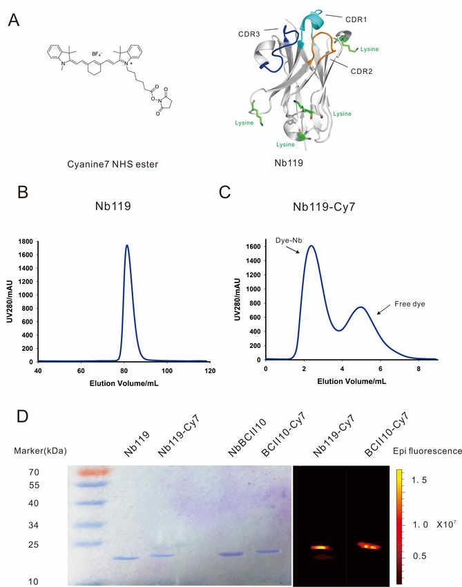
Int. J. Mol. Sci. 2019, 20, 3347                                                                                      3 of 14

Int. J. Mol. Sci. 2019, 20, x                                                                                         3 of 13

of an arthritis immune response, including its initiation, maintenance, regulation and termination.
of CD86 positive subpopulations in inflamed joints of arthritis mice models and can be used for
In previous studies, we have shown that specific probes targeting Vsig4 co-stain a subset of CD86
SPECT/CT molecular imaging to monitor and even predict the development of RA [26,27]. The Nb119
positive subpopulations in inflamed joints of arthritis mice models and can be used for SPECT/CT
also specifically stains the F4/80high and CD11bintermediate Kupffer cells (MHCII+CD68+CD64+
molecular imaging to monitor and even predict the development of RA [26,27]. The Nb119 also
macrophages profile) in naive mice liver. It can further monitor Kupffer cells non-invasively in an
specifically stains the F4/80high and CD11bintermediate Kupffer cells (MHCII+ CD68+ CD64+ macrophages
acute liver mouse model [28]. In the present study, we extend the use of nanobodies for imaging the
profile) in naive mice liver. It can further monitor Kupffer cells non-invasively in an acute liver
in vivo biodistribution of resident macrophages by targeting Vsig4 for usage as a florescence tool for
mouse model [28]. In the present study, we extend the use of nanobodies for imaging the in vivo
NIRF. We show that allophycocyanin (APC) conjugated Nanobodies raised against Vsig4 could bind
biodistribution of resident macrophages by targeting Vsig4 for usage as a florescence tool for NIRF.
on peritoneal macrophages of WT C57b/6 mice by using flow cytometry. The Nb119 labelled with
We show that allophycocyanin (APC) conjugated Nanobodies raised against Vsig4 could bind on
NIR dyes Cyanine 7 (Cy7) specifically target the inflamed joints in mice with CIA in NIRF imaging.
peritoneal macrophages of WT C57b/6 mice by using flow cytometry. The Nb119 labelled with NIR dyes
The in vivo biodistribution of Vsig4 was specifically focusing on synovial macrophages, which
Cyanine 7 (Cy7) specifically target the inflamed joints in mice with CIA in NIRF imaging. The in vivo
correlated to Alexa fluor647 labeled Nanobody staining of these cells by using histochemistry. Thus,
biodistribution of Vsig4 was specifically focusing on synovial macrophages, which correlated to Alexa
Nbs represent elegant fluorescence targeting probes for NIRF imaging of specific macrophages in
fluor647 labeled Nanobody staining of these cells by using histochemistry. Thus, Nbs represent elegant
inflamed joints.
fluorescence targeting probes for NIRF imaging of specific macrophages in inflamed joints.
2. Results
2. Results

2.1. In Vitro Nb119-Cy7 Experiment
2.1.
     The Vsig4-specific
     The   Vsig4-specific Nbs (Nb119) (Nb119) were produced
                                                         produced inin Escherichia
                                                                        Escherichia coli,
                                                                                     coli, purified via affinity
chromatography     and  size  exclusion     chromatography     (SEC), as described
chromatography and size exclusion chromatography (SEC), as described before [29].   before  [29]. Flow cytometry
analysis confirmed
analysis confirmedthatthatthe
                            theNb119,
                                   Nb119,butbutnot
                                                 notthe
                                                      the isotype
                                                        isotype   control
                                                                control   BCII10,
                                                                        BCII10,    recognized
                                                                                recognized       Vsig4
                                                                                             Vsig4        expressed
                                                                                                     expressed   on
      + CD11b
on F4/80
F4/80          + MHCII
         + CD11b   + MHCIIintermediate    peritoneal macrophages but not on F4/80lowlowMHCII
                              intermediate peritoneal macrophages but not on F4/80          MHCII  high
                                                                                                     high peritoneal
                                                                                                         peritoneal
population (Figure
population   (Figure 1).
                      1). The Nb119 staining was confirmed confirmed by
                                                                      by anti-Vsig4
                                                                         anti-Vsig4 antibody
                                                                                      antibody (Supplementary
                                                                                                 (Supplementary
Figure S1).
Figure  S1). Flow cytometry using antibody recognizing Vsig4 specifically detects expression of the              the
target antigen  on  peritoneal    macrophages      in
target antigen on peritoneal macrophages in naive mice.naive mice.

      Figure 1. Nb119 binds to F4/80+ CD11b+ MHCIIintermediate peritoneal macrophages. The peritoneal
      Figurecells
      cavity 1. Nb119  binds
                  from B7     to mice
                          naive  F4/80were
                                      + CD11b+ MHCIIintermediate peritoneal macrophages. The peritoneal cavity
                                           gated according to MHCII and CD11b expression. MHCIIintermediate
      cellsCD11b
      and  from B7   naive mice shows
                   + population  were gated
                                        F4/80according  to MHCII
                                              positive and  Nb119 and    CD11b
                                                                    positive.   expression.
                                                                              F4/80         MHCII
                                                                                    low MHCII    highintermediate
                                                                                                      population  and
      CD11b  + population   shows   F4/80 positive  and  Nb119    positive. F4/80 low  MHCII  high
      is Nb119 negative. Flow cytometry histogram plots of APC-labeled Nb119 (blue) and are shown inpopulation       is
      Nb119   negative.  Flow  cytometry   histogram  plots of  APC-labeled    Nb119
      comparison with an APC-labeled rat IgG2a kappa isotype control antibody (red).  (blue) and    are   shown     in
      comparison with an APC-labeled rat IgG2a kappa isotype control antibody (red).
     In our previous study, the crystal structure of Nb119 shows that four lysines are located in
     In our previous
the framework          study,
                 but not       theantigen
                          in the   crystal binding
                                           structureCDR
                                                     of Nb119  shows
                                                         regions       that
                                                                  of the    four lysines
                                                                         nanobody    and are located
                                                                                          could       in the
                                                                                                be further
framework   but  not in the antigen  binding  CDR  regions of the nanobody    and could  be further
labeled by Cy7 [29] (Figure 2A). NB119 was successfully purified in large yield and homogenously    labeled
by Cy7 [29] (Figure 2A). NB119 was successfully purified in large yield and homogenously by
Superdex 75pg column (Figure 2B) and conjugated with Cy7, followed by a desalting purification to
further isolate the Cy7 labeled Nb119 from the free Cy7 (Figure 2C). The Nanobody labeling was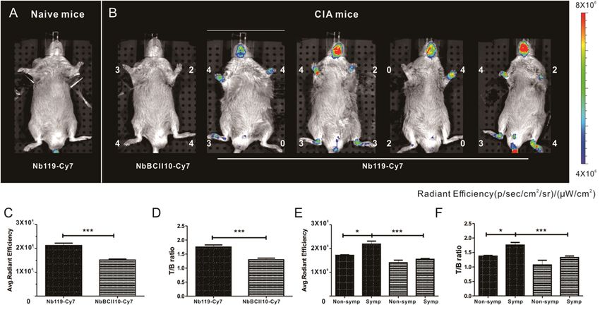
Int. J. Mol. Sci. 2019, 20, 3347                                                                                               4 of 14

by Superdex 75pg column (Figure 2B) and conjugated with Cy7, followed by a desalting purification
to further isolate the Cy7 labeled Nb119 from the free Cy7 (Figure 2C). The Nanobody labeling was
furtherInt. J. Mol. Sci. 2019,
        validated              20, x
                          by using     SDS-PAGE and NIRF imaging. A single band around 15 kDa of4 of                13 purified
                                                                                                                  the
nanobody       or conjugated nanobody was noted for Coomassie stained or NIRF imaged views after
       further validated by using SDS-PAGE and NIRF imaging. A single band around 15 kDa of the
SDS-PAGE.
       purified This      size correlates
                    nanobody                  to the
                                   or conjugated      predicted
                                                   nanobody       molecule
                                                              was noted       mass of stained
                                                                        for Coomassie   a nanobody.
                                                                                                or NIRFConjugated
                                                                                                        imaged viewsNbBCII0
and Nb119       were slightly
       after SDS-PAGE.           Thislarger    than thetounconjugated
                                       size correlates                     nanobody.
                                                           the predicted molecule   massConjugated     NbBCII0
                                                                                          of a nanobody.           and Nb119
                                                                                                          Conjugated
showed NbBCII0
          positive   andbands
                           Nb119visualized
                                     were slightlyby
                                                   larger
                                                      NIRFthan the unconjugated
                                                             imaging             nanobody.
                                                                       (excitation    710 nmConjugated   NbBCII0
                                                                                              and emission    805and
                                                                                                                   nm) which
       Nb119     showed      positive   bands   visualized
confirmed the Cy7 conjugation of the nanobody (Figure 2D). by NIRF  imaging  (excitation 710 nm and  emission 805 nm)
         which confirmed the Cy7 conjugation of the nanobody (Figure 2D).

              2. Purification
      FigureFigure                andand
                     2. Purification    purity
                                           puritystudy
                                                   study of
                                                          of Nb119   Cy7conjugates.
                                                             Nb119 Cy7    conjugates.     (A) Schematic
                                                                                     (A) Schematic           representation
                                                                                                     representation  of       of
      Cyanine   7 NHS
            Cyanine      esterester
                       7 NHS     (Cy7)   and
                                     (Cy7)    Nb119
                                            and Nb119protein     structure
                                                        protein structure   CDR1
                                                                          CDR1      in Cyan,
                                                                                in Cyan,   CDR2CDR2
                                                                                                 in blueinand
                                                                                                            blue  andinCDR3 in
                                                                                                               CDR3
      orange,orange,
               the 4 the  4 lysines
                      lysines       in the
                                in the     frameworkregion
                                        framework      region are
                                                               areindicated in green.
                                                                    indicated          (B) Superdex
                                                                               in green.             75 purification
                                                                                            (B) Superdex             of
                                                                                                             75 purification  of
      Nb119.Nb119.   (C) Superdex
               (C) Superdex          75 purificationof
                                  75 purification     of Cy7
                                                         Cy7 conjugated
                                                              conjugated  Nb119;
                                                                           Nb119;dye-conjugated    nanobody
                                                                                      dye-conjugated           and free and free
                                                                                                          nanobody
            dye are indicated. (D) Coomassie-stained gel and NIRF image of unconjugated Nb119, BCII10 and
      dye are indicated. (D) Coomassie-stained gel and NIRF image of unconjugated Nb119, BCII10 and
            respective Cy7 conjugated nanobody.
      respective Cy7 conjugated nanobody.
         2.2. NIRF-Imaging Experiments in Vivo
2.2. NIRF-Imaging Experiments in Vivo
               Vsig4 has been reported to be an interesting target for imaging the progression of arthritis and
      Vsig4  has been reported
       the macrophages              to be
                        involved [27].   We anaimed
                                                interesting  target
                                                     to evaluate       for imaging
                                                                 the suitability      the progression
                                                                                 of Cy7-conjugated      of arthritis
                                                                                                    nanobody for     and
the macrophages     involved
       in vivo imaging           [27].
                       of arthritic     We
                                    joints    aimed
                                           in the      to evaluatearthritis
                                                  collagen-induced    the suitability   of of
                                                                              (CIA) model  Cy7-conjugated     nanobody
                                                                                              RA in DBA/1J mice.
for in vivo imaging of arthritic joints in the collagen-induced arthritis (CIA) model of RA in DBA/1J
mice. This is a commonly used experimental model of inflammatory joint arthritis caused by a T-cell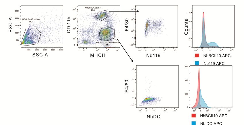
Int. J. Mol. Sci. 2019, 20, 3347 5 of 14
dependent, antibody-mediated auto immune response directed against cartilage type II collagen. After
CIA mice Int. J.immunization,
Mol. Sci. 2019, 20, x some mice remained asymptomatic and others are symptomatic, 5 of 13and the
arthritic joints among mice are not often homogeneous before day 40 post immunization [30]. Therefore,
This is a commonly used experimental model of inflammatory joint arthritis caused by a T-cell
we intravenously injected Cy7-labeled Nb119 and NbBCII10 into mice at a dose of 100 µg at 28 days
dependent, antibody-mediated auto immune response directed against cartilage type II collagen.
after subcutaneous
After CIA miceinjection of collagen
immunization, II inremained
some mice the tail asymptomatic
of the animals. andImaging
others arewas performed
symptomatic, and3 h after
injectiontheofarthritic
the fluorochrome
joints amongconjugates
mice are not(Figure 3). NIRF-imaging
often homogeneous before day was40used
post to gauge the targeting
immunization [30]. of
the Nb119-Cy7
Therefore, towe theintravenously
inflamed joints of CIA
injected mice. The
Cy7-labeled Nb119uptake of Nb119-Cy7
and NbBCII10 in the
into mice at a arthritic
dose of 100paws
μg of CIA
mice is at 28 days after subcutaneous
significantly higher than injection
in the pawsof collagen II in the
of naive tail of
mice. Thetheresults
animals.showed
Imaging was performed
efficient and specific
3 h after injection of the fluorochrome conjugates (Figure 3). NIRF-imaging was used to gauge the
labeling of arthritic joints with Nb119-Cy7 but not with NbBCII10-Cy7, reflected by a higher signal in
targeting of the Nb119-Cy7 to the inflamed joints of CIA mice. The uptake of Nb119-Cy7 in the
arthritic joints for Nb119-Cy7 as compared to NbBCII10-Cy7. No strong signals of Nb119-Cy7 and
arthritic paws of CIA mice is significantly higher than in the paws of naive mice. The results showed
BCII10-Cy7 were
efficient andobserved in liver
specific labeling of or kidneys,
arthritic jointsreflecting that the
with Nb119-Cy7 butfluorescent signals of the
not with NbBCII10-Cy7, systemically
reflected
administered
by a higherNbssignal
did not pass through
in arthritic joints forthe fur of mice.
Nb119-Cy7 As a result,
as compared NIRF imaging
to NbBCII10-Cy7. Noof Nb119-Cy7
strong signals allows
of Nb119-Cy7
the specific measurement and BCII10-Cy7 were observed
of Vsig4 levels in inflamedin liver or kidneys,
joints. When the reflecting
CIA micethat the
werefluorescent
scored (0–4 for
signals of the systemically administered Nbs did not pass through
severe inflammation) for arthritis severity and subsequently injected with Nb119-Cy7 or the fur of mice. As a result, NIRF
NbBCII10-
imaging of Nb119-Cy7 allows the specific measurement of Vsig4 levels in inflamed joints. When the
Cy7, Nb119-Cy7 uptake was readily visualized in arthritic lesions from the symptomatic CIA mice and
CIA mice were scored (0–4 for severe inflammation) for arthritis severity and subsequently injected
correlated with the clinical scoring (Figure 3A,B). Semi-quantitative ROI analyses confirmed a rapidly
with Nb119-Cy7 or NbBCII10- Cy7, Nb119-Cy7 uptake was readily visualized in arthritic lesions
increasing
fromradiant efficiencyCIA
the symptomatic andmice target/background
and correlated with (T/B)
the ratio
clinicalofscoring
arthritic joints3A,B).
(Figure afterSemi-
injection of
Nb119-Cy7 at 3 h post-injection
quantitative (Figurea3C,D).
ROI analyses confirmed rapidlyThe T/B ratio
increasing of symptomatic
radiant arthritic joints detected
efficiency and target/background
(T/B) ratio of
with Nb119-Cy7 wasarthritic joints afterhigher
significantly injection of Nb119-Cy7
than at 3 h post-injectionarthritic
that of non-symptomatic (Figure 3C,D).
jointsThe T/B
detected with
ratio of symptomatic arthritic
Nb119-Cy7 at 3 h post-injection (Figure 3E,F). joints detected with Nb119-Cy7 was significantly higher than that of
non-symptomatic arthritic joints detected with Nb119-Cy7 at 3 h post-injection (Figure 3E,F).
Figure 3. In vivo specific NIRF imaging with Cy7-labeled Nb119 tracer visualizes Vsig4 expression
Figure 3. In vivo specific NIRF imaging with Cy7-labeled Nb119 tracer visualizes Vsig4 expression in
in arthritic joints of CIA mice. Representative NIRF image of CIA mice, 3 h after injection with Cy7
arthritic joints of CIA mice. Representative NIRF image of CIA mice, 3 h after injection with Cy7
labeledlabeled
Nb119Nb119or control BCII10.
or control BCII10.(A)(A)DBA/1
DBA/1 naive micedid
naive mice didnotnot
showshowuptakeuptake
of Cy7 oflabeled
Cy7 labeled
Nb119. Nb119.
(B) Mice(B)displaying symptoms
Mice displaying symptoms of arthritis
of arthritisshowed specificuptake
showed specific uptake of Cy7-labeled
of Cy7-labeled Nb119Nb119 in inflamed
in inflamed
joints in correlation with clinical scores but showed no uptake of Cy7-labeled
joints in correlation with clinical scores but showed no uptake of Cy7-labeled BCII10 control nanobody. BCII10 control
Clinicalnanobody.
scores are Clinical scores next
indicated are indicated
to eachnext to each
joint. joint.intensities
Signal Signal intensities
of allofinjected
all injected mice
mice areall
are all equally
equally leveled to allow direct and fair visual comparison. Representative images of five mice per
leveled to allow direct and fair visual comparison. Representative images of five mice per group
group and at least three independent experiments are shown using National Institutes of Health color
and at least three independent experiments are shown using National Institutes of Health color scale.
scale. NIRF imaging was performed at 3 h after injection. ROIs were drawn around arthritic joints
NIRF imaging was using
and T/B ratios performed at 3 hinafter
NIRF imaging injection.
the absence ROIs were
of potentially drawn around
confounding signals fromarthritic joints and
hind limb
T/B ratios
for using NIRF imaging
semi-quantitative analyses.in the absenceefficiencies
(C) Radiant of potentially
and (D) confounding
calculated T/Bsignals
ratios offrom hind limb for
Cy7-Nb119
(solid fill) and
semi-quantitative NbBCII10
analyses. (pattern
(C) Radiant fill)efficiencies
are shown, nand = 5 (D)
per group. (E) Radiant
calculated T/B ratiosefficiencies and (F) (solid
of Cy7-Nb119
fill) andcalculated
NbBCII10 T/B(pattern
ratios of fill)
Cy7-Nb119
are shown, = 5and
(solid nfill) perNbBCII10
group. (pattern fill) inefficiencies
(E) Radiant non-symptomaticand (F) joints
calculated
(non-symp) and symptomatic joints (symp) of five mice are compared. Data are presented as mean ±
T/B ratios of Cy7-Nb119 (solid fill) and NbBCII10 (pattern fill) in non-symptomatic joints (non-symp)
and symptomatic joints (symp) of five mice are compared. Data are presented as mean ± SEM from at
least three independent experiments for each group. Levels of statistical significance are indicated by
asterisks (*= p < 0.05, *** = p < 0.001).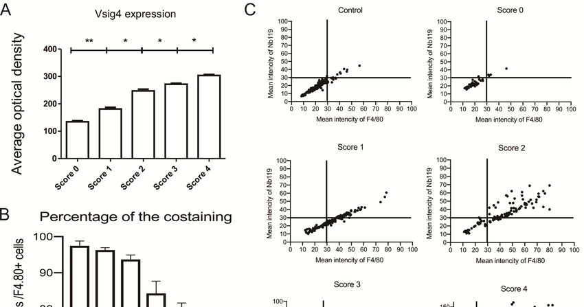
Int. J. Mol. Sci. 2019, 20, x 6 of 13
Int. J. SEM
Mol. Sci.
from2019, 20, 3347
at least three independent experiments for each group. Levels of statistical significance are6 of 14
indicated by asterisks (*= p < 0.05, *** = p < 0.001).
2.3. Histopathology
2.3. Histopathology Staining
Staining of
of Arthritis
Arthritis and
and Fluorescence
Fluorescence Microscopy
Microscopy ex
ex Vivo
Vivo
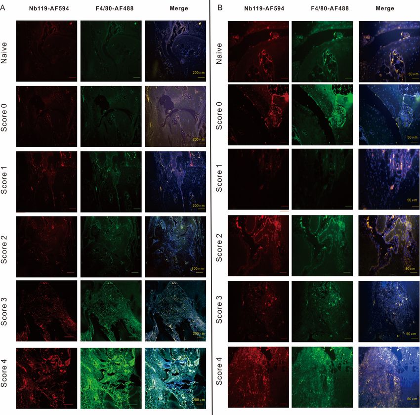
We further
We further analyzed
analyzed Vsig4
Vsig4 expression
expression after
after immunofluorescence
immunofluorescence analysis. Compared with
analysis. Compared with
nonimmunized naive control joints, joints in CIA mice exhibited destruction typicalThe
nonimmunized naive control joints, joints in CIA mice exhibited destruction typical of RA. of inflamed
RA. The
hind ankle
inflamed sections
hind anklewere evaluated
sections by histopathology
were evaluated staining. Hematein
by histopathology eosin staining
staining. Hematein eosin revealed
staining
inflammatory cells infiltration, pannus formation, bone erosion and cartilage distraction
revealed inflammatory cells infiltration, pannus formation, bone erosion and cartilage distraction in ankle
in
sections
ankle with different
sections clinicalclinical
with different score (Figure 4).
score (Figure 4).
Figure 4.
4.Representative
Representative images
images of histopathology sections.
of histopathology NaiveNaive
sections. and arthritic joint from
and arthritic jointeach
fromgroup,
each
stained with hematoxylin
group, stained and eosin
with hematoxylin and(left panel
eosin (leftmagnification, ×100; right
panel magnification, ×100;panel
rightmagnification, ×200).
panel magnification,
×200). Photomicrographs
Photomicrographs of leftankles
of left hind hind ankles from
from five five representative
representative mice
mice are are shown
shown forclinical
for each each clinical
score
score group.
group.
Nb119- Alexa
Nb119- Alexa Fluor
Fluor594
594was
wasnotnotdetected
detectedininthe
thesynovial
synovial tissue
tissue sections
sections of of score
score 0 CIA
0 CIA mice.
mice. In
In contrast, Nb119- Alexa Fluor 594 was highly unregulated in the inflamed region
contrast, Nb119- Alexa Fluor 594 was highly unregulated in the inflamed region lining the CIA lining the CIA
synovium where
synovium where it
it was
was co-localized
co-localized with
with the
the macrophage
macrophage marker,
marker, F4/80
F4/80(Figure
(Figure5).
5).
Moreover, quantitation of Nb119 expression levels further confirmed the marked up-regulation
of Nb119 associated with the clinical scoring and the severity of arthritis indicated expansion of the
synovial macrophages in the late stage of arthritis (Figure 6A). The percentage of Vsig4 and F4/80
contained within F4/80 positive cells in ankle sections of CIA mice confirmed that the majority of the
Vsig4+ cells are F4/80 positive and the Vsig4 low infiltrating macrophages increases in the late stage
of arthritis (Figure 6B). Immunofluorescence analysis of the scatter plotting of each score indicated
a strong correlation of the F4/80 and Vsig4 staining at the lower score group and a more heterogeneous
distribution in the later stage of the inflamed joints (Figure 6C).Int. J. Mol. Sci. 2019, 20, 3347 7 of 14
Int. J. Mol. Sci. 2019, 20, x 7 of 13
Figure5.5.Confocal
Figure Confocalmicroscopy
microscopy demonstrates
demonstrates that Vsig4+ +F4/80
that Vsig4 F4/80 + macrophages
+ macrophages increased according
increased to
according
the severity of arthritis. Immunofluorescence microscopy of CIA joints left
to the severity of arthritis. Immunofluorescence microscopy of CIA joints left hind ankles having hind ankles having
differentclinical
different clinicalscores.
scores.DBA-1
DBA-1 mice
mice were
were immunized
immunized withwith type
type IIII collagen
collagen inin complete
completeFreund’s
Freund’s
adjuvant.Slides
adjuvant. Slideswere
wereincubated
incubated with
with Nb119
Nb119 labelled
labelled by
by AF549
AF549 (red)
(red) and
and AF488-labelled
AF488-labelled anti-F4/80
anti-F4/80
(green).Cell
(green). Cellnuclei
nucleiwere
werestained
stainedwith
with44′,6-diamino-2-phenylindole
0 ,6-diamino-2-phenylindole (DAPI)
(DAPI) dyedye (blue),
(blue),colocalization
colocalizationofof
F4/80 and Vsig4 were shown in the third panel (white). (A) Scale bar = 200 µm. (B) Scalebar
F4/80 and Vsig4 were shown in the third panel (white). (A) Scale bar = 200 μm. (B) Scale bar==400
400μm.
µm.
Photomicrographs of left hind ankles from representative 5 mice are shown for each
Photomicrographs of left hind ankles from representative 5 mice are shown for each clinical score group. clinical score
group.
Moreover, quantitation of Nb119 expression levels further confirmed the marked up-regulation
of Nb119 associated with the clinical scoring and the severity of arthritis indicated expansion of the
synovial macrophages in the late stage of arthritis (Figure 6A). The percentage of Vsig4 and F4/80
contained within F4/80 positive cells in ankle sections of CIA mice confirmed that the majority of the
Vsig4+ cells are F4/80 positive and the Vsig4 low infiltrating macrophages increases in the late stage
of arthritis (Figure 6B). Immunofluorescence analysis of the scatter plotting of each score indicated a
strong correlation of the F4/80 and Vsig4 staining at the lower score group and a more heterogeneous
distribution in the later stage of the inflamed joints (Figure 6C).Int. J. Mol. Sci. 2019, 20, 3347 8 of 14
Int. J. Mol. Sci. 2019, 20, x 8 of 13
Figure 6. Immunofluorescence analysis in ankle sections of CIA mice having different clinical scores:
Figure
(A) Vsig46.expression
Immunofluorescence
in total cells;analysis
(B) the in ankle sections
percentage of CIA
of Vsig4 mice having
and F4/80 containeddifferent
withinclinical scores:
F4/80 positive
(A) Vsig4 expression in total cells; (B) the percentage of Vsig4 and F4/80 contained
cells. Data are presented as mean SEM. Levels of significance were calculated using one-way ANOVA. within F4/80
positive cells. Data are presented as mean SEM. Levels of significance were calculated
Values are means ± SEM (*= p < 0.05, ** = p < 0.01, n = 5). (C) The scatter plots of immunofluorescenceusing one-way
ANOVA.
analysis for Values are means
Vsig4 mean intensity± vs.
SEM (*=mean
F4/80 p < 0.05, ** =inpankle
intensity < 0.01, n = 5).
sections (C) mice
of CIA The scatter plots of
having different
immunofluorescence
clinical analysis
scores, scatter plots forhind
of left Vsig4 meanfrom
ankles intensity vs. F4/80 mean
five representative intensity
mice are shownin ankle sections
for each of
clinical
CIA mice
score group.having different clinical scores, scatter plots of left hind ankles from five representative mice
are shown for each clinical score group.
3. Discussion
3. Discussion
Imaging techniques will improve the understanding of the arthritis pathogenesis, assessing the
courseImaging techniquesailments
of inflammatory will improve the understanding
and permitting treatment offollow-up
the arthritisforpathogenesis,
these diseases. assessing
Examples the
course of inflammatory ailments and permitting treatment follow-up for
include the labeling of antibodies or antibody fragments against markers on macrophages such as these diseases. Examples
include
F4/80, MMRthe [31]
labeling of antibodies
and Vsig4 or antibody
[27], or against fragments cell
the endothelial against
markermarkers on macrophages
E-selectin [32]. A marker such as
such
F4/80,
as folateMMR [31] and
receptor, thatVsig4
binds[27],folicor against
acid withthe
highendothelial
affinity, iscell marker E-selectin
associated with metabolic[32]. Aactivity
markerinsuchthe
as folate
joints and receptor,
is uniquely that binds folic acid
overexpressed with highmacrophages
on activated affinity, is associated with metabolic
[12,33]. Probes correlatedactivity in the
with reactive
joints and
oxygen is uniquely
species production, overexpressed
cathepsins or oninflammation-associated
activated macrophages [12,33]. enzymesProbessuch as correlated
MMPs orwith cell
reactive oxygen species production, cathepsins or inflammation-associated
death were assessed in CIA by using a near-infrared PSVue 794 dye [34]. enzymes such as MMPs
or cell death wereimaging
Fluorescence assessedhas in the
CIAadvantages
by using a ofnear-infrared PSVue
high sensitivity and794 dye [34].and lower instrument
resolution
Fluorescence
cost compared imaging imaging
to molecular has the with
advantages
SPECT of high techniques,
or PET sensitivity although
and resolution and lower
tissue penetration
instrument cost compared to molecular imaging with SPECT or PET techniques,
represents the main challenge of optical imaging. This limitation can be partially resolved by adopting although tissue
penetration represents the main challenge of optical imaging. This limitation
NIR light, which provides a wider dynamic range and minimal background with lower scattering can be partially resolved
by adopting
than NIR light, which
visible fluorescence provides
detection. NIRF a wider dynamic
imaging range
has been and minimal
frequently usedbackground
in athymic withnudelower
mice.
scattering than visible fluorescence detection. NIRF imaging has been
However, the DBA/1 or C6 mice, which are frequently used to establish the RA model, cause highfrequently used in athymic
nude mice.signal
unspecific However, the DBA/1
originating fromortheir
C6 mice, whichthe
fur. Since areinflamed
frequently areaused to establish
of RA is located theinRA
themodel,
paws,
cause high unspecific signal originating from their fur. Since the inflamed area
which are hardly influenced by the autofluorescence of their fur, NIRF imaging has been widely used of RA is located in the
paws, which are hardly influenced by the autofluorescence of their fur,
in preclinical arthritis studies nowadays. An ideal florescence probe could be multi-functional andNIRF imaging has been
widely different
support used in preclinical
purposes such arthritis studies
as flow nowadays.
cytometry, An idealmicroscopy
fluorescence florescenceand probe could
optical be multi-
imaging.
functional and support different purposes such as flow cytometry, fluorescence microscopy and
optical imaging.Int. J. Mol. Sci. 2019, 20, 3347 9 of 14
Nbs can be an easy and effective way to identify macrophages from the complex in vivo
condition based on the detection of Vsig4 expressed on the macrophages. Recently, we developed
and characterized Nb119 and evaluated Nb-based Technetium-99m radio-labelled tracers for
SPECT/micro-CT and fluorine-18 labelled tracers for PET [35]. In a previous study, we confirmed that
Vsig4-specific nanobody targeting synovial macrophages constitutes a specific tool for non-invasive
SPECT/CT imaging as a way of detecting early signs of inflammation and assessing inflammation in
multiply arthritis models in vivo [26,27]. In this study, we wanted to assess the ability of the Vsig4
nanobody to act as an optical probe for NIRF imaging. Here, we used NIRF-dye Cy7-conjugated
nanobody and isotype control nanobody for a direct comparison of in vivo and ex vivo analyses.
A limitation of our study is that we did not optimize the amount of fluorescent dyes per
nanobody, which might affect the maximum achievable signal for imaging and fluorescence. Molecular
random conjugation could cause further loss of affinity when lysine residues are located in or close
to the antigen-binding region [13]. Recently, we crystalized the Vsig4-Nb119 complex and solved
high-resolution atomic structure, showing that Nb119 has four lysines dispersed in the framework
and away from the antigen-binding region [29]. Accordingly, although the labeling strategy can
be improved by site-specific conjugation of the NIRF dye, Vsig4 nanobody random conjugation to
primary amine groups did not affect binding performance for in vivo imaging. In the CIA model,
which is readily inducible in DBA/1 mice, Nb119-Cy7 specifically detected the inflamed lesions that
were found in the arthritic mice but not in the naive mice. Nb119 signals formed a positive hot spot
only in inflamed paws featuring disease development in the CIA model. Radiant efficiency and T/B
ratio of Cy7-Nb119 in inflamed paws was significantly higher as compared to Cy7-BCII10. Similar
signals were even detected in the non-symptomatic joints group as in the symptomatic joints group by
using Cy7-BCII10.
In this study, we injected fixed doses of 100 µg nanobody regardless of the weight of the animals,
instead of performing weight-adapted injections. The differences in weight of individual mice (range
20.6–24.2 g) may explain some of the observed signal variations within the Cy7-Vsig4 and Cy7-BCII10
groups. The large size of conventional antibodies impedes tissue penetration and renal elimination,
resulting in suboptimal in vivo targeting. The nanobody often shows higher contrast in a short time
as alternatives to monoclonal antibodies as theranostics [17,18]. Our study confirmed that Vsig4
nanobody allowed 3 h post injection imaging with high target-to-background ratio, which indicates
that small single-domain nanobodies are best suited for short-term uses, such as noninvasive imaging.
These data are in line with the histochemistry in the naive and arthritic joint from each group,
stained with hematoxylin and eosin corresponding to the rise of clinical scores (Figure 5). In our
previous research, we showed Vsig4 expression on a subset of CD68 positive synovial macrophages [27].
CD68 is a broad marker and highly expressed by cells including circulating macrophages, osteoclasts
and tissue macrophages. In the current study, we used a commonly used more specific macrophage
marker F4/80 for symposium macrophages staining. The increasing signal of immunofluorescence
staining with Alexa Fluor 549 labelled Nb119 and Alexa Fluor 488 labelled F4/80 confirms the massive
increase of synovial macrophages in the inflamed joints. The Nb119 and F4/80 positive signals were
highly correlated at the low score joints but showed less percentage of co-staining in the more serve
inflamed joints, suggesting increasing heterogeneity in the population of infiltrating macrophages.
Synovial macrophages are attractive targets for imaging because they seem to play a pivotal role in
maintaining the chronic inflammation state in arthritis. As such, using fluorescent Vsig4 nanobody
can indeed provide molecular information via flow cytometry, immunofluorescence microscopy and
in vivo imaging as compared to the clinical score.Int. J. Mol. Sci. 2019, 20, 3347 10 of 14
4. Materials and Methods
4.1. Mice and Cells
Eight-week old male C57Bl/6 and DBA/1 mice were purchased from Charles River Company
(Wilmington, Massachusetts, USA). The experiments were approved by the local ethics committee
under approval code 2015-246 on March 4th, 2015. For Figure 3, five mice per group were used for
Cy7-BCII10 and Cy7-Nb119 injection. For Figures 4–6, 30 left hind paws were selected from at least
three individual experiments in total according to each score group (naïve, score 0–4), and 5 paws
fell into each group after evaluation. The peritoneal macrophages (PECs) were obtained by injecting
8 mL of cold RPMI containing 2% endotoxin-free FCS in the peritoneum using a 10 mL syringe with
an 18-gauge needle and drawing the fluid back into the syringe. PECs were washed three times by
centrifugation at 300 g for 10 min at 4 ◦ C and resuspended in PBS containing 2% FCS.
4.2. Induction of CIA Model and Assessment of Arthritis
CIA was induced as described before. Briefly, 2 mg/mL chicken collagen type II (Sigma-Aldrich,
Shanghai, China) in 0.1 M acetic acid was emulsified in complete Freund’s adjuvant (Difco Laboratories,
Detroit, Michigan, USA). Mice were sensitized with a subcutaneous injection at the base of the tail
with 100 µL emulsion. A boost injection with 2 mg/mL chicken collagen type II in incomplete Freund’s
adjuvant was performed at day 21. Each limb was scored for severity of arthritis as follows: 0 = normal;
1 = redness/swelling of one joint; 2 = redness/swelling of more than one joint; 3 = swelling of entire
paw; 4 = ankylosis and/or deformity. The arthritis mice at day28 post induction were used in the NIRF
imaging experiments.
4.3. Nanobody
Vsig4-specific Nbs (Nb119) were prepared from lymphocytes isolated from an alpaca immunized
with the recombinant mouse Vsig4 protein, selected via phage display and biopanning and produced as
described previously [27]. The screened Nb119 gene sequence was inserted into the pHEN6c plasmid
and transfected in the E. coli WK6 host cell. Nbs against the β-lactamase BCII enzyme of Bacillus cereus
(BCII10) were used as a negative control throughout the study. The pHEN6C display vectors permits
the inducible periplasmic expression of Nanobodies as soluble C-terminally His6 tagged proteins in
E. coli strain WK6. All the proteins were loaded on a NI-NTA column and further buffer exchanged
and purified by size exclusion chromatography (SEC) for further usage [29].
4.4. Cytofluorometry Analysis
Nb119 and NbBCII10 monoclonal antibody were labelled using alexa fluor-647 lightning-link APC
conjugation kit (Innova biosciences, San Diego, CA, USA). Non-activated macrophages were harvested
from the peritoneal cavity of mice. About 5 × 10⁵ cells were washed three times with PBS-2% FCS
and resuspended in a total volume of 100 µL. Anti-F4/80 (Cl:A3-1)/Alexa fluor 488 and Anti-Mouse
CD11b (M1/70)/PE antibodies were purchased from AbD Serotec and Ebioscience. Anti-Vsig4 (NLA14)
monoclonal antibody and rat IgG2a kappa isotype control were purchased from Ebioscience. Five µg of
antibody or nanobody were used per 1 × 10⁶ cells for 20 min at 4 ◦ C. Excess fluorescein labelled antibody
was removed by washing with PBS-2% BSA. Stained cells were further analyzed by flow cytometry
and histograms were prepared using FlowJo software (Becton Dickinson, San Jose, CA, USA).
4.5. Nanobody-Cy7 Labeling and NIRF Imaging in Vivo Analysis
Nbs were labelled using the Cyanine7 NHS ester (Lumiprobe, Excitation maximum, nm: 750,
Emission maximum, nm: 773) according to the manufacturers’ instructions. Briefly, 2 mg nanobody
was incubated overnight on ice with × 8 molar excess Cyanine7 NHS ester in 0.1 M Sodium bicarbonate
solution at pH 8.5. The Nanobody-Cy7 solution was purified by size-exclusion chromatography andInt. J. Mol. Sci. 2019, 20, 3347 11 of 14
then passed through a Millex-GV4 0.22-mm filter (Millipore, Burlington, MA, USA). The purity of
Vsig4 and BCII10 nanobody was assessed by SDS-PAGE size fractionation, followed by Coomassie
brilliant blue stain.
4.6. NIRF Imaging
C57BL/6J mice were injected intravenously with 100 µL of 100 µg Nanobody-Cy7. At 3 h post
injection, mice were anesthetized with 3% isoflurane using an XGI-8 anesthesia system in the induction
chamber; then, 1%–2% isoflurane was maintained for the duration of the imaging procedure using the
isoflurane manifold housed inside the imaging chamber and the mice were positioned in the imaging
chamber of the small-animal NIRF-imaging system XENOGEN IVIS® SPECTRUM (Beijing, China).
Nanobody-Cy7 accumulation in each paw of the mice was quantified as average radiant efficiency
normalized for the size of the region of interest. Joints to background ratio was calculated by dividing
the values of the joints uptake by the background value determined from the hind limb.
4.7. Microscopy
Inflamed joints of CIA mice were fixed in 3% paraformaldehyde (pH 7.4) and then decalcified
for two weeks in 10% EDTA (pH 7.4). The joints were embedded in Tissue-Tek OCT and frozen in
liquid nitrogen. Cryostat sections (5 µm) were either stained with hematoxylin and eosin as standard
protocol and either incubated in 1% PBS/BR containing detection antibodies for immunofluorescence
microscopy. Anti-F4/80 (Cl: A3-1)/Alexa Fluor 488 was purchased from AbD Serotec and Nb was
labelled by Alexa Fluor 594 (Alexa Fluor 594 NHS Ester, Shanghai, China) purchased from Invitrogen.
2 µg of antibody and Nb in 1% PBS-BR were used per slide. Fluoro-Gel II/DAPI (Electron Microscopy
Sciences, Shanghai, China) mounted slides were used for fluorescence microscopy with an UPlanFI 10×,
20× or 30× objective on an OLYMPUS BX51 microscope with OLYMPUS DP71 CCD and OLYMPUS
DP Controller software (Shanghai, China).
4.8. Statistical Analysis
Statistical analyses were conducted using the Student’s t test and one-way ANOVA assuming
unequal variances. Prism 6.0 (Graphpad software, San Diego, CA, USA) was used for statistical
analyses and graph creation. p-values ≤ 0.05 were considered significant.
5. Conclusions
Our studies have confirmed that nanobody targeting Vsig4 constitute a specific tool for
non-invasive fluorescence in vivo imaging as a way of assessing inflammation in arthritis models
in vivo. The flow cytometry and immunofluorescence microscopy studies also indicate that fluorescence
labelled NbVsig4 appears to serve as a convenient probe for distinguishing macrophages. We showed
that fluorescent nanobodies are well suited as diagnostic tools for rapid and specific in vivo detection
of macrophages with clear tissue penetration when performing optical imaging in vivo, providing
complementary and more molecular information as compared to paw swelling and clinical scoring.
As it is nonradioactive, highly sensitive, inexpensive, and comparatively easy use to produce targeted
probes, we advocate the use of the NIRF imaging technique for evaluation of nanobody in preclinical
molecular imaging experiments.
Supplementary Materials: Supplementary materials can be found at http://www.mdpi.com/1422-0067/20/13/
3347/s1.
Author Contributions: Conceptualization, F.Z. and Y.W.; Data curation, F.Z.; Investigation, F.Z., S.L., Z.O., J.Z.
and H.M.; Methodology, F.Z., S.M., P.D.B. and G.R.; Writing—original draft, F.Z. and Y.W.; Writing—review &
editing, S.S., S.M., P.D.B., G.R. and Y.W.
Funding: This work was supported by National Natural Science Foundation of China (NO. 81501527 to F.Z.,
NO. 31870132, NO. 81772745 to YW).Int. J. Mol. Sci. 2019, 20, 3347 12 of 14
Acknowledgments: The authors thank Fujun Zhang for immunofluorescence microscopy technical and assistance.
Conflicts of Interest: The authors declare no conflict of interest
Abbreviations
NIRF Near-infrared fluorescence
Vsig4 V-set and Ig domain-containing 4
CIA Collagen-induced arthritis
References
1. Chen, Z.; Bozec, A.; Ramming, A.; Schett, G. Anti-inflammatory and immune-regulatory cytokines in
rheumatoid arthritis. Nat. Reviews. Rheumatol. 2019, 15, 9–17. [CrossRef] [PubMed]
2. Udalova, I.A.; Mantovani, A.; Feldmann, M. Macrophage heterogeneity in the context of rheumatoid arthritis.
Nat. Reviews. Rheumatol. 2016, 12, 472–485. [CrossRef] [PubMed]
3. Rocha, B.; Ruiz-Romero, C.; Blanco, F.J. Mass spectrometry imaging: A novel technology in rheumatology.
Nat. Reviews. Rheumatol. 2017, 13, 52–63. [CrossRef]
4. Ornelas, A.; McCullough, C.R.; Lu, Z.; Zacharias, N.M.; Kelderhouse, L.E.; Gray, J.; Yang, H.; Engel, B.J.;
Wang, Y.; Mao, W.; et al. Induction of autophagy by arhi (diras3) alters fundamental metabolic pathways in
ovarian cancer models. BMC Cancer 2016, 16, 824. [CrossRef] [PubMed]
5. Scales, H.E.; Ierna, M.; Smith, K.M.; Ross, K.; Meiklejohn, G.R.; Patterson-Kane, J.C.; McInnes, I.B.; Brewer, J.M.;
Garside, P.; Maffia, P. Assessment of murine collagen-induced arthritis by longitudinal non-invasive duplexed
molecular optical imaging. Rheumatology 2016, 55, 564–572. [CrossRef]
6. Kelderhouse, L.E.; Mahalingam, S.; Low, P.S. Predicting response to therapy for autoimmune
and inflammatory diseases using a folate receptor-targeted near-infrared fluorescent imaging agent.
Mol. Imaging Biol. 2016, 18, 201–208. [CrossRef]
7. Reum Son, A.; Kim, D.Y.; Hun Park, S.; Yong Jang, J.; Kim, K.; Ju Kim, B.; Yun Yin, X.; Ho Kim, J.; Hyun
Min, B.; Keun Han, D.; et al. Direct chemotherapeutic dual drug delivery through intra-articular injection for
synergistic enhancement of rheumatoid arthritis treatment. Sci. Rep. 2015, 5, 14713. [CrossRef]
8. Muyldermans, S.; Baral, T.N.; Retamozzo, V.C.; De Baetselier, P.; De Genst, E.; Kinne, J.; Leonhardt, H.;
Magez, S.; Nguyen, V.K.; Revets, H.; et al. Camelid immunoglobulins and nanobody technology.
Vet. Immunol. Immunopathol. 2009, 128, 178–183. [CrossRef]
9. Hassanzadeh-Ghassabeh, G.; Devoogdt, N.; De Pauw, P.; Vincke, C.; Muyldermans, S. Nanobodies and their
potential applications. Nanomedicine 2013, 8, 1013–1026. [CrossRef]
10. Vaneycken, I.; Devoogdt, N.; Van Gassen, N.; Vincke, C.; Xavier, C.; Wernery, U.; Muyldermans, S.;
Lahoutte, T.; Caveliers, V. Preclinical screening of anti-her2 nanobodies for molecular imaging of breast
cancer. FASEB J. 2011, 25, 2433–2446. [CrossRef]
11. Mwangi, T.K.; Berke, I.M.; Nieves, E.H.; Bell, R.D.; Adams, S.B.; Setton, L.A. Intra-articular clearance of
labeled dextrans from naive and arthritic rat knee joints. J. Control. Release 2018, 283, 76–83. [CrossRef]
[PubMed]
12. Slooter, M.D.; Bierau, K.; Chan, A.B.; Lowik, C.W. Near infrared fluorescence imaging for early detection,
monitoring and improved intervention of diseases involving the joint. Connect. Tissue Res. 2015, 56, 153–160.
[CrossRef] [PubMed]
13. Debie, P.; Van Quathem, J.; Hansen, I.; Bala, G.; Massa, S.; Devoogdt, N.; Xavier, C.; Hernot, S. Effect of dye
and conjugation chemistry on the biodistribution profile of near-infrared-labeled nanobodies as tracers for
image-guided surgery. Mol. Pharm. 2017, 14, 1145–1153. [CrossRef] [PubMed]
14. Debie, P.; Vanhoeij, M.; Poortmans, N.; Puttemans, J.; Gillis, K.; Devoogdt, N.; Lahoutte, T.; Hernot, S.
Improved debulking of peritoneal tumor implants by near-infrared fluorescent nanobody image guidance in
an experimental mouse model. Mol. Imaging Biol. 2018, 20, 361–367. [CrossRef] [PubMed]
15. van Driel, P.B.; van der Vorst, J.R.; Verbeek, F.P.; Oliveira, S.; Snoeks, T.J.; Keereweer, S.; Chan, B.; Boonstra, M.C.;
Frangioni, J.V.; van Bergen en Henegouwen, P.M.; et al. Intraoperative fluorescence delineation of head and
neck cancer with a fluorescent anti-epidermal growth factor receptor nanobody. Int. J. Cancer 2014, 134,
2663–2673. [CrossRef]Int. J. Mol. Sci. 2019, 20, 3347 13 of 14
16. Bannas, P.; Lenz, A.; Kunick, V.; Fumey, W.; Rissiek, B.; Schmid, J.; Haag, F.; Leingartner, A.; Trepel, M.;
Adam, G.; et al. Validation of nanobody and antibody based in vivo tumor xenograft nirf-imaging experiments
in mice using ex vivo flow cytometry and microscopy. J. Vis. Exp. 2015, e52462. [CrossRef]
17. Bannas, P.; Lenz, A.; Kunick, V.; Well, L.; Fumey, W.; Rissiek, B.; Haag, F.; Schmid, J.; Schutze, K.;
Eichhoff, A.; et al. Molecular imaging of tumors with nanobodies and antibodies: Timing and dosage are
crucial factors for improved in vivo detection. Contrast Media Mol. Imaging 2015, 10, 367–378. [CrossRef]
18. Bannas, P.; Well, L.; Lenz, A.; Rissiek, B.; Haag, F.; Schmid, J.; Hochgrafe, K.; Trepel, M.; Adam, G.;
Ittrich, H.; et al. In vivo near-infrared fluorescence targeting of t cells: Comparison of nanobodies and
conventional monoclonal antibodies. Contrast Media Mol. Imaging 2014, 9, 135–142. [CrossRef]
19. Benson, R.A.; McInnes, I.B.; Brewer, J.M.; Garside, P. Cellular imaging in rheumatic diseases.
Nat. Reviews. Rheumatol. 2015, 11, 357–367. [CrossRef]
20. Vogt, L.; Schmitz, N.; Kurrer, M.O.; Bauer, M.; Hinton, H.I.; Behnke, S.; Gatto, D.; Sebbel, P.; Beerli, R.R.;
Sonderegger, I.; et al. Vsig4, a b7 family-related protein, is a negative regulator of t cell activation.
J. Clin. Investig. 2006, 116, 2817–2826. [CrossRef]
21. Condeelis, J.; Pollard, J.W. Macrophages: Obligate partners for tumor cell migration, invasion, and metastasis.
Cell 2006, 124, 263–266. [CrossRef] [PubMed]
22. Li, J.; Diao, B.; Guo, S.; Huang, X.; Yang, C.; Feng, Z.; Yan, W.; Ning, Q.; Zheng, L.; Chen, Y.; et al. Vsig4
inhibits proinflammatory macrophage activation by reprogramming mitochondrial pyruvate metabolism.
Nat. Commun. 2017, 8, 1322. [CrossRef] [PubMed]
23. Roh, J.; Jeon, Y.; Lee, A.N.; Lee, S.M.; Kim, Y.; Sung, C.O.; Park, C.J.; Hong, J.Y.; Yoon, D.H.; Suh, C.; et al.
The immune checkpoint molecule v-set ig domain-containing 4 is an independent prognostic factor for
multiple myeloma. Oncotarget 2017, 8, 58122–58132. [CrossRef] [PubMed]
24. Liao, Y.; Guo, S.; Chen, Y.; Cao, D.; Xu, H.; Yang, C.; Fei, L.; Ni, B.; Ruan, Z. Vsig4 expression on macrophages
facilitates lung cancer development. Lab. Investig. 2014, 94, 706–715. [CrossRef] [PubMed]
25. Byun, J.M.; Jeong, D.H.; Choi, I.H.; Lee, D.S.; Kang, M.S.; Jung, K.O.; Jeon, Y.K.; Kim, Y.N.; Jung, E.J.;
Lee, K.B.; et al. The significance of vsig4 expression in ovarian cancer. Int. J. Gynecol. Cancer 2017, 27, 872–878.
[CrossRef] [PubMed]
26. Zheng, F.; Perlman, H.; Matthys, P.; Wen, Y.; Lahoutte, T.; Muyldermans, S.; Lu, S.; De Baetselier, P.;
Schoonooghe, S.; Devoogdt, N.; et al. Specificity evaluation and disease monitoring in arthritis imaging
with complement receptor of the ig superfamily targeting nanobodies. Sci. Rep. 2016, 6, 35966. [CrossRef]
[PubMed]
27. Zheng, F.; Put, S.; Bouwens, L.; Lahoutte, T.; Matthys, P.; Muyldermans, S.; De Baetselier, P.; Devoogdt, N.;
Raes, G.; Schoonooghe, S. Molecular imaging with macrophage crig-targeting nanobodies for early and
preclinical diagnosis in a mouse model of rheumatoid arthritis. J. Nucl. Med. 2014, 55, 824–829. [CrossRef]
28. Zheng, F.; Devoogdt, N.; Sparkes, A.; Morias, Y.; Abels, C.; Stijlemans, B.; Lahoutte, T.; Muyldermans, S.;
De Baetselier, P.; Schoonooghe, S.; et al. Monitoring liver macrophages using nanobodies targeting vsig4:
Concanavalin a induced acute hepatitis as paradigm. Immunobiology 2015, 220, 200–209. [CrossRef] [PubMed]
29. Wen, Y.; Ouyang, Z.; Schoonooghe, S.; Luo, S.; De Baetselier, P.; Lu, W.; Muyldermans, S.; Raes, G.;
Zheng, F. Structural evaluation of a nanobody targeting complement receptor vsig4 and its cross reactivity.
Immunobiology 2017, 222, 807–813. [CrossRef] [PubMed]
30. Brand, D.D.; Latham, K.A.; Rosloniec, E.F. Collagen-induced arthritis. Nat. Protoc. 2007, 2, 1269–1275.
[CrossRef] [PubMed]
31. Put, S.; Schoonooghe, S.; Devoogdt, N.; Schurgers, E.; Avau, A.; Mitera, T.; D’Huyvetter, M.; De Baetselier, P.;
Raes, G.; Lahoutte, T.; et al. Spect imaging of joint inflammation with nanobodies targeting the macrophage
mannose receptor in a mouse model for rheumatoid arthritis. J. Nucl. Med. 2013, 54, 807–814. [CrossRef]
[PubMed]
32. Gompels, L.L.; Madden, L.; Lim, N.H.; Inglis, J.J.; McConnell, E.; Vincent, T.L.; Haskard, D.O.; Paleolog, E.M.
In vivo fluorescence imaging of e-selectin: Quantitative detection of endothelial activation in a mouse model
of arthritis. Arthritis Rheum. 2011, 63, 107–117. [CrossRef] [PubMed]
33. Zhang, N.; Xu, C.; Li, N.; Zhang, S.; Fu, L.; Chu, X.; Hua, H.; Zeng, X.; Zhao, Y. Folate receptor-targeted mixed
polysialic acid micelles for combating rheumatoid arthritis: In vitro and in vivo evaluation. Drug Deliv. 2018,
25, 1182–1191. [CrossRef] [PubMed]Int. J. Mol. Sci. 2019, 20, 3347 14 of 14
34. Put, S.; Westhovens, R.; Lahoutte, T.; Matthys, P. Molecular imaging of rheumatoid arthritis: Emerging
markers, tools, and techniques. Arthritis Res. Ther. 2014, 16, 208. [CrossRef] [PubMed]
35. Cleeren, F.; Lecina, J.; Bridoux, J.; Devoogdt, N.; Tshibangu, T.; Xavier, C.; Bormans, G. Direct fluorine-18
labeling of heat-sensitive biomolecules for positron emission tomography imaging using the al(18)f-resca
method. Nat. Protoc. 2018, 13, 2330–2347. [CrossRef] [PubMed]
© 2019 by the authors. Licensee MDPI, Basel, Switzerland. This article is an open access
article distributed under the terms and conditions of the Creative Commons Attribution
(CC BY) license (http://creativecommons.org/licenses/by/4.0/).You can also read