Bruton's tyrosine kinase regulates macrophage induced inflammation in the diabetic kidney via NLRP3 inflammasome activation
←
→
Page content transcription
If your browser does not render page correctly, please read the page content below
INTERNATIONAL JOURNAL OF MOLECULAR MEDICINE 48: 177, 2021

           Bruton's tyrosine kinase regulates macrophage‑induced
                  inflammation in the diabetic kidney via
                      NLRP3 inflammasome activation
               JING ZHAO1, JUAN CHEN1, YUAN‑YUAN LI1, LING‑LING XIA2* and YONG‑GUI WU1*

                     Departments of 1Nephropathy and 2Infectious Diseases, The First Affiliated Hospital of
                                 Anhui Medical University, Hefei, Anhui 230022, P.R. China

                                        Received January 22, 2021; Accepted June 18, 2021

                                                    DOI: 10.3892/ijmm.2021.5010

Abstract. It has been previously reported that macrophages            end‑stage renal disease (1). DN is mainly manifested as the
may be involved in diabetic nephropathy (DN) development.             thickening of the glomerular basement membrane (GBM),
Furthermore, Bruton's tyrosine kinase (BTK) may participate           the proliferation of mesangial cells and extracellular matrix,
in macrophage activation and lead to the release of inflam‑           and renal tubulointerstitial fibrosis (2). Furthermore, its
matory mediators. The main aim of the present study was               pathogenesis is complex, involving hereditary, metabolic and
to analyze the association between renal BTK expression               hemodynamic changes, as well as inflammation. A recent
and clinical indicators. Moreover, BTK knockout mice were             study has reported the correlation between DN and inflamma‑
used to establish a diabetic model for further research. The          tion (3). Macrophages are a type of immune cell and are derived
results demonstrated that BTK was activated in the kidneys            from monocytes. Macrophages have multiple functions, such
of patients with DN and was associated with the progression           as phagocytosing cell fragments, releasing chemokines factors
of proteinuria, creatinine levels, estimated glomerular filtra‑       and secreting inflammatory mediators (4). Furthermore, it
tion rate and pathological changes in the kidneys of patients         has been established that macrophages play a key role in the
with DN. Furthermore, BTK knockout was observed to reduce             occurrence and development of DN (5,6).
urinary protein excretion, alleviate renal injury and decrease            Bruton's tyrosine kinase (BTK) is an intracellular
renal inflammation in diabetic mice. This protection may be           non‑receptor tyrosine kinase, and is one of the five members of
attributed to BTK‑induced suppression of the activation of the        the Tec tyrosine kinase family (7). BTK is expressed in myeloid
Nod‑like receptor (NLR) family pyrin domain containing 3              cells, such as macrophages and tissue plasma cells. Moreover,
inflammasome. Collectively, it has been demonstrated in               BTK signaling molecules have been well‑documented and
the present study that BTK may be a potential target for DN           it has been considered as an important signal in immuno‑
treatment.                                                            regulation (8). It has recently demonstrated that BTK is crucial
                                                                      for the proliferation, development and differentiation of
Introduction                                                          B cells (9). In addition, BTK may be involved in human innate
                                                                      immunity, particularly in relation to B cell and macrophage
Diabetic nephropathy (DN) is a severe microvascular compli‑           involvement (10). It has also been demonstrated that BTK can
cation in diabetic patients and has been the main cause of            conduct cell signals by regulating Toll‑like receptor (TLR)2
                                                                      and TLR4 in macrophages (11). Previous studies have proved
                                                                      that BTK‑regulated inflammation is involved in various renal
                                                                      diseases, such as IgA nephropathy and Lupus nephritis (12,13).
Correspondence to: Professor Ling‑Ling Xia, Department of             The BTK inhibitor PCI‑32765, also known as ibrutinib, has
Infectious Diseases, The First Affiliated Hospital of Anhui Medical   been widely used as an antitumor drug for the treatment of
University, 218 Jixi Road, Hefei, Anhui 230022, P.R. China            chronic lymphocytic leukemia (14). It has been reported that
E‑mail: 13966684365@163.com                                           ibrutinib can reduce MAPK and NF‑κ B pathway activation,
                                                                      and then decrease the release of inflammatory mediators in
Professor Yong‑Gui Wu, Department of Nephropathy, The First
Affiliated Hospital of Anhui Medical University, 218 Jixi Road,       macrophages (15). Moreover, a recent study demonstrated that
Hefei, Anhui 230022, P.R. China                                       a BTK inhibitor may serve as an effective therapeutic strategy
E‑mail: wuyonggui@medmail.com.cn                                      in severe COVID‑19, due to its excellent anti‑inflammatory
                                                                      effect (16).
Key words: diabetic nephropathy, Bruton's tyrosine kinase,                The inflammasome is a multi‑protein complex expressed
Nod‑like receptor family pyrin domain containing 3, macrophage,       in myeloid cells, and is mainly composed of sensors,
inflammation                                                          apoptosis‑associated speck like protein containing a caspase
                                                                      recruitment domain (ASC), and the caspase protease.
                                                                      Inflammasomes exist in the cytoplasm of multiple cell types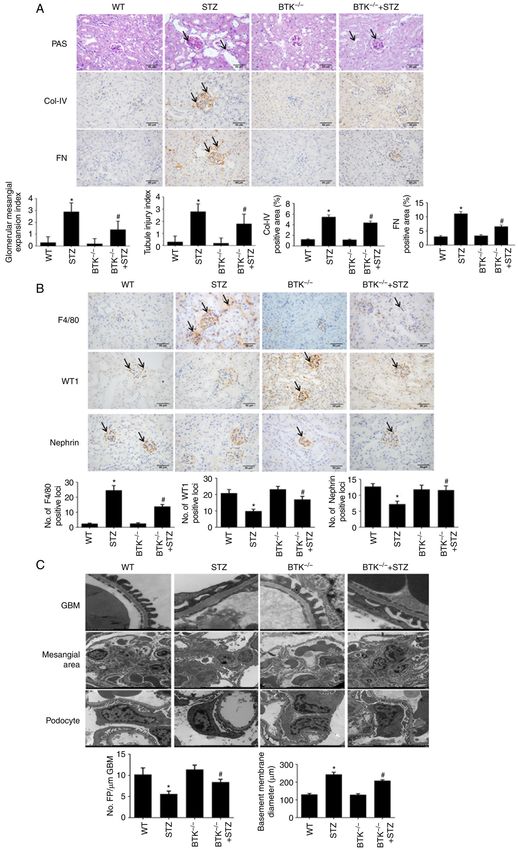
2 ZHAO et al: BRUTON'S TYROSINE KINASE REGULATES INFLAMMATION IN THE DIABETIC KIDNEY
and have the ability to induce the innate immune responses Center of Nanjing Medical University. The embryos were
by sensing damage signals and microbial attacks (17). The removed from liquid nitrogen and were resuscitated in a water
Nod‑like receptor (NLR) family pyrin domain containing 3 bath for 2 min at 35˚C, then immediately placed in a Petri dish
(NLRP3) inflammasome is the most widely studied inflamma‑ with thawing solution (0.1 M sucrose PBS with 6% glycerin)
some, and is composed of NLRP3, ASC and pro‑caspase‑1 (18). for 1 min, followed by diluent solution (0.1 M sucrose PBS with
Moreover, the NLRP3 inflammasome can be activated and 3% glycerin) for 3 min. The embryos were then transferred to
is known to participate in the development of inflamma‑ the washing solution (0.1 M sucrose PBS with 0% glycerin) for
tion by cleaving the inactive cytokines, IL‑1β precursor and 3 min twice. The embryos were then cultured in HTF medium
IL‑18 precursor, into active IL‑1β and IL‑18 (19,20). Recent (cat. no. MR‑070‑D; EMD Millipore) in an incubator at 37˚C
studies have reported that the activation of the NLRP3 inflam‑ with 5% CO2. Following 24 h of culture at 37˚C, the normal
masomes plays an important role in diabetic kidney disease embryos were selected and transplanted into the uterus of
(DKD) (21,22). Additionally, inflammasome activation marker 15 female C57BL/6J mice (age, 6‑8 weeks; weight, 18‑22 g,
expression levels, such as caspase‑1, IL‑1β and IL‑18, have obtained from the Experimental Animal Center of Nanjing
been shown to be positively associated with the severity of Medical University) (30,31). The offspring from the female
albuminuria in patients with DN (23,24). mice (the BTK‑floxed mice) were then crossed with 6 male and
Although the therapeutic potential of NLRP3 is undisputed, 9 female CMV‑cre transgenic mice (age, 6‑8 weeks; weight,
currently no clinically approved therapies exist that target the 18‑22 g; C57BL/6J genetic background, obtained from the
NLRP3 inflammasome directly, at least to the best of our Experimental Animal Center of Nanjing Medical University),
knowledge (25). Recently, BTK has attracted increased atten‑ and the offspring were self‑crossed to obtain BTK knockout
tion as a regulator of NLRP3. It has been demonstrated that mice (BTK‑/‑ mice).
the inhibition of BTK can reduce the inflammatory response Streptozotocin (STZ) was purchased from Sigma‑Aldrich
by decreasing the activation of the NLRP3 inflammasome in (Merck KGaA). The mice were administered STZ daily,
numerous diseases, such as ischemic brain injury, diet‑induced at a dose of 50 mg/kg of body weight for 5 days. In total,
metabolic inflammation and polymicrobial sepsis (26‑28). 28 mice were randomly divided into 4 groups as follows:
However, the role of BTK in DN remains to be elucidated. On i) Wild‑type (WT) group (WT littermate C57BL/6J mice;
this basis, the present study aimed to investigate the effect of n=6); ii) STZ group (WT littermate C57BL/6J mice + STZ;
BTK on DN and its association with the NLRP3 inflamma‑ n=8); iii) BTK‑/‑ group (n=6); and iv) BTK‑/‑ + STZ group
some in the kidneys. (BTK‑/‑ + STZ; n=8). All mice were maintained under standard
feeding conditions (temperature, 20±2˚C; humidity, 45‑55%;
Materials and methods 12‑h light/dark cycle) and were granted free access to food
and water. After 12 weeks of rearing, all mice were anesthe‑
Recruitment. A total of 49 patients with type 2 DN and 18 tized with an intraperitoneal injection of sodium pentobarbital
healthy individuals were recruited from the Department of (50 mg/kg body weight) and euthanized via exsanguination by
Nephrology in The First Affiliated Hospital of Anhui Medical drawing 1.2 ml blood. All mice were observed for 20 min to
University between January 1, 2017 and January 1, 2020. All verify death, according to the criterion of breathing cessation
patients exhibited no fever symptoms or infectious diseases, and cardiac arrest. The experimental humane endpoints that
and were diagnosed via renal biopsy. Renal tissue sections from were established in the present study, included loss of appetite,
patients with DN were provided by the Kidney Pathology Center rapid weight loss (>20% of body weight within a week), severe
at The First Affiliated Hospital of Anhui Medical University. infections, weakness and organ failure.
The para‑carcinoma tissues from patients with renal carcinoma
was used as the control group (29). The clinical characteristics Genotype identification. After euthanizing the mice, 3 mm
of all the study subjects are presented in Tables SI and SII. mouse tail was obtained and used for DNA extraction
Serum samples from patients and healthy individuals were (cat. no. 10185ES50, Shanghai Yeasen Biotechnology Co.,
provided by the Clinical Laboratory of The First Affiliated Ltd.). Following amplification in a QuantStudio™ 6 Flex
Hospital of Anhui Medical University. All the patients and Real‑Time PCR System (Thermo Fisher Scientific, Inc.), gene
healthy individuals had signed informed consent forms and all expression was detected by 1.5% agarose gel electrophoresis.
experiments were approved by the Ethics Committee of Anhui The PCR conditions were as follows: 95˚C for 5 min, followed
Medical University (approval number: 5101309). by 40 cycles of 95˚C for 30 sec, 58˚C for 30 sec and 72˚C for
30 sec. The primers (Shanghai Sangon Biotech Co., Ltd.) were
Animals and experimental groups. A total of 28 male as follows: BTK‑FRT‑tF1, CTGCATAAGGCAGGTGCCACT
C57BL/6J mice (age, 6‑8 weeks; weight, 18‑22 g) were AA, and BTK‑FRT‑tR1, CATCAGA AGCAGG CCACCCA;
purchased from the Experimental Animal Center of Nanjing BTK‑loxP‑tF1, TTGCATA AAG GCAGCA ATACAACAG,
Medical University. The animal experiment was performed in and BTK‑loxP‑tR1, TAGCTCCAGA ACTCAATGACAA AG
accordance with guidelines, ‘Principles of Laboratory Animal A; lacZ3F4, CCG G TC G CT ACC ATT ACC AGT; IMP
Care and Use in Research’ (Ministry of Health, Beijing, C‑loxptR, ATGG CGAGCTCAGACCATA AC; Cre KT 119,
China). The experiments were approved by the Anhui Medical TGCCACGACCAAGTGACAG CAATG, and Cre KT 120,
University Ethics Committee (approval number: 2020064). A ACCAGAGACGGAAATCCATCGCTC.
total of 30 BTK‑floxed (Macrophage‑specific knockout) mouse
frozen embryos were purchased from The European Mouse Immunohistochemical staining. The paraffin‑embedded
Mutant Archive and resuscitated at the Experimental Animal kidney tissues from mice were cut into 3‑µm‑thick sections.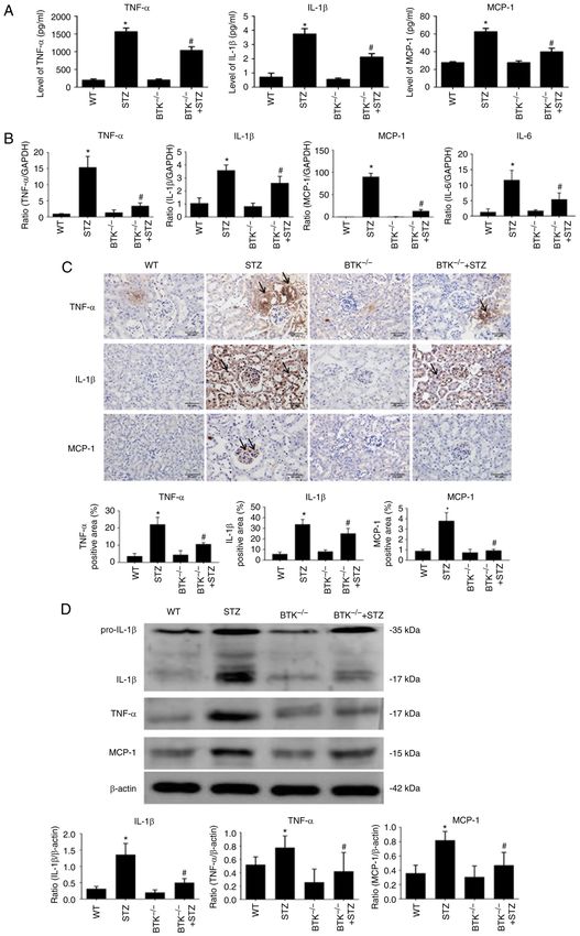
INTERNATIONAL JOURNAL OF MOLECULAR MEDICINE 48: 177, 2021                                         3

All sections were deparaffinized, rehydrated and subjected        Technology, Inc.), anti‑NF‑κ B p65 (1:1,000; cat. no. 8242;
to antigen retrieval in 0.01 M citrate buffer (pH 6.0)            Cell Signaling Technology, Inc.), anti‑nicotinamide adenine
by microwaving. After blocking with 10% goat serum                dinucleotide phosphate oxidase 1 (NOX1) (cat. no. ab131088;
(cat. no. SL038; Beijing Solarbio Science & Technology            1:1,000; Abcam), anti‑nicotinamide adenine dinucleotide
Co., Ltd.) at room temperature, the sections were incubated       phosphate oxidase 4 (NOX4) (1:1,000; cat. no. bs‑1091R;
with anti‑CD68 (cat. no. ab213363; 1:300; Abcam), anti‑BTK        Beijing Boaosen Biotechnology, Co., Ltd.), anti‑NLRP3
(1:200; cat. no. 8547; Cell Signaling Technology, Inc.),          (1:1,000; cat. no. ab214185; Abcam), anti‑caspase‑1
anti‑F4/80 (1:200; cat. no. ab111101; Abcam), anti‑collagen       (1:1,000; cat. no. ab138483; Abcam) and anti‑ASC (1:200;
(Col)‑IV (1:200; cat. no. ab236640; Abcam), anti‑fibronectin      cat. no. sc‑376916; Santa Cruz Biotechnology, Inc.). After
(FN; 1:200; cat. no. ab2413; Abcam), anti‑Wilms Tumor 1           washing, the membrane was incubated with HRP‑labeled
(WT1) transcription factor (WT1; 1:200; cat. no. ab267377;        secondary antibodies (cat. nos. SA00001‑1 and SA00001‑2;
Abcam) and anti‑Nephrin (1:200; cat. no. ab216341; Abcam)         1:2,000; ProteinTech Group, Inc.) at room temperature for 1 h.
antibodies overnight at 4˚C. Secondary biotin‑labeled goat        The signals were detected with the use of a chemiluminescent
anti‑mouse/rabbit IgG antibodies (1:200; cat. no. PV‑6000;        gel imaging system and the relative ratio was semi‑quantified
OriGene Technologies, Inc.) were then added for 2 h at room       using ImageJ 1.0 software (National Institutes of Health).
temperature. Finally, 3,3‑diaminobenzidine (cat. no. ZLI‑9017;
Sigma‑Aldrich; Merck KGaA) and hematoxylin at room                Reverse transcription‑quantitative (RT‑qPCR). TRIzol®
temperature were used for staining. All images were captured      reagent (Thermo Fisher Scientific, Inc.) was used to extract the
with a Zeiss microscope (Zeiss AG) and analyzed using             total RNA in renal tissues. cDNA was synthesized from total
ImageJ 1.0 software (National Institutes of Health). Six fields   RNA using a Reverse Transcription kit (Vazyme Biotech Co.,
were selected for statistical analysis in each sample. The mean   Ltd.). cDNA was amplified and detected in a QuantStudio™ 6
value was calculated and used as the final sample of the immu‑    Flex Real‑Time PCR System (Thermo Fisher Scientific, Inc.)
nohistochemistry data. The mean values of all samples were        using the miScript SYBR‑Green PCR kit (Qiagen GmbH)
used for final statistical analysis.                              to determine the quantity of mRNA. The conditions were as
                                                                  follows: 95˚C for 10 min, followed by 40 cycles of 95˚C for
ELISA. The levels of TNF‑α, IL‑1β and monocyte chemoat‑           10 sec, and 60˚C for 20 sec. The expression levels of all genes
tractant protein‑1 (MCP‑1) in serum were detected with the        were standardized with the reference gene GAPDH using the
use of human and mouse ELISA kits (cat. no. P06804, P10749,       2‑ΔΔCq method (32). The primer sequences used were selected
P10148; RayBiotech, Inc.). The urinary albumin excretion          according to the previous studies (15,33).
rate (UAER) was detected using a mouse albumin ELISA
kit (cat. no. ab108792, Abcam). The detection methods were        Histological staining. The kidney tissue was fixed in
executed according to the manufacturer's instructions.            4% paraformaldehyde for 16 h at room temperature and then
                                                                  dehydrated through an ethanol gradient. Subsequently, the
Detection of HbA1c and blood glucose. HbA1c was                   kidney tissue was paraffin‑embedded and cut into 3‑µm‑thick
detected using the Glycosylated hemoglobin A1c Assay kit          sections. The sections were dewaxed and stained with periodic
(cat. no. H464‑1, Nanjing Jiancheng Bioengineering Institute).    acid‑Schiff (PAS) staining kit (cat. no. G1281; Beijing Solarbio
The blood glucose levels were detected using the glucose meter    Science & Technology Co., Ltd.) and the Masson's staining kit
(Contour TS, Bayer). The detection methods were executed          (cat. no. G1340; Beijing Solarbio Science & Technology Co.,
according to the manufacturer's instructions.                     Ltd.). The staining methods were executed according to the
                                                                  manufacturer's instructions. All images were acquired using a
Western blot (WB) analysis. Renal tissues were lysed in           light microscope (BX51, Olympus Corporation). The glomer‑
RIPA lysis buffer (cat. no. P0013B, Beyotime Institute of         ular mesangial expansion, tubulointerstitial injury index and
Biotechnology). The mixture was centrifuged at 15,294 x g         fibrosis area were evaluated and graded using ImageJ 1.0
at 4˚C for 30 min and then the pellet was removed. Protein        software (National Institutes of Health).
samples were separated via 10 or 15% SDS‑PAGE, and trans‑
ferred to the nitrocellulose membrane (cat. no. HATF00010,        Transmission electron microscopy (TEM). Kidney tissue was
Merck KGaA). The band distribution was observed via               quickly placed in 2% glutaraldehyde for 24 h. The tissue was
Ponceau staining. The membrane was then blocked with              then fixed with 1% acetic acid at room temperature for 2 h,
5% skim milk powder (containing TBS‑Tween‑20) at room             followed by acetone (50, 70, 80, 90 and 100%; twice for 15 min
temperature for 2 h, followed by incubation with the following    each time) for dehydration; sections were embedded in epoxy
primary antibodies at 4˚C for 12 h: Anti‑β ‑actin (1:35,000,      resin. The resin block was cut into ultra‑thin sections of 70 nm,
cat. no. 66009‑1‑Ig; ProteinTech Group, Inc.), anti‑TNF‑ α        stained with lead citrate at 37˚C and then observed under a
(1:1,000; cat. no. ARG10158; arigo Biolaboratories Corp.),        transmission electron microscope (H‑7700; Hitachi, Ltd.).
anti‑IL‑1β (1:1,000; cat. no. 12242; Cell Signaling Technology,   GBM thickness and foot process fusion were analyzed using
Inc.), anti‑MCP‑1 (1:1,000; cat. no. ARG57649; arigo              ImageJ 1.0 software (National Institutes of Health).
Biolaboratories Corp.), anti‑BTK (1:1,000; cat. no. ab25971;
Abcam), anti‑phosphorylated (p)‑BTK (1:1,000; cat. no. 87457;     Statistical analysis. Data were statistically analyzed using
Cell Signaling Technology, Inc.), anti‑inducible nitric           SPSS 23.0 (IBM Corp.). Normally distributed data are
oxide synthase (iNOS; 1:1,000; cat. no. ab178945; Abcam),         presented as the mean ± SD, and non‑normally distributed
anti‑p‑NF‑κ B p65 (1:1,000; cat. no. 8242; Cell Signaling         data were expressed as the median (p25, p75). Pearson's test
4 ZHAO et al: BRUTON'S TYROSINE KINASE REGULATES INFLAMMATION IN THE DIABETIC KIDNEY Table I. Clinical indicators of mice (n=28). Clinical indicator WT STZ BTK‑/‑ BTK‑/‑ + STZ Body weight 28.88±0.57 22.56±1.93a 27.16±1.02 23.66±1.35 Kidney/body ratio (%) 0.62±0.04 1.00±0.11a 0.70±0.06 0.97±0.06 BG (mmol/l) 6.51±0.68 22.17±1.69a 7.22±0.60 22.76±2.85 HbA1c (%) 4.42±0.65 6.98±0.72a 4.77±0.89 7.14±1.10 UAER (µg/24 h) 28.41±14.48 449.87±101.43a 26.31±18.22 304.39±173.74b All the data are expressed as the mean ± SD. Significant differences were analyzed by one‑way ANOVA with Bonferroni's post hoc analysis. a P
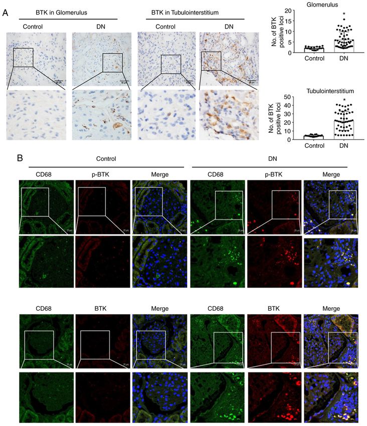
INTERNATIONAL JOURNAL OF MOLECULAR MEDICINE 48: 177, 2021                                                         5

Figure 1. BTK is notably activated in renal macrophages in the kidneys of patients with DN. (A) Immunohistochemistry of BTK expression in the glomerulus
and tubulointerstitium (scale bar, 50 µm; *P
6                    ZHAO et al: BRUTON'S TYROSINE KINASE REGULATES INFLAMMATION IN THE DIABETIC KIDNEY

Figure 2. BTK expression is correlated with clinical indicators and pathological changes in the kidneys of patients with DN. (A) Correlation analysis of renal
BTK expression in glomerulus with proteinuria in patients with type 2 DN. (B) Correlation analysis of renal BTK expression in glomerulus with serum creati‑
nine in patients with type 2 DN. (C) Correlation analysis of renal BTK expression in glomerulus with eGFR in patients with type 2 DN. (D) Correlation analysis
of renal BTK expression in tubulointerstitium with proteinuria, in patients with type 2 DN. (E) Correlation analysis between renal BTK expression in tubu‑
lointerstitium and serum creatinine in patients with type 2 DN. (F) Correlation analysis of renal BTK expression in tubulointerstitium with eGFR in patients
with type 2 DN. (G) Correlation analysis between renal BTK expression in glomerulus and Tervaert classification in patients with type 2 DN. (H) Correlation
analysis between renal BTK expression in tubulointerstitium and IFTA in patients with type 2 DN. (I) Correlation analysis of renal BTK expression between
tubulointerstitium and interstitial inflammation in patients with type 2 DN. Pearson's test was used in (C and F) for correlation analysis. Spearman's test was
used in (A, B, D, E, G H and I) for correlation analysis. BTK, Bruton's tyrosine kinase; DN, diabetic nephropathy; eGFR, estimated glomerular filtration rate;
IFTA, interstitial fibrosis and tubular atrophy.

BTK knockout alleviates renal inflammation in diabetic mice.                     expression levels of TNF‑α, IL‑1β and MCP‑1 were signifi‑
The change in the inflammatory response was also evalu‑                          cantly decreased in the BTK‑/‑ + STZ group compared with
ated in the mice. The results of ELISA demonstrated that the                     the STZ group (Fig. 4C). On the whole, the results indicated
TNF‑ α, IL‑1β and MCP‑1 levels were increased in serum                           that diabetic mice exhibited additional severe inflammation
from the STZ group. However, this change was significantly                       and macrophage infiltration in serum and kidney, while BTK
reduced in the BTK‑/‑ + STZ group (Fig. 4A). Subsequently,                       knockout alleviated this phenomenon.
changes in inflammatory mediators were detected in the
mouse kidneys. The data indicated that the mRNA and                              BTK knockout suppresses the activation of the NLRP3
protein expression levels of TNF‑α, IL‑1β and MCP‑1 were                         inflammasome in the kidneys of diabetic mice. To further
increased in the STZ group (Fig. 4B‑D). Moreover, these                          observe the mechanisms of BTK in renal inflammation, WB
changes were significantly reduced when BTK was knocked                          analysis was conducted to observe the activation of the NLRP3
out. The immunohistochemistry results also revealed that the                     inflammasome in the mouse kidneys. The WB analysis data
INTERNATIONAL JOURNAL OF MOLECULAR MEDICINE 48: 177, 2021                                                           7

Figure 3. BTK knockout alleviates the pathological changes in mice with STZ‑induced diabetes. (A) Glomerular mesangial expansion and tubular injury were
detected using PAS staining. Immunohistochemistry of ColIV and FN expression was performed in mouse kidneys (scale bar, 50 µm). (B) Immunohistochemistry
of F4/80, WT1 and FN expression levels in mouse kidneys (scale bar, 50 µm). (C) The thickness of GBM, mesangial expansion and podocyte injury were
detected via transmission electron microscopy (*P
8                   ZHAO et al: BRUTON'S TYROSINE KINASE REGULATES INFLAMMATION IN THE DIABETIC KIDNEY

Figure 4. BTK knockout alleviates kidney inflammation in diabetic mice. (A) TNF‑α, IL‑1β and MCP‑1 levels of in mouse serum were detected using ELISA
kits. (B) TNF‑α, IL‑1β, MCP‑1 and IL‑6 mRNA expression levels in mouse kidneys were detected by RT‑qPCR. (C) TNF‑α, IL‑1β and MCP‑1 immunohisto‑
chemistry analysis of protein expression was conducted in mouse kidneys (scale bar, 50 µm). (D) Protein expression levels of TNF‑α, IL‑1β and MCP‑1 were
detected by western blot analysis (*P
INTERNATIONAL JOURNAL OF MOLECULAR MEDICINE 48: 177, 2021                                                           9

Figure 5. BTK knockout suppresses the oxidative stress and the activation of the NLRP3 inflammasome in the kidneys of diabetic mice. (A) p‑BTK/BTK
expression levels in mouse kidneys were detected via WB analysis. (B) iNOS, p‑p65 and p65 expression levels in mouse kidneys were detected via WB analysis.
(C) NOX1 and NOX4 expression levels in mouse kidneys were detected via WB analysis. (D) NLRP3, caspase‑1 and ASC expression levels in mouse kidneys
were detected via WB analysis (*P
10                ZHAO et al: BRUTON'S TYROSINE KINASE REGULATES INFLAMMATION IN THE DIABETIC KIDNEY

the STZ group compared with the WT group. However, BTK                diabetic mice. Collectively, these results suggested that BTK
deletion significantly alleviated this effect (Fig. 5C). Moreover,    knockout alleviated kidney injury in diabetic mice.
it was found that the expression levels of NLRP3, caspase‑1 and           The NLRP3 inflammasome is a multiprotein complex
ASC were significantly decreased in the BTK‑/‑ + STZ group            that regulates innate immune responses to infection and cell
compared with the STZ group (Fig. 5D). These results thus             stress (18). In recent studies, it was reported that the activa‑
suggested that BTK knockout decreased oxidative stress and            tion of NLRP3 may be associated with disease progression
the activation of the NLRP3 inflammasome in the kidneys of            in patients with DKD (21,22), Currently, there are no existing
diabetic mice.                                                        clinically approved therapies that target the NLRP3 inflamma‑
                                                                      some directly (24). BTK has been proven to serve as a critical
Discussion                                                            regulator of NLRP3 inflammasome activation (25). It has been
                                                                      observed that the regulatory effects of BTK inhibition on the
DN is a severe complication of diabetes mellitus, and its treat‑      NLRP3 inflammasome vary in different mouse models. BTK
ment remains challenging (1). Previous studies have reported          deficiency has been shown to promote NLRP3 inflammasome
that the development of DN is closely associated with inflam‑         activation and induce IL‑1β‑mediated colitis, but it also allevi‑
mation, and that macrophages are the main immune cell type            ates other diseases, such as ischemic brain injury, diet‑induced
involved in this process (5,6). In the early stages of DN, patients   metabolic inflammation and polymicrobial sepsis (26‑28).
present an increase in serum inflammatory mediator levels.            On this basis, the association between BTK and the NLRP3
Subsequently, macrophages can be recruited into the kidneys           inflammasome in DN was examined in the present study. It
by these mediators, such as C‑C motif chemokine ligand 2 and          was revealed that no significant side‑effects were noted in BTK
MCP‑1 (34,35). This process was verified in the present study         knockout mice. Moreover, it was noted that BTK knockout
and culminated in an increased macrophage infiltration in             markedly reduced the NLRP3, caspase‑1 and ASC expression
kidneys. The results of the present demonstrated that either in       levels in the kidneys of diabetic mice. Those results suggested
the serum of patients with DN or in the serum of diabetic mice,       that the protective effect of BTK knockout was mainly attrib‑
inflammatory mediator expression levels were significantly            uted to the reduced activation of the NLRP3 inflammasome.
increased. Moreover, macrophage infiltration and activation               In conclusion, the present study demonstrated that there was
were increased in the kidneys of patients with DN and diabetic        a notable association between BTK and the clinical indicators
mice. It was also identified that the levels of inflammatory          and pathological changes of patients with DN. Moreover, BTK
cytokines released by macrophages in kidneys were increased.          knockout reduced inflammation in diabetic mice, and this
These findings indicated that macrophages can be recruited            protection was a result of the suppression of NLRP3 inflam‑
into the kidneys and activated by hyperglycemia, leading to           masome activation. However, the lack of analysis of IL‑6
the aggravation of inflammation.                                      protein and NF‑κ B mRNA expression also present a limitation
    BTK is an important factor in innate immunity and has             of the present study. To date, to the best of our knowledge,
been closely associated with inflammation (8,11). It has been         there is no effective treatment available for DN. BTK inhibitor
previously revealed that high glucose levels may promote the          had been widely used in clinical practice. The present study
phosphorylation of BTK, leading to the release of inflammatory        demonstrated that BTK deletion effectively reduced the acti‑
mediators via the NF‑κ B signaling pathway (15). It was also          vation of the NLRP3 inflammasome; thus, this may prove to
demonstrated in the present study that BTK was activated in           be a potential method for the treatment of DN. The findings
renal macrophages in patients with DN. Moreover, BTK was              presented herein may have important clinical significance;
associated with clinical indicators and pathological changes,         BTK inhibitor may be an effective therapeutic strategy against
which indicated that BTK played a crucial role in the disease         DN. The authors aim to perform further studies in the future
progression of DN. In in vivo experiments, it was identified that     in order to provide further suggestions which may aid in the
BTK knockout reduced UAER, the accumulation of extracel‑              development of novel treatment strategies for DN.
lular matrix and macrophage infiltration in diabetic mice.
The fibrotic lesions were also observed by Masson's staining,         Acknowledgements
although no significant changes were observed in any of the
groups analyzed. This may be attributed to an early diabetic          The authors would like to thank the Research and
pathology change in mouse kidneys observed at 12 weeks of             Experimental Center of Anhui Medical University, for
feeding, which manifested through mesangial matrix prolif‑            providing experimental technical support.
eration and tubulointerstitial damage. Podocytes are important
intrinsic cells in the kidney and are known as the most vulnerable    Funding
cells in the kidney (36). Podocyte loss and fusion of foot process
can occur in early stage DN, and it is also the main cause of         The present study was supported financially by the National
proteinuria (37,38). Therefore, it was considered that podocyte       Natural Science Foundation of China (grant no. 81470965)
injury could be a favorable choice to evaluate early renal injury,    and the Natural Science Foundation of Anhui Province (grant
thus WT1 and Nephrin were used the present study as podocyte          no. 1808085MH236).
markers (39). The results of the present study demonstrated
that BTK knockout protected the diabetic mice from podocyte           Availability of data and materials
injury, further confirmed through TEM. It was also noted that
the thickened basement membrane, proliferated extracellular           All data generated or analyzed during this study are included
matrix and podocyte injury were alleviated in BTK knockout            in this published article.
INTERNATIONAL JOURNAL OF MOLECULAR MEDICINE 48: 177, 2021 11
Authors' contributions 12. Wei J, Wang Y, Qi X and Wu Y: Enhanced Bruton's tyrosine
kinase activity in the kidney of patients with IgA nephropathy.
Int Urol Nephrol 53: 1399‑1415, 2021.
JZ and YGW conceived and designed the study. JC and YYL 13. Kong W, Deng W, Sun Y, Huang S, Zhang Z, Shi B, Chen W,
collected the data and performed the animal experiments. JZ Tang X, Yao G, Feng X and Sun L: Increased expression of
Bruton's tyrosine kinase in peripheral blood is associated with
and JC analyzed the data and wrote the manuscript. YGW lupus nephritis. Clin Rheumatol 37: 43‑49, 2018.
and LLX conducted the animal experiments and reviewed the 14. Jain N, Keating M, Thompson P, Ferrajoli A, Burger J,
manuscript. JZ, LLX and YGW confirmed the authenticity of Borthakur G, Takahashi K, Estrov Z, Fowler N, Kadia T, et al:
Ibrutinib and venetoclax for first‑line treatment of CLL. N Engl
all the raw data. All authors have read and approved the final J Med 380: 2095‑2103, 2019.
manuscript. 15. Fan Z, Wang Y, Xu X and Wu Y: Inhibitor of Bruton's
tyrosine kinases, PCI‑32765, decreases pro‑inflammatory
mediators' production in high glucose‑induced macrophages. Int
Ethics approval and consent to participate Immunopharmacol 58: 145‑153, 2018.
16. Roschewski M, Lionakis MS, Sharman JP, Roswarski J,
All patients and healthy individuals had signed informed Goy A, Monticelli MA, Roshon M, Wrzesinski SH, Desai JV,
Zarakas MA, et al: Inhibition of Bruton tyrosine kinase in
consent forms and all experiments were approved by the patients with severe COVID‑19. Sci Immunol 5: eabd0110, 2020.
Ethics Committee of Anhui Medical University (approval 17. Rathinam VA and Fitzgerald KA: Inflammasome complexes:
number: 5101309). The animal experiments were performed in Emerging mechanisms and effector functions. Cell 165: 792‑800,
2016.
accordance with guidelines, ‘Principles of Laboratory Animal 18. Wang L and Hauenstein AV: The NLRP3 inflammasome:
Care and Use in Research’ (Ministry of Health, Beijing, Mechanism of action, role in disease and therapies. Mol Aspects
Med 76: 100889, 2020.
China). The experiments were approved by the Anhui Medical 19. Hooftman A, Angiari S, Hester S, Corcoran SE, Runtsch MC,
University Ethics Committee (approval number: 2020064). Ling C, Ruzek MC, Slivka PF, McGettrick AF, Banahan K, et al:
The immunomodulatory metabolite itaconate modifies NLRP3 and
inhibits inflammasome activation. Cell Metab 32: 468‑478.e7, 2020.
Patient consent for publication 20. Liu D, Yang P, Gao M, Yu T, Shi Y, Zhang M, Yao M, Liu Y and
Zhang X: NLRP3 activation induced by neutrophil extracellular
Not applicable. traps sustains inflammatory response in the diabetic wound. Clin
Sci (Lond) 133: 565‑582, 2019.
21. Han Y, Xu X, Tang C, Gao P, Chen X, Xiong X, Yang M, Yang S,
Competing interests Zhu X, Yuan S, et al: Reactive oxygen species promote tubular
injury in diabetic nephropathy: The role of the mitochondrial
ros‑txnip‑nlrp3 biological axis. Redox Biol 16: 32‑46, 2018.
The authors declare that they have no competing interests. 22. Mulay SR: Multifactorial functions of the inflammasome
component NLRP3 in pathogenesis of chronic kidney diseases.
Kidney Int 96: 58‑66, 2019.
References 23. Tang SCW and Yiu WH: Innate immunity in diabetic kidney
disease. Nat Rev Nephrol 16: 206‑222, 2020.
1. Cole JB and Florez JC: Genetics of diabetes mellitus and diabetes 24. Ram C, Jha AK, Ghosh A, Gairola S, Syed AM, Murty US,
complications. Nat Rev Nephrol 16: 377‑390, 2020. Naidu VGM and Sahu BD: Targeting NLRP3 inflammasome
2. Tervaert TW, Mooyaart AL, Amann K, Cohen AH, Cook HT, as a promising approach for treatment of diabetic nephropathy:
Drachenberg CB, Ferrario F, Fogo AB, Haas M, de Heer E, et al: Preclinical evidences with therapeutic approaches. Eur
Pathologic classification of diabetic nephropathy. J Am Soc J Pharmacol 885: 173503, 2020.
Nephrol 21: 556‑563, 2010. 25. Weber ANR: Targeting the NLRP3 inflammasome via BTK.
3. Liang G, Song L, Chen Z, Qian Y, Xie J, Zhao L, Lin Q, Zhu G, Front Cell Dev Biol 9: 630479, 2021.
Tan Y, Li X, et al: Fibroblast growth factor 1 ameliorates diabetic 26. Ito M, Shichita T, Okada M, Komine R, Noguchi Y, Yoshimura A
nephropathy by an anti‑inflammatory mechanism. Kidney Int 93: and Morita R: Bruton's tyrosine kinase is essential for NLRP3
95‑109, 2018. inflammasome activation and contributes to ischaemic brain
4. Guilliams M, Thierry GR, Bonnardel J and Bajenoff M: injury. Nat Commun 6: 7360, 2015.
Establishment and maintenance of the macrophage niche. 27. Purvis GSD, Collino M, Aranda‑Tavio H, Chiazza F,
Immunity 52: 434‑451, 2020. O'Riordan CE, Zeboudj L, Mohammad S, Collotta D, Verta R,
5. Klessens CQF, Zandbergen M, Wolterbeek R, Bruijn JA, Guisot NE, et al: Inhibition of Bruton's TK regulates macrophage
Rabelink TJ, Bajema IM and IJpelaar DHT: Macrophages in NF‑κ B and NLRP3 inflammasome activation in metabolic
diabetic nephropathy in patients with type 2 diabetes. Nephrol inflammation. Br J Pharmacol 177: 4416‑4432, 2020.
28. O'Riordan CE, Purvis GSD, Collotta D, Krieg N, Wissuwa B,
Dial Transplant 32: 1322‑1329, 2017. Sheikh MH, Ferreira Alves G, Mohammad S, Callender LA,
6. Fu J, Akat KM, Sun Z, Zhang W, Schlondorff D, Liu Z, Tuschl T, Coldewey SM, et al: X‑Linked immunodeficient mice with no func‑
Lee K and He JC: Single‑Cell RNA profiling of glomerular cells tional bruton's tyrosine kinase are protected from sepsis‑induced
shows dynamic changes in experimental diabetic kidney disease. multiple organ failure. Front Immunol 11: 581758, 2020.
J Am Soc Nephrol 30: 533‑545, 2019. 29. Li XQ, Chang DY, Chen M and Zhao MH: Deficiency of C3a
7. Smith CI, Islam TC, Mattsson PT, Mohamed AJ, Nore BF and receptor attenuates the development of diabetic nephropathy.
Vihinen M: The Tec family of cytoplasmic tyrosine kinases: BMJ Open Diabetes Res Care 7: e000817, 2019.
Mammalian Btk, Bmx, Itk, Tec, Txk and homologs in other 30. Liu J, Lee GY, Biggers JD, Toth TL and Toner M: Low cryo‑
species. BioEssays 23: 436‑446, 2001. protectant concentration rapid vitrification of mouse oocytes and
8. Weber ANR, Bittner Z, Liu X, Dang TM, Radsak MP and embryos. Cryobiology 98: 233‑238, 2021.
Brunner C: Bruton's Tyrosine Kinase: An emerging key player in 31. Weinerman R, Ord T, Bartolomei MS, Coutifaris C and
innate immunity. Front Immunol 8: 1454, 2017. Mainigi M: The superovulated environment, independent of
9. Pal Singh S, Dammeijer F and Hendriks RW: Role of Bruton's tyro‑ embryo vitrification, results in low birthweight in a mouse model.
sine kinase in B cells and malignancies. Mol Cancer 17: 57, 2018. Biol Reprod 97: 133‑142, 2017.
10. Haselmayer P, Camps M, Liu‑Bujalski L, Nguyen N, Morandi F, 32. Livak KJ and Schmittgen TD: Analysis of relative gene expres‑
Head J, O'Mahony A, Zimmerli SC, Bruns L, Bender AT, et al: sion data using real‑time quantitative PCR and the 2(‑Delta Delta
Efficacy and pharmacodynamic modeling of the BTK inhibitor C(T)) method. Methods 25: 402‑408, 2001.
evobrutinib in autoimmune disease models. J Immunol 202: 33. Birnbaum Y, Bajaj M, Yang HC and Ye Y: Combined SGLT2
2888‑2906, 2019. and DPP4 Inhibition Reduces the Activation of the Nlrp3/ASC
11. Rip J, de Bruijn MJW, Appelman MK, Pal Singh S, Hendriks RW inflammasome and attenuates the development of diabetic
and Corneth OBJ: Toll‑Like receptor signaling drives nephropathy in mice with type 2 diabetes. Cardiovasc Drugs
BTK‑mediated autoimmune disease. Front Immunol 10: 95, 2019. Ther 32: 135‑145, 2018.12 ZHAO et al: BRUTON'S TYROSINE KINASE REGULATES INFLAMMATION IN THE DIABETIC KIDNEY
34. Moreno JA, Gomez‑Guerrero C, Mas S, Sanz AB, Lorenzo O, 38. Zhou L, Chen X, Lu M, Wu Q, Yuan Q, Hu C, Miao J, Zhang Y,
Ruiz‑Ortega M, Opazo L, Mezzano S and Egido J: Targeting Li H, Hou FF, et al: Wnt/β‑catenin links oxidative stress to podo‑
inflammation in diabetic nephropathy: A tale of hope. Expert cyte injury and proteinuria. Kidney Int 95: 830‑845, 2019.
Opin Investig Drugs 27: 917‑930, 2018. 39. Puelles VG, Bertram JF and Moeller MJ: Quantifying podocyte
35. Wen Y and Crowley SD: The varying roles of macrophages depletion: Theoretical and practical considerations. Cell Tissue
in kidney injury and repair. Curr Opin Nephrol Hypertens 29: Res 369: 229‑236, 2017.
286‑292, 2020.
36. Nagata M: Podocyte injury and its consequences. Kidney Int 89: This work is licensed under a Creative Commons
1221‑1230, 2016. Attribution-NonCommercial-NoDerivatives 4.0
37. Agrawal S, He JC and Tharaux PL: Nuclear receptors in podocyte International (CC BY-NC-ND 4.0) License.
biology and glomerular disease. Nat Rev Nephrol 17: 185‑204, 2021.You can also read