The effect of ivermectin alone and in combination with cobicistat or elacridar in experimental Schistosoma mansoni infection in mice
←
→
Page content transcription
If your browser does not render page correctly, please read the page content below
www.nature.com/scientificreports
OPEN The effect of ivermectin alone
and in combination with cobicistat
or elacridar in experimental
Schistosoma mansoni infection
in mice
Belén Vicente1,5, Julio López‑Abán1,5, Juliane Chaccour2, Juan Hernández‑Goenaga1,
Patricia Nicolas3, Pedro Fernández‑Soto1, Antonio Muro1 & Carlos Chaccour2,3,4*
Schistosoma mansoni is less susceptible to the antiparasitic drug ivermectin than other helminths.
By inhibiting the P-glycoprotein or cytochrome P450 3A in mice host or parasites in a murine model,
we aimed at increasing the sensitivity of S. mansoni to the drug and thus preventing infection. We
assigned 124 BALB/c mice to no treatment, treatment with ivermectin only or a combination of
ivermectin with either cobicistat or elacridar once daily for three days before infecting them with 150
S. mansoni cercariae each. The assignment was done by batches without an explicit randomization
code. Toxicity was monitored. At eight weeks post-infection, mice were euthanized. We determined
number of eggs in intestine and liver, adult worms in portal and mesenteric veins. Disease was
assessed by counting granulomas/cm2 of liver and studying organ weight indices and total weight.
IgG levels in serum were also considered. No difference between groups treated with ivermectin only
or in combination with cobicistat or elacridar compared with untreated, infected controls. Most mice
treated with ivermectin and elacridar suffered severe neurological toxicity. In conclusion, systemic
treatment with ivermectin, even in the presence of pharmacological inhibition of P-glycoprotein or
cytochrome P450 3A, did not result in effective prophylaxis for S. mansoni infection in an experimental
murine model.
Progress in the control or elimination of schistosomiasis must be approached from different angles: accurate,
fast and cheap diagnosis; affordable, safe and effective treatment; and well-established prevention and control
strategies. WHO has recognized the need to identify new compounds as an alternative to praziquantel, the single
therapeutic agent in use today. Although praziquantel remains effective, concerns arise about potential drug
resistance stemming from its continuous use in mass drug administration campaigns in endemic a reas1,2. Dif-
ferent artemisinin-derived compounds have been developed and tested alone or combined with p raziquantel3.
Mefloquine has also been used, with good results in experimental m odels4, and edelfosine alone or in combina-
tion with praziquantel, with good results in reducing granulomatous inflammation both in vitro and in vivo5,6.
rug7. It is the first-line treatment for strongyloidiasis, scabies and
Ivermectin is a widely used antiparasitic d
onchocerciasis, part of the regimen for lymphatic filariasis (LF) and has demonstrated efficacy against other
soil-transmitted helminths8. In the mouse model of schistosome infection, adult worm load can be reduced by
very high doses of ivermectin9, as it appears that Schistosoma mansoni is much less susceptible to ivermectin
than other helminths10. It is unknown whether S. mansoni cercariae differ in sensitivity to ivermectin, to date
no study has described a prophylactic model.
Decreased susceptibility to ivermectin can be brought about by ATP-binding cassette (ABC) transport
proteins and their interaction with cytochrome P450 (CYP) 3A11,12. We hypothesize that the combination of
1
Infectious and Tropical Diseases Group (e‑INTRO), IBSAL‑CIETUS (Biomedical Research Institute of
Salamanca‑Research Centre for Tropical Diseases at the University of Salamanca), Faculty of Pharmacy, University
of Salamanca, Salamanca, Spain. 2Clinica Universidad de Navarra, Pamplona, Spain. 3ISGlobal, Barcelona Institute
for Global Health, Hospital Clínic, Universitat de Barcelona, Rosello 132, 5ª 2ª, 08036 Barcelona, Spain. 4Ifakara
Health Institute, Ifakara 67501, United Republic of Tanzania. 5These authors contributed equally: Belén Vicente
and Julio López-Abán. *email: carlos.chaccour@isglobal.org
Scientific Reports | (2021) 11:4476 | https://doi.org/10.1038/s41598-021-84009-y 1
Vol.:(0123456789)
www.nature.com/scientificreports/

                                            Figure 1.  Study timeline. Ivermectin or ivermectin and cobicistat or ivermectin and elacridar were
                                            administered daily for three days before infecting BALB/c mice with 150 cercariae of S. mansoni. After 8 weeks
                                            of infection, parasite load in organs and IgG production against SoSmAWA antigen were examined. Treatment
                                            tolerance was monitored throughout.

                                            ivermectin and elacridar, a P-glycoprotein inhibitor, or cobicistat, a CYP3A inhibitor, could increase the sus-
                                            ceptibility of S. mansoni invading schistosomula to ivermectin. The objective of this work was to evaluate the
                                            capacity of ivermectin alone and in combination with elacridar or cobicistat to prevent S. mansoni infection in
                                            an experimental model using BALB/c mice.

                                            Materials and methods
                                            Parasites and animals. To maintain the Schistosoma mansoni (LE strain) cycle, freshwater snails Biompha-
                                            laria glabrata were used as intermediate hosts, and CD1 mice as definitive hosts. Snails of 4–8 mm in diameter
                                            were infected with seven miracidia each. They were kept in 25 °C water for 30 days until the emission of fur-
                                            cocercariae was induced with light and a temperature of 26 °C for 2 h. Triplicate counts were done to obtain a
                                            dose of 150 cercariae in 0.7–1.2 ml of chlorine-free water.
                                                For the infection experiment, 124 SPF BALB/c mice (Charles River, Lyon, France) with a weight of 18.5–21.3 g
                                            and an age of seven weeks were used. The animals were kept in a controlled temperature and humidity environ-
                                            ment with a 12:12 h light:dark cycle. They were supplied with water and food ad libitum in the facilities of the
                                            Animal Experimentation Service of the University of Salamanca according to the current Spanish legislation on
                                            animal experimentation (L32/2007, L6/2013 and RD 53/2013) and the transposition of the rules of the Euro-
                                            pean Union (Di 2010/63/CE). All experiments with animals were approved by the Bioethics Committee of the
                                            University of Salamanca (Registration number CBE-225). Humane endpoints were applied when an evidence of
                                            severe pain, excessive distress, suffering or an impending death was observable in any of the animals, which were
                                            then euthanized. All mice were euthanized at the end of the experiment by intraperitoneal injection of sodium
                                            pentobarbital in PBS (100 mg/kg). The status of all mice was checked daily using a composite score including
                                            vitality, secretions, fur quality, mobility, dyspnea, ascites, neurological signs, and ability to ingest water or food.
                                                All methods were carried out in accordance with relevant guidelines and regulations. All animal handling
                                            and methods complied with the ARRIVE guidelines.

                                            Experimental design. We used the BALB/c mouse model of infection by Schistosoma mansoni established
                                            in the IBSAL-CIETUS of the University of Salamanca, Spain. Mice were divided into five experimental groups:
                                            untreated uninfected (G1 Untr, n = 9), infected (G2 Inf, n = 45), treated with 1000 μg/kg of ivermectin daily for
                                            three days by oral catheter before infection (G3 Iv, n = 30), treated with 1000 μg/kg of ivermectin and 25 mg/kg of
                                            cobicistat daily for three days by oral catheter before infection (G4 Iv + Co, n = 30), and treated with 1000 μg/kg
                                            of ivermectin and 2.5 mg/kg of elacridar daily for three days by oral catheter before infection (G5 Iv + El, n = 10)
                                            (Fig. 1). In G4 and G5, ivermectin was administered 2 h after cobicistat and elacridar.
                                                After each drug administration, the behavior of the animals was observed for potential signs of toxicity
                                            (nervous, neuromuscular, digestive). On the third day of treatment and 4 h after administering the ivermectin
                                            dose, the animals in group 2, 3, 4 and 5 were infected with 150 cercariae of S. mansoni each. The infection was
                                            performed after anesthesia with a mixture of ketamine (50 mg/kg), diazepam (5 mg/kg) and atropine (1 mg/
                                            kg), in a final volume of 100 µl by intraperitoneal injection in order to immobilize the animals. They were placed
                                            supine, a plastic ring was placed on the shaved abdomen, attached with adhesive tape, and the water solution

          Scientific Reports |   (2021) 11:4476 |                https://doi.org/10.1038/s41598-021-84009-y                                                      2

Vol:.(1234567890)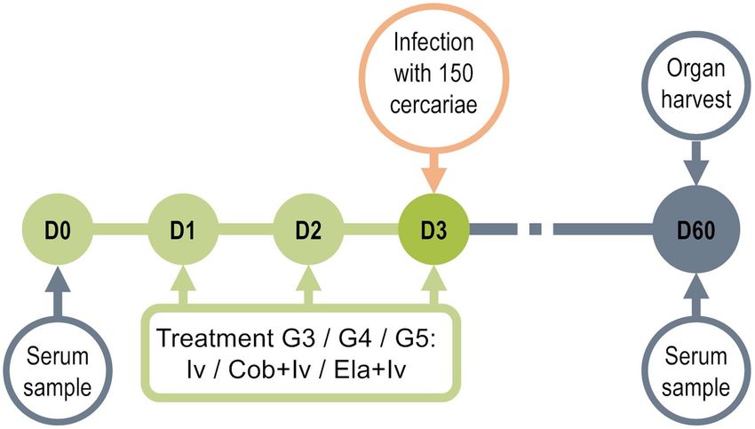
www.nature.com/scientificreports/
was applied with cercariae. After 45 min the rings were removed from the abdomen of the mice and the water
dried. Once the mice recovered mobility after sedation, the mice were returned to their corresponding cages.
The animals were weighed and had blood samples taken before starting the experiment, before infection and
before the necropsies. Eight weeks post-infection, the mice were euthanized. Serum was obtained from the blood
samples by centrifugation at 8000 rpm at 4 °C for 8 min. Samples were stored at − 20 °C until use.
To recover adult worms from the portal vein and mesenteric veins, livers were sectioned and perfused with
a 0.9% saline solution with heparin through the left ventricle. Adults worms were recovered with tweezers and
the number of Schistosoma couples and single male and female worms was counted.
A part of the intestine and part of the liver of each mouse were also separated and weighed. Subsequently,
they were digested to determine the egg load. For this, both liver and intestine samples were incubated at 37 °C
for 24 h in 5% KOH while stirring. The digested samples were centrifuged at 800g for 5 min and the supernatant
was removed, leaving 5 ml of concentrated egg solution. A McMaster chamber was used to count each sample
in triplicate and calculate the number of eggs per gram of intestine or liver. Spleen, bowel and liver indices were
obtained from the weights of these organs and the live weight of the mice to determine their degree of inflam-
mation. Photographs were taken where macroscopic lesions of the livers were observed to estimate the number
of granulomas per square centimeter. The ImageJ v.1.51k, PowerPoint and Adobe Photoshop CC 2017 programs
were used to count granulomas and estimate the affected liver surface.
ELISA for determination of S. mansoni soluble somatic antigen (SoSmAWA)‑specific IgG. In
order to detect specific antibodies against S. mansoni infection, the mouse sera were analyzed by indirect enzyme
immunoassay (ELISA), for specific immunoglobulin G (IgG) levels. We used soluble adult somatic antigen from
Schistosoma mansoni (SoSmAWA) according to Abán et al. (1999)13.
We coated 96-well polystyrene plates (Costar 3369, Corning Inc.) with 2.5 µg of SoSmAWA per well in a final
volume of 100 µl of carbonate buffer at pH 9.6 and incubated them for 18 h at 4 °C. Plates were washed three
times with 200 µl of 1X PBS per well at pH 7.2 and 0.05% Tween (PBS-T) to remove residues of unbound anti-
gen, and blocked with 2% BSA in PBS-T (100 µl per well) at 37 °C for 1 h to avoid nonspecific antibody-binding
and washed three times. Serum samples from each mouse were incubated for 1 h at 37 °C at a 1:100 dilution in
PBS-T, and washed as in the previous steps. The secondary mouse anti-IgG antibody (Sigma Aldrich, anti-mouse
IgG-peroxidase, A5906) was added at a 1:1000 dilution in PBS-T, incubated for 1 h at 37 °C, and washed. The
plates were then incubated with o-phenylenediamine dihydrochloride (OPD) as a peroxidase substrate, and
H2O2 as an oxidizing agent in citrate buffer at pH 5.0. The reaction was stopped with 50 µl per well of 3 N H2SO4.
Absorbance was measured at 492 nm in a spectrophotometer (ThermoScientific Multiskan GO 1510, Finland).
Statistical analysis. Data are presented as means and standard errors of the mean. A Bartlett test was car-
ried out to verify the homogeneity of the data distribution of all the variables. A t-unpaired two-tailed ANOVA
analysis was done and, if statistically significant, the data was analyzed in a post hoc Fisher’s Least Significant
Difference test to determine the existence of significant differences between the study groups. P-values below
0.05 were considered significant. A multivariate analysis of main variables was also carried out. For these analy-
ses and work chart, we used Simfit V7.3.0 and Statview V5.0 statistical packages.
Results
Effect of Iv and Iv‑Co on the weight of BALB/c mice after infection by S. mansoni. Weight loss
after the administration of a xenobiotic can be an indicator of toxicity in the protocol used. Healthy animals
gained an average of 3.2 ± 0.6 g during the experimental period, whereas infected animals gained 3.6 ± 0.3 g. Ani-
mals treated with ivermectin after infection gained 3.5 ± 0.5 g, whereas those treated with ivermectin + cobicistat
only gained 2.5 ± 0.5 g. These differences were not statistically significant (Fig. 2).
Neurotoxicity. After administering the combination of ivermectin and elacridar to G5 as detailed in “Mate-
rials and methods”, the animals showed seizures, circular movements, and loss of ambulatory capacity. Six ani-
mals in the ivermectin and elacridar group died or were euthanized according to the end-point rules of the pro-
tocol. Only four out of ten mice survived the administration of the ivermectin and elacridar drug combination.
No deaths or signs of neurotoxicity were seen in the group that was administered ivermectin alone or combined
with cobicistat.
The four mice surviving in the elacridar and ivermectin group were infected and necropsy performed at eight
weeks post infection. In the necropsy, no differences in worms recovered (8.5 ± 3.5 males, 6.0 ± 3.8 females) and
egg in tissues were (1992 ± 1082 egg per gram of liver and 1493 ± 801 egg per gram of small intestine) with respect
to other groups were seen, no statistical test was performed given the sample size (n = 4).
Effect of Iv and Iv‑Co on worm recovery. The parasite load was used to estimate the prophylactic capac-
ity of the drug or drug combinations against infection by S. mansoni. Mice treated prophylactically with iver-
mectin showed no significant reduction in the total recovery of adult worms. The animals treated with the
combination of ivermectin and cobicistat had a non-significant 8% reduction in worm load compared with the
control group of infected mice. A similar situation occurred in the separate recovery of male and female worms
(6–12% reduction) (Fig. 3).
Effect of Iv and Iv‑Co on the number of eggs retained in liver and intestine. Schistosome eggs in
liver and intestine are the most relevant indicator of the severity of the disease. Mice treated with ivermectin or
Scientific Reports | (2021) 11:4476 | https://doi.org/10.1038/s41598-021-84009-y 3
Vol.:(0123456789)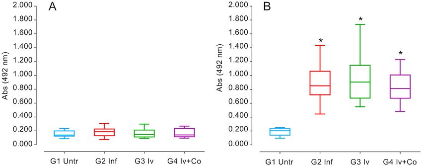
www.nature.com/scientificreports/
Figure 2. Weight gain throughout experimental period of eight weeks post-infection. Mice treated with
ivermectin (Iv) or ivermectin and cobicistat (Iv + Co), infected with 150 cercariae of S. mansoni (inf) and control
without treatment and infection (Untr).
Figure 3. Male and female worm recovery in the necropsy eight weeks post-infection of mice treated with
ivermectin (Iv) or ivermectin and cobicistat (Iv + Co), infected with 150 cercariae of S. mansoni (inf).
the combination of ivermectin and cobicistat did not reveal reductions in the number of eggs per gram of liver
or intestine compared with the infected control group (Fig. 4).
Effect of Iv and Iv‑Co on S. mansoni granulomas and organ indexes. Granuloma formation is an
immune response to the presence of eggs in tissues and it is an indicator of severe disease. There was no reduc-
tion in granulomas per liver surface in animals that were administered ivermectin When using the combined
ivermectin + cobicistat, the reduction was 3.6% but this was not statistically significant when compared with
infected mice (Fig. 5). Hepatic and intestinal indices are indicators of the relative inflammation of each of these
organs against the eggs trapped. In schistosome infection, hepato-splenomegaly is described as one of the most
prominent signs and is present in severe forms of the disease. Infected mice showed a 350–360% increase in
spleen weight (ANOVA F (3.105) 4.439, p = 0.006) and a 45–59% increase in liver weight (ANOVA F(3.105) 16.670,
p < 0.001) compared with the untreated infected mice. The intestinal index increased by 64–72%, which was not
statistically significant (Fig. 6).
IgG levels against SoSmAWA in infected animals. Immunoglobulin levels during infection with S.
mansoni serve to reveal the status of the infection and the response against the infection of the animals. High
levels of specific IgG against the SoSmAWA antigen were observed in all infected groups at eight weeks post-
infection. The infected group did not differ significantly from the groups given preventative treatment (Fig. 7).
Scientific Reports | (2021) 11:4476 | https://doi.org/10.1038/s41598-021-84009-y 4
Vol:.(1234567890)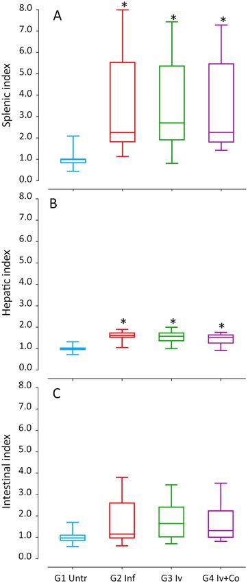
www.nature.com/scientificreports/

                                  Figure 4.  Eggs per gram of liver or thin intestine at the necropsy eight weeks post-infection of mice treated
                                  with ivermectin (Iv) or ivermectin and cobicistat (Iv + Co), infected with 150 cercariae of S. mansoni (inf).

                                  Figure 5.  Granulomas in liver surface at the necropsy eight weeks post-infection of mice treated with
                                  ivermectin (Iv) or ivermectin and cobicistat (Iv + Co), infected with 150 cercariae of S. mansoni (inf).

                                  Multivariate analysis. We included the following variables for the study of principal components (PC):
                                  total recovered worms, eggs in tissue, granulomas in the liver, organ indices, weight variation during the experi-
                                  ment and antibody levels at eight weeks post-infection. Between the principal components 1 and 2 (PC1 and
                                  PC2), 74% of the information was collected (Fig. 8). We found no differences between the infected groups
                                  previously treated with ivermectin (G3 Iv), ivermectin + cobicistat (G4 Iv-Co) or untreated (G2 Inf). Only the
                                  untreated and uninfected group (G1 Untreated) differed (Fig. 5). All variables affected both principal compo-
                                  nent 1 and 2 (Fig. 9).

                                  Regulatory compliance. All methods were carried out in accordance with relevant guidelines and regula-
                                  tions.

Scientific Reports |   (2021) 11:4476 |                https://doi.org/10.1038/s41598-021-84009-y                                                  5

                                                                                                                                              Vol.:(0123456789)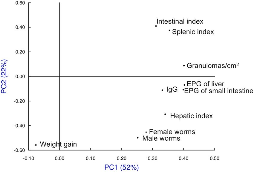
www.nature.com/scientificreports/
Figure 6. Splenic index (A), hepatic index (B) and intestinal index (C) of mice treated with ivermectin (Iv) and
Ivermectin + cobicistat (Iv + Co) and infected with 150 cercariae by S. mansoni (Inf) at 8 weeks post-infection.
*Statistically significant differences p < 0.05 compared to untreated controls (Untr).
Discussion
The discovery of glutamate-mediated signaling in S. mansoni raised hopes for new drug targets in this species14.
Macrocyclic lactones such as ivermectin target the neuronal glutamate-gated chloride channel that is expressed
by arthropods, nematodes and t rematodes15. Schistosomes express these channels, yet the evidence base for the
efficacy of ivermectin against S. mansoni is inconclusive, which might be explained by a relatively low affinity of
the S. mansoni glutamate-gated chloride channel for ivermectin10. Nevertheless, one study reported a marked
reduction in adult worm load after high-dose treatment with ivermectin, which was attributed to the tegumental
Scientific Reports | (2021) 11:4476 | https://doi.org/10.1038/s41598-021-84009-y 6
Vol:.(1234567890)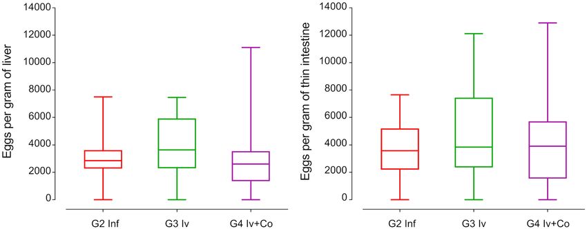
www.nature.com/scientificreports/
Figure 7. ELISA detection of specific IgG antibodies to SoSmAWA antigen at the beginning of the experiment
(A) and 8 weeks post-infection (B) of mice treated with ivermectin (Iv) and Ivermectin + cobicistat (Iv + Co)
and infected with 150 cercariae by S. mansoni (Inf). *Statistically significant differences p < 0.05 compared to
untreated controls (Untr).
Figure 8. Multivariate study of principal components highlighting each study group and searching their
reference centroid at eight weeks post-infection. Groups: mice treated with ivermectin and infected with 150
cercariae of, S. mansoni (Iv); treated with ivermectin + cobicistat (Iv + Co) and infected; infected only with 150
cercariae of (Inf): and an untreated and uninfected control group (Untr).
damage inflicted on the worms9. The slight, yet non-significant, reduction in adult worm load observed in our
study might indicate a possible dose-dependent effect, as Taman et al. used a 25-fold higher dose of ivermectin.
These extremely high doses are contraindicated for systemic human use due to safety concerns. It could be
worthwhile exploring a potential alternative approach for targeting the development of schistosome in tissue.
We are not aware of data showing differential expression of GluCl channels at different life stages of S. mansoni.
l-Glutamate-containing neurons have been reported in cercaria that have not been identified in other life stages16.
It is, however, unknown if ivermectin treatment affects these life stages differently. Using an inhaled formulation
Scientific Reports | (2021) 11:4476 | https://doi.org/10.1038/s41598-021-84009-y 7
Vol.:(0123456789)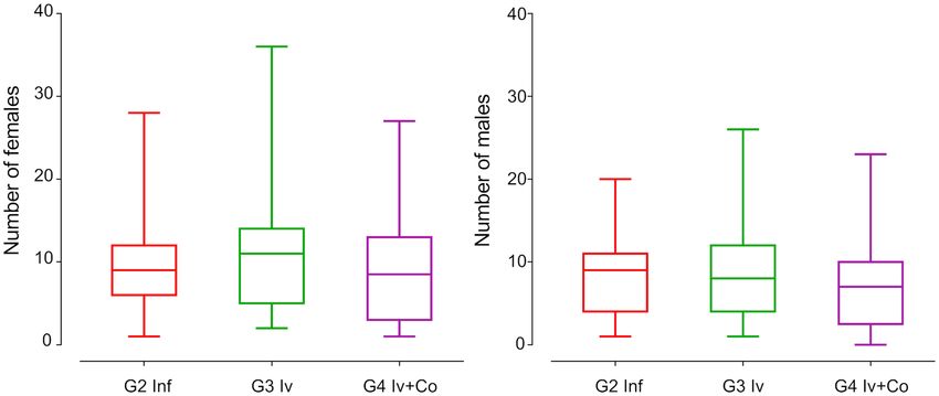
www.nature.com/scientificreports/
Figure 9. Multivariate study of principal components, loads associated with each of the variables used in the
study at eight weeks post-infection. Groups: mice treated with ivermectin and infected with 150 cercariae of,
S. mansoni (Iv); treated with ivermectin + cobicistat (Iv + Co) and infected; infected only with 150 cercariae of
(Inf): and an untreated and uninfected control group (Untr). EPG eggs per gram.
against schistosomula stages in the lung might allow for higher concentrations against a developmental stage
that is not often targeted17.
In C. elegans, the susceptibility to ivermectin can be modulated by targeting ABC transporters or the P-gp18,19.
We previously showed in a pharmaco-enhancement model of mosquitoes feeding on treated pigs that the simul-
taneous inhibition of cytochrome p450 3A and the P-gp enhanced the effect of ivermectin by two mechanisms:
by increasing plasma levels in the pig host and by increasing the susceptibility of the mosquito. This combina-
tion resulted in prolonged target insecticidal concentrations of the drug able to kill Anopheles mosquitoes20.
Given the inconclusive evidence regarding the efficacy of ivermectin treatment in S. mansoni-infected mice,
we hypothesized that P-gp and CYP3A inhibitors may also alter the susceptibility of S. mansoni cercariae to
ivermectin in a prophylaxis model.
Despite pharmacological inhibition of CYP and P-gp in our study, ivermectin did not have any significant
effect on the numbers of S. mansoni eggs or adult worms in the mouse nor on the host immune response as
evidenced by a lack of differences in IgG levels or granuloma formation. This finding stands in contrast to Kasi-
nathan et al. who showed a significant effect of P-gp inhibitors in combination with praziquantel on both egg
numbers and granuloma size in S. mansoni-infected mice, and suggested effects on parasite immunomodulatory
factors to the h ost21. These differences point to different mechanisms involved in the metabolism of praziquantel
and ivermectin.
Ivermectin has an excellent safety profile demonstrated over decades in global mass drug administration cam-
paigns. Its safety is partly explained by the activity of P-gp in the capillaries of blood–brain barrier that excludes
ivermectin from the mammalian central nervous system. Only a few cases of encephalopathy were described in
rare population genotypes (mdr-1 gene) in Loa loa massive drug administration c ampaigns22.
In our study, the combined use of ivermectin and P-gp inhibitor elacridar led to seizures and disturbances in
movement defined as final point criteria in most animals in the treated group. As elacridar disrupts the function
of P-gp in the murine blood–brain barrier, elevated ivermectin levels in the brain lead to important neurological
toxicity23,24. It serves as a reminder that in states with an impaired blood–brain barrier, as is the case in hyper-
inflammatory states, ivermectin could cause significant neurological toxicity.
A limitation of this study pertains to our choice of model animal. Although ivermectin does not affect physi-
ological parameters in mice, they are one of the rodent species most susceptible to ivermectin t oxicity25. The data
on lethal dose included by Merck in the ivermectin label were determined in murine m odels26. This dose poten-
tially does not represent the true toxicity in larger mammals or humans. The vulnerability of mice to ivermectin
toxicity excludes the possibility of using higher doses that might produce more pronounced prophylactic effects
against Schistosoma infection. Another limitation concerns the standard time span and cercaria dose in which the
experiments were conducted to make comparable with other studies. In field conditions people became infected
in several times with lower daily doses and measurement time point is difficult to establish. The drug-treated
groups were not treated all simultaneously, which might have a small influence on the results.
Scientific Reports | (2021) 11:4476 | https://doi.org/10.1038/s41598-021-84009-y 8
Vol:.(1234567890)www.nature.com/scientificreports/
Conclusions
Ivermectin did not show prophylactic properties against experimental infection with S. mansoni cercariae. Para-
site load, granulomatous lesions, or antibody responses in the ivermectin-treated group were comparable to the
untreated control group. Combining ivermectin with cobicistat to prevent infection by S. mansoni did not result
in differences in parasite load, granulomatous lesions, or antibody responses compared to the untreated control
group. The combination of ivermectin and elacridar led to severe toxicity.
Financial support
This study was supported by the University of Navarra with a generous donation from Jeffery and Heart Deal.
CCh and PN received salary support from Unitaid through the BOHEMIA grant to ISGlobal. ISGlobal acknowl-
edges support from the Spanish Ministry of Science and Innovation through the “Centro de Excelencia Severo
Ochoa 2019–2023” Program (CEX2018-000806-S), and support from the Generalitat de Catalunya through the
CERCA Program. USAL acknowledges support from the Instituto de Salud Carlos III, ISCIII, Spain (http://www.
isciii.es) under grants: RICET RD16/0027/0018 and PI19/01727; Ministerio de Ciencia e Innovación (MICINN)
Spain RTI2018-099474-B-I00 and European Union co-financing by FEDER (Una manera de hacer Europa, Sis-
tema Nacional de Garantía Juvenil, Cofinanciación por FEDER, and Iniciativa de Empleo Juvenil BDNS: 427002).
Data availability
All study data is contained within this manuscript and the supplementary material.
Received: 6 November 2020; Accepted: 19 January 2021
References
1. Wang, W., Wang, L. & Liang, Y.-S. Susceptibility or resistance of praziquantel in human schistosomiasis: A review. Parasitol. Res.
[Internet] 111, 1871–1877. https://doi.org/10.1007/s00436-012-3151-z (2012).
2. Lotfy, W. M., Hishmat, M. G., El Nashar, A. S. & Abu El Einin, H. M. Evaluation of a method for induction of praziquantel resist-
ance in Schistosomamansoni. Pharm. Biol. [Internet] 53, 1214–1219. https://doi.org/10.3109/13880209.2014.970289 (2015).
3. Xiao, S. H. Development of antischistosomal drugs in China, with particular consideration to praziquantel and the artemisinins.
Acta Trop. 96, 153–167 (2005).
4. Keiser, J. et al. Mefloquine—An aminoalcohol with promising antischistosomal properties in mice. PLoS Negl. Trop. Dis. [Internet]
3, e350. https://doi.org/10.1371/journal.pntd.0000350 (2009).
5. Yepes, E. et al. In vitro and in vivo ant-schistosomal activity of the alkylphospholipid analog edelfosine. PLoS ONE 9, e109431
(2014).
6. Yepes, E. et al. Inhibition of granulomatous inflammation and prophylactic treatment of schistosomiasis with a combination of
edelfosine and praziquantel. PLoS Negl. Trop. Dis. [Internet] 9, e0003893. https://doi.org/10.1371/journal.pntd.0003893 (2015).
7. Ōmura, S. & Crump, A. Ivermectin: panacea for resource-poor communities? Trends Parasitol. [Internet] 30, 445–455. http://www.
sciencedirect.com/science/article/pii/S1471492214001263 (2014).
8. Merck. Stromectrol. TGA-Australia approved Package insert [Internet]. 2014 [cited 31 Aug 2020]. https://www.ebs.tga.gov.au/ebs/
picmi/picmirepository.nsf/pdf?OpenAgent&id=CP-2011-PI-02659-3&d=2016071016114622483.
9. Taman, A., El-Beshbibi, S., El-Tantawy, N., El-Hawary, A. & Azab, M. Evaluation of the in vivo effect of ivermectin on Schistosoma
mansoni in experimentally-infected mice. J. Coast Life Med. 2, 817–823 (2014).
10. Dufour, V., Beech, R. N., Wever, C., Dent, J. A. & Geary, T. G. Molecular cloning and characterization of novel glutamate-gated chlo-
ride channel subunits from Schistosomamansoni. PLOS Pathog. [Internet] 9, e1003586. https://doi.org/10.1371/journal.ppat.10035
86 (2013).
11. Seif El-Din, S. H., Abdel-Aal Sabra, A.-N., Hammam, O. A. & El-Lakkany, N. M. Effect of ketoconazole, a cytochrome P450 inhibi-
tor, on the efficacy of quinine and halofantrine against Schistosomamansoni in mice. Korean J. Parasitol. [Internet] 51, 165–175
(2013).
12. Ziniel, P. D. et al. The Schistosomamansoni cytochrome P450 (CYP3050A1) is essential for worm survival and egg development.
PLoS Negl. Trop. Dis. [Internet] 9, e0004279. https://doi.org/10.1371/journal.pntd.0004279 (2016).
13. Abán, J. L. et al. A fatty acid binding protein from Fasciola hepatica induced protection in C57/BL mice from challenge infection
with Schistosoma bovis. Vet Parasitol. Netherlands 83, 107–121 (1999).
14. Mendonça-Silva, D. L., Fittpaldi Pessôa, R. & Noël, F. Evidence for the presence of glutamatergic receptors in adult Schistoso-
mamansoni. Biochem. Pharmacol. 64, 1337–1344 (2002).
15. Wolstenholme, A. Ion channels and receptor as targets for the control of parasitic nematodes. Int. J. Parasitol. Drugs Drug Resist.
1, 2–13 (2011).
16. Solis-Soto, J. M. & de Jong-Brink, M. Immunocytochemical study on biologically active neurosubstances i daughter sporocysts
and cercariae of Trichobilharziaocellata and Schistosomamansoni. Parasitology 108, 301–311 (1994).
17. Chaccour, C. et al. Nebulized ivermectin for COVID-19 and other respiratory diseases, a proof of concept, dose-ranging study in
rats. Sci. Rep. [Internet] 10, 17073. https://doi.org/10.1038/s41598-020-74084-y (2020).
18. James, C. & Davey, M. Increased expression of ABC transport proteins is associated with ivermectin resistance in the model
nematode Caenorhabditiselegans. Int. J. Parasitol. 39, 213–220 (2009).
19. Ardelli, B. & Prichard, R. Inhibition of P-glycoprotein enhances sensitivity of Caenorhabditiselegans to ivermectin. Vet. Parasitol.
191, 264–275 (2013).
20. Chaccour, C. et al. Cytochrome P450/ABC transporter inhibition simultaneously enhances ivermectin pharmacokinetics in the
mammal host and pharmacodynamics in Anophelesgambiae. Sci. Rep. 7, 8535 (2017).
21. Kasinathan, R. S., Sharma, L. K., Cunningham, C., Webb, T. R. & Greenberg, R. M. Inhibition or knockdown of ABC transporters
enhances susceptibility of adult and juvenile schistosomes to praziquantel. PLoS Negl. Trop. Dis. [Internet] 8, e3265. https://doi.
org/10.1371/journal.pntd.0003265 (2014).
22. Bourguinat, C. et al. Analysis of the mdr-1 gene in patients co-infected with Onchocercavolvulus and Loaloa who experienced a
post-ivermectin serious adverse event. Am. J. Trop. Med. Hyg. 83, 28–32 (2010).
23. Schinkel, A. H. et al. Disruption of the mouse mdr1a P-glycoprotein gene leads to a deficiency in the blood–brain barrier and to
increased sensitivity to drugs. Cell [Internet] 77, 491–502. https://doi.org/10.1016/0092-8674(94)90212-7 (1994).
24. Mealey, K. L., Bentjen, S. A., Gay, J. M. & Cantor, G. H. Ivermectin sensitivity in collies is associated with a deletion mutation of
the mdr1 gene. Pharmacogenet. Genomics [Internet]. 2001;11. https://journals.lww.com/jpharmacogenetics/Fulltext/2001/11000
/Ivermectin_sensitivity_in_collies_is_associated.12.aspx
Scientific Reports | (2021) 11:4476 | https://doi.org/10.1038/s41598-021-84009-y 9
Vol.:(0123456789)www.nature.com/scientificreports/
25. Davis, J. A. et al. Behavioral effects of ivermectin in mice. Lab. Anim. Sci. 49, 288–296 (1999).
26. BV Dohme Merck Sharp & Stromectol. FDA-approved package insert [Internet]. 2007 [cited 6 Oct 2020]. https: //www.merck. com/
product/usa/pi_circulars/s/stromectol/stromectol_pi.pdf.
Acknowledgements
We thank Ana Gómez Sánchez and Marta González Leal for their technical help with the S. mansoni life cycle,
ELISA and animal management.
Author contributions
Conceptualization: C.Ch. Data curation: B.V., J.L.A., J.H.G., P.F.S. Formal analysis: B.V., J.L.A., A.M. Funding
acquisition: C.Ch. Investigation: B.V., J.L.A., A.M. Methodology: C.Ch., P.N., P.F.S., J.H.G., B.V., J.L.A., A.M.
Supervision: A.M. Writing—original draft: J.L.A., B.V., J.Ch., C.Ch. Writing—review and editing: all authors
contributed, reviewed and approved the last draft.
Competing interests
The authors declare no competing interests.
Additional information
Correspondence and requests for materials should be addressed to C.C.
Reprints and permissions information is available at www.nature.com/reprints.
Publisher’s note Springer Nature remains neutral with regard to jurisdictional claims in published maps and
institutional affiliations.
Open Access This article is licensed under a Creative Commons Attribution 4.0 International
License, which permits use, sharing, adaptation, distribution and reproduction in any medium or
format, as long as you give appropriate credit to the original author(s) and the source, provide a link to the
Creative Commons licence, and indicate if changes were made. The images or other third party material in this
article are included in the article’s Creative Commons licence, unless indicated otherwise in a credit line to the
material. If material is not included in the article’s Creative Commons licence and your intended use is not
permitted by statutory regulation or exceeds the permitted use, you will need to obtain permission directly from
the copyright holder. To view a copy of this licence, visit http://creativecommons.org/licenses/by/4.0/.
© The Author(s) 2021
Scientific Reports | (2021) 11:4476 | https://doi.org/10.1038/s41598-021-84009-y 10
Vol:.(1234567890)You can also read