Targeting CDK9 for Anti-Cancer Therapeutics - MDPI
←
→
Page content transcription
If your browser does not render page correctly, please read the page content below
Review

Targeting CDK9 for Anti-Cancer Therapeutics
Ranadip Mandal 1, Sven Becker 1 and Klaus Strebhardt 1,2,*

                                          1 Department of Gynecology and Obstetrics, Johann Wolfgang Goethe University, Theodor-Stern-Kai 7, 60590
                                            Frankfurt am Main, Germany; ranadip.mandal@kgu.de (R.M.); sven.becker@kgu.de (S.B.)
                                          2 German Cancer Consortium (DKTK), 69120 Heidelberg, Germany

                                          * Correspondence: strebhardt@em.uni-frankfurt.de; Tel.: +49-(049)-069-6301-6894; Fax: +49-(049)-069-6301-6364

                                          Simple Summary: CDK9, in combination with Cyclin T1, is one of the major regulators of RNA
                                          Polymerase II mediated productive transcription of critical genes in any cell. The activity of CDK9
                                          is significantly up-regulated in a wide variety of cancer entities, to aid in the overexpression of genes
                                          responsible for the regulation of functions, which are beneficial to the cancer cells, like proliferation,
                                          survival, cell cycle regulation, DNA damage repair and metastasis. Enhanced CDK9 activity, there-
                                          fore, leads to poorer prognosis in many cancer types, offering the rationale to target it using small-
                                          molecule inhibitors. Several, increasingly specific inhibitors, have been developed, some of which
                                          are presently in clinical trials. Other approaches being tested involve combining inhibitors against
                                          CDK9 activity with those against CDK9’s upstream regulators like BRD4, SEC and HSP90; or down-
                                          stream effectors like cMYC and MCL-1. The inhibition of CDK9’s activity holds the potential to be
                                          a highly effective anti-cancer therapeutic.

                                          Abstract: Cyclin Dependent Kinase 9 (CDK9) is one of the most important transcription regulatory
                                          members of the CDK family. In conjunction with its main cyclin partner—Cyclin T1, it forms the
                                          Positive Transcription Elongation Factor b (P-TEFb) whose primary function in eukaryotic cells is
Citation: Mandal, R.; Becker, S.;         to mediate the positive transcription elongation of nascent mRNA strands, by phosphorylating the
Strebhardt, K. Targeting CDK9 for
                                          S2 residues of the YSPTSPS tandem repeats at the C-terminus domain (CTD) of RNA Polymerase II
Anti-Cancer Therapeutics. Cancers
                                          (RNAP II). To aid in this process, P-TEFb also simultaneously phosphorylates and inactivates a
2021, 13, 2181. https://doi.org/
                                          number of negative transcription regulators like 5,6-dichloro-1-β-D-ribofuranosylbenzimidazole
10.3390/cancers13092181
                                          (DRB) Sensitivity-Inducing Factor (DSIF) and Negative Elongation Factor (NELF). Significantly en-
Academic Editor: Claudia Maletzki
                                          hanced activity of CDK9 is observed in multiple cancer types, which is universally associated with
                                          significantly shortened Overall Survival (OS) of the patients. In these cancer types, CDK9 regulates
Received: 31 March 2021                   a plethora of cellular functions including proliferation, survival, cell cycle regulation, DNA damage
Accepted: 29 April 2021                   repair and metastasis. Due to the extremely critical role of CDK9 in cancer cells, inhibiting its func-
Published: 1 May 2021                     tions has been the subject of intense research, resulting the development of multiple, increasingly
                                          specific small-molecule inhibitors, some of which are presently in clinical trials. The search for
Publisher’s Note: MDPI stays neu-         newer generation CDK9 inhibitors with higher specificity and lower potential toxicities and suitable
tral with regard to jurisdictional        combination therapies continues. In fact, the Phase I clinical trials of the latest, highly specific CDK9
claims in published maps and institu-
                                          inhibitor BAY1251152, against different solid tumors have shown good anti-tumor and on-target
tional affiliations.
                                          activities and pharmacokinetics, combined with manageable safety profile while the phase I and II
                                          clinical trials of another inhibitor AT-7519 have been undertaken or are undergoing. To enhance the
                                          effectiveness and target diversity and reduce potential drug-resistance, the future of CDK9 inhibi-
                                          tion would likely involve combining CDK9 inhibitors with inhibitors like those against BRD4, SEC,
Copyright: © 2021 by the authors. Li-
                                          MYC, MCL-1 and HSP90.
censee MDPI, Basel, Switzerland.
This article is an open access article
distributed under the terms and con-
                                          Keywords: CDK9; Cyclin T1; RNAP II; transcription; BRD4; MYC; apoptosis
ditions of the Creative Commons At-
tribution (CC BY) license (http://crea-
tivecommons.org/licenses/by/4.0/).

Cancers 2021, 13, 2181. https://doi.org/10.3390/cancers13092181                                                        www.mdpi.com/journal/cancers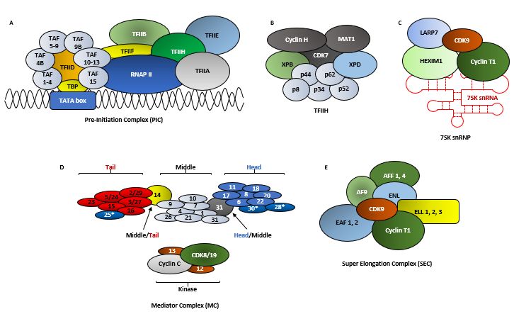
Cancers 2021, 13, 2181                                                                                                          2 of 33

                                 1. Cyclin Dependent Kinases (CDKs)
                                       Cyclin Dependent Kinases (CDKs) are a family of serine/threonine kinases which re-
                                 quire a regulatory cyclin (with the exception of CDK5, which require p35/p39) subunit to
                                 attain their kinase activity. Presently, this family comprises of 21 members, further classi-
                                 fied into three sub-types, broadly based on their regulatory functions: (1) regulators of the
                                 cell-cycle—CDKs 1, 2, 4 and 6; (2) regulators of transcription—CDKs 7, 8, 9, 12, 13 and 19;
                                 (3) regulators of diverse or as yet undefined functions—CDKs 5, 10, 11, 14, 15, 16, 17, 18
                                 and 20 [1–3]. At present, 29 cyclins have been identified in humans. While, some CDKs
                                 can have multiple cyclin partners, some cyclins can also partner with multiple CDKs.
                                 There are still some orphan CDKs whose cyclin partners have not been identified (Table 1).
                                 This review article is primarily going to focus on CDK9.

      Table 1. The different known members of the CDK family, their regulating cyclin partners (except CDK5) and their general
      functions.

             Cyclin/Regulating
                                                                      Functions                                        Reference
                  Partners
                                  Cell-cycle regulation—promotes G2/M transition, regulates G1 progress and G1/S
 CDK1              Cyclin B1                                                                                              [4,5]
                                                                         transition
                                  Cell-cycle regulation—G1/S transition, exit from S-phase; initiation of DNA syn-
 CDK2         Cyclins A/D1/E1                                                                                             [6]
                                                                            thesis
                                   Cell-cycle regulation—G1-phase transition; partial phosphorylation of Rb with
 CDK4             Cyclin D                                                                                                [7]
                                                                            CDK6
                                  All aspects of neuronal physiology; immune response; angiogenesis; myogenesis;
 CDK5              p35/p39                                                                                               [6,8,9]
                                                    melanogenesis and regulation of insulin levels
                                   Cell-cycle regulation—G1-phase transition; partial phosphorylation of Rb with
 CDK6             Cyclin D                                                                                                [7]
                                                                            CDK4
                                 CDK Activating Kinase (CAK)—phosphorylates cell-cycle regulating kinases; tran-
 CDK7             Cyclin H       scription regulation—S5 phosphorylation on RNAP II CTD to initiate transcription         [10]
                                                                          initiation
                                  Part of Mediator Complex (MC), regulates the phosphorylation transcription fac-
 CDK8      Cyclin C/Med12/Med13                                                                                           [11]
                                                            tors, their activity and turn-over
CDK9         Cyclins T1/T2a/T2b                      Positive regulation of transcription elongation                      [12]
CDK10             Cyclin M                            Cell-cycle regulation and tumor suppressor                        [13,14]
CDK12             Cyclin K                           Positive regulation of transcription elongation                    [15,16]
CDK13             Cyclin K                           Positive regulation of transcription elongation                    [15,17]
CDK14             Cyclin Y                  Regulation of cell-cycle, proliferation, migration and invasion               [18]
CDK15             Unknown                       Inhibits apoptosis by phosphorylating Survivin on T34                     [19]
                                 Promotes proliferation in medulloblastoma, prostate, breast, melanoma and cervi-
CDK16             Cyclin Y         cal cancers, inhibits apoptosis by down-regulating the tumor suppressor p27 in       [20–22]
                                                                           NSCLC
CDK17             Unknown               Down-regulation causes poor prognosis in glioma. Unknown functions                [23]
                                  Negative regulator of cell migration and adhesion, prevents the accumulation of
CDK18         Cyclins A2 and E                                                                                          [24–26]
                                                          DNA damage and genome instability
                                  CDK8 homolog, part of Mediator Complex (MC), promotes proliferation and mi-
CDK19             Cyclin C         totic gene expression in the absence of CDK8 expression, negative regulation of        [11]
                                                                    NOTCH signaling
           Cyclin H and CK2 (ge-
                neric CDK20);
CDK20                             Cell-cycle regulator (generic CDK20) and promotes cell survival (cardiac CDK20)         [27]
            KCNIP2 and SNAPIN
              (cardiac CDK20)
                                 Regulates spermatogonial proliferation and meiosis progression and germ line cell
CDK21             Unknown                                                                                                 [28]
                                                    activation in testis; unknown function in cancer
Cancers 2021, 13, 2181                                                                                            3 of 33

                              2. CDK9
                              2.1. The Structure of CDK9
                                   CDK9 was originally discovered by Graña X. et.al. in 1994, in the quest to identify
                              potential CDC2 (CDK1) related kinases, as a 42 kDa, 372 amino acid (aa) protein, termed
                              PITALRE due to the presence of a Pro-Ile-Thr-Ala-Lue-Arg-Glu containing motif [29].
                              This motif corresponded with the highly conserved PSTAIRE box, seen in multiple CDKs
                              (Figures 1 and 2A) [30].
                                   CDK9 exists in two isoforms, the originally identified and more abundant one of 42
                              KDa and the less abundant one of 55 kDa, the latter having an additional 117 aa at its N-
                              terminus [12]. These two isoforms are transcribed from the same CDK9 gene, but by two
                              different promoters, located more than 500 bp apart on the CDK9 gene, with the 42 kDa
                              promoter being significantly stronger than the 55 kDa one [31]. Within the nucleus,
                              CDK942 is primarily localized within the nucleoplasm while CDK955 primarily accumu-
                              lates within the nucleolus [12] (Figure 2A). All amino acid positions and other aspects of
                              CDK9 mentioned henceforth would be about CDK942.

      Figure 1. The PITALRE (Pro-Ile-Thr-Ala-Lue-Arg-Glu) sequence of CDK9 which aligned with the highly conserved
      PSTAIRE box, observed in multiple CDKs.
Cancers 2021, 13, 2181                                                                                                          4 of 33

      Figure 2. The domain structures of key proteins involved in P-TEFb regulated transcription. (A) The two CDK9 isoforms
      expressed in most cells. The CDK955 is longer than the CDK942 by 117 amino acids (aa) at the N-terminus (NT) as the
      former isoform is transcribed by a different promoter,
Cancers 2021, 13, 2181 5 of 33
structure of P-TEFb revealed that T186 was in close proximity and interacts with the argi-
nine residues R148 and R172 [32] and R65 [38] (Figure 2A), to form an intra-molecular H-
bond network. T186 was also reported to coordinate and stabilize an inter-molecular salt-
bridge between R65 and R172 of CDK9 and E96 of Cyclin T1 (Figure 2B). Like T186A [39],
mutations at these three arginine residues (R65A, R148A and R172A) resulted in an almost
complete loss of interaction with Cyclin T1, while R65A and R172A also led to a loss of pT186,
suggesting a mutual stabilization mechanism. On the other hand, E96A and E96K mutations
of Cyclin T1 caused a partial and complete loss of P-TEFb formation, respectively [38].
Initially, it was reported that, when synthesized, CDK9 undergoes auto-phosphory-
lation at the T186 residue on its T-loop. When Kim et al. had expressed a mutated CDK9
(T186A) in insect cells using the Baculovirus expression system, no phosphorylation could
be detected, even in the presence of Cyclin T1, although, the kinase activity of the mutant
remained unimpeded. Incubating CDK9–WT; T186A; and D167N (kinase dead) with the
CDK Activating Kinase (CAK; CDK7/Cyclin H), showed a phosphorylation signal only in
the CDK9WT but in none of the mutants. Even in the presence of Cyclin T1, CAK couldn’t
enhance the phosphorylation signal of CDK9 above the background level, allowing them
to conclude that CDK9 underwent auto-phosphorylation, irrespective of the presence of
Cyclin T1 and this phosphorylation was not critical for the kinase activity of CDK9 [42].
CDK9 T186 auto-phosphorylation was also confirmed by Baumli et al. following the mass
spectrometric analysis of full-length CDK9, in complex with Cyclin T1 [32].
Larochelle et al. had demonstrated that insect cell-derived CDK9D167N, both as mono-
mer or in combination with Cyclin T1, purified from bacteria, were phosphorylated at
T186 [37], partially contradicting the earlier observation [42]. Similarly, generated CDK9WT
also showed pT186 and could not be further activated by CAK. However, unphosphory-
lated WT CDK9, synthesized in vitro by programming rabbit reticulocyte lysates, could
not be phosphorylated at T186, even in the presence of Cyclin T1 and ATP, but elicited
basal activity towards SPT5. When CAK was introduced into the reaction, pT186 appeared
as well as a great enhancement in SPT5 phosphorylation. Similar trend in T186 phosphor-
ylation was also seen with CDK9D167N, albeit at much lower levels and no activity towards
SPT5 (Figure 2C) (explained in the following sections). Introducing CAK to this reaction,
phosphorylated WT CDK9 at T186 and increased its activity towards SPT5. The CDK9D167N
was also phosphorylated at T186, albeit at much lower level and had no kinase activity,
proving that Cyclin T1 is critical for the activity of CDK9. In HCT116 cell lines, CAK was
also shown to be responsible for almost all of pT186, as well as the activation of CDK9 on
transcribed chromatin. Additionally, the selective inhibition of CDK7 diminished CDK9
mediated pS2 of RNAP II (Figure 2G) [37].
Furthermore, an siRNA library screen of 78 ubiquitously expressed serine/threonine
kinases, to identify potential CDK9 T-loop kinases, had revealed the Ca2+/Calmodulin-
dependent Kinase 1D (CaMK1D) as one such target. Interestingly, although the siRNA
mediated knock-down of CaMK1D led to the reduction in pT186, this effect could not be
reversed neither by the overexpression of ectopic CaMK1D, nor by increasing Ca2+ levels
in HeLa cells. CDK9 and CaMK1D were also not found to interact in either the cytosol or
nucleus of HeLa cells, indicating that CaMK1D indirectly affects the CDK9 pT186. In con-
trast, inhibiting CaMK1D activity by the CaMK inhibitor KN-93 or the CaM inhibitor W-
7, led to 59% and 29% reduction in pT186 levels, respectively, in HeLa cells. A key takea-
way from this work was the revelation that the knock-down of expression or inhibition of
activity of CaMK1D also led to the corresponding reduction in total CDK9 levels. This
reduction was revealed to be due to the proteasomal degradation of CDK9, which could be
partially reversed by treating HeLa cells for 1 h with the protease inhibitor MG-101 [41].
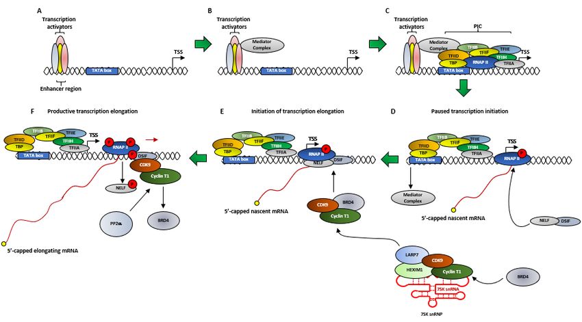
2.3. RNA Polymerase II-Mediated Transcription
The molecular function of CDK9 cannot be described without briefly explaining tran-
scription. In human cells, the expressions of many protein coding genes are regulated by
Cancers 2021, 13, 2181                                                                                         6 of 33

                         the RNA Polymerase II (RNAP II), at several steps like pre-initiation, initiation, elonga-
                         tion, RNA processing and termination. For the sake of this review, we will briefly focus
                         on some of these steps. Transcription initiation by RNAP II begins with the formation of
                         the Pre-Initiation Complex (PIC) (Figures 3A and 4A–C), for which RNAP II is assisted by
                         General Transcription Factors (GTFs) like TFIIA, TFIIB, TFIID, TFIIE, TFIIF and TFIIH.
                         TFIID in-turn is composed of the TATA box-Binding Protein (TBP), which is required for the
                         transcription from every promoter, and several TBP Associated Factors (TAFs) (Figure 3A),
                         which are required for promoter specific transcription [43–45].
                              TFIIH is a multi-protein complex, comprising of, in its transcription initiator role—
                         10 proteins (XPB, XPD, p62, p52, p44, p34 and p8 forming the core, plus CAK with CDK7,
                         Cyclin H and MAT1) [46] (Figure 3B). After the formation of the PIC, the transcription of
                         a gene initiates subsequent to the TFIIH mediated—(1) phosphorylation of RNAP II, at
                         the S5 (pS5) residues of the Y1S2P3T4S5P6S7 tandem repeats and (2) ‘promoter melting’
                         which involves opening-up ~10 bp of the promoter dsDNA, allowing RNAP II to access
                         the template strand [43,47,48]. The human RNAP II CTD possesses 52 of this heptad re-
                         peats [49]. The pS5 also allows the removal of the CDK8 containing Mediator Complex
                         (MC) (Figures 3D and 4D) and the release of RNAP II from the PIC [45]. During transcrip-
                         tion initiation, RNAP II generates a nascent transcript ~50 ribonucleotides from the pro-
                         moter and then pauses due to the combined effect of two negative regulators—(1) NELF
                         and (2) DSIF (Figure 4D) [50]. One final consequence of the pS5 is the recruitment and
                         activation of capping enzymes which perform a multi-step 5’-capping of the nascent
                         mRNA strand (Figure 4D). This capping prevents the degradation of the nascent mRNA
                         strand by nucleases like XRN2 (5′-3′ exoribonuclease 2) and the resulting termination of
                         the promoter proximally paused RNAP II [45,51,52]. The next step in RNAP II mediated
                         transcription is transcriptional elongation by P-TEFb. Interestingly, the regulation of the
                         expressions of most genes in human cells occurs during their transcription elongation,
                         rather than at their initiation phase [45,53,54]. Thus, CDK7 aids in both pausing transcrip-
                         tion and relieving it by activating CDK9 [38] (Figure 4).

                         2.4. The Molecular Functions of CDK9
                              A great deal of the initial information about the function of P-TEFb was obtained by
                         studying its function in HIV-1 transcription, which is regulated by the viral Trans-Activa-
                         tor protein Tat. In the absence of Tat, RNAP II undergoes promoter proximal pausing, in
                         a region adjacent to the Trans-Activation Response element (TAR) RNA sequence, to syn-
                         thesize short transcripts of ~60 ribonucleotides [38,55]. When present, Tat promotes HIV-
                         1 transcription by first binding to the bulge of the TAR hairpin-loop and then recruiting
                         P-TEFb, in complex with the Super Elongation Complex (SEC) (Figure 3E), to the HIV-1
                         promoter. When P-TEFb was knocked-down by siRNA in HeLa cells, it did not cause the
                         cell death but inhibited Tat activation and HIV-1 transcription [47,56]. The previously
                         mentioned phosphorylation sites in CDK9-S347, T350, S353, S357 and T354 were found to
                         be essential for the binding of P-TEFb to TAR, as P-TEFb with a C-terminal truncated
                         CDK9 (Δ323), bound to TAR significantly weakly, as compared to CDK9WT [40,57]. In fact,
                         the then newly found protein was called Cyclin T due to its unique ability to bind with
                         Tat [58]. After two additional Cyclin Ts (T2a and T2b), which cannot interact with Tat,
                         were discovered, the original was termed Cyclin T1 [59].
                              As mentioned in the previous section, DSIF and NELF are responsible for the pro-
                         moter proximal pausing of RNAP II (Figure 4F). P-TEFb plays an essential role in promot-
                         ing transcription elongation by performing three functions that release RNAP II from its
                         pause—(1) phosphorylating NELF, causing its dissociation from the paused RNAP II, (2)
                         phosphorylating the SPT5 subunit of DSIF (Figures 2C and 4F), at T4 of its CTD
                         GS(Q/R)TP residue [60], which subsequent to NELF dissociation, converts into a positive
                         elongation factor and (3) phosphorylating RNAP II at the S2 (pS2) residues of the
                         Y1S2P3T4S5P6S7 repeats at its CTD. Following the release of RNAP II from its promoter
Cancers 2021, 13, 2181                                                                                                            7 of 33

                                  proximal pause, the kinase activity of P-TEFb is rendered unnecessary, although it still
                                  travels along the Transcription Elongation Complex (TEC) [38,40,50,61].

      Figure 3. The structures of key complexes involved in RNAP II mediated transcription and regulation of CDK9 activity.
      (A) Pre-Initiation Complex (PIC)–the PIC is composed of RNAP II which is assisted by General Transcription Factors
      (GTFs) like TFIIA, TFIIB, TFIID, TFIIE, TFIIF and TFIIH. TFIID in-turn is composed of the TATA box-Binding Protein
      (TBP) and upto 15 TBP Associated Factors (TAF). TFIID, assisted by TFIIA, first recognizes the core promoter by scanning
      for and subsequently associating with the TATA box sequence on the promoter DNA. TBP then recruits the TFIIB which
      subsequently loads the RNAP II-TFIIF complex on the promoter. Eventually, TFIIE and TFIIH facilitates the opening of
      the transcription bubble [62]. (B) The TFIIH is a heterodecameric protein complex comprising of the proteins XPB, XPD,
      p62, p52, p44, p34 and p8 forming the core, plus CAK with CDK7, Cyclin H and MAT1. It is an essential part of PIC and
      is also involved in DNA repair. The DNA helicase XPD and the dsDNA translocase activity of XPB are both required for
      opening of the transcription bubble, although XPD is not critical for transcription initiation but for DNA damage repair.
      CAK (CDK7/Cyclin H) phosphorylates S5 of RNAP II, whereas MAT1 both assists in the interaction between CDK7 and
      Cyclin H and recruits CAK to the TFIIH core, by interacting with XPD and XPB. The function of XPB is regulated by p52
      and p8 while that of XPD is regulated by p44 [46]. (C) The 7SK snRNP is composed of the non-coding 7SK snRNA, serving
      as as scaffold for the RNA binding proteins HEXIM1/HEXIM2, LARP7 and MePCE. (D) The Mediator Complex (MC) is
      comprised of upto 30 subunits in humans, which are generally divided into the head, middle, tail and kinase modules.
      The head is composed of the mediators 6, 8, 11, 17, 18, 20 and 22 in the head (blue); 1, 4, 7, 9, 10, 21 and 31 in the middle
      (black); and 15, 16, 23, 2/29, 3/27 and 5/24 in the tail (red). The mediators 31 and 14 connect the head with middle and tail
      with middle, respectively. The kinase domain is composed of the mediators 12 and 13, CDK8 or its paralogue CDK19 and
      Cyclin C. The functions of the mediators 25, 28 and 30 (*) are yet undefined. The MC promotes the assembly of the PIC by
      serving as a functional bridge between RNAP II and the above mentioned GTFs [63]. (E) The Super Elongation Complex
      (SEC) is composed of CDK9/Cyclin T1, the Eleven-nineteen Lysine-rich Leukemia (ELL) proteins 1, 2 and 3, the AF4/FMR2
      Family members 1 and 4 (AFF 1 and 4), Eleven-Nineteen Leukemia (ENL), ALL1-Fused gene from chromosome 9 (AF9)
      and the ELL-associated factors 1 and 2 (EAF 1 and 2) [64,65].
Cancers 2021, 13, 2181                                                                                                          8 of 33

      Figure 4. The simplified model of the regulation of transcription by P-TEFb. (A) At the beginning of transcription, Tran-
      scription Factors (TFs) bind to the enhancer regions, upstream of the core promoter region (represented by the TATA box).
      The TATA box in-turn is located upstream of the Transcription Start Site (TSS). (B) TFs recruit the Mediator Complex (MC)
      (Simplified here from Figure 3). (C) The MC then assists in the recruitment and assembly of the PIC on the DNA strand
      which, as mentioned before, starts promoter melting and form the transcription bubble, to allow to the RNAP II to access
      the DNA template strand. (D) The TFIIH subunit of PIC also phosphorylates the S5 residue at the CTD of RNAP II to
      initiate transcription from the TSS to generate a nascent mRNA transcript of ~50 ribonucleotides but pauses due to recruit-
      ment of two negative regulators of transcription DSIF and NELF. The S5 phosphorylation also enables the dissociation of
      the MC and freeing-up of the RNAP II from the PIC and promotes the recruitment of capping enzymes to cap the 5′-end
      of the nascent mRNA strand in a multi-step process, to prevent the nascent strand from being degraded by nucleases like
      XRN2. The cap remains until transcription finishes and the mRNA is properly processed. (E) When it is ideal for the cells
      to carry-out productive transcription, BRD4 recruits P-TEFb (CDK9/Cyclin T1) from its negative regulatory complex—
      7SK snRNP to the RNAP II. However, the kinase activity of the BRD4 bound CDK9 remains transiently inhibited due to
      the phosphorylation of T29 of CDK9 by BRD4 (F) The phosphatase PP2α is then recruited which dephosphorylates T29,
      restoring the kinase activity of CDK9. This causes the additional P-TEFb mediated phosphorylations of RNAP II at S2, the
      SPT5 subunit of DSIF at T4 and NELF. This allows NELF to dissociate from RNAP II, which along with SPT5 phosphory-
      lation, transforms DSIF to a positive elongation factor. These events relieve RNAP II from transcription pause and allows
      it to progress along with the DNA template to initiate productive transcriptional elongation. As the transcription elonga-
      tion gears-up, BRD4 was released from the P-TEFb.

                                 2.5. The Regulation of CDK9 Activity
                                       In order to regulate a highly choreographed process like transcription, the activation
                                 and activity of transcription are tightly regulated. In the nuclear extract of HeLa cells,
                                 ~80% of CDK9 was found to be associated with Cyclin T1 (P-TEFb) and 10% each with
                                 Cyclins T2a and T2b [59]. Of these, ~50% of P-TEFb is sequestered in the 7SK snRNP (small
                                 nuclear Ribonuclear Protein) [50,66,67] (Figure 3C), which negatively regulates its kinase
                                 activity. The 7SK snRNP is composed of the RNAP III transcribed, non-coding 7SK
                                 snRNA, which serves as the scaffold for the RNA binding proteins HEXIM1/HEXIM2
                                 (Hexamethylene bis-acetamide inducible proteins) (Figure 2D), LARP7 (La-Related Pro-
                                 tein) (Figure 2E) and MePCE (Methylphosphatase Capping Enzyme) [30,50,68]. While
                                 HEXIM1 inhibits P-TEFb’s kinase activity in a 7SK dependent manner [69], LARP7 stabi-
                                 lizes the 7SK snRNA and protects it from exonuclease meditated degradation by binding
Cancers 2021, 13, 2181                                                                                         9 of 33

                         to its 3′-UUUU-OH sequence, as evident from the fact that the knock-down of LARP7
                         resulted in the complete degradation of 7SK snRNA [70,71]. MePCE was originally
                         thought to add a unique γ-monomethyl phosphate cap to the 5′-end of 7SK snRNA [72],
                         however, recent work had shown that this function existed only in a LARP7 free environ-
                         ment. The presence of LARP7, on one hand, inhibited the capping activity of MePCE, but
                         on the other hand, promoted its interaction with LARP7, to co-operatively stabilize 7SK
                         snRNA [73]. Here too, the CDK9 pT186 played a critical role as CDK9T186A or T186D (Figure
                         2A) inhibited the associations of the resulting CDK9s to HEXIM1 and LARP7 by ~99%, as
                         compared to CDK9WT, in Jurkat cells [39] as well as in HeLa cells [74]. Similarly, CDK9R65A,
                         R148A and R172A and Cyclin T1E96K (Figure 2B) were completely incapable of interacting with

                         HEXIM1 and LARP7 [38].
                               The remaining 50% P-TEFb was associated with BRD4 (Figure 2F) or with the SEC
                         (Figure 3E). BRD4 is the only member of the Bromodomain and Extraterminal (BET) pro-
                         tein family capable of binding with P-TEFb [30,75]. Unlike the inactive P-TEFb in the 7SK
                         snRNP complex, P-TEFb associated with BRD4 was active and was recruited to various
                         promoters via BRD4’s interactions with acetylated histones-H3, H4 and MC [76,77]. Inter-
                         estingly, P-TEFb remained associated with BRD4 since the formation of PIC until the ini-
                         tiation of transcription elongation [30]. Under physiological conditions, the EAF1 and 2 of
                         SEC (Figure 3E) interacted with the med26 subunit of the MC (Figure 3D) to get directly
                         recruited to genes like MYC and HSP70. ChIP-seq analysis following med26 depletion
                         resulted in lower occupancy of SEC components throughout the complete transcribed re-
                         gions of the aforementioned two genes, as well as reduced levels of S2 RNAP II on these
                         genes. However, with some genes, SEC recruitment required the pre-occupancy of BRD4,
                         while in others, the RNAP II associated factor (PAF1) recruited SEC by interacting with
                         AF9 or ENL [64,65].
                               Multiple hypothesis had been put forth to explain the interaction between BRD4 and
                         P-TEFb. Zhou et al. had reported that BRD4, bound to P-TEFb, was recruited to HIV PIC
                         (Figures 3A and 4E) where, in order to subsequently facilitate the formation of an efficient
                         TEC, transiently inhibited the kinase activity of P-TEFb by auto-phosphorylating the T29
                         of CDK9. As the PIC gears-up for transcription elongation, between positions +1–+14 for
                         HIV-1, BRD4 was released from the TEC. The phosphatase PP2α was then recruited to the
                         TEC, which de-phosphorylated T29, restoring the kinase activity of P-TEFb [78]. Schröder
                         et al. had proposed a ‘two-pronged’ binding mechanism between BRD4 and P-TEFb,
                         where on one hand, the N-terminal Bromodomain (BD2) of BRD4 (Figure 2F) interacted
                         with at least 3 of the 4 known acetylated lysine residues of Cyclin T1 (K380ac, K386ac and
                         K390ac), while P-TEFb was still bound to HEXIM1 and 7SK snRNA. On the other hand,
                         the C-terminal 54 aa long P-TEFb Interacting Domain (PID) of BRD4 (Figure 2F) interacted
                         with the active P-TEFb, devoid of both HEXIM1 and 7SK snRNA, by dissociating HEXIM1
                         from Cyclin T1. This marked the transition between inactive and active P-TEFb in the
                         presence of BRD4. Of note, BD1 showed no binding affinity towards Cyclin T1 and the
                         affinity of BD2 towards the tri-acetylated Cyclin T1 was similar to its affinity towards
                         acetylated histones [79]. This finding was further corroborated by Itzen et al. who had
                         demonstrated that in vitro, PID stimulated the kinase activity of P-TEFb by ~1.3 folds over
                         its basal activity, even in the presence of HEXIM1 (Figure 2D). By performing Isothermal
                         Titration Calorimetry, they had also demonstrated that PID did not bind to Cyclin T1, in
                         the absence of CDK9, thereby proposing that the Bromodomain and PID interacted syn-
                         ergistically with Cyclin T1 and CDK9 of P-TEFb, respectively, forming a stable clamp ~900
                         aa apart. Interestingly, PID-P-TEFb interaction did not alter the phosphorylation specific-
                         ity and substrate preference for CDK9 at the CTD of RNAP II [80]. Yang et al. had also
                         reported the importance of a new CDK9 phosphorylation site–S175, in the biding of P-
                         TEFb with BRD4. When mutated to CDK9S175A/D in HeLa cells, the association of the mu-
                         tant CDK9 with BRD4 was completely abolished, without affecting the association with
                         either Cyclin T1 or HEXIM1, indicating that the pS175 is necessary for the association of
                         P-TEFb with BRD4 and its resulting dissociation from the inactive 7SK snRNP complex
Cancers 2021, 13, 2181                                                                                       10 of 33

                         [74,81]. This was also confirmed by Mbonye et al. in T-cells [38,39]. On the other hand,
                         CDK9T186A/E almost completely abolished the association of the mutant CDK9 with
                         HEXIM1 but not with Cyclin T1 or BRD4, confirming the need for this phospho-site for
                         HEXIM1 binding [81,82], although Mbonye et al. had shown that CDK9T186A couldn’t in-
                         teract with Cyclin T1 [38]. The kinase responsible for the pS175 was eventually identified
                         as CDK7. Treatment of Jurkat cells with PMA led to the partial dissociation of P-TEFb
                         from the 7SK snRNP (Figure 3C) and therefore pS175 level, which was effectively inhib-
                         ited by the selective CDK7 inhibitor THZ1. pT186 remained unaffected [38].
                              From all these and other works, it has become evident that BRD4 and other promoter-
                         specific TFs are necessary to retrieve the active P-TEFb from the 7SK snRNP complex and
                         carry out transcription elongation.

                         2.6. The Other Pool of CDK9
                               As mentioned earlier, ~80% of CDK9 existed as P-TEFb, whether sequestered as an
                         inactive complex with 7SK snRNP or as an active complex with BRD4 or other TFs. But,
                         what about the remaining 20% CDK9, unbound to Cyclin T1? O’Keeffe et al. had demon-
                         strated that free CDK9 was unstable within the cells and got degraded rapidly. In 293T
                         cells, stably expressing a HA-tagged CDK9, ~80% of the newly synthesized HA-CDK9
                         was degraded within the first 6–12 h, whereas the remaining ~20% HA-CDK9 did not
                         show any significant degradation until 48 h. They were the first to identify that CDK9
                         bound to two chaperones—the more general HSP70 and the substrate specific
                         HSP90/CDC37 complex, which acted in a sequential manner. Initially, HSP70 stabilized
                         any newly synthesized CDK9 by correctly folding it. Once the nascent CDK9 attained a
                         certain folded configuration, it was handed over to the HSP90/CDC37 complex, which
                         established and maintained the CDK9 for the follow-up binding to Cyclin T1, thereby
                         completing the final assembly of P-TEFb. The inhibition of HSP90’s interaction potential
                         by the inhibitor Geldanamycin led to ~12.5 folds increase in HSP70 bound HA-CDK9. The
                         erstwhile mentioned stable ~20% CDK9 comprised of these three complexes [83]. More
                         recently, Mbonye et al. had confirmed these findings in T-cells. They had also described
                         that in the absence of Cyclin T1 expression, as in resting T-cells, HSP90/CDC37 bound
                         CDK9 was predominantly localized within the cytosol and lacked any pT186. Upon un-
                         dergoing TCR activation, resulting in Cyclin T1 expression, CDK9 was imported to the
                         nucleus to get activated. Once again, the treatment of Th17 cells with the Geldanamycin
                         derivative-17-AAG significantly reduced the formation of P-TEFb and the expression of
                         Cyclin T1, possibly through proteasomal its degradation. Additionally, the association of
                         CDK9 with HSP90/CDC37 showed a minor, but significant, uptick in CDK9T186A [38]. In-
                         deed, Napolitano et al., had demonstrated that CDK9 could be detected in both the cytosol
                         and the nucleus, while Cyclin T1 was predominantly localized within the nucleus. Our
                         own work in this direction has also confirmed this in multiple cervical, breast and ovarian
                         cancer cell lines (un-published work) [84,85]. It was also shown that CDK9 could shuttle
                         between the nucleus and cytosol, via the CRM1/exportin protein and inhibiting the asso-
                         ciation between CDK9 and CRM1 by the nuclear export inhibitor LMB (Leptomycin B)
                         enriched nuclear CDK9. Additionally, it was demonstrated that the expression of Cyclin
                         T1 promoted the nuclear localization of CDK9 [86,87].

                         3. The Clinical Relevance of CDK9
                               CDK9 expression had been implicated in the prognosis and resistance to anti-cancer
                         therapeutics in a large number of cancer types like those of breast, lung, prostate, endo-
                         metrium, apart from melanoma, osteosarcoma, myeloid leukemia, soft tissue sarcomas
                         etc. [1,88–92]. We will briefly explain some of these cancer entities here.
Cancers 2021, 13, 2181 11 of 33
3.1. Breast Cancer
ERα+ endocrine therapy-resistant breast cancers over-expressed the cMYC oncogene
and genes regulated by it [93] which correlated with poor RFS, but not when the patients
had only undergone chemotherapy [89]. In a study on breast cancer cell lines, Sengupta
et al. had demonstrated that in multiple aromatase inhibitor-resistant MCF-7 cancer cell
lines, the levels of cMYC mRNA and proteins were significantly higher than the parental
MCF-7 cell line. In these cell lines, the cMYC promoter showed higher recruitment of pS2,
but not pS5 in RNAP II, as compared to MCF-7 cells, which also correlated with higher
levels of both pT186 and CDK9 (2.5 and 3.1 folds, respectively). Lastly, the inhibition of
CDK9 activity, in the resistant cell lines, with the small-molecule inhibitor CAN-508,
showed a dose-dependent inhibition of cell growth, in conjunction with ~60% reduction
in cMYC mRNA and proteins, highlighting the critical role of CDK9 in the up-regulation
of cMYC expression in endocrine therapy-resistant ERα+ breast cancer patients [89].
3.2. Osteosarcoma
Similarly, Ma et al. had demonstrated that, of 70 osteosarcoma patient derived sam-
ples, 67.1% exhibited high CDK9 expression, which significantly correlated with meta-
static disease, non-survival, worse OS (Overall Survival) and DFS (Disease Free Survival)
and poor post-neoadjuvant chemotherapy necrosis. CDK9 expression was also higher in
osteosarcoma cell lines, as compared to the osteoblast cell line HOB-c. Down-regulating
CDK9 (siCDK9) expression led to decreased cell proliferation, as well as reduced levels of
pS2 RNAP II and MCL-1 whereas, treating the osteosarcoma cell lines with the ATP com-
petitive CDK9 inhibitor LDC000067 significantly reduced the levels of pS2 RNAP II and
MCL-1 and their clonogenicity, in a dose-dependent manner, in conjunction with in-
creased apoptosis. 12 days of treatment with 10µM of LDC000067 also significantly re-
duced the diameters of the 3D spheroids of U2OS (57.4%) and KHOS (48.8%) cell lines, as
compared to their untreated counterparts [88,94].
3.3. Endometrial Cancer
Of the 32 endometrial cancer patient samples (carcinosarcoma-2, clear cell-2, serous-
4, mixed cell-2, mucinous-1 and endometrioid-21), analyzed by He et al., 59.4% had low
and 40.6% had high CDK9 expression, but demonstrated no correlation between these
expression values and the pathological stage, grade and sub-type of endometrial tumor.
As seen before, high CDK9 expression significantly worsened the OS (39 vs. 14 months)
and PFS (Progression Free Survival) (96 vs. 26 months). Concentration dependent knock-
down of CDK9 expression and dose-dependent inhibition of CDK9 activity by LDC000067
led to correspondingly significant reductions in cell viability and MCL-1 expression while
BAX expression and PARP cleavage were enhanced. Additionally, 14 days of treatment
with increasing concentrations of LDC000067 resulted in significant reductions of clono-
genicity and 2D migration of endometrial cancer cell lines [90].
3.4. AML
The dysregulation of the CDK9 pathway was also reported by various groups in
AML. Increased expressions of cMYC and MCL-1 had been associated with the patho-
genicity of AML due to the enhanced survival and expansion of the AML cells. High MCL-
1 expression was also observed in and associated with poor prognosis in ~50% of R/R
AML cases. Translocation products of the MLL (Mixed Lineage Leukemia) gene were
found to associate with P-TEFb in AML to trigger constitutively active transcription [95].
Its levels were found to be ~2 folds higher in recurrent patients as compared to pre-treat-
ment patients, while its down-regulation led to the death of both human and murine AML
cells [96,97]. As a result, targeting the anti-apoptotic BCL-2 family members, to which
MCL-1 belongs, had been attempted. Venetoclax is one such inhibitor which was ap-
proved by the FDA in 2020, for use against newly diagnosed AML patients of 75 years orCancers 2021, 13, 2181 12 of 33
older, in combination with Azacitidine, Decitabine or Low-Dose Cytarabine (LDAC). Un-
fortunately, MCL-1 up-regulation enables Venetoclax resistance and relapse. Combining
Venetoclax with the CDK9 inhibitor Alvociclib had shown synergistic effect against both
Venetoclax resistant and sensitive AML patients, in a pre-clinical study [97,98].
On the other hand, in primary AML samples, the mRNAs of HEXIM1 and cMYC
were found to be over-expressed with mutual exclusivity [99]. HEXIM1 served as the
binding partner of several other proteins, nearly half of which has been recognized for
their roles in cancers. In AML, the cytoplasmic-mislocated mutant of the p53 regulator
NPM (Nucleoplasmin)-NPMc+, was observed in ~35% of all AML patients. Both NPM and
NPMc+ were HEXIM1 binding proteins. NPM overexpression induced the proteasomal
degradation of HEXIM1, thereby freeing-up P-TEFb to regulate enhanced transcription
elongation. NPMc+ had a disrupted NLS and an extra NES, thereby localizing the resulting
protein to the cytoplasm instead of the nucleoli, as seen with NPM. NPMc+ sequestered
HEXIM1 from its usual nuclear location to the cytoplasm, again hampering the formation
of the negative regulatory 7SK snRNP complex, leading to the increased P-TEFb mediated
transcription elongation [100].
3.5. Lung Cancer
The pro-inflammatory cytokine TNFα stimulated the expression of the Matrix Met-
alloproteinase-9 (MMP-9) in an NF-κB dependent manner, which promoted cancer cell
migration and invasion, by degrading basement membrane, and also angiogenesis, by
vascular structure remodeling and the release of angiogenic factors like VEGF (Vascular
Endothelial Growth Factor) [101,102]. Shan et al. had demonstrated that in the lung ade-
nocarcinoma cell line A549, TNFα stimulated the transcription of the MMP-9 promoter.
To achieve this, TNFα enhanced the associations of both CDK955 and CDK942 to Cyclin T1,
as well as the binding of P-TEFb to the MMP9 gene which led to increased levels pS2
RNAP II bound to MMP9. The P-TEFb inhibitor DRB substantially inhibited TNFα stim-
ulated MMP-9 mRNA expression by ~76 folds and its activity by ~80%, but not those of
TGF-ß1 stimulated MMP-2. Similarly, the over-expression of a double-negative CDK9
(CDK9dn) or an siCDK9 lowered the TNFα stimulated MMP-9 activity by more than half.
Additionally, CDK9 positively regulated the NF-κB response element, through which
TNFα stimulated the transcription of the MMP-9 promoter. Once again, DRB as well as
CDK9dn blocked the activation of NF-κB by TNFα [103]. More recently a novel highly
selective CDK9 inhibitor-21e, was shown to inhibit CDK9 activity (IC50 11 nM) in NSCLC
cells as well as cell lines of other cancer entities. It suppressed the clonogenicity of two
NSCLC cell lines while inducing apoptosis by down-regulating the expression of BCL-2
and up-regulating the pro-apoptotic protein BIM, as well as increasing Caspase-3 activa-
tion. 21e also significantly inhibited pS2 RNAP II. Inhibiting CDK9 activity also sup-
pressed the expressions of the stemness markers in the two NSCLC cell lines-SOX2, OCT4,
NANOG and KIF4, while also significantly inhibiting other stemness phenotypes like Side
Population (SP) and serum-free suspension culture. Lastly, 21e also inhibited the tumor
growth of a mouse xenograft model derived from the NSCLC cell line H1299, once more
validating the significance of CDK9 in lung cancer [104].
3.6. Prostate Cancer
Androgen Receptor (AR), a steroid receptor TF for testosterone and di-hydrotestos-
terone, played a critical role in prostate cancer, especially in the castration resistant sub-
type. AR mutations, Post-Translational Modifications (PTMs), over-expression and splice
variants were some of the ways through which prostate cancers evaded Androgen Abla-
tion Therapy (AAT) [105]. AR was compartmentalized in the cytoplasm in an inactive
form, in complex with HSP90, which sequestered it from migrating into the nucleus and
binding to the DNA. In the presence of suitable steroid ligands, AR underwent a confor-
mational change and detached from the HSP90. The conformationally changed AR thenCancers 2021, 13, 2181 13 of 33
underwent homo-dimerization and translocated to the nucleus, where it bound to Andro-
gen Response Element (ARE) located at the promoters of the target genes. The phosphor-
ylation of AR is one PTM which resulted in its altered-activation, transcriptional activity,
stability, nuclear retention and cell growth. 18 such phosphorylation sites are currently
known [106], among which, S81 is most frequently phosphorylated in response to andro-
gen stimulation, by kinases like CDK1 [107,108], CDK5 [109] and CDK9 [110]. Mutating
this phosphorylation site to S81A resulted in the reduced cell growth and altered the tran-
scription of the AR regulated genes in a promoter specific manner. In vitro, CDK9 phos-
phorylated S81 exclusively and ex vivo, under basal conditions, WT-CDK9/Cyclin T1
could but CDK9dn/Cyclin T1 could not phosphorylate S81, but in the presence of the syn-
thetic androgen R1881, both constructs could phosphorylate S81, albeit to a lower level for
CDK9dn/Cyclin T1. siCDK9 treatment also led to a significant reduction in pS81, in the
presence of R1881. Similarly, inhibiting CDK9 activity by DRB or Flavopiridol treatment
reduced pS81 signal in a dose-dependent manner, in conjunction with the reduced tran-
scription of the AR regulated early response genes—SGK1 and NKX3.1 [110]. Chen et al.
had shown that while CDK1 mediated pS81 increased during mitosis, CDK9 performed
this function during the interphase of androgen responsive LNCaP and non-responsive
PC3 prostate cancer cell lines. Additionally, CDK9 mediated pS81 stabilized AR chroma-
tin binding for transcription [107].
3.7. Melanoma
CDK9 has been reported to be overexpressed in many melanoma cell lines [111] while
CDKs 7 and 9 were also shown to be over-expressed in multiple uveal melanoma cell lines.
Using the potent CDK7/9 inhibitor—SNS-032, it was demonstrated that pS2/5/7 at RNAP
II, by these two kinases, could be inhibited in a dose-dependent manner, resulting in re-
duced cell proliferation, migration, invasion, cancer stemness, in vivo tumor formation,
increased MMP-9, but not MMP-2, expression, in dose- and time-dependent manners. Un-
der similar conditions, increased apoptosis of the treated cells was also observed, both ex
and in vivo, accompanied by enhanced PARP and Caspase-3 cleavages and reduced ex-
pressions of several anti-apoptotic proteins. Lastly, SNS-032 also led to reduced transcrip-
tion and expressions of YAP (Yes Associated Protein) and its active form pYAP (S127). All
these alterations were attributed directly to the inhibition of CDK7/9 by SNS-032 [112].
Recently, Zhang, et al. had demonstrated that the CSN6, a member of the Constitu-
tive Photomorphogenesis 9 (COP9) Signalosome (CSN) complex family, was over-ex-
pressed in malignant melanoma cell lines and tissues, driving their proliferation, migra-
tion and invasion. CSN6 was shown to interact with CDK9, stabilizing and protecting the
latter from being ubiquitinated and subsequently degraded by UBR5 (Ubiquitin Protein
ligase E3 component n-recognin 5), in the malignant melanoma cell lines A375 and MV3.
Mouse xenograft models of A375 cells with stable knock-down of CSN6 manifested sig-
nificantly smaller tumors and CDK9 positive cells, as compared to those derived from
CSN6 expressing cells. These effects of CSN6 knock-down could be significantly rescued
by concurrent knock-down of UBR5, highlighting the significance of CDK9 in melanoma
[113]. As CSN6 expression is up-regulated in multiple cancer entities [113–115], it stands to
reason that this effect, demonstrated in melanoma could occur in other cancer types too.
3.8. Ovarian Cancer
Amongst 26 primary ovarian cancer patient samples (serous-20, squamous-1, high-
grade serous-1, endometroid-1, transitional cell-1, endometroid and serous-1 and endo-
metroid and clear cell-1), Wang et al. had shown significantly lower CDK9 expression in
primary cancer tissues than patient-paired metastatic and recurrent cancer tissues. Of
these, 13 patients with lower CDK9 expression had better DFS and OS, as compared to
those with higher expression. No correlations were observed between CDK9 expression
and the stage, grade, subtype and ascites of the ovarian cancer patients. Similar to endo-Cancers 2021, 13, 2181 14 of 33
metrial cancer and osteosarcoma, use of siCDK9 and LDC000067 resulted in a dose-de-
pendent inhibition of cell-viability and a decrease in pS2 of RNAP II, MCL-1 levels and an
increase in BAX expression and PARP cleavage. Additionally, LDC000067 treatment led
to significant reductions of clonogenicity, 2D migration and 3D spheroid formations of
two ovarian cancer cell lines—SKOV-3 and OVCAR-8, once again highlighting the signif-
icance of CDK9 in the poor prognosis of ovarian cancer [116].
4. Inhibitors of CDK9
A number of CDK9 inhibitors have been reported over the years, including many
pan-CDK inhibitors (Table 2). Cassandri et al., in their review article, had listed a number
of natural CDK targeting compounds like Indirubins, isolated from several indigo pro-
ducing plants. A couple of synthetic derivatives of Indirubins like 6-bromoindirubin and
6-bromoindirubin-3′-monooxime were also reported [1]. Another natural compound Ro-
hitukine, with anti-inflammatory and anti-immunomodulatory properties, derived from
the Dysoxylum binectariferum plant, served as the basis for one of the earliest pan-CDK
inhibitor Flavopiridol [117]. In this review, we would briefly describe Flavopiridol, as it
was the first CDK9 inhibitor to enter clinical trials [118], and some of the other more recent,
selective inhibitors, although not all of them entered the clinical trials phase (Table 2).
4.1. Flavopiridol
Flavopiridol (Alvocidib) (Table 2) is a semi-synthetic flavonoid alkaloid derivative of
Rohitukine, discovered following an NCI based screening of 72,000 compounds against
60 human cancer cell lines [119]. Initially, Flavopiridol showed inhibitory activities against
EGFR and PKA (IC50 21 and 122 µM, respectively), however, during the NCI screen, it
exhibited growth inhibition at IC50 66 nM, ~1000 folds lower than the concentrations re-
quired for EGFR and PKA inhibition [120]. Its remarkable anti-proliferative and growth
inhibitory effects, in vivo and ex vivo, respectively, were initially attributed to its activity
against CDKs 1, 2, 4 and 7. Eventually, it was found to be most effective against CDK9
[121]. Flavopiridol is an ATP-competitive inhibitor, which showed promising in vitro ac-
tivity against a number of cancer entities, but demonstrated only limited in vivo activity,
except in hematologic cancers like MCL and CLL, resulting in the cessation of its further
development [1,122]. Presently, only three trials, involving Flavopiridol, are active,
against AML, advanced solid tumors and Myelodysplastic Syndromes (MDS). Dinaciclib
(SCH-727965) was developed as next generation inhibitor, based on Flavopiridol. Di-
naciclib inhibited CDKs 1, 2, 5 and 9 with respective in vivo IC50 of 3, 1, 1 and 4 nM, as
compared to 3, 12, 14 and 4 nM for its predecessor. It also triggered apoptosis and arrested
cell-growth in >100 tumor cell lines [123,124]. However, preliminary results from clinical
trials have been reported to be less encouraging [122]. Further trials are ongoing (Table 2).
4.2. AZD-4573
The development of the highly selective CDK9 inhibitor AZD-4573 (Table 2) was
based on another selective, oral inhibitor of the amidopyridine series—AZ-5576, which
was reasonably potent against the AML cell line MV-4-11, both ex and in vivo, but exhib-
ited lower solubility in PBS and rate of metabolism in human microsomes, limiting its
ability to provide a high therapeutic dose and optimal duration of target engagement
within the clinical scenario. AZD-4573 showcased excellent selectivity against CDK9 with
in vitro IC50, measured using the Thermofisher Kinase Panel, at 10 µM against CDKs 1,Cancers 2021, 13, 2181 15 of 33
4/6/7 and 2/4/6, respectively. Both the mRNA and protein levels of MCL-1 were down-
regulated in a dose-dependent manner, without affecting the same for other anti-apop-
totic proteins like BCL-2 and BCL-XL. Caspase-3 cleavage was also observed in MV-4-11
and MOLM-8 cell lines, suggesting that MCL-1 is the major initiator of AZD-4573 driven
apoptosis. Against a diverse panel of cancer cell lines, 6 and 24 h of AZD-4573 treatment
generated significant Caspase-3 activation and reduction in viability, more pronouncedly
in hematological cancer cell lines than in solid tumor cell lines. The pharmacokinetics (PK)
and pharmacodynamics (PD) of AZD-4573 also improved in vivo over AZ-5576 [125,126].
Presently, AZD-4573 is subject to two phase-I clinical trials against advanced hematolog-
ical cancers (NCT04630756) [127] and relapsed/refractory hematological cancers
(NCT03263637) [128] (Table 2).
4.3. BAY-1143572 (Atuveciclib)
Atuveciclib (Table 2) was discovered by Lücking et al. in search of a highly selective,
orally applicable and exclusive P-TEFb inhibitor. The basis for Atuveciclib design was the
triazine BAY-958, which demonstrated in vitro IC50 of 11 nM against CDK9 and a
CDK2/CDK9 IC50 ratio of 98, but slow absorption and low oral bioavailability in vivo.
Atuveciclib displayed in vitro IC50 of 13 nM against CDK9 and an IC50 ratio of 100. Ex vivo
IC50 against CDK9 were 0.92, 0.31 and 0.89 µM in HeLa, MOLM-13 and MV-14 cell lines,
respectively, with excellent anti-proliferative effect. In vivo, BAY-1143572 showed mark-
edly lower blood clearance and improved oral bioavailability. In MV-14 AML cell line
derived rat xenograft, daily oral administration of 12 mg/kg of BAY-1143572 for 14 days
resulted in no tumor re-growth in 9/12 animals [129]. Owing to these promising pre-clin-
ical results, BAY-1143572 was subjected to two Phase-I studies against advanced malig-
nancies (lymphomas and solid tumors; NCT01938638) and acute leukemia
(NCT02345382). In both cases however, due to high drug related TEAE (Treatment Emer-
gent Adverse Effect) and lack of clinal response at the dose tested, both trials were pre-
maturely terminated [130,131] (Table 2).
4.4. BAY-1251152
BAY-1251152 (Table 2) was a follow-up, P-TEFb inhibitor of BAY-1143572, with in-
creased potency, with IC50 against CDK9 under both in vitro and ex vivo (MOLM-13 cells)
conditions being 3 and 29 nM, respectively; enhanced selectivity against CDK2; permea-
bility; solubility; and no efflux. BAY-1251152 also demonstrated impressive in vivo effi-
cacy in MOLM-13 derived xenograft mouse and rat models [132]. In our own studies,
BAY-1251152 had demonstrated IC50 of 19.06, 16.57 and 74.64 nM against HeLa, SiHa and
OVCAR-3 cells, respectively, as compared to comparative BAY-1143572 IC50 of 1.27 and
0.64 µM for HeLa and SiHa cells (un-published work). Until the development of AZD-
4573, BAY-1251152 was the only selective CDK9 inhibitor currently undergoing clinical
trials [133] against advanced malignancies (NHL and solid tumors; NCT02635672). This
phase-I study revealed significant, dose-dependent reductions in the mRNA levels of
MYC, PCNA and MCL-1 and manageable safety. There were also signs of anti-tumor ac-
tivity viz. stable disease (SD) in 9/31 (29%) and durable SD in 3/31 (9.7%) patients [134].
Another phase-I trial against advanced hematological malignancies (NCT02745743) is on-
going. Preliminary results from AML patients revealed significant, but short-lived post-
treatment reductions in the mRNA levels of MYC (0.5–4 h), PCNA (1–4 h) and MCL-1 (1–
3 h). No objective responses were achieved at any doses, despite achieving CDK9 inhibi-
tion [135]. Additionally, BAY-1251152 monotherapy was declared to generate durable re-
missions of over two years in 2/7 patients with very aggressive relapsed/refractory double-
hit DLBCL (Diffused Large B-Cell Lymphoma), following a phase-I trial [136] (Table 2).Cancers 2021, 13, 2181 16 of 33
4.5. SNS-032 (BMS-387032)
SNS-032 (Table 2), was originally shown as a selective CDK2 inhibitor, but was later
shown to be able to inhibit CDK7 and CDK9, with modest inhibitory activities against
CDKs 1, 4, 5 and 6. In vitro kinase profiling revealed IC50 values of 38–48 nM (CDK2), 62
nM (CDK7), 4 nM (CDK9), 480 nM (CDK1), 925 nM (CDK4), 340 nM (CDK5) and >1000
nM (CDK6) [137], while Albert et al. reported these values, respectively, at 6 nM (CDK2),
68 nM (CDK7), 1.4 nM (CDK9), 52 nM (CDK1), 355 nM (CDK4) and 3404 nM (CDK6) (no
CDK5 values were reported) [138]. Ex vivo IC50 values were 231 and 192 nM against CDKs
7 and 9, respectively, in RPMI-8226 cells, along with reductions in pS5 and 2 of RNAP II,
XIAP and MCL-1 levels and increased PARP cleavage [137]. In a phase-I clinical trial in-
volving patients with advanced solid tumors, high SNS-032 concentrations inhibited pS5
and 2 of RNAP II and MCL-1 expression, in their PBMCs. However, all 20 participants of
this study discontinued treatment due to relapse or disease progression (65%); deteriora-
tion without progress (20%); adverse effect (5%); drug-related toxicity (5%); and patient
request (5%). Only 3 patients exhibited SD following treatment [139]. Another phase-I
pharmacological study (NCT00446342) against 19 CLL and 18 MM patients, identified
MTD (Maximum Tolerated Dose) and DLT of 75 and 100 mg/m2, respectively, for the CLL
patients. MTD could not be established while DLT was not observed for the MM patients.
Overall, SNS-032 was well tolerated by both patient types, at all infusion doses and in-
duced reductions in pS5 and 2 of RNAP II, within 2 h of starting the infusion, reverting to
baseline levels after 24 h. At 6 h, when the infusion was stopped, the reduction of pS2 level
was more pronounced (64%) than pS5 level (35%), along with XIAP (10%) and MCL-1
(56%) levels and increased PARP cleavage. However, SNS-032 only exhibited limited anti-
tumor activities in both the CLL (1 partial and 1 stable diseases) and MM cohorts (1 stable
disease), with the treatment of all three stable cases terminated due to drug-related toxic-
ities, patient request or investigator decision [140]. A third clinical trial of SNS-032 against
solid tumors was also completed, although its results have not yet been posted
(NCT00292864) (Table 2).
Olson et al. had recently demonstrated that conjugating a Thalidomide group to SNS-
032, to produce THAL-SNS-032, could selectively and potently degrade CDK9, but no
other CDK targets of SNS-032, in a CRBN dependent manner. Compared to SNS-032,
THAL-SNS-032 exhibited in vitro IC50 values of 171 nM (CDK1), 62 nM (CDK2), 398 nM
(CDK7), 4 nM (CDK9). Against a panel of leukemia cell lines, THAL-SNS-032 demon-
strated anti-proliferative effects at IC50 values 2–6.5 folds lower than those of SNS-032. It
also inhibited pS2, in MOLT4 AML cells, in a time- and dose-dependent manner, but pS5
and 7 levels remained completely unaffected. While both SNS-032 and its derivative sim-
ilarly induced PARP and Caspase-3 cleavages, only THAL-SNS-032 induced MCL-1 deg-
radation and γH2A.X activation in MOLT4 cells [141], unlike in RPMI-8226 cells [139].
Reduction in global gene expression was also significantly more pronounced after THAL-
SNS-032 treatment, as compared to its predecessor [141]. In short, addition of the Thalid-
omide group to SNS-032, turned a pan-CDK inhibitor into a highly selective CDK9 inhib-
itor. Due to the CRBN-mediated ubiquitination and degradation of CDK9, the mecha-
nisms of THAL-SNS-032-mediated induction of apoptosis and inhibition of the transcrip-
tional machinery was also different than that of SNS-032, which only inhibited the activa-
tions of CDKs 2, 7 and 9. No clinical trials of THAL-SNS-032 has been initiated till date
(Table 2).
4.6. AT-7519
AT-7519 (Table 2) is a pan-CDK inhibitor developed by Wyatt et al., that selectively
inhibited CDKs with in vitro IC50 values of 190 nM (CDK1), 47 nM (CDK2), 67 nM (CDK4)
and 18 nM (CDK5). It showed low oral bioavailability. Intraperitoneal injection for 8 days
BID of AT-7519 in A2780 derived ovarian cancer xenograft mouse model produced 86%
tumor growth inhibition at 7.5 mg/kg dose [142]. Squires et al. later identified CDK9 as aCancers 2021, 13, 2181 17 of 33
target for AT-7519 with in vitro IC50 values of 210 nM (CDK1), 47 nM (CDK2), 100 nM
(CDK4), 13 nM (CDK5), 170 nM (CDK5) andCancers 2021, 13, 2181 18 of 33
phosphorylation of CDK9 at T186 and RNAP II at S5; XIAP and BCL-2 levels but inhibited
pS2 (EC50 < 100 nM), MCL-1 and cMYC levels, in a dose-dependent manner. Treatment
for 24 h induced PARP and Caspase-3 cleavages in all three cell lines, as well as cell cycle
arrest at the G0/G1 phase. In vivo, Tmax and T1/2 were 2 and 3.33 h; and 1.55 and 3.37 h in
mice and rats, respectively. JSH-150 was also orally bio-available with bio-availabilities of
45.01 and 45.10% in mice and rats, respectively. MV4-11 derived mouse xenograft models,
20 and 30 mg/kg/day of JSH-150 treatment for 14 days, suppressed tumor progression,
without causing general cytotoxicities or tumor recurrence, one week after the treatment
was stopped [149]. No clinical trial of JSH-150 has been reported so far (Table 2).
4.9. LY-2857785
LY-2857785 (Table 2) is a reversible, ATP-competitive inhibitor, demonstrating in
vitro IC50 values of 246 nM (CDK7), 16 nM (CDK8) and 11 nM (CDK9). Against a panel of
114 kinases, it showed IC50 of 10 µM (CDK6/7), as against 5.2,
15, 305 and 103 nM, respectively, for Flavopiridol [138]. As has been mentioned in the
previous sections, due to the high specificity of LDC000067 against CDK9, it has proved
to be a valuable tool in pre-clinical studies against multiple and diverse cancer entities
[88,90,94,116]. However, no clinical trials of LDC000067 were ever undertaken (Table 2).
4.11. CDKI-73 (LS-007)
The novel CDK9 inhibitor CDKI-73 was the lead in class compound of a class of 5-
substituted 4-(thiazol-5-yl)-2-(phenylamino) pyrimidines, designed by Wang et al., which
targeted the ATP gatekeeper residue F30 and ribose-binding pocket of CDK9 [151,152].
Under in vitro conditions, CDKI-73 exhibited IC50 of 8.17 nM (CDK1), 3.27 nM (CDK2),
8.18 nM (CDK4), 37.68 nM (CDK6), 134.26 nM (CDK7) and 5.78 nM (CDK9) [153]. Against
patient derived CLL cells, CDKI-73 was demonstrated to be significantly more potent than
Flavopiridol or its analog Fludarabine. However, when these same cells were protected
from the effects of cytotoxic drugs, by culturing them with CD40L-expressing MEFs,
CDKI-73 retained its cytotoxic effects while that of Fludarabine was completely annulled.
It was equally effective against these primary CLL cells (n = 28) as against those derived
from clinically relapsed CLL patients (n = 10), including those harboring p53 deletion (n = 3)
while exhibiting superior bioavailability than Flavopiridol. Once again, treatment of the
primary CLL cells for 4 h with 0.1 µM of CDKI-73 inhibited pS2 and T186 phosphoryla-
tions of RNAP II and CDK9, respectively, as well as depleted MCL-1 expression. Interest-You can also read