Stem Cell Research & Regenerative - 4th Annual Conference on Medicine - 48 pages
←
→
Page content transcription
If your browser does not render page correctly, please read the page content below
4th Annual Conference on
Stem Cell Research
& Regenerative
  Medicine
   F EBRUA RY 15 –16, 2018

                             Host Institution
Overview
Mission and Vision
About us
RegenMed SA is a citywide organization in
San Antonio designed to facilitate networking
and to promote interactions among individuals,
institutions, center, installations, companies and
foundations with interests in stem cell research,
tissue engineering, regenerative medicine and / or
biotechnology related to these areas.
RegenMed SA includes representation of individuals,
facilities, and resources within San Antonio
related to stem cell research and regenerative
medicine. Those organziations include UT Health
San Antonio™, UTSA™, the Southwest Research
Institute, Texas Biomedical Research Institute
and the Southwest National Primate Research
Center, the U.S. Army Institute of Surgical Research, the National Trauma Institute, GenCure™, a subsidiary of
BioBridge Global,, StemBiosSys and many other biotechnology companies, among others.
Mission and vision
RegenMed SA’s mission is to facilitate and promote communication, interaction, and collaboration among the
many people and facilities within San Antonio and neighboring regions that share interests and resources related
to the areas of stem cell research, tissue engineering and regenerative medicine. The San Antonio conference
on Stem Cell Research & Regenerative Medicine is designed to provide a forum for the exchange of information
describing ongoing research, education, innovation, clinical application and product development in these areas.
Steering Committee
Tiziano Barberi, PhD, Texas Biomedical Research Institute
Becky Cap, MBA, GenCure
Sy Griffey, PhD, StemBioSys
Jian Ling, PhD, Southwest Research Institute
John McCarrey, PhD, UTSA
Sharon Smith, MS, National Trauma Institute
Christi Walter, PhD, UT Health San Antonio
Erik Weitzel, MD (Col., USAF), US Army Institute of Surgical Research
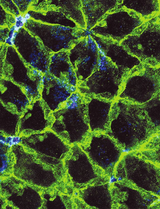
On the The image, courtesy of Erzsebet Kokovay, Ph.D., Assistant Professor, Department of Cell Systems &
cover Anatomy, UT Health San Antonio, shows the neural stem cell niche. Adult neural stem cells send a process
(blue) through the brick-like structure of the ependymal cells (green) to sample signals in the ventricle.
Contents

Contents
           Inside                                      12       Keynote Speakers
     Front Cover      Overview
                                                       13       Poster Presentations
                 3    Contents
                                                       45       Notes
                 4    Sponsors
                                                 Inside
                 8    Program               Back Cover          Acknowledgements

Map
1 Day One
2 Reception/Posters
3 Day Two
P Parking
HP    Handicap                                         2
      Parking                                                           3

                                                                  HP
                                                                         P
                                                            1

                                                           P

                        Free     Guests may connect to the HSCGuest wireless network by
                        Public   simply selecting HSCGuest from the available connections
                        Wifi     on your device and accepting the terms of use.

                                         #RegenMedSA2018

                                                                                                  3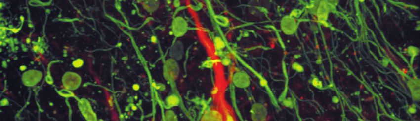
Sponsors

     Welcome to the Fourth Annual
        Conference on Stem Cell
    Research & Regenerative Medicine
       organized by RegenMed SA
               Very special thanks to our conference sponsors

                                   Platinum
                        San Antonio Life Sciences Institute
                             UT Health San Antonio
                                     UTSA

                                      Gold
                  San Antonio Economic Development Corporation /
               City of San Antonio Economic Development Department

                                     Silver
                                     Acelity
                                 StemBioSys, Inc.

                                    Bronze
                                   Gencure™
                        Texas Biomedical Research Institute

4

Advanced Technologies
   Driving Discovery
   C O N S U LTAT I O N · C O L L A B O R AT I O N · E D U C AT I O N

                                       RESEARCH INSTITUTIONAL CORE LABS

                                • Bioanalytics & Single-Cell – new     • Macromolecular Interactions
                                  Cy-TOF™ with MaxPar metal
                                                                       • Mass Spectrometry
                                  labeling technology
                                • Biobanking & Genome Analysis         • Micro CT
                                • Biomolecular NMR                     • Optical Imaging
                                • Flow Cytometry                       • X-Ray Crystallography

Research core labs are your partner in discovery to advance human health

                       Get started.
                     Visit www.uthealthsa.org/iCores
    or call the Director of Institutional Core Facilities (210) 567-2059.

                 @UTHresearch         UT Health San Antonio Research

Program

                   Fourth Annual Conference
                     on Stem Cell Research
                    & Regenerative Medicine
                                               Thursday, February 15
                        Pestana Lecture Hall, UT Health San Antonio, Long Campus

                                                  Friday, February 16
                           Holly Auditorium, UT Health San Antonio, Long Campus

    Thursday, February 15 – Pestana Lecture Hall
    8:00 a.m. – Welcome and Remarks
                 William L. Henrich, MD, MACP, President (UT Health San Antonio)
                 Introduced by Christi A. Walter, PhD (UT Health San Antonio)

                 Taylor Eighmy, PhD, President (UTSA)
                 Introduced by President Henrich

                 Robert A. Hromas, MD, FACP, Dean (Joe R. and Teresa Lozano Long School of Medicine)
                 Introduced by President Henrich

    8:15 a.m. – Keynote Lecture #1
                 Introduced by Daniel Lodge, PhD (UT Health San Antonio)

                 Stewart Anderson, MD (Perelman School of Medicine, University of Pennsylvania,
                 Children’s Hospital of Philadelphia)
                 Accelerating Human Stem Cell Derived Neuronal Maturation by Conditional Activation of mTOR Signaling

    Session 1
                 [Session Chair – Tiziano Barberi, PhD, Texas Biomedical Research Institute]

    9:15 a.m. – Daniel Oh, PhD (Columbia University)
                Bone-like Scaffold: Principles and Applications

    9:40 a.m. – Marie-Claire Gauduin, PhD (Texas Biomedical Research Institute)
                Engineering of Macaque CD4+ T Cells and CD34+ Hematopoietic Stem Cells resistant to in vitro
                VIS Infection using Zinc Finger Nucleases

8
Program

10:00 a.m. – Break

10:30 a.m. – Anibal Diogenes, DDS, MS, PhD (UT Health San Antonio)
            Stem Cell Therapy for Dental Infections

10:55 a.m. – Keren Cheng, PhD (UTSA)
            Epigenetic Programming of Foundational Spermatogonial Stem Cells

11:15 a.m. – James Bynum, PhD (United States Army Institute of Surgical Research)
             Cellular Therapeutics for Treatment of Trauma-related Injuries: A Path to the Clinic

11:35 a.m. – Xiao-Dong Chen, MD, PhD (StemBioSys and UT Health San Antonio)
             Presence of Cyr61 in Native Bone Marrow-derived Extracellular Matrix is Critical for Retention
             of Mesenchymal Stem Cell Properties

12:00 p.m. – Luncheon and networking – Pestana Lecture Hall
Session 2
             [Session Chair –Erzsebet Kokovay, PhD, UT Health San Antonio]

1:00 p.m. – Xing Guo Cheng, PhD (Southwest Research Institute)
            A Novel Method for Single Cell Layer-by-Layer Encapsulation

1:20 p.m. – Richard LeBaron, PhD (UTSA)
            Investigation of TGF-B1 the Extracellular Matrix Protein BIGH3, and BIGH3-derived Integrin Ligand
            Peptides in Tissue Injury and Apoptosis

1:45 p.m. – Daniel Lodge, PhD (UT Health San Antonio)
            Cell-Based Therapies for the Treatment of Psychiatric Disease

2:10 p.m. – Ben Antebi, PhD (United States Army Institute of Surgical Research)
            The Effect of Acute Respiratory Distress Syndrome on Bone-Marrow Mesenchymal Stem Cells

2:35 p.m. – Break

3:05 p.m. – Anand Srinivasan, PhD (GenCure)
            Manufacturing Human-Derived Platelet Lysate for Xeno-Free Expansion of Mesenchymal Stem Cells

3:25 p.m. – Marcel Daadi, PhD (Texas Biomedical Research Institute)
            Standards for deriving Nonhuman Primate Induced Pluripotent Stem Cells, Neural Stem Cells
            and Dopaminergic Lineage

                                                                                                                        9
Program
Session 2 (continued)
3:45 p.m. – Keynote Lecture #2
Introduced by Sy Griffey, PhD (StemBioSys)
Joachim Kohn, PhD (Rutgers University and New Jersey Center for Biomaterials)
Understanding and Predicting the effect of Biomaterials on Cell and Stem Cell Differentiation
4:45 p.m. – Adjourn to Poster Presentations & Reception (ALTC Rm 2.205)
5:00–7:00 p.m. – Poster Presentations and Networking Reception (ALTC Rm 2.205)
Friday, February 16 – Holly Auditorium
Session 3
[Session Chair – Jian Ling, PhD, Southwest Research Institute]
8:30 a.m. – Erzsebet Kokovay, PhD (UT Health San Antonio)
Vascular Regulation of Adult Neural Stem Cells
8:55 a.m. – Eric Brey, PhD (UTSA)
Vascularization of Biomaterials for Regenerative Medicine
9:20 a.m. – Rebecca Bricker, MS, PhD Candidate (Texas Biomedical Research Institute, UT Health San Antonio)
Towards the derivation of Olfactory Placodal Cells from Human Pluripotent Stem Cells
9:40 a.m. – Chris Delavan, MS (United States Army Institute of Surgical Research)
Comparison of Direct vs Transwell MLR Assays for Evaluation of Human Mesenchymal Stromal Cell
Immunomodulation Activity
10:05 a.m. – Break
10:30 a.m. – Brian Hermann, PhD (UTSA)
Single-Cell Transcriptomes as Tools for Regenerative Medicine Research
10:55 a.m. – Becky Robinson-Zeigler, PhD (Advanced Regenerative Manufacturing Institute)
The Cake is Not a Lie: Delivering on the Promise of Regenerative Medicine
10Program
11:20 a.m. – Donna Lehman, PhD (UT Health San Antonio)
Mapping the Chromatin Landscape in the Developing Human Hypothalamus using an iPSC-based
Model reveals Neuronal Activity-dependent Regulation of Bone
11:45 a.m. – Mike Fiske, MS (GenCure)
Overcoming Challenges in Developing GenCure’s Biomanufacturing Center
12:00 p.m. – Luncheon and networking
Session 4
[Session Chair – Bijay Parida, PhD, United States Army Institute of Surgical Research]
1:00 p.m. – Stephen Harris, PhD (UT Health San Antonio)
Periodontal Stem Cell Differentiation: Lineage Tracing, Transcriptome and Epigenetic Regulation
at the Single Cell Level
1:25 p.m. – Arezoo Mohammadipoor, PhD (United States Army Institute of Surgical Research)
Extracellular Vesicles are as Potent Anti-inflammatory Mediators as their Mesenchymal Stem Cell Counterparts
1:50 p.m. – Nicole Edwards, PhD Candidate (Southwest Research Institute)
An Advanced Collagen Wound Matrix Combined with Adipose-derived Stem Cells improves Cutaneous
Wound Healing in a Mouse Model of Type 2 Diabetes
2:10 p.m. – Chris Rathbone, PhD (UTSA)
Vascularization Strategies for Injured and Diseased Skeletal Muscle
2:30 p.m. – Pratap Khanwilkar, PhD (InCube Labs)
Stack the Deck in your Favor: Increasing the Probability that your Translational Idea will Succeed
2:55 p.m. – Break
3:15 p.m. – Keynote Lecture #3
Introduced by John McCarrey, PhD (UTSA)
Jenny Hsieh, PhD (UT Southwestern Medical Center & UTSA Brain Health Consortium)
Precision Models for Brain Health and Disease
4:15 p.m. – Conference adjourns
11Keynote Speakers
Keynote Speakers
Joachim Kohn, Ph.D. Jenny Hsieh, Ph.D.
Board of Governors Professor of Associate Professor, Depts. of Molecular Biology and Neurology
Chemistry and Chemical Biology, & Neurotherapeutics, UT Southwestern Medical Center
Rutgers University Semmes Foundation Endowed Chair in Cell Biology & Director
Director, New Jersey Center for of the UTSA Brain Health Consortium, UTSA
Biomaterials
Dr. Jenny Hsieh, a nationally recognized researcher,
Dr. Joachim will join the UTSA faculty this spring to lead the
Kohn, Director UTSA Brain Health Initiative as the Semmes
of the NJ Center Foundation Chair in Cell Biology.
for Biomaterials,
Dr. Hsieh has a doctorate in biology from Johns
is a leader in
Hopkins University and completed a postdoctoral
biomaterials science
fellowship at the Salk Institute for Biological Studies.
and widely known
for the development Her research focuses on how to make neurons
of tyrosine-derived, replicate themselves so a brain affected by disease or injury can replace its own
resorbable polymers, which are now used in damaged cells and heal. She tackles the challenge using molecular and genetic
several FDA-approved medical devices. tools and is focused on understanding the factors that control the brain’s stem
cells so she can manipulate and stimulate new growth.
Currently about 250,000 patients in the
USA, Canada, Latin America, and Europe Keynote Title: Precision models for brain health and disease
are using implants containing tyrosine-
derived, resorbable polymers which are being
commercialized by REVA Medical, TYRX, Stewart Anderson, M.D.
and Medtronic.
Professor, Department of Psychiatry, Perelman School of Medicine,
University of Pennsylvania
Dr. Kohn’s current research efforts focus on
the development of new discovery paradigms Research Director, Child and Adult Psychiatry, The Children’s Hospital of
Philadelphia
for revolutionary biomaterials using
combinatorial and computational methods Dr. Stewart Anderson, Professor of Psychiatry, at
to optimize the composition, properties, and the University of Pennsylvania’s Perelman School of
cellular responses of biomaterials for specific Medicine. His medical training at the University of
applications, particularly tissue engineering Connecticut include two research fellowships at the
and drug delivery. National Institute of Mental Health.
Dr. Kohn has published extensively, The main focus of his laboratory concerns the
including over 200 major, peer-reviewed molecular and cellular mechanisms that govern the
publications, 40 book chapters, and over development of the mammalian forebrain.
70 issued US patents. Many of his recent New directions in the Anderson lab include the study of mitochondria
publications focus on methods to control the in interneuron migration, maturation, and function. In addition, they are
behavior of cells (including stem cells) in the generating mouse and human stem cell-derived interneurons for use in cell-
context of regenerative medicine. based therapies for seizures, psychotic disorders, and as tools for the study of
gene-gene and gene-environment interactions in neuropsychiatric disease.
Keynote Title: Understanding the effect
of biomaterials on cell and stem cell Keynote Title: Accelerating the maturation of human stem cell derived
differentiation neurons by transient activation of mTOR signaling
12Poster Presentations Poster Presentations Comparison of Preterm and Term Wharton’s Jelly-Derived Mesenchymal Stem Cell Properties in Differing Oxygen Tensions Saloni Agarwala, Caitlyn Wintera, Alexis Corrala, Shamimunisa B Mustafaa, Peter Hornsbyb, Alvaro Moreiraa University of Texas Health-San Antonio, aDepartment of Pediatrics, bDepartment of Cellular and Integrative Physiology, 7703 Floyd Curl Drive, San Antonio, TX 78229 USA ABSTRACT: Mesenchymal stem cells (MSCs) have shown promise as therapeutic agents in treating morbidities associated with premature birth. MSCs derived from human umbilical cords are easy to isolate, have low immunogenicity, and a robust ability to secrete paracrine factors. To date, there are no studies evaluating preterm versus term umbilical cord tissue-derived MSCs. Therefore, our aim was twofold: 1) To compare stem cell properties in preterm versus term MSCs, and 2) Examine the impact of oxygen tension on stem cell behavior. Umbilical cord tissue was obtained from 5 preterm and 5 term neonates. The cells were isolated and characterized as MSCs in accordance to The International Society for Cellular Therapy. We exposed MSCs to differing oxygen tensions to examine the impact of environmental factors on cell performance. We studied the following stem cell properties: i) motility, ii) proliferation, iii) senescence, iv) cell viability, v) colony forming unit efficiency, and vi) inflammatory cytokine expression. Under normoxia (21% O2), the cells from preterm and term infants had similar properties. Under hypoxic conditions (1% O2), term MSCs had better cell proliferation; however, cells exposed to hyperoxia (90% O2) had the slowest motility and lowest cell viability (p
Poster Presentations

     Jamie Archambault, Caitlyn Winter, Saloni Agarwal, Dawn McDaniel, Karli McCoy, Matthew Willis, Alvaro Moreira
     Department of Pediatrics, Division of Neonatology
     University of Texas Health San Antonio

     Effects of Mesenchymal Stromal Cell Conditioned Media on TGF-β1 Expression in an In
     Vitro Model of Bronchopulmonary Dysplasia

     Background: Bronchopulmonary dysplasia (BPD) is a devastating respiratory condition that
     develops in premature neonates exposed to mechanical ventilation and supplemental oxygen. A
     proposed mechanism for the development of this disease involves impaired alveolar wound healing.
     In healthy newborn lungs, alveolar type II (AT2) cells respond to injury by spreading, migrating,
     and proliferating to mend the affected area. Transforming growth factor beta 1 (TGF-β1) is a
     regulatory cytokine that plays an inhibitory role in the repair of injured AT2 cells. TGF-β1 is
     clinically relevant, as studies have documented elevated levels in tracheal samples from human
     neonates with BPD. Emerging evidence in regenerative medicine suggests that mesenchymal
     stromal cells (MSCs) have the capacity to improve repair processes through the release of paracrine
     factors. It has been theorized that these factors are secreted into the media during cell culture; thus,
     highlighting MSC conditioned media (MSC-CM) as a novel therapeutic agent for BPD. The
     purpose of this study is to: a) establish the relationship between TGF-β1 and AT2 cell injury in an
     in vitro model of BPD, b) determine the optimal duration and concentration of MSC-CM for
     treatment following an injury that mimics BPD, and c) demonstrate the effects of MSC-CM on AT2
     cell proliferation and motility.

     Methods: Cells were enzymatically digested from umbilical cord tissue and met the definition of
     MSCs per ISCT criteria. Rat AT2 (RAT2) cells were exposed to either normoxia (21% O2) or
     hyperoxia (90% O2) with escalating concentrations of H2O2 to establish an in vitro model of BPD.
     RNA was collected and RT-PCR was performed to quantify TGF-β1 expression. After establishing
     the BPD model, injured RAT2 cells were exposed to MSC-CM for durations of 1, 2, and 4 hours.
     TGF-β1 expression was again measured using RT-PCR technique. RAT2 cells were then divided
     into 3 experimental groups (control, injury, and injury + treatment). Cell proliferation was measured
     by plating cells and culturing with different concentrations of MSC-CM. Absorbance was measured
     at 420-480 nm and 650 nm following 1 minute of shaking. Finally, a motility assay was performed
     by plating cells, allowing them to form a confluent monolayer, and creating a scratched cell-free
     area. Scratch size was analyzed at the 2, 4, and 6 hour time points to assess cell motility. Statistical
     analysis was performed on Microsoft Excel using one tailed T-tests, with p-values
Poster Presentations
Investigating the loss of terminal neuronal differentiation as a novel mechanism
driving neuronal death in Alzheimer’s disease and related tauopathies
Adrian Beckmann1, Habil Zare2 and Bess Frost3
1
Institute of Biotechnology, Barshop Institute of Longevity and Aging, University of Texas Health
Science Center at San Antonio 2 Department of Computer Science, Texas State University 3
Department of Cell Systems and Anatomy, Barshop Institute of Longevity and Aging, University
of Texas Health Science Center at San Antonio
Tauopathies are a class of neurodegenerative disorders associated with deposits of insoluble
tau protein within the brain. At over 5 million cases currently diagnosed among Americans,
Alzheimer’s disease (AD) is the most common type of tauopathy. With no therapies that
significantly slow or alter the disease course for AD, the number of Americans diagnosed with
AD is expected to increase to 16 million by the year 2050. As one of two hallmark pathologies of
AD, pathogenic tau has emerged as a promising target for therapeutic targeting. Mutations in
the tau gene are associated with dominantly inherited familial tauopathies termed
frontotemporal dementia with parkinsonism linked to chromosome 17 (FTDP-17), demonstrating
that tau dysfunction is sufficient to drive neurodegeneration. In my preliminary analyses, I have
identified prospero and staufen, two proteins that orchestrate the expression and silencing of
genes that maintain terminal neuronal differentiation, among the top ten significantly
downregulated genes in brains of a Drosophila model of tauopathy. In addition, I also identified
51 genes that are differentially expressed in tau transgenic Drosophila that are known to be
regulated by prospero and staufen, suggesting that pathogenic tau may disrupt the cellular
program that maintains terminal neuronal differentiation. When this program is perturbed, post-
mitotic neurons can re- activate the cell cycle, a known process to sufficiently induce neuronal
death. In addition, previously identified cellular phenotypes in tauopathy share many
overlapping phenotypes with dedifferentiated cancerous cells and neural stem cells, including
presence of nuclear envelope invaginations, heterochromatin decondensation, expression of
development-associated genes, and cell cycle activation. Therefore, I hypothesize that
pathogenic tau leads to neuronal death by dysregulating prospero and staufen, thereby
disrupting the cellular program that maintains terminal neuronal differentiation in neurons. In
Aim I, I will determine the mechanistic cause and downstream consequences of prospero
depletion. In Aim 2, I will determine if loss of terminal neuronal differentiation mediates neuronal
death in tau transgenic Drosophila. In Aim 3, I will determine whether maintaining neuronal
differentiation is a viable therapeutic strategy for preventing neuronal death in tauopathy. My
findings will identify new targets for therapeutic interventions for AD and uncover the cellular
processes that tau perturbs to mediate neurodegeneration.
15Poster Presentations
Bianca Cerqueira PhD, Shatha Dallo PhD, Lauren Cornell MS
NovoThelium
Tissue-engineered Matrix for Nipple Areolar Regeneration After Mastectomy
One in eight women will be diagnosed with breast cancer during her lifetime and most
choose to have reconstruction following mastectomy. Nipple reconstruction is typically
the last stage of breast reconstruction and positively impacts final appearance of and
patient satisfaction with breast reconstruction. Research has shown that specifically
nipple reconstruction aids in women’s emotional well-being and improved body image
after mastectomy. Currently, the only option for nipple reconstruction is cutting and
suturing the skin on the breast to create a scar mound with a subsequent tattoo for
desired pigmentation. Unfortunately, this method results in nipple flattening by 50-75%
within two years due to retraction forces of the underlying tissue and contraction of scar
tissue. Additional materials may be used to augment the flap reconstruction, including
autologous fat or cartilage, calcium hydroxylapatite, hyaluronic acid, poly-L-lactic acid
microparticles, acellular porcine small intestine muscosa matrix, and acellular dermal
matrix. Despite augmentation, surgical flap reconstructed nipples still flatten over time.
While patient satisfaction is high following the initial reconstruction, eventual flattening of
the reconstructed nipple causes satisfaction rates to drop as low as 16% within one to
two years. It has been reported that the loss of nipple projection and poor color match
are the main causes of dissatisfaction and loss of emotional benefits of this surgery. We
are developing a tissue-engineered matrix for regeneration of the entire nipple areolar
complex after mastectomy. Our matrix is derived from decellularized allograft nipple
areolar tissue and enables the patient to regenerate an anatomical nipple made from
her own cells that maintains shape and projection. This matrix retains the unique
microstructure of the nipple that may promote melanocyte repopulation and natural
repigmentation. Additionally, this regenerative matrix can be used in new breast
reconstruction procedures or in revisions of previously reconstructed breasts. Through
this research we aim to provide patients with innovative technologies to restore form
and sense of self after breast cancer treatment.
16Poster Presentations
Tiffani Chance1,2, James Bynum (PhD)2, Christopher Rathbone (PhD)1, Barbara Christy (PhD)2,
Larry Estlack2, Christopher Delevan2, Andrew P. Cap (MD, PhD)2
(1) The University of Texas at San Antonio
(2) The United States Army Institute of Surgical Research
The Homogeneity and Functionality of Exosomes Isolated from Different Preparations
Introduction: Exosomes are internally derived micro-vesicles, containing proteins and multiple
types of RNAs,that are typically 30-200 nm in size. Recently, exosomes have gained interest as
microRNA carriers, biomarkers for diseases such as cancer, and potential therapeutics for injury.
Exosome isolation and characterization, however, is still considered a major scientific challenge-
with no true optimal technique. Here, we assess the characterization of spheroid and monolayer
human bone marrow mesenchymal stem cell (hMSC) derived exosomes isolated by
ultracentrifugation (traditional) and by a commercial kit (modern). We also utilize a 4.5 hour
angiogenesis tube formation assay to measure exosome functionality.
Methods: hMSCs were grown to about 95-100% confluence in hMSC high performance basal
media (Rooster Bio; USA) supplemented with 1:100 glutamax (GIBCO, ThermoFisher; USA).
Monolayer and spheroid (seeded at a density of 50,000 cells per well of a pluronic covered 48-
well plate) grown cells were switched to serum free media (1:100 glutamax to DMEM; GIBCO)
and left to incubate for 48 hours (37 °C under 5% CO2). Afterwards, the collected conditioned
media was split into three categories for exosome isolation: spheroid or monolayer isolation by
ultracentrifugation, or monolayer isolation using Qiagen Exo Easy Maxi Kit (Qiagen; USA). The
size distribution of exosomes was analyzed by NanoSight (LM14C, Malvern; UK; n = 5). Exosome
samples were normalized by protein content, and human umbilical vein endothelial cell (HUVEC)
angiogenesis tube formation assays (n = 3) were carried out either in ibidi angiogenesis slides
(ibidi; USA) or in 24 well tissue culture plates pre-coated with Geltrex LDEV Growth Factor
Reduced Basement Membrane Matrix (BD Bioscience; USA). The number of tubes formed was
quantified using ImageJ. Samples were then ran through Seahorse (Agilent; USA) to assess the
bioenergetics profile of exosomes (n = 5). Statistical analysis was performed using one-way
ANOVA tests.
Results: Exosomes were successfully isolated from the three different methods of preparation.
From NanoSight analysis, the sizes of the particles isolated from spheroid and monolayer
ultracentrifugation fell within the appropriate range for exosomes. The particles isolated using
the Qiagen Exo Easy Maxi Kit fell within the range of 10-400 nm, indicating that the sample is
not as pure as those from ultracentrifugation. Additionally, monolayer derived exosomes formed
a greater number of tubes, showed increased oxygen consumption rate and enhanced spare
respiratory capacity, and had a higher bioenergetics health index than spheroid exosomes.
Conclusion: Based on NanoSight analysis, using ultracentrifugation to isolate exosomes from
conditioned media resulted in a more homogenous population of particles that fell within the size
range of exosomes (30-200 nm), indicating that ultracentrifugation isolated a cleaner population
of exosomes than the commercial kit. Monolayer derived exosomes formed more tubes and
displayed increased mitochondrial capacities than spheroid derived exosomes, possibly due to
the lack of stress from being placed in a new environment (well plate) during serum free
incubation.
17Poster Presentations
I-Chung Chen1, Yu-Huey Lin1, Lorena Roa-De-La-Cruz1, Kazadi Mutoji1, Brain P.
Hermann1, Jon M. Oatley2 and John R. McCarrey1
1
The University of Texas at San Antonio; 2Washington State University
Generation and Characterization of Induced Pluripotent Stem Cells Derived from
ID4-EGFPBright Spermatogonial Stem Cells
Spermatogonial stem cells (SSCs) are unipotent stem cells that sustain spermatogenesis
throughout the male reproductive lifespan by undergoing asymmetric divisions leading to
self-renewal of SSCs and the production of progenitors that enter the spermatogenic
differentiation pathway to ultimately form spermatozoa. Recently, FACS-sorted
EGFPBright spermatogonia recovered from mice carrying the Id4-eGfp transgene were
shown to represent an essentially pure population of SSCs on the basis of validation by
the spermatogonial transplantation assay3. Previously, it was shown that at a very low
frequency SSCs maintained in culture have the potential to undergo spontaneous
reprogramming to form embryonic stem cell-like (ES-like) cells4. More recently
contrasting reports have claimed that SSCs can5 or cannot6 be reprogrammed to form
induced pluripotent stem cells (iPSCs). However, neither of these studies used ID4-eGFP
as a marker to recover SSCs, therefore the purity of the starting populations of SSCs
used for these studies was less than optimal, raising questions about the identity of the
cells that actually underwent reprogramming in each case. We used a breeding scheme
to generate mice hemizygous for the Id4-eGfp transgene (Id4-eGfp tg/+) that encodes the
eGFP marker protein expressed in SSCs, and homozygous for the 4F2A transgene which
harbors a polycistronic cassette encoding the four Yamanaka pluripotency factors under
the control of an inducible doxycycline (DOX) promotor (4F2A tg/tg). These double
transgenic mice (Id4-eGfp tg/+ / 4F2A tg/tg) were then used to generate iPSCs from
known SSCs. Specifically, we used flow cytometry to recover ID4-eGFP+Bright
spermatogonia from postnatal day 6 (P6) testes on the basis of GFP intensity (highest
30% of GFP intensity = Bright), and then placed these cells in culture under conditions
known to sustain SSCs7. Reprogramming of these cells was then induced by the addition
of DOX as described8. Characteristic iPSC colonies appeared at ~14 days after the
addition of DOX, and two putative iPSC lines were established with a reprogramming
efficiency of 0.004%. Each of these putative iPSC lines is now being characterized for
expression of pluripotency markers including alkaline phosphatase, OCT4, SOX2,
NANOG and SSEA1, as well as for teratoma formation and karyotype normalcy.
Confirmed SSC-derived iPSC lines will then be assessed for genome-wide DNA
methylation patterns with a particular focus on allele-specific methylation patterns at
imprinted loci. Once optimized, this SSC à iPSC transition system will be useful as an
in vitro model of epigenetic reprogramming associated with the normal germ cell à
blastocyst transition in vivo.
3
Helsel et al. Development. 2017 Feb 15;144(4):624-634. doi: 10.1242/dev.146928. Epub 2017
Jan 13.
4
Kanatsu-Shinohara et al. Cell. 2004 Dec 29;119(7):1001-12.
5
Bermejo-Alvarez et al. Sci Rep. 2015 Sep 2; 5:13691. doi: 10.1038/srep13691.
6
Corbinaeu et al. Oncotarget. 2017 Feb 7;8(6):10050-10063. doi: 10.18632/oncotarget.14327.
7
Kanatsu-Shinohara et al. Biol Reprod. 2003 Aug;69(2): 612-6. doi:
10.1095/biolreprod.103.017012
8
Carey et al. Nat Methods. 2010 Jan;7(1):56-9. doi: 10.1038/nmeth.1410. Epub 2009 Dec 13.
18Poster Presentations
DIFFUSION-BASED LOADING OF THERAPEUTIC POLY(LACTIC-
CO-GLYCOLIC ACID) (PLGA) NANOPARTICLES INTO AN
ARTIFICIAL SKIN SUBSTITUTE
Nicholas Clay, Andrew Kowalczewski, Christine Kowalczewski, Ryan Clohessy,
Robert J. Christy
US Army Institute of Surgical Research, Fort Sam Houston, TX
Background: In recent years, the use of artificial skin substitutes has become an
attractive treatment option for full-thickness burns in situations where skin grafts are
unavailable. Unfortunately, skin substitutes are prone to microbial infection or immune
rejection, resulting in a reduced likelihood of graft incorporation and a subsequent
revision surgery. To improve the success rate of skin substitutes, methods to
incorporate different therapeutic agents (e.g., anti-microbials or pro-wound healing
growth factors) within the interior of existing skin substitutes are needed. We
hypothesized that poly(lactic-co-glycolic acid (PLGA) nanoparticles could be loaded with
growth factors or antimicrobials and then mixed with Integra, an FDA-approved skin
substitute.
Materials & Methods: Fluorescent PLGA nanoparticles were suspended in
aqueous media at a constant concentration and then mixed with a piece of Integra at
room temperature for 24 h. Pieces of the Integra-PLGA composite were imaged with
confocal microscopy.
Results: Preliminary results suggest that PLGA nanoparticles can readily diffuse
throughout the Integra matrix. Future work will examine the viability of dermal fibroblasts
in this biomaterial. As we move toward clinical applications of this material, we will seek
to apply the Integra-PLGA composite to a full-thickness burn in a porcine model.
Conclusions: We believe this facile approach to incorporate antimicrobials and
growth factors within an existing skin substitute will reduce the rate of skin substitute
rejection and infection, in turn leading to improved wound healing outcomes for injured
service members. A variety of novel or well-established therapeutic agents can be
encapsulated in PLGA nanoparticles and then incorporated into Integra, in turn leading
to customized, multifunctional skin substitutes.
Acknowledgement: This research was supported in part by an appointment to
the Postgraduate Research Participation Program at the U.S. Army Institute of Surgical
Research administered by the Oak Ridge Institute for Science and Education through
an interagency agreement between the U.S. Department of Energy and USAISR.
19Poster Presentations
Ryan M Clohessy, Jaideep Banerjee, Christine J Kowalczewski, Shanmugasundaram Natesan,
Robert J Christy
US Army Institute of Surgical Research
Upregulation of miR-429 Reduces the Pro-Fibrotic Response in an In Vitro Model of
Human Dermal Myofibroblast Differentiation
miR-429 is one of the microRNAs that have been reported to be significantly down regulated in
a burnt denatured dermis as compared to normal skin. In this work, we investigated the role of
miR-429 in an in vitro model of hypertrophic scarring. Primary human fibroblasts were
maintained at 37’C with 5% CO2. For all experiments, cell cycles were synchronized for 24 hrs
in serum-free medium prior to stimulation with 10ng/ml TGF-β1 and miR-429 or a scrambled
sequence. The effects of miR-429 were evaluated through the development of stress fibers,
proliferation rate, migration, and production of contractile and oxidative stress markers alpha
smooth muscle actin, fibronectin and Kelch-like ECH-associated protein 1. All experiments were
done with n=4 samples and results considered significant if pPoster Presentations
UTILITY OF MAGNETIC NANOPARTICLES FOR TARGETED ENDOTHELIAL
TRANSPLANTATION IN AN EX VIVO MODEL
L.E. Cornell1, J.S. McDaniel1, B.J. Lund1, D.O. Zamora1
1
United States Army Institute of Surgical Research, Ocular Trauma Department, JBSA Fort Sam
Houston, TX
Purpose: The corneal endothelium is responsible for maintaining corneal clarity.
However, this cell layer poses great challenges for clinicians due to its location, lack of
regenerative potential, and reducing cell population with age. This study investigates the
potential of human corneal endothelial cells (HCEC), loaded with iron-based nanoparticles, to
be magnetically-directed to injured regions of the cornea.
Methods: Cultures of human HCEC were maintained in human endothelial serum free
media containing 10 ng/ml FGF-2 and plated at 75,000 cells in a 48 well plate. Cells were then
exposed to 50nm dextran-coated biotin conjugated super paramagnetic iron oxide nanoparticles
(SPIONP) at 37°C for up to 72 hrs. SPIONP uptake was evaluated via Atomic Emission
Spectroscopy (ICP). Mathematical modeling based upon stokes law, gravity, and magnetic field
strength was used to determine optimum SPIONP cell loading in relation to magnetic field
strength for induced cellular movement within the aqueous chamber. Mathematical modeling
efficacy was then evaluated by injecting SPIONP loaded HCECs onto a denuded human
corneal endothelium in the presence of an applied magnetic field.
Results: HCEC were successfully cultured and maintained their in-vivo cell-specific
marker expressions. ICP analysis revealed that SPIONP internalization by HCEC was increased
by magnetic exposure during cell-MNP loading. When SPIONP loaded-HCEC were placed in
solution with the denuded cornea, up to 1 million cells/mL, the cells showed targeted movement
through the solution towards the externally applied magnetic field of 1.23 Tesla.
Conclusions: These studies show that HCEC maintained their lineage and readily
incorporated SPIONPs. Proof of concept studies performed here indicate that cells with
internally-loaded SPIONP can be directed and manipulated through an aqueous solution to a
predetermined area when a magnetic field is applied. Mathematical modeling of the cell loading
capacity and magnetic strength needed for this movement to occur can be an effective tool for
tailoring specific therapeutic needs for patients. Results of this study may lead to the
development of a non-surgical technique to replenish this vital cell layer.
21Poster Presentations
Angiogenic potential of human umbilical vein endothelial cells (HUVECs) in co-culture with
cellular therapy products
Larry E. Estlack1, Christopher Delavan1, Maryanne C. Herzig1, Barbara A. Christy1,2, James A. Bynum1
and Andrew P. Cap1,3
1
Coagulation and Blood Research, US Army Institute of Surgical Research
2
Department of Molecular Medicine, UT Health San Antonio
3
Department of Surgery, UT Health San Antonio
Background. Mesenchymal stromal cells (MSCs) show tremendous promise for the treatment of military
and civilian trauma patients, based on their ability to regulate inflammation, lessen secondary damage
and promote wound healing. Previous data indicates that differences in tissue factor (TF) expression and
pro-coagulant activity exist between MSC preparations and sources, suggesting that not all MSCs are
equivalent in their safety profiles. Our laboratory is developing a panel of potency assays that can be
used to evaluate individual MSC products to inform selection of superior cell products for preclinical and
clinical testing. One MSC characteristic likely to be important for clinical benefit in wound healing is the
ability of MSCs to affect angiogenesis. In this study we implement an assay designed to compare the
angiogenic potential of MSCs and other cellular therapy products and investigate the relationship to
cellular metabolism.
Methods. Human umbilical endothelial vein cells (HUVEC) were obtained from LONZA Inc. and grown
following manufactures conditions. Human bone marrow and adipose-derived MSCs (BM-MSCs and AD-
MSCs) were obtained from several sources and grown under standard conditions. Angiogenesis tube
formation assays were carried out in either ibidi angiogenesis slides (ibidi USA) or in 24 well tissue culture
plates precoated with Geltrex LDEV Growth Factor Reduced Basement Membrane Matrix (BD
Bioscience). Tube Formation Assays were performed in the presence of conditioned media, exosomes or
in co-culture with MSCs plated in transwell inserts. Angiogenesis was determined by quantitation of tube
formation using ImageJ. Expression of mRNA from genes involved in angiogenesis (VEGF, FLT4),
signaling (JMJD8) damage response (OSGIN1, BBC3) and TF were evaluated by qRT-PCR in HUVECs
after incubation with MSC or conditioned media. The expression of cellular proteins was visualized by
Western blot using Licor secondary antibodies and detection and quantitation on an Odyssey scanner.
Bioenergetic analysis via extracellular flux measurements were carried out on confluent cells in a Sea
Horse XFe24 bio analyzer (Agilent).
Results. HUVECs respond to MSCs with increased TF protein expression as well as mRNA expression
of VEGF, FLT4 and JMJD8. Co-culture with BM-MSCs or AD-MSCs for 24 hr. altered the bio-energetic
profile; increasing both the oxygen consumption ratio (OCR) and the spare respiratory capacity of the
HUVEC cells. Co-culture with either BM-MSCs or AD-MSCs increased tube formation by HUVECs (22%
and 40% increase, respectively).
Conclusions. We have established a functional assay designed to evaluate the angiogenic potential of
MSCs and other cell therapy products. This assay can be used to evaluate potency of multiple cell
products derived from different sources in order to choose the best cell product for preclinical testing and
clinical trials. Although analysis of multiple MSCs from different sources will be needed, our preliminary
results suggest that potency differences exist between bone marrow-derived MSCs and adipose-derived
MSCs. Direct contact between HUVECs and MSCs is not required for stimulation of angiogenesis by
MSCs, suggesting that a paracrine mechanism is responsible. Evaluation of the bio-energetic profile in
the co-cultured samples allows investigation of the relationships between angiogenesis and cellular
metabolism.
22Poster Presentations
Cellular Therapy Products Enhance Angiogenic Potential of Human Umbilical Vein
Endothelial Cells
Larry E. Estlack1, Christopher Delavan1, Maryanne C. Herzig1, Barbara A. Christy1,2, James A.
Bynum1 and Andrew P. Cap1,3
1
Coagulation and Blood Research, US Army Institute of Surgical Research
2
Department of Molecular Medicine, UT Health San Antonio
3
Department of Surgery, UT Health San Antonio
Background. Mesenchymal stromal cells (MSCs) show tremendous promise for the treatment of military
and civilian trauma patients, based on their ability to regulate inflammation, lessen secondary damage
and promote wound healing. Previous data indicates that differences in tissue factor (TF) expression and
pro-coagulant activity exist between MSC preparations and sources, suggesting that not all MSCs are
equivalent in their safety profiles. Our laboratory is developing a panel of potency assays that can be
used to evaluate individual MSC products to inform selection of superior cell products for preclinical and
clinical testing. One MSC characteristic likely to be important for clinical benefit in wound healing is the
ability of MSCs to affect angiogenesis. In this study we implement an assay designed to compare the
angiogenic potential of MSCs and other cellular therapy products and investigate the relationship to
cellular metabolism.
Methods. Human umbilical endothelial vein cells (HUVEC) were obtained from LONZA Inc. and grown
following manufactures conditions. Human bone marrow and adipose-derived MSCs (BM-MSCs and AD-
MSCs) were obtained from several sources and grown under standard conditions. Angiogenesis tube
formation assays were carried out in either ibidi angiogenesis slides (ibidi USA) or in 24 well tissue culture
plates precoated with Geltrex LDEV Growth Factor Reduced Basement Membrane Matrix (BD
Bioscience). Tube Formation Assays were performed in the presence of conditioned media, exosomes or
in co-culture with MSCs plated in transwell inserts. Angiogenesis was determined by quantitation of tube
formation using ImageJ. Expression of mRNA from genes involved in angiogenesis (VEGF, FLT4),
signaling (JMJD8) damage response (OSGIN1, BBC3) and TF were evaluated by qRT-PCR in HUVECs
after incubation with MSC or conditioned media. The expression of cellular proteins was visualized by
Western blot using Licor secondary antibodies and detection and quantitation on an Odyssey scanner.
Bioenergetic analysis via extracellular flux measurements were carried out on confluent cells in a Sea
Horse XFe bio analyzer (Agilent).
Results. HUVECs respond to MSCs with increased TF protein expression as well as mRNA expression
of VEGF, FLT4 and JMJD8. Co-culture with BM-MSCs or AD-MSCs for 24 hr. altered the bio-energetic
profile; increasing both the oxygen consumption ratio (OCR) and the spare respiratory capacity of the
HUVEC cells. Co-culture with either BM-MSCs or AD-MSCs increased tube formation by HUVECs (22%
and 40% increase, respectively).
Conclusions. We have established a functional assay designed to evaluate the angiogenic potential of
MSCs and other cell therapy products. This assay can be used to evaluate potency of multiple cell
products derived from different sources in order to choose the best cell product for preclinical testing and
clinical trials. Although analysis of multiple MSCs from different sources will be needed, our preliminary
results suggest that potency differences exist between bone marrow-derived MSCs and adipose-derived
MSCs. Direct contact between HUVECs and MSCs is not required for stimulation of angiogenesis by
MSCs, suggesting that a paracrine mechanism is responsible. Evaluation of the bio-energetic profile in
the co-cultured samples allows investigation of the relationships between angiogenesis and cellular
metabolism.
23Poster Presentations
Title: Development of an in vitro model of proliferative vitreoretinopathy using induced
pluripotent stem cells
Whitney Greene1, Patricia Sanchez-Diaz 2, Teresa Burke1, Ramesh Kaini1, Heuy-Ching Wang1
1
United States Army Institute of Surgical Research, San Antonio TX USA. 2University of the
Incarnate Word, San Antonio TX USA
Purpose: Proliferative vitreoretinopathy (PVR) is the result of abnormal wound healing and
fibrosis after retinal detachments and perforating eye injuries. Studies of combat-related eye
injuries indicate that PVR occurs after 60% of open- globe injuries. The only treatment option
is vitreoretinal surgery with poor visual outcome. To identify therapeutic targets and develop
effective treatment options, an in vitro model that recapitulates in vivo pathology is essential.
Retinal pigment epithelium derived from induced pluripotent stem cells (iPS-RPE) provides an
accurate in vitro model to study the cellular mechanisms that underlie PVR: migration,
proliferation, and contraction.
Methods: iPS-RPE was grown to confluency on matrigel-coated transwells. Monolayers were
scratched to create a wound. Conditioned media was collected for ELISA to detect secreted
proteins. In a separate experiment, iPS-RPE was treated with 5% vitreous. iPS-RPE was labeled
by immunofluorescence for α-smooth muscle actin and β-catenin. Western blot analysis was
performed to detect proteins that regulate epithelial-mesenchymal transition (EMT). Microarray
analysis was performed to measure expression of pro-fibrotic genes during wound healing.
Results: iPS-RPE secreted a multitude of factors, including TGFβ, VEGF, PDGF-C, MMP-2
and TIMPs. Immunofluorescence analysis confirmed expression of genes that regulate EMT and
α-smooth muscle actin. Expression of genes that regulate EMT and promote fibrosis was
confirmed by western blot and microarray. Vitreous exposure induced the expression of multiple
pro-fibrotic genes α-smooth muscle actin, COL1A2, SERPINE1, and TIMP4.
Conclusions: The wound healing model using iPS-RPE accurately recapitulates in vivo events
that lead to development of PVR. These results indicate that iPS-RPE provide a physiologically
relevant model that can be used to screen pharmacological compounds for ability to inhibit
pathogenesis of PVR.
Funding: This work was supported by U.S. Army Clinical Rehabilitative Medicine Research
Program (CRMRP) and Military Operational Medicine Research Program (MOMRP).WG was
supported by National Research Council and the Metis Foundation.
24Poster Presentations
Zachary S. Jordan, Shehreeze Ali, Landry J. Johnson, Charles L. Hutchinson, Asif M. Maroof
University of Texas at San Antonio
Patterning of human pluripotent stem cells to various forebrain domains through sonic
hedgehog signaling
Neurological disease progression involves the dysfunction and eventual death of specific
populations of forebrain neural subtypes. As current animal models are limited in recapitulating
aspects of human pathophysiology, human induced pluripotent stem (iPS) cells and their
differentiated progeny provide an unlimited source of human neurons useful in cell-based
therapies or in vitro disease modeling. In order to generate forebrain neural subtypes, we
directed the differentiation of human iPS cells by inhibiting both the SMAD and WNT pathways
with small molecules. However, there is a large degree of heterogeneity when further patterning
forebrain neural progenitor cells (NPC) into regionally specified domains along the dorsal/ventral
axis. Therefore we varied Sonic Hedgehog (SHH) signaling by treating our in vitro cultures with
small molecules that either activate or inhibit the SHH pathway. We hypothesized that inhibiting
SHH signaling biases patterning toward pallial (dorsal) regions whereas activating SHH
signaling biases patterning toward subpallial (ventral) regions. Using human iPS cells that
contain the transgene green fluorescent protein (GFP) regulated by either pallial (FEZF2) or
subpallial (NKX2.1) promoters, we optimized the differentiation conditions to pattern to specific
regions of the telencephalon. Our results suggest that certain SHH inhibitors increase FezF2-
GFP expression, yet not more so than without any exogenous factors, suggesting that the
default fate specification is towards the pallium. Additionally, exogenously activating SHH
signaling dramatically decreased FezF2 expression. We intend to repeat the experiments with
the NKX2.1 reporter cell line and include another cell line with a similar reporter for Islet1 in
order to gain a more detailed understanding of the specific subpallial structures these treatment
conditions pattern toward. Patterning human iPS cells to generate precise neural subtypes of
the brain will minimize the heterogeneity in fate specification, which would be essential for
developing transformative cell-based therapies or for creating a platform to model neurological
disease.
25Poster Presentations
GENOME WIDE PROFILING OF 5-HYDROXYMETHYLCYTOSINE
THROUGHOUT NEURONAL DIFFERENTIATION AND ITS
ASSOCIATION WITH BIPOLAR DISORDER
Ashish Kumar1, Mark Z Kos2, Donna Roybal3, Melanie A Carless1
1. Department of Genetics, Texas Biomedical Research Institute.
2. South Texas Diabetes and Obesity Institute, University of Texas Rio Grande
Valley.
3. Departments of Psychiatry and Pediatrics, UT Health.
5-hydroxymethylcytosine (5hmC) is a relatively understudied epigenetic marker, which
has garnered much attention in recent years due to its dynamic nature as an intermediary
molecule during the demethylation of 5-methylcytosine, its role as a key player in the
pluripotency of embryonic stem cells, and its association with priming of transcriptionally
active genes. 5hmC is most highly expressed in the brain and has been implicated in
psychosis, acute stress, schizophrenia, autism, addiction, and Alzheimer’s disease
through both human and animal studies. Currently, we do not know how genome-wide
5hmc levels might be modulated during development to increase risk for psychiatric
disorders. We therefore conducted a pilot study to generate genome-wide 5hmc profiles
during neuronal differentiation in unaffected individuals and those with bipolar disorder
(BD). We established four induced pluripotent stem cell (iPSC) lines from two adolescents
diagnosed with BD and their unaffected siblings (one male and one female sib-pair).
These were differentiated into neuronal stem cells (NSCs). We performed Reduced
Representation Hydroxymethylcytosine Profiling (RRHP) in each iPSC and differentiated
NSC. Samples were sequenced on the HiSeq2500 and aligned to the human genome
using Bowtie v.2.2.5. We identified 1.6 million 5hmC sites and saw increased 5hmC in
NSCs compared to iPSCs (pPoster Presentations
Lap Man Lee1, Ketan H. Bhatt1, Dustin W. Haithcock1, Balabhaskar Prabhakarpandian1, Kapil
Pant1, George J. Klarmann2, Luis M. Alvarez3, and Eva Lai4
1
CFD Research Corporation, 2Geneva Foundation, 3PMR WRNMMC, 4US Army MRMC
High-Throughput and Label-free Isolation of Adipose-derived Stem Cells using a
Continuous Microfluidic Sorter Cascade
Human adipose tissue is a rich source of mesenchymal stem cells (MSCs) with important clinical
applications. Current methods for stem cell isolation are time-consuming, labor-intensive and
invasive. CFD Research Corporation (CFDRC) is developing a continuous microfluidic sorter
cascade which enables high throughput and rapid isolation of stem cells from human tissue-
digested cell samples. This novel automated microfluidic system sorts targeted stem cells from
other tissue cells using distinctive biophysical cues, e.g. cell size, elasticity, among others. No
additional biochemical labeling steps are required for device operation. The continuous
microfluidic sorter cascade is composed of a spiral-shaped inertial and deterministic lateral
displacement (DLD) sorters. CFDRC-developed multi-physics simulation software CFD-ACE+
was utilized to design the inertial and DLD microfluidic sorters to determine optimal parameters
including channel geometry and flow rates. Soft-lithography techniques were utilized to fabricate
sorter prototypes. Prototypes were tested with spiked adipose tissue-digest samples. Stem cell
enrichment ration of ~14× and non-target cell discard of >95% were achieved within 10 minutes
of operation using the microfluidic sorter cascade. The purified stem cells products maintain high
viability (>95%), assimilate immunophenotyping expression (Sca-1/Ly6+, CD45-, CD29+), and
retain important bio-functionality in both proliferation and differentiation. Currently, efforts are
underway to develop a stand-alone integrated instrument for automated operation of the
microfluidic sorter cascade. The successful implementation of this high-throughput, label-free
microfluidic sorter cascade will significantly streamline the workflow for stem cells sample
preparation steps from human samples, as well as, improve the efficacy of stem cell-based
therapeutics for “patient-specific” autologous and allogeneic transplantation in regenerative
medicine.
27Poster Presentations
By: Jake Lehle, Seetha Raju, Eric Nilsson, Michael K. Skinner, John R. McCarrey
The University of Texas at San Antonio
Title: Epimutations – The Ghosts in Your Genes!
Epimutations differ from standard genetic mutations in that they are typically manifest as defects in the
epigenome rather than defects in the genome. Epimutations can be caused from environmental effects
such as famine, stress, or specific exposures to various chemicals – especially endocrine disruptor
chemicals (EDCs) such as vinclozolin. The epigenome is particularly susceptible because it is made up
of reversible modifications controlling DNA packaging. Once incurred, epimutations can be transmitted
from one generation to the next by epigenetic inheritance. Previously, two types of epimutations were
described – primary epimutations (a direct disruption to the epigenome that is then transmitted by
epigenetic inheritance) and secondary epimutations (an initial genetic mutation that disrupts the
epigenome and can be transmitted by either epigenetic or genetic inheritance). To determine if exposure
in utero to the endocrine disruptor vinclozolin induces primary or secondary epimutations, we exposed
pregnant female rats carrying the lacI mutation-reporter transgene to vinclozolin and assessed the
frequency of genetic mutations in kidney tissue and sperm recovered from F1 and F3 generation progeny.
Our results indicate that vinclozolin induces primary epimutations rather than secondary epimutations, but
also suggest that some primary epimutations can predispose a subsequent accelerated accumulation of
genetic mutations in F3 generation descendants. These effects have the potential to contribute to
transgenerational phenotypes. We therefore propose the existence of third type of epimutation, “tertiary
epimutations,” defined as an initial primary epimutation that subsequently promotes genome instability
leading to an accelerated accumulation of genetic mutations.
28Poster Presentations
Zane R Lybrand1,2,3, Jingfei Zhu2,3, Mahafuza Aktar2,3, Ling Zhang2,3, Parul Varma1,2,3, Karthik
Rajasekaran3, Kyung-Ok Cho4, Shaoyu Ge5, Jenny Hsieh1,2,3*
1
Department of Biology, The University of Texas at San Antonio, San Antonio, Texas
2
Department of Molecular Biology, UT Southwestern Medical Center, Dallas, Texas
3
Department of Neurology and Neurotherapeutics, UT Southwestern Medical Center, Dallas, Texas
4
Department of Pharmacology, Catholic Neuroscience Institute, The Catholic University of Korea, Seoul
06591, South Korea
5
Department of Neurobiology & Behavior, Stony Brook University, Stony Brook, New York
Miswiring of adult-born neurons during a critical period drives epilepsy
Acute brain injury, such as status epilepticus, results in abnormal development of adult-born granule cells
(GCs) in the hippocampus and spontaneous recurrent seizures (SRS). We used optogenetics and
chemogenetics to alter activity during initial stages of GC maturation to investigate the pathological
remodeling of hippocampus circuitry by adult-born GCs. Early activation promoted ectopic GC migration
and abnormal dendritic development associated with a hyperexcitable physiology. These cellular
properties were sufficient to promote SRS. Furthermore, silencing aberrant adult-born GCs in a temporal
lobe epilepsy model during this early maturation period prevented abnormal development and SRS
development that was not seen when mature GCs were silenced. Using monosynaptic rabies virus tracing
we identified that silencing aberrant neurogenesis prevented recurrent CA3 back-projections and restored
proper cortical connections to the hippocampus circuitry. Our results reveal a mechanism in which
pathological activity promotes abnormal GC development sufficient to miswire hippocampus circuitry to
cause spontaneous seizures.
29You can also read