Physiology of odor and flavor perception
←
→
Page content transcription
If your browser does not render page correctly, please read the page content below
Physiology of odor and flavor perception
George Vierra December 2013
Any question about terms can be found in the Appendix under Glossary of Neuroscience Words.

It is important to know how the brain creates the flavors that we perceive. Foods do not contain
flavors. They contain flavor molecules. The flavors of those molecules are created in our brains.

When we sense the flavor of food in our mouths, it is not by sniffing it, but by breathing out we
send puffs of smell back to our mouths and up through our nasal passages as we chew and swallow.
This backdoor approach is called retronasal smell (retro=backward). It can also be called mouth
smell. It is different than sniffing smell, or orthonasal smell (ortho=forward) which happens when
breathing in.

When delivered by the retronasal route, smell dominates flavor. In stating how a food “tastes” it
really means tastes, smells and feels. Taste is correctly defined as sensitivity to sweet, salt, sour,
bitter and, for foods, umami. We should actually be talking about food flavor. Flavor is due mainly
to retronasal smell.

Tastes are hardwired from birth. Retronasal smells, and hence flavors, are learned and open to
individual preferences. Think of the difference cuisines of the world with drastically different
flavors.

Studies have shown (Gordon Shepherd among many) that sniffing in a smell gives rise to spatial
pattern activity in the brain. These patterns have been called images of smell or odor images.
Different smells create different images. Think of how visually, different faces create different
images. One is subconscious and one conscious. Human brains are very good at recognizing faces.
It is felt the brain is also good at recognizing different patterns representing different smells.

What makes this possible is that humans have very big brains. Humans are short on receptor cells
or molecules in comparison to other mammals but our big brain compensates. Humans do not have
ears or eyes as sensitive as other animals, but our large brain allowed us to make tools, create
languages and be creative. We can say the humans have the highest sense of flavor because of the
complex processing that occurs in our large brains. This large brain allows humans to process
memory, emotion, higher cognitive processing and language.

                                                                                                         1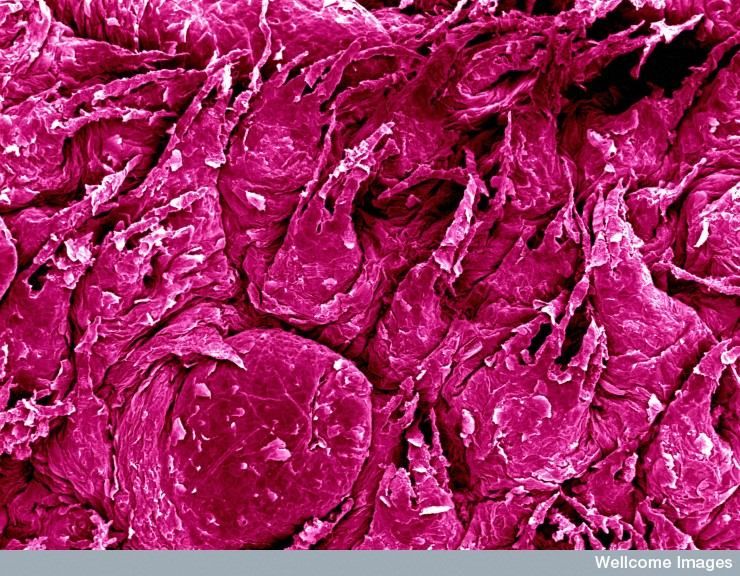
Nose and Smell

The sense of smell has never been regarded as being important. Aristotle wrote 2,500 years ago
“Our sense of smell is inferior to that of other living creatures, and also inferior to all the other
senses we possess.”

How can the importance of senses be weighed? How about examining daily behavior?

It was not until 1826 when The Physiology of Taste; Or, Meditations on Transcendental
Gastronomy: Theoretical, Historical and Practical Work was published by Jean Anthelme
Brillat-Savarin. He wrote about flavor in a very scientific manner about the physiological and
psychological processes responsible for perception of taste and flavor. He acknowledged the
dominant role of smell in taste and flavor. He recognized that smell’s contribution to taste could
only come from the smell arising at the back of the mouth and being swept up in the nasal chamber.
We call that the retronasal route. He wrote this was the “chimney” of taste.

In 1982 Paul Rozin stated we needed to recognize smell as a dual sense; comprising orthonasal
(breathing in) and retronasal (breathing out) senses. He found that the same smell was not the
same when perceived ortho- and retronasally.

How does retronasal smelling work?

Start by studying dogs as compared with humans.
Engineering
Dog’s nose is designed for orthonasal smell and humans for retronasal smell.
Dog smelling. Begins with nostrils, called the nares (něr-ēz). Look directly into a dog’s snout. A
central round opening encircled by membranes called alar folds and a curved slit to the side. Like a
comma on its side. When a dog sniffs the ground, air is drawn in through the central opening by
muscles of the alar folds that enlarge the opening. But it breathes out by contracting other
muscles that direct the outflow through the slits on the side. So that, the air that exits doers not
interfere with the air (and smell molecule) that enters.

When a dog sniffs the surrounding air, the spherical diameter is about 4 inches (10 cm) around the
opening of the nares. This is the fine tune sniffing. Nose to the ground stuff. This is the reach of
the nose. If the dog lifts its head and sniffs the air, it changes to long inspirations. Mouth is open to
                                                                                                        2
ease air being drawn slowly over the olfactory membranes for careful detection. Makes a valued
hunting partner.

A pointer. The dog’s nose acts more like a motor than sensory organ; agitates environment to free
smell molecules from source, adjusting proximity of nares to the source to enhance concentration of
odors from the source, and separating in and out air to control balance.

So, what happens inside the dogs’ snout?

When mammals arose over 200 million years ago, they were all small animals, like today’s mice.
Snouts were a major contact with their world. With present day dogs, olfactory receptors are
located at the back of the nasal cavity. These receptors line bony surfaces to maximize the sensory
area. Between these receptors and the nostrils one finds additional respiratory membranes.
These membranes are formed by the ethmo- and maximal-turbinal bones. These membranes serve
as a kind-of atmospheric filter. They provide warming, moisturizing and cleaning services.

           The arrows show the sniffing pathways for orthonasal and retronasal routes.
                   Note long length of retronasal pathway. Gorgon Shepherd Neurogastronomy

Warming and moisturizing the inhaled air brings it into balance with air in the mouth and respiratory
tract, and cleaning removes bugs and particulates. A bit of HVAC. These filtering mechanisms
are in all mammals, except primates, including humans.

Inhaled air is manipulated with aid of snout muscles and directed into the snout. One path is into
the middle of the HVAC cartridge during normal breathing. It also may direct air more to the
cartridges side so it can reach the olfactory sensory sheet, called the olfactory epithelium. This
sheet is at the back of the nasal cavity. This is where sensing occurs.

                                                                                                     3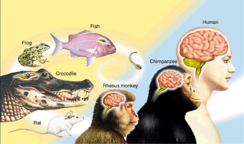
When dogs actively sniff objects, the sniffing rate goes up. During normal breathing there are one
or two respirations per second. That goes up to six to eight per second when the hunt is on. Rats
and other small rodents can top ten to twelve per second. Our limits are about four per second.

The orthonasal path way is efficient and short. The drawing shows the retronasal pathway is long
and relatively slow; from the back of the mouth, through the nasopharynx to the nasal cavity. Dogs
are more highly adapted to orthonasal smelling.

How did human noses get where they are today?

As ancestral humans rose up from walking on the knuckles and legs about 4 million years ago, many
things began to change. They were lifted away from the disease and particulate infested ground.
Their complicated air-cleaning apparatus was no longer as important. The snout started to shrink
and the cleaning apparatus lost its capacity. We ended up with our relatively modest nose and
nasal cavity. There are other theories on the history of our nose and smelling, but this one will do
for now.

With loss of snout, the relation of the nasal cavity to the back of the mouth has changed. Let’s
compare dogs and humans. The dog has an elaborate cleaning system for orthonasal smelling, a
long tube, the nasopharynx, connecting its nasal cavity to its pharynx at the back of the mouth for
retronasal smell. Humans have a short orthonasal pathway and a short nasopharynx for retronasal
smelling.

    The arrows show the pathways in humans for sniffing smells by the orthonasal route and for
  sensing smells from the mouth by the retronasal routes. Note the direct and shorter pathway in
                       comparison to that of dogs. Gorgon Shepherd Neurogastronomy

                                                                                                       4
This short retronasal pathway enables odors released from foods and drinks in the mouth to reach
the nasal cavity smell receptors.

When food or drink comes into the mouth, the retronasal route to the smell organ begins. The
food is moved about with the tongue and chewed. The tongue taste buds go into action. The
mouth senses the tactile characters. When the chewer exhales, air leaves the lungs and passes
through the open epiglottis into the nasopharynx at the back of the mouth. The air absorbs odors
from the food and drink that’s on the cheeks, coats the tongue and that have already become
volatile due to the warm, moist masticated mass. Because the mouth is closed, the odor-saturated
air is forced into the back of the nasal chamber and out through the nostrils. These turbulent puffs
head up to the olfactory sensory neurons to leave their mark. Voila! What a great salmon or
Beaujolais.

No studies on dog retronasal pathway efficiency have been done. But they do have long, narrow
nasopharynx to the olfactory epithelium. In human’s, the pathway is much shorter.
Why is retronasal smell so important for humans?

As our ancestors became bipedal, they started to travel. Long distances. Soon, they came upon
new dietary staples and flavors. These were appreciated retronasally.

About 400,000 years ago, humans started to cook food. New odors and tastes appeared in their
mouths. These were appreciated retronasally.

Soon humans discovered fermentation and its resulting products. They were more exotic and
intense. These were appreciated retronasally.

These new experiences happened among the early hunter-gatherer human cultures and lasted
through the ice ages. About 10,000 years ago hunter gathering slowed and farming grew. Culture
became more stable. Humans domesticated animals and grew crops. They still occasionally hunted
and gathered, but those were not their primary food sourcing activities.

All of the new farming and domesticating led to a broader choice of foods. The flavors from which
could be very complex and exotic and, to a large degree, controlled by humans. These changes and
the motivations for them were appreciated retronasally.
                                                                                                   5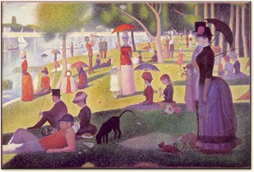
Does the brain know the messenger?

How important is smell? Let’s run a test. These are normally called nose-pinch tests. See The
Taste Committee in the Appendix.

What did we just learn?

   1. If there is no breathing out, there is no smell or flavor. These were appreciated retronasally.
   2. Identifying flavors is dependent upon smelling them. These were appreciated retronasally.
   3. Flavor has always been married to taste and texture. Retronasal smell has never been
       invited.
   4. The actual flavor of candy or pie or roast beef, has nothing to do with how it tastes. Flavors
       were appreciated retronasally.
   5. Even though you cannot perceive flavor when your nose is pinched, when releasing your
       nose pressure, when the smell is properly married with taste and touch, the flavor appears to
       come from the mouth. The mouth gets all the credit. But we know the flavors were
       appreciated retronasally.
   Why is the mouth the teacher’s pet?

   We really don’t know. But some observations.

   When we first take in food and drink, we put it in our mouths. First in line. Next we feel the food
   and beverage in the mouth and roll it about with our tongues and chew it. Before were accept
   this just put in our mouth it must pass an aversion test.
   Too salty, bitter, sour or sweet (rarely)? Too mushy, hard, hot, cold or spicy? Doesn’t pass,
   out it goes. Before were can begin to chew and tongue roll and liberate odors.

   If it passes muster, is it something we like or hardly accept, but need?

   All our attention is to the mouth. It’s the center of attention. Our perception follows our
   attention.

   If our appreciation happens because of retronasal smell perception, we pay no attention.

                                                                                                    6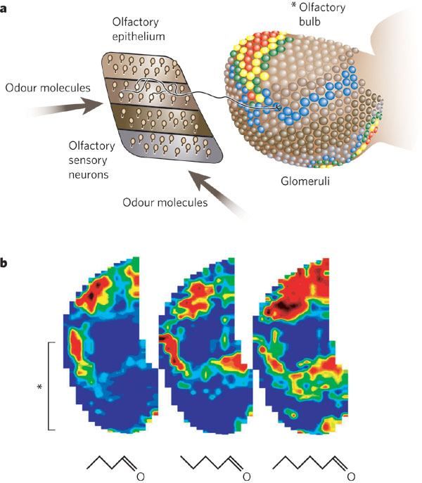
Because the smell in the nose is sensed as if coming from the mouth, it is part of the nervous
system known to as referred sensation. A sense appears to come from one place but actually
comes from somewhere else.

Separating smell from flavor

Smell is two senses; one for breathing in and one for breathing out. We all think of the one for
breathing in, through our nose, as the only sense of smell. The one for breathing out is never
recognized as a separate sense. Probably because it is always associated with taste and touch;
all that going on simultaneously in the mouth. This combined sense we call flavor. But again,
the mouth is given credit for this. They are combined, but separate. In what way? How does the
smell pathway discriminate its smell molecules? How does this contribute to flavor?

Flavor building blocks

Almost 200 years ago Jean Anthelme Brillat-Savarin wrote:

The number of tastes is infinite, since every soluble body has a special flavor which does not
wholly resemble any other…Up to the present time there is not a single circumstance in which a
given taste has been analysed with stern exactitude…Men who will come after us will know much
more than we of this subject, and it cannot be disputed that it is chemistry which will reveal the
causes of the basic elements of taste.

Men and women have not stopped trying to make sense of taste. Let’s first look at flavor
molecules to try to understand what we now know.

Fruit aromas

Nestlé Research published work in 2003 that had analyzed volatile aromas in the mouth during
eating ripe and unripe bananas. The retronasal smell of the banana comes with the first
outward breath. Dozens of different smells are released from the chewed bananas. Chief
among them are simple alcohols, such as the ethyl alcohol in wine. These simple alcohols give
rise to a sensation we call sweet. This is only an analogy to the sweet taste. This show how
language and smell are related. Often alcohol in wine is said to have a “side taste” of sweetness.

                                                                                                     7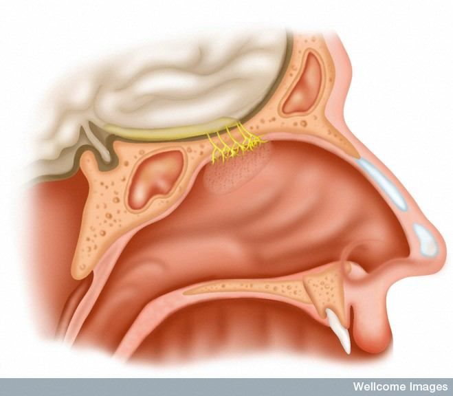
What is Flavor?

In Brillat-Savarin’s section “Analysis of the Sensation of Tasting” he writes:

He who eats a peach…is first of all agreeably struck by a perfume which it exhales; he puts a
piece of it into his mouth, and enjoys a sensation of tart freshness which invites him to continue;
but it is not until the instant of swallowing, when the mouthful passes under his nasal channel,
that the full aroma is revealed to him…Finally, it is not until it has been swallowed that the man,
considering what he has just experienced, will say to himself, “Now there is something really
delicious!”

Now let’s taste a peach. Squeeze nostrils shut. Put peach in mouth and chew and move about
with your tongue. What do you taste? Free nostril passages. What do you taste?

Fruitiness generally comes from esters – molecules with double-bond oxygen on an internal
carbon – that are produced when an acid is combined with an alcohol through enzymatic action
in a ripening plant.
                             RCO2H + R'OH             RCO2R' + H2O
When chewing food, retronasal smells are added resulting from enzymes in our saliva. The
sensory profile is filled from all these sources. Slight variations in this profile allow us to identify
ripeness, greenness and fruitiness.

The volatiles are described in terms of the main notes (fruity, candy…) and secondary notes
(apple, cheesy, pineapple, caramel, mushroom…). Is this telling us there is a smell “image” for ripe
bananas that overlap with the smell images for other foods, like say, melons? There are specific
molecules that are the signature for ripe bananas. We can identify those smells. Our brain has
patterns of these ripe fruit, and other food and beverage, signatures that we can perceive.

Each food and beverage has its characteristic molecular composition, modified by how it is
prepared-cooked, baked, fermented, aged, etc. By themselves, foods have no flavor. They are
the raw materials out of which the brain creates flavor.

                                                                                                      8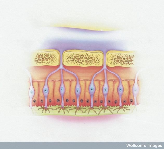
Making pictures of smell

Smell begins with the action of smell molecules on the receptor molecules in the nose. Until
1991 little was known about the receptor molecules.

How do receptor molecules work?

For more than a century, biologists have used the “lock-and-key” concept as how the two
molecules interact with each other. An odor molecule is made up of different types of atoms,
giving each an independent structure. The odor molecule functions as the key. How does the
lock work? This is the major question. How does the information in the odor molecule get
translated to an image in the brain?

Let’s start by looking at vision and hearing. Individual photons activate rhodopsin molecules in
the photoreceptor cells in our retina. Also in hearing, sound waves are first converted into inner
ear vibrations, which activate the hair cells in our cochleas [kok-lee-uhs].

Sensory stimulation is much more complex. Monitoring smell takes instruments. It’s not the
same as the added advantage of hearing or seeing the stimuli we are delivered. The rector cells
are hidden and buried in the nasal cavity. The cells are easily fatigued by repeated stimuli.
There are thousands of odors to identify, with different perception thresholds. All work is
done with orthonasal smell with puffs of smell. Very slow stuff.
What are smell molecules like?

Smells sniffed orthonasally range in size and structure. They vary widely. Retronasal smells are
mainly smaller molecules that evaporate from the liquids and foods released from within our
mouth.

What part of the smell molecules stimulates the receptors? Changes in a single molecule
feature, such as a single atom, can change smell perception.

Odor molecules may vary in length (straight chain aliphatic groups may have one to a dozen or
more carbon atoms), terminal function groups (acids, alkanes, aldehydes, etc.), whether a
functional group is within the carbon atom chain (such as an O atom in a ketone), have side
                                                                                                   9
groups attached to the carbon chain (such as a phenol ring), chirality (right-handed or left-
handed), molecule geometric shape (like ring like conformation of a terpene) and overall size.
The ability to detect these differences of single atoms within these smell molecules make
detection sensitive and complex.

The race for smell receptors

What kind of receptor molecule can sort through all this? We don’t know for sure. Several
good proposals.

The lock-and-key idea is that receptors are tuned to the shape of the odor molecules. They
call this the stereochemical theory. Even if true, the nature of the receptors, the specific kinds
of proteins in the cell membranes, remains unknown. Many different experiments took place
around the world. Repeated attempts to identify the receptors met with no success.

In 1991 Linda Buck and Richard Axel reported they had found olfactory receptor genes that
carry the genetic code the cell uses to make olfactory receptor proteins. They won the 2004
Nobel Prize.

Odor-binding pocket

How does this fit the lock-in-key model? It was suggested that the molecular interaction does
not take place in a narrow lock that responds to only one key, but in a larger space called a
binding pocket. A given receptor cell might carry just one type of receptor, which would require
that the receptor does not have a narrow affinity for a given molecule, but rather a broad
spectrum of affinities reflecting the known broad odor responses of olfactory receptor cells.
This make the olfactory receptor cell similar to photoreceptor cell of the retina, each which
carries only one of the three types of color receptors, but with broad responsiveness to
different wavelengths of light.

Forming a sensory image

Research over the past half-century has shown that the brain represents smell molecules by
spatial patterns. These patterns are formed in the olfactory bulb. These patterns are the smell

                                                                                                 10
images processed by the brain for smell perception. How can a nonspatial stimulus received by
the receptors in the nose be represented as a spatial pattern specific for that odor molecule?
Smell images seem strange. Visual images are easy to accept. But, how does that happen. A
lab test on horseshoe crabs gives some answers. The crab has two small eyes in its hard shell.
Each visual receptor cell is contained in a microcartridge that takes in part of the visual scene.
Combining all the receptors gives a good pattern of the light falling on the eye. By blocking
part of the retinas, the visual perception will not be the same. It is seen that a strong cell gets
stronger and a weak cell weaker. There are lateral inhibitory connections between the receptor
cells. Through these connections, the strongly excited cells at the border more strongly inhibit
the weakly excited cells, and the more weakly stimulated cells more weakly inhibit the strongly
excited cells. This mechanism is called lateral inhibition. The effect is called contrast
enhancement. This is contrast enhancement in space. Contrast enhancements also exist in
time.
Lateral inhibition has other functions. The final result is that in the horseshoe crab the final
image is not the image of a camera but an abstracted, high contrast image, in which the edges in
the scene are abstracted and enhanced and the rest of the field is suppressed. Studies on
Mammalian eyes have confirmed this effect.
The eyes set up a two dimensional representation of the visual world. A visual image. The
nervous system can process the image.
Images of smell
Once at the olfactory receptor cells, the information pathway goes through a series of regions:
the olfactory bulb, the olfactory cortex, and the orbitofrontal olfactory cortex.
11The olfactory bulb
The olfactory bulb is shaped like a light bulb. It sticks out in front of the frontal lobe of the
brain.
An olfactory pathway in a rat follows. See illustration. When smell molecules invade a receptor
binding pocket in an olfactory receptor neuron (orn), all an individual cell “knows” is how much
the invading molecule have excited its binding sites. The more excitement, the more the cell
responds by generating impulse. The frequency impulses sent to the olfactory nerves (on) tell
little about what the smell is. This means the code for smell molecules, the code the brain reads
must lie in the differences between the responses of the different cells.
12The Smell Pathway. Left side. Operations to process smell input from reception in the nose
to perception in the cerebral cortex.
Right side. The smell pathway carrying out these operations. In the olfactory epithelium, the
main type of cell is the olfactory receptor neuron (orn)
In the olfactory bulb, the main type of cells are the mitral cell (mc); tufted cell (tc); periglomerular
cell (pg); and the granule cell (gc).The cells lie in different layers: olfactory nerve (on); glomeruli
(glom);external plexiform layer (epl); mitral cell body layer (ml); and granule cell layer (gcl). In the
olfactory cortex, pyramidal cells receive input from the olfactory bulb and connect to
interneurons. Central fibers extending out to modulate the olfactory bulb cells arise in the
nucleus of the horizontal limb of the diagonal band (NLDB). The orbitofrontal cortex
(OFC) is represented by a single pyramidal cell to conserve space. Gorgon Shepherd Neurogastronomy
The olfactory bulb, the fibers from several thousand receptor cells, all containing the same type
of olfactory receptor, converge on a single site, called a glomerulus (glom). Connecting to each
module within the olfactory bulb are some large cells called mitral cells (mc). The mitral cells
send their fibers to the olfactory cortex. They are joined by smaller versions of mitral cells
called tufted cells (tc). Together, they provide a straight pathway. There are also numerous
interneurons, cells with short branches that are in place; periglomerular cells (pg) and granuale
cells (gc). Through the pattern of its input and interaction between its neurons, the olfactory
bulb creates the code for representing the stimulating odor molecules.
How the olfactory bulb represents smell
There are specific patterns of metabolically active sites within the olfactory bulb associated
with different odors. This implies that a specific topographical pattern of neuronal activity
might be associated with processing of different odor information. The olfactory bulb is
approximately like a sphere, except for the part attached to the brain.
13On the surface of the bulb, projections are used like a world map. Flat map odor images are
found on the surface. Each is a focus. Activity foci take place within the glomerular layer. The
foci are overlapping, but different for different odors.
A smell is like a face
In the figure below, under a, the smell molecule activates the receptor within a binding pocket.
The figure shows a sheet of receptor cells in the nose. Each cell sends its impulse response
through its fiber (axon) to a glomerulus in the olfactory bulb. All the cells containing the same
receptor molecule send their fiber usually to a pair of glomeruli on the medial and lateral sides of
the olfactory bulb. Whenever a particular receptor is activated, the responses of all the cells
are focused on these modules. Cells more or less strongly activated cause more or less strong
activation of their corresponding glomeruli, resulting in a pattern such as shown in the figure.
14How the smell image is formed
Above: The olfactory receptor cells lie in zones in the olfactory epithelium. Representative
cells are shown sending their axons to converge on a glomerulus in the olfactory bulb. The
different shades of gray indicate different activity levels of activity in the glomerular layer.
Below: Flattened maps of patterns of activity in the glomerular layer produced by stimulation
with three different odor molecules differing only by a single carbon atom. The bracket
indicates the extent of the glomerular pattern on the medial side of the olfactory bulb shown
above. Gorgon Shepherd Neurogastronomy
In the figure above, under b, three different aldehyde molecules had their pictures taken. The
images of the activity patterns in the glomeruli sheet were constructed from imaging of their
functional MRI responses. These molecules vary only in number of carbon atoms, from four to
six.
These images carry the identifying information of the odor molecules. Odor images extend
over much of the olfactory bulb; they are overlapping, different and become more extensive with
increased concentration.
See Odor Images Concentration Differences in Appendix
Pointillist images of smell
There are two ways to put colored paint on a canvas to elicit the perception of color in the
mind of the observer.
1. Mix the paints to achieve a particular color impression. White plus red to get pink.
2. Place different colors in individual small “points” of color and let the effect of color mixing
arise from a distance, where the colors blend in the mind of the observer.
This second method is the method of pointillism. It was perfected by Georges Seurat. His most
famous painting is A Sunday on La Grande Jatte (1884). The closer one gets, it is easier to see
the individual dots or points of color.
15Sunday Afternoon on the Island of La Grande Jatte (Un dimanche après-midi à l’Ile de la Grande Jatte), Georges Seurat,
1884-1886.
Look closer at the shadow line just above the dog.
Detail of the Grand Jatte, above the dog
Look closer
. Close-up of the paint on canvas in the Grand Jatte.
Reproduction by dots also became a method of publishing photographs and other graphic art in
newspapers and magazines.
It has been considered that this may be how smell is perceived. The illuminated dots of colored
paint reflecting different wavelengths of light are analogous to modules called glomeruli in the
olfactory bulb. Each activated differentially by different odor.
The perception of smell would require an odor mixing of the neighboring modules. The brain is
organized to carry out such mixing as discussed in the smell pathway above.
The sight and smell similarities are interesting. The perception of color arises from electromagnetic
waves of different wave lengths. They activate our photoreceptors in different ways. It is actually
the brain that creates the perception of color in how it manages the signals according to the
different wavelengths. Smell is not present in the odor molecules that stimulate the smell receptors.
The brain creates the perception of smell from different odor molecules.
16Processing the pointillist images
The brain goes through a series of stages to extract information needed to detect and discriminate
among the different stimuli.
First, the odor image in the olfactory bulb is discerned at the layer of the odor modules forming the
odor image. This first odor image is enhanced by a system of lateral inhibitory microcircuits. The
enhanced image is sent to the olfactory cortex. There, a cortical microcircuit, with widespread
connections, reformats the image into a “content-addressable” memory. Finally, this memory image
is sent to the highest centers in the neocortex. Here a complex cortical microcircuit creates
conscious perception.
In a nutshell, form a pointillist image; locally process it; globally format it; represent it in memory;
enhance it with emotion; and perceive it consciously. Each step has its own microcircuits.
The glomerulus: a Holmes-like smell-detector
The analogy to Seurat’s points of paint in his painting is the olfactory glomeruli. The glomeruli is
the most distinct multicellular unit in the brain. A glomerulus is an overcrowded meeting place, where
nose signals are transferred to the brain. The fiber terminals (axons) from the nose’s receptor cells
connect to short branches (dendrites) of nerve cells in the olfactory bulb. A single glomerulus
receives thousands of incoming nerve fibers from the receptor cells. How many? Rabbits have
about 50 million olfactory receptor cells. And about 2,000 glomeruli. 25,000 cells for each
glomerulus.
As Linda Buck and colleagues showed, all the fibers coming to one glomerulus express only one
type of olfactory receptor in their cilia in the nose. All the fibers are carrying the same information!
A bit of overkill.
Imagine if 25,000 people are talking to you at the same time. If this was at a football game or street
rally, it would all be “noise.” However, if you were performing at a concert and you were alone on
stage and it was your birthday and they all sang the Happy Birthday song, it would come through
loud and clear. This is technically the “signal.” A signal-to-noise ratio of 25,000 would make the
message more distinct.
17It is felt by Gordon Shepherd that the signal-to-noise enhancement is the key operation of the
glomerulus. This enhancement is used in orthonasal smell to detect and discriminate among specific
signals in the environment that may be critical for survival. It is likely used in retronasal smell to
detect and discriminate the volatile compounds of food when in the mouth.
The difference between the sight, noise and smell system is worth noting. While looking or
listening for things, all animals, including humans, have lots of background noise and changing
images to sort through to isolate a source. In smelling, the sources come to the animal. Oxford
philosopher, Isaiah Berlin, gave analogies. He stated the visual system is like the fox. Each
location knows many things. The smell system is like the hedgehog. Each glomerulus knows one
thing. The hedgehog spends much less expensive neural tissue to do their tracking than does the
fox.
How effective are humans?
Odor image fineness depends on several factors.
Number of receptor cell. Dogs have up to 100 million. About 10 time those of rodents and
humans.
Types of receptors. Rodents have over 1,000. Dogs about 800. Humans 350.
Number of glomeruli. Dogs several thousand. Humans around 6,000.
All are important.
However, greater brain complexity in analyzing odor images is the critical factor.
How can the brain sort it all out?
Humans have several thousand independently acting modules. The brain cannot make any sense
of the information until it can be compared. What is the mechanism for sorting and correlating?
Lateral interactions are needed. These interactions begin through periglomerular cells (pg).
These are interneurons which connect to neighboring glomeruli. See figure below.
pg respond to odor input with single impulses or with impulse bursts. There are inhibitions of
neighboring cells (mitral and tufted dendrites). Possibly this enables more active glomerulus to
inhibit its less active neighbors. A kind of lateral inhibition.
18Excitation may also occur interglomerularly. Study as to how this occurs is ongoing.
One effect of these lateral interactions is to begin the extraction of the spatial pattern so that it
can be read more effectively by the next level of microcircuits involving the olfactory granule cells.
Enhancing the odor image
We have discussed the processing in the olfactory bulb carried out by the glomerular layer. This
forms an image representing the smell molecules, performs signal-to-noise operations and lateral
interactions to begin the imaging process.
The image is then sent to the second level within the olfactory bulb. It is by means of the mitral cell
(mc) and tufted cell (tc) that the levels are connected. These cells collect input in their dendritic
branched in the glomerulus. The processed signal is then transferred, after further processing, to
the next level and out to the olfactory cortex.
Note: Dendrites are the branched projections of a neuron that act to
conduct the electrochemical stimulation received from other neural cells to the cell body of the neuron from which the
dendrites project. Electrical stimulation is transmitted onto dendrites by upstream neurons (usually their axons)
via synapses which are located at various points throughout the dendritic tree.
19Before the odor image can be sent on, it must first be coordinated with all the other glomerular
modules and lateral inhibition between coordinated glomerular modules must occur. This takes
place through long dendrites of the mc and tc. First, these dendrite do not interact with each other,
but with interneurons called granule cell (gc). It is through the gc that correct format to the odor
image is corrected for output to the next stage, the olfactory cortex.
Glomerular coordinating
Image processing begins in the localized connections of pgs between glomeruli. How is it completed
through the gcs?
Odor image extends broadly within the olfactory glomeruli layer. This is true even when excited by
a single odor molecule. The mc and tc , far apart, must be coordinated such that the lateral
inhibition can occur to enhance the image. Over both long and short distances, how does effective
lateral inhibition occur?
How are smells created?
The output fibers from the olfactory bulb all bundle up in the lateral olfactory tract. The tract,
about an inch long, reaches from the olfactory bulb in front of the nasal cavity to the olfactory
cortex on the brains underside.
What is the olfactory cortex? Why is it between the olfactory bulb and the neocortex, the highest
cortical level? Research has shown the olfactory cortex represents the transition from smell stimuli
to the perception creation of a smell; the meeting of the outside world with the perceptual world. If
you smell something or taste something, it is because of the olfactory cortex. How does it work?
The main nerve cell of the olfactory cortex is the pyramidal cell (pc). Its cell body is shaped like a
pyramid. The pc sends impulses to excite the interneurons. The interneurons feed back inhibition
on to an excited pyramid cell, controlling its output and on to neighboring pyramid cells to sharpen
contrast. The axon collaterals also feed back excitation onto an excited pyramid cell and its
neighbors. In normal function this feedback excitation is counterbalanced by the feedback
inhibition through inhibitory interneurons.
20Many cells in the brain are excited by their inputs and in turn excite the targets of their outputs
(through axons) to other regions. Within their own regions two things can happen. They can be
driven weakly, in which case their response can be strengthened and made more complex by
excitatory connections back onto themselves and their neighbors. With stronger inputs they may
become too excited and actually go into epilepsy seizures; to prevent this they have Inhibitory
feedback connections to reduce their excitation. The inhibitory feedback can also be aimed at
surrounding cells, in which case it can inhibit less active cells and thereby bring about enhancement
of contrast between themselves and they neighbors.
Contrast enhancement is fundamental for every sensory system. It enables us to discriminate
between two points of touch on our skin, between two spots or fields in our vision, and between the
tastes of two wines.
In neurobiology, lateral inhibition is the capacity of an excited neuron to reduce the activity of its
neighbors. Lateral inhibition disables the spreading of action potentials from excited neurons to
neighboring neurons in the lateral direction. This creates a contrast in stimulation that allows
increased sensory perception.
Remember that input in to the olfactory bulb where receptor cells with the same response
sensitivities converge on to one glomerular module; the same “molecular receptive range. Now, the
output is distributed across the olfactory cortex to many pyramidal cells. The information is
changed from a mosaic image to a distributed presentation of that image.
It's another reason to think that we form a spatial "image" of the smell molecules, so that when we
discriminate between two wines it involves discriminating between two complex images like
faces. The more experience we get the better we are at discriminating between wines by training
our lateral inhibition to enhance the contrast between them.
Distributed image
The “olfactory cortex serves as a content-addressable memory for association of odor stimuli with
memory traces of previous odor stimuli” was reported by Lewis Haberly in 1985. All the necessary
properties (large number of pyramid cells and synapses, well distributed input from olfactory bulb
fibers, positive feedback) are found in the basic olfactory cortical microcircuit.
21Olfactory cortex matches input to memory
Donald Wilson and Richard Stevenson wrote that whereas the representation of smells in the
olfactory bulb is driven by stimuli, the representation in the olfactory cortex is memory based.
The olfactory cortex reacts especially to changes in its input signals from the olfactory bulb.
The olfactory cortex can adapt to the same smell. Increase or decrease that same smell or input
another smell, the olfactory cortex reacts.
The system learns. Different smells result in a better performing cortical system. The system can
store changes as a memory. The data base expands.
The changes allow the system to better match input with stored patterns.
Changes allow the system to improve its signal-to-noise ratio such that individual smells are more
easily identified in a crowded aroma background.
The olfactory cortex microcircuit functions to take an input reflecting many diverse stimuli and
construct a coherent odor object. A whole can be made of some parts.
The odor object can be combined with other sensory inputs to produce a sensation of flavor.
This occurs in the orbitofrontal cortex.
Smell and flavor
The olfactory cortex starts the process of a brain representation of a smell of what we are
consuming. But, the message must be read.
The neocortex. (The newest type of cortex created in mammals.) The neocortex has three main
parts.
1. The areas that connect with the sensory and motor pathways. The primary sensory and
motor areas. Very large in humans. Carry out initial processing of sensory input and
movement at the neocortical level.
2. The area that elaborates properties of a given sense. Visually; color, movements, faces…
And motor act coordination. Association areas.
223. The higher association areas are where our highest mental faculties occur; language,
reasoning, planning… Our sensory and motor worlds are expanded. Here the smell images in
olfactory cortex are fashioned.
Neocortical smell perception
The thalamus is often called the gateway to the neocortex. All vision, hearing, touch and taste
senses must pass through.
The human smell system.
OR=olfactory receptor cells; OB=olfactory bulb; OC=olfactory cortex;
MOFC=medial orbitofrontal cortex; LOFC=lateral orbitofrontal cortex
Gorgon Shepherd Neurogastronomy
The thalamus transmits forward and the cortical regions transmit back; they function in a
coordinated manner. Visions, hearing, touch and taste senses are located in the middle and back of
the brain.
The olfactory cortex (OC) sends a small number of fibers to the thalamus, but most go directly to
the orbitofrontal cortex because the OC is situated in the most prefrontal part of the brain, just
above the eye orbits (sockets).
23The prefrontal cortex contains the circuits that subserve most of our highest human cognitive
functions. It is the peak level in the human brain. The OC output is aimed precisely there.
The sense of smell is uniquely privileged. It has direct input to the prefrontal cortex, arrives through
a short pathway (involving only olfactory receptor cells, mitral cells and olfactory cortical pyramidal
neurons) and is it is situated in the center of the brain.
The volatile molecules released from everything we eat are so important that they are evaluated
quickly in the highest level of the human brain.
Human’s big olfactory brain
Humans have fewer receptor cells than many other animals. However, despite the declining number
of receptor genes, the brain processing mechanisms of the smell pathway, culminating in the
neocortex, bequeath a richer world of smells and flavors on humans than on any other animal.
Compare the brain of a mouse with a human. In actual fact, the human’ olfactory bulb is nearly as
big as the entire mouse brain. The mouse has way more receptor genes. However, the human smell
pathway has been maintained in size, whereas the amount of brain to process the signals in that
pathway has increased enormously. This increased brain power give humans enhanced processing
of its smell and flavor input.
24The human brain compared to a mouse brain for relative size.
Locations of different brain systems related to different functions and behaviors are shown.
Gorgon Shepherd Neurogastronomy
Information processing and connectivity
25How does the neocortex enhance human meaning to odor images in the form of a smell object
arriving from the olfactory cortex? The possibilities? The neocortex construction enables it to
carry out enhanced smell input processing or its increased connections with other brain areas go
far beyond what has been available to the olfactory cortex.
How does the smell pathway create the perception of smell?
1. Some cells are tuned specifically to odors.
2. Most cells do not respond to changes in odor intensity. This is in contrast to cells in the
olfactory bulb and olfactory cortex.
3. Some cells respond to both odor and smell stimuli. Can be called sensory fusion. The first
step in creating the combined perception of flavor.
4. Some cells respond preferentially to “pleasant” smells and others to “unpleasant” smells.
The different responding cells are grouped in different parts of the orbitofrontal cortex.
5. The qualities of pleasant and unpleasant are in turn reflective of their reward value.
Smell
Color artwork sagittal section of the nasal area.
This shows the nerves from the olfactory bulb and tract connecting with the superior nasal concha
26Diagram of olfactory cell in the nose
Flavor is not just defined by your taste buds. Your sense of smell also comes in to
play. And so, if your olfactory nerves (nerves inside your nose) can't function
normally because of say, a cold, your sense of how food tastes also seems affected.
But in reality, there is a large amount of odor that goes up the back of your throat
into your nasal cavity as you eat. This component is missing when you have a cold,
and so your stuffed nose reveals to you only what you can truly taste with your taste
buds.
The olfactory system is similar and different to the taste buds. Olfactory receptor
cells are continually renewed, and this is thought to decline with age. Again, there
may be underlying genetic variation in olfactory cell turnover. There are reported sex
differences in olfactory behavior (a lot of pheromone work in all sorts of animals), but
much of the anatomical underpinnings of these differences are more central, in the
brain.
This critical interplay between rapid cell growth and cell death in taste buds and
olfactory epithelial cells is critical to maintain normal taste and smell function.
Saliva is the active source of these factors for the taste bud, while nasal mucus in the
active source of these factors for the olfactory epithelium.
Taste and smell functions are critical to maintain a homeostatic balance: Taste acts
as the guardian of what we eat before entering the gastrointestinal tract; smell acts
as the guardian of what we breathe into our lungs and respiratory tract. These
senses allow us to eat foods and to drink beverages that are both non-toxic and
nourishing, and to breathe air that is free from pollutants and contaminants.
27Saliva (also referred to as spit, spittle, drool or slobber) is the watery and usually
frothy substance produced in the mouths of humans and most other animals. In
mammals, saliva is produced in and secreted from the three pairs of major salivary
glands, and hundreds of minor salivary glands. Human saliva is composed of
98% water, while the other 2% consists of other compounds such as electrolytes,
mucus, antibacterial compounds, and various enzymes. As part of the initial process
of food digestion, the enzymes in the saliva break down some of the starch and fat in
the food at the molecular level. Saliva also breaks down food caught in the teeth,
protecting them from bacteria that cause decay. Furthermore, saliva lubricates and
protects the teeth, the tongue, and the tender tissues inside the mouth. Saliva also
plays an important role in tasting food, by trapping thiols produced from odorless
food compounds by anaerobic bacteria living in the mouth.
The amount of saliva that is produced in a healthy person per day; estimates range
from 0.75 to 1.5 liters per day while it is generally accepted that during sleep the
amount drops to almost zero. In humans, the sub-mandibular gland contributes
around 70-75% of secretion, while the parotid gland secretes about 20-25 % and
small amounts are secreted from the other salivary glands.
Nasal mucus: A slippery fluid produced by the membranes lining the nose.
Excessive nasal mucus is the basis of a runny nose. Mucus is the Latin word for "a
semifluid, slimy discharge from the nose.”
Taste buds and olfactory receptor cells are the fastest growing and most rapidly
regenerating cells in the body. Taste buds regenerate completely in a 24 hour
period. The entire bud is replaced by new cells on a daily basis. This process
depends upon stimulation of the basal cells or stem cells of the taste bud by several
families of growth factors (carbonic anhydrase VI, adenylyl cyclases,etc), hormones
(thyroxine, carbohydrate-active steroids, etc.), trace metals (zinc, copper, magnesium,
etc), and many other proteins and substances present in saliva.
28There is also rapid control of the regeneration that takes the form of programmed
cell death or apoptosis. This process is controlled by several families of “death
factors” in saliva including tumor necrosis factor alpha (TNF Alpha), TRAIL, and
other cytokines which inhibit growth. This push-pull function is carefully
orchestrated by a process which preserves taste function despite other processes
which act to inhibit growth or activate cell death, not unlike putting hot pizza into the
mouth which burns the tongue or palate. Other factors include oral diseases such as
gingivitis, fungal infections, and other processes which inhibit cell growth or activate
cell death.
By contrast, there is also rapid control of cell growth through secretion of “death
factors” such as TNF alpha, TRAIL, and other cytosine which inhibit growth. As
in the taste bud, there is a delicate balance between cell growth and cell death so
that olfactory receptor cells can function normally and preserve smell function. As in
the mouth, there are many processes which are both physiological and pathological
which can either inhibit growth or activate cell death. These processes include
inhalation of smoke or toxic gases, catching a cold or some viral illness which
produces inflammation and infection in the nasal cavity and other such processes.
Studies have shown that nerves are not required for initial taste bud development,
but that maintenance of taste buds is dependent on nerve innervation. If the nerve
innervating taste buds is cut, the taste buds degenerate, and do not reappear until
the nerve reinnervates the tissue. It is not known how the nerve activates taste bud
formation in regeneration, and only specific nerves will regenerate taste buds. As
stated, cells within the taste bud have a limited life span of approximately ten days to
two weeks. Therefore, new taste cells must continually differentiate to replace dying
taste cells, and cells at a variety of different developmental stages are present within
a single taste bud. In consequence, the synapses between taste receptor cells and
nerves must constantly be remodeled. Several morphologically distinct cells can be
29distinguished within a single taste bud; these different cell types may represent
different stages in the development of a taste cell.
It seems true that regeneration of bud cell and olfactory receptor neurons slows
down with age. There is some difference in sexes, but it is more affected by period or
pregnancy in females, making it a little harder to define other differences between
males and females. Cultural differences are possible. But "culture" is too abstract
and too broad, and instead, "living style" or "habit" might be a better word to
describe this aspect, e.g., smoking could alter both smell and taste sensitivity. Liking
of spicy food could also change taste.
3031
Olfactory System

Primary sensory neurons in the nasal cavity olfactory epithelium project through the cirbiform plate
and to the olfactory bulb. Secondary neurons in the olfactory bulb project to olfactory cortex
regions including the anterior olfactory nucleus, piriform cortex, olfactory tubercle, cortical nucleus
of the amygdale, and enteorhinal cortex.

Receptorcells continuously degenerate and are replaced by new ones every 60 days. Basal cells
develop into receptor cells.

The olfactory system uses as many as 950 different odorant receptor genes that express
chemoreceptors on the olfactory epithelium.

Different groups of glomeruli are activated by different odorants, but not in any simple pattern.
Glomeruli are the functional units for processing of odor information. Olfactory receptor neurons
having the same receptor project to the same glomerulus.

Receptor cells terminals synapse with mitral cell dendrites in glomeruli – complex oval structures
containing thousands of synapses. Mitral cells are located in a specific layer in the olfactory bulb
and have dendrites that project superficially to glomeruli and axons that project to olfactory cortex.

Periglomerular cells are interneurons located near glomeruli and have inhibitory effects on signal
conduction, using DA and GABA neurotransmitters. Granule cells are also interneurons
but lack axons, making inhibitory dendrodendritic synapses with mitral cells via GABA. These
cells act as local feedback mechanisms on mitral cells.

Tuff cells are located deep to the glomeruli and have dendrites that project to glomeruli (glutamine
and aspartate neurotransmitters) and axons projecting to olfactory cortex.

The olfactory cortex has 3-4 layers (as opposed to 6 layers in neocortex). The main ascending
inputs project to the superficial part of layer I (instead of mainly layer IV). Pyramidal cells are in
layer II and apical dendrites are oriented perpendicular to the olfactory cortex layers. Inputs from
olfactory bulb end on the distal parts of the apical dendrites. Association inputs from other
cortical regions end on the proximal parts of apical dendrites. Because association fibers end more
                                                                                                         32
proximally to the apical dendrites, it suggests that association fibers from other cortical regions
have a greater influence on the perception of smell than the actually primary sensory fibers
themselves.
The human brain flavor system
Brillat-Savarin wrote “The tongue of an animal is comparable in its sensitivity to his intelligence.”
The human brain flavor system has basically two stages. The sensory system that feed it and the
action systems that draws on the human brain systems that generate and control our behavior. The
sensory systems are shown on the following drawing.
We can summarize the flavor perception system
Smell ► Goes directly to the olfactory cortex in the forebrain limbic system. There it forms
distributed memories of the smell stimuli represented as odor objects. There, the smell objects have
direct access to brain systems for memory and emotion. The olfactory cortex also projects to the
orbitofrontal cortex at the brain’s front where it connects to uniquely human capacities for
judgement and planning.
Taste ► Taste pathways head to the brain stem. They have access to hardwired expressions of
the emotional qualities of the taste stimuli. From there they go to their cortical areas and interact
with the other messages at the core of flavor.
Mouth-feel ► Also to the brain stem, go the different types of touch that food and liquid exhibit.
From the stem to the thalamus and their cortical receiving and association areas. Both the tongue
and mouth have great impact in the cortex. This accounts for how dominant perception of the food
or drink in our mouth is and the illusion that its smell is coming from its taste.
Sight ► Before consuming, the sight of food and drink activates the visual pathway that passes
through the thalamus to the visual areas in the back of the brain. This has a highly significant
influence on we judge flavor.
Sound ► The sound of our food as we eat is important to flavor appreciation.
Taste and flavor
Smell arising from food in the mouth contributes to flavor. The sense of smell is a dual system;
orthonasal and retronasal. Retronasal smell is never sensed by itself. It interacts with virtually every
33other sense in the mouth. “Taste” is the obvious contributing system to our sense of taste. It
generally gets all the credit for resulting flavor.
Taste buds
When the taste buds on the tongue and the back of the mouth are stimulated by food or drink, the
taste system begins. They are called buds because they are composed of taste cells crammed
together to make a kind of cartridge or bud. Each taste cell ending in fine hairs that carries
receptor for different stimuli. The taste buds are located in different folds (papillae) of the tongue
surface.
Taste buds respond to five different kinds of stimuli; salts, acids, sugars, bitter compounds and the
amino acid glutamate, linked to umami. Umami is described as being savory or meatlike. Each
stimulus acts on a special type of receptor.
Gorgon Shepherd Neurogastronomy
Taste encoding
Taste nerves enter the brain stem. This part of the brain is responsible for heart rate, breathing
and other vital activities. Remember the olfactory nerves enter at the front of the brain; closest to
the highest cognitive centers.
How are tastes represented in the brain? Two models are suggested. 1. Each type of stimulus
has a labeled line into the brain, leading to its distinct perception. 2. A nerve fiber responds best to
34one type of taste stimulus, but responds to two or three other stimuli to a lesser extent. These are
label line and across-fiber pattern theories. The figure below (as shown above) shows that the
label line model fits in most cases.
In the brain stem, the fibers connect to a group of cells called the nucleus of the solitary tract.
From there, the path leads to the thalamus. The thalamus pushes it on to the cerebral cortex.
The human taste system
7=facial nerve; 9=glossopharyngeal nerve; 10=vagus nerve
Gorgon Shepherd Neurogastronomy
35The primary cortical areas in humans are the anterior insula and frontal operculum. In these areas,
conscious perception of taste is suggested to occur.
Taste qualities
The five basic tastes are salt, sour, sweet, bitter and umami. Note that all these tastes have
perceptual contributions from other types of receptors; e.g., sour has its major contribution from
acid receptors, but also contributions from other receptors.
Gorgon Shepherd Neurogastronomy
Taste and retronasal smell together
Taste stimuli usually occur in conjunction with retronasal smells. Together, they create flavor.
Sometimes they seem to cross reference each other. We might say something “smells sweet.” This
is called sensory fusion. It is often found that these two stimuli, when acting together, activate
areas of the brain that are not activated when they act independently. They call in a lot of help.
Flavor perception seems to be more complex that taste and smell alone.
The Gustatory System
36You can also read