Potential Role of Flavonoids in Treating Chronic Inflammatory Diseases with a Special Focus on the Anti-Inflammatory Activity of Apigenin - MDPI
←
→
Page content transcription
If your browser does not render page correctly, please read the page content below

Review

Potential Role of Flavonoids in Treating Chronic
Inflammatory Diseases with a Special Focus on the
Anti-Inflammatory Activity of Apigenin
Rashida Ginwala, Raina Bhavsar, DeGaulle I. Chigbu, Pooja Jain and Zafar K. Khan *
 Department of Microbiology and Immunology, and Center for Molecular Virology and Neuroimmunology,
 Center for Cancer Biology, Institute for Molecular Medicine and Infectious Disease, Drexel University College
 of Medicine, Philadelphia, PA 19129, USA; rag78@drexel.edu (R.G.); rmb345@drexel.edu (R.B.);
 dic26@drexel.edu (D.I.C.); pj27@drexel.edu (P.J.)
 * Correspondence: zkk22@drexel.edu

 Received: 28 November 2018; Accepted: 30 January 2019; Published: 5 February 2019

 Abstract: Inflammation has been reported to be intimately linked to the development or worsening
 of several non-infectious diseases. A number of chronic conditions such as cancer, diabetes,
 cardiovascular disorders, autoimmune diseases, and neurodegenerative disorders emerge as a
 result of tissue injury and genomic changes induced by constant low-grade inflammation in and
 around the affected tissue or organ. The existing therapies for most of these chronic conditions
 sometimes leave more debilitating effects than the disease itself, warranting the advent of safer, less
 toxic, and more cost-effective therapeutic alternatives for the patients. For centuries, flavonoids and
 their preparations have been used to treat various human illnesses, and their continual use has
 persevered throughout the ages. This review focuses on the anti-inflammatory actions of flavonoids
 against chronic illnesses such as cancer, diabetes, cardiovascular diseases, and neuroinflammation
 with a special focus on apigenin, a relatively less toxic and non-mutagenic flavonoid with
 remarkable pharmacodynamics. Additionally, inflammation in the central nervous system (CNS)
 due to diseases such as multiple sclerosis (MS) gives ready access to circulating lymphocytes,
 monocytes/macrophages, and dendritic cells (DCs), causing edema, further inflammation, and
 demyelination. As the dearth of safe anti-inflammatory therapies is dire in the case of CNS-related
 disorders, we reviewed the neuroprotective actions of apigenin and other flavonoids. Existing
 epidemiological and pre-clinical studies present considerable evidence in favor of developing
 apigenin as a natural alternative therapy against chronic inflammatory conditions.

 Keywords: natural products; flavonoids; apigenin; dendritic cells; neuroinflammation; chronic
 inflammation

1. Introduction
     Cellular inflammation can be the driving factor in many diseases, leading to either untimely cell
death, causing organ-specific damage, or cell stimulation, initiating the formation of various tumors.
Chronic inflammation is seen to be integral to the development of various diseases including
diabetes, heart disease, cancer, digestive disorders, autoimmune diseases, or neurodegenerative
disorders [1,2]. Because inflammation is the result of the immune system’s protective response to
invading pathogens or endogenous signals like damaged cells, it has long been associated with the
symptomatology of infectious diseases. However, a growing body of epidemiological evidence
suggests that inflammation may also be linked to non-infectious diseases because of an imbalance in
physiological immune responses [1,3]. According to the World Health Organization (WHO), chronic
inflammation and its related diseases pose the greatest threat to public health, and a steady rise in
Antioxidants 2019, 8, 35; doi:10.3390/antiox8020035                         www.mdpi.com/journal/antioxidants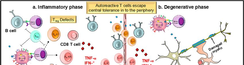
Antioxidants 2019, 8, 35 2 of 30
the prevalence of such diseases is anticipated for the next 30 years in the United States alone [4]. Thus,
recognizing and understanding the involvement of inflammatory processes underlining different
disorders will pave the way to development of a new class of drugs to help curb the tide of chronic
inflammatory diseases.
Inflammation is characterized by the protective response of the immune system that involves
the recognition of highly conserved pathogenic structures (pathogen-associated molecular patterns
(PAMPs)) or endogenous non-infectious molecules (damage-associated molecular patterns (DAMPs)
or alarmins or cell-death associated molecules) by pathogen-recognition receptors (PRRs) [2,3,5]. The
activation of these receptors leads to the production of various pro-inflammatory cytokines such as
tumor necrosis factor (TNF)-, interleukin (IL)-1, and chemokines through the induction of the
nuclear factor-kappa B (NF-B) pathway and NLRP3 (NOD-, LRR-, and pyrin domain-containing 3)
inflammasome activation. The inflammatory pathways, such as the mitogen-activated protein kinase
(MAPK), Janus kinase/signal transducers and activators of transcription (JAK-STAT), and especially
the NF-B pathway, help orchestrate the inflammatory responses through the production of
inflammatory cytokines and mediators, cell proliferation and survival, T-cell differentiation, and
dendritic cell (DC) maturation [6]. The role of the cytokines and chemokines is to recruit additional
immune cells to the site of infection, including circulating neutrophils that enhance microbial killing
through the production of interferon (IFN)-, proteases, and reactive oxygen species (ROS). Cytokines
also induce the production of cyclooxygenase-2 (COX-2), an enzyme that catalyzes the production of
prostaglandins, which are key mediators of inflammation [7]. Additionally, dendritic cells, the most
potent antigen presenting cells of the immune system, also aid in activating the adaptive immune
response through naïve T-cell polarization and B-cell activation. Elimination of the
foreign/endogenous agent and reprogramming of the effector cells to effectively end the production
of inflammatory mediators then leads to resolution of inflammation and return to homeostasis.
However, failure to do so leads to prolonged periods of unresolved inflammation that becomes a
contributing factor in almost all chronic or degenerative diseases. Therefore, there is a need to
develop therapies targeting underlying inflammation to achieve therapeutic advances targeting most
of these degenerative disorders that currently have no cure.
Chronic inflammation is the leading cause of death worldwide, where three of every five
individuals die as a result of chronic inflammatory diseases like diabetes, heart disorders, cancer,
stroke, and obesity [4]. It constitutes a significant economic burden due to life-long debilitation
leading to high therapy cost and lost wages [8,9]. Furthermore, existing therapies are rarely curative,
mostly disease-modifying with low success rates, and have adverse and sometimes life-threatening
side effects [10,11]. Commonly prescribed anti-inflammatory drugs include Metformin, non-steroidal
anti-inflammatory drugs (NSAIDs), statins, and corticosteroids, which alleviate inflammation
through several mechanisms [4,10]. Novel therapies targeting specific cells of the immune system,
for example, T-cell targeted therapies (Laquinimod, Tacrolimus, Edratide), various anti-B-cell targets
(Rituximab, BLyS), and cytokine inhibitors (adalimumab, infliximab), have also been employed in
disease management and treatment with varying results [11–14]. However, there is still a pressing
need to develop safer and cost-effective therapeutic alternatives. Natural products, which are
classically defined as compounds derived from natural sources such as plants, animals, and micro-
organisms, have been used for many millennia to treat a number of human ailments [15,16]. These
compounds have historically served as important leads for pharmaceutical companies in the
development of synthetic drugs, which were initially produced in the form of crude compilations
and more recently, with the advent of combinatorial chemistry and sophisticated techniques like
genomics and proteomics, consist of purified compounds [17–20]. Also, synthetic derivates of natural
compounds with certain enhanced characteristics can also be engineered. In fact, about 34% of the
U.S. Food and Drug Administration (FDA)-approved medicines between 1981 and 2010 are natural
products or derivatives of natural products including anti-cancer drugs and immunosuppressants
[21]. Although, combinatorial chemistry has also resulted in the relative ease with which synthetic
libraries of small molecule drugs can be generated, scaling back natural product-based drug
discovery [22,23]. However, seminal discoveries like that the first naturally derived medicine,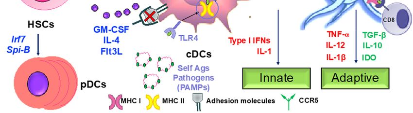
Antioxidants 2019, 8, 35 3 of 30
morphine, to those of penicillin and streptomycin and the more recent anti-parasitic drugs
avermectins and artemisinin, show that natural products are definitely the best source of drugs
[24,25]. With newer techniques comes the knowledge of the structures of the different natural
compounds, which in turn allows for understanding their specific mechanism of action in health and
disease.
As seen with their role in the treatment of several diseases including malaria, cancer, and
diabetes, natural products have been reported to have inhibitory effects on inflammation. In the past
several decades, numerous studies have reported the anti-inflammatory activities of various plants,
plant extracts, or purified compounds derived from plant and other natural sources [26–28]. Various
plant-derived compounds including curcumin, resveratrol, and capsaicin inhibit inflammation
through reduction in the levels of several cytokines including IL-1, IL-6, and TNF-, and the
suppression of COX-2, prostaglandins, and inflammatory pathways [29]. Active organosulphur
compounds in garlic such as ajoene, alliin, and allicin work by reducing levels of pro-inflammatory
cytokines while increasing levels of anti-inflammatory IL-10 [30]. Natural products derived from
marine flora including those of coral and algal origin also inhibit inflammation through suppression
of IL-6, TNF-, and nitric oxide (NO) release and inhibition of COX-2, inducible NO synthase (iNOS),
and NF-B activity [31,32]. Another group of plant-derived natural products is the polyphenolic
bioactive components of various plants and vegetables known as ‘flavonoids’. The word flavonoid is
a derivative of the latin word flavus, meaning ‘yellow’, indicating the color of these compounds in
their natural form [33–35]. Flavonoids, as secondary metabolites in numerous fruits, herbs, root,
stems, bark, flowers, grains, tea, and wine, impart both color and protection to the plants and make
them safe for consumption, for which they are also termed as ‘phytonutrients’. Because of their broad
spectrum of biological activity and attractive properties such as anti-oxidant, anti-mutagenic, anti-
inflammatory, and anti-viral effects, these compounds present an indispensable library of
compounds that can be developed as therapeutic entities. This review will focus on the anti-
inflammatory activities of various flavonoids that can potentially work against several chronic
diseases. We will then briefly showcase the known anti-inflammatory properties of a comparatively
less-toxic flavonoid, apigenin, to assess its potential as a drug lead against chronic
neuroinflammatory diseases.
2. Flavonoids in Health and Disease
Flavonoids are a multi-functional group that possess substantial characteristics that can be
exploited for the development of therapeutic agents targeting several chronic diseases. They have
been seen to exert a wide range of pharmacological effects, such as anti-oxidant, anti-tumor, anti-
viral, anti-allergic, anti-inflammatory, and anti-viral effects. These protective biological properties are
mostly due to the phenolic structure of these flavonoids.
2.1. Chemical Structure
Flavonoids belong to the group of polyphenolic natural compounds, with more than 4000
identified varieties. This variation is the result of the modifications that are possible to the carbon
skeleton, which is common to all flavonoids and consists of a flavan system of two benzene rings
(denoted as A and B) that are linked together by a heterocyclic pyrene ring (denoted as C) [36]. The
chemical diversity of flavonoids is based on two structural variations; namely the pattern of
substitution of the C ring that depends on the carbon on which the B ring is attached, and the degree
of oxidation of the C ring [33,37]. The basic flavonoid structure is aglycone, but they typically occur
in nature as glycoside and methylated derivatives, which are products of secondary metabolism in
plants [36].
Certain structural modifications can also favor the anti-inflammatory activities of the flavonoid
families. Current understanding of the structural requirements dictates that the unsaturation of the
C ring, the presence of a carbonyl group on C-4, the number and position of hydroxyl groups, and
glycosylation status affect the anti-inflammatory properties of flavonoids [38,39]. For example, the
presence of a catechol group in the B ring of the flavonoid quercetin confers potent anti-inflammatory
Antioxidants 2019, 8, 35                                                                       4 of 30

activity, while the addition of one hydroxyl group on position 2′ of the B ring of morin abolishes any
anti-inflammatory properties [38]. Additionally, the hydroxylation pattern of the B ring of certain
flavonoids promotes the inhibition of cytokine secretion by mast cells and macrophages [40].
Glycosylation of flavonoids has been linked to a reduction in the inhibitory effect on inflammation
because glycoside derivatives are more readily absorbed than aglycones.

2.2. Subclasses
      On the basis of the molecular structure, flavonoids can be divided into different subclasses (as
described in detail in Table 1) as follows: flavonols (e.g., quercetin, kaempferol, myricetin, and
fisetin), flavones (e.g., apigenin and luteolin), flavanones (e.g., hesperetin and naringenin),
flavononols, anthocyanidins, and isoflavones [33,36]. The various compounds within a specific
subclass differ in the pattern of substitution of the A and B rings. The process of flavonoid
biosynthesis is conserved in plant, where the action of enzymes modifies the basic flavonoid
structure, leading to various intermediary compounds or flavonoid subclasses. The C ring of the
flavonoid precursors, chalcones, closes to form a chromone unit, resulting in the formation of
flavanones. Oxidation at the third carbon of flavanones produces the flavanols. Flavones are then
formed by a double bond at C-2. The reduction of flavonols produces flavan-3-ols such as
anthocyanidins [38]. These subclasses have varying distribution among the different natural sources,
for example, flavonols and flavones are typically abundant in onions and tea.
Antioxidants 2019, 8, x FOR PEER REVIEW 5 of 30
Table 1. Subclasses of flavonoids. ERK—extracellular signal-regulated kinases, NF-B—nuclear factor-kappa B; MAPK—mitogen-activated protein kinase; ROS—reactive
oxygen species; COX-2—cyclooxygenase-2; IL—interleukin; TNF—tumor necrosis factor; iNOS—inducible NO synthase; PKC—protein kinase C, MDA—malondialdehyde
, MMP—matrix metalloproteinase, FAK—focal adhesion kinase.
Class of Flavonoids Chemical Structure Dietary Source Compound Molecular Targets Biological Function Reference
Tea, red wine, red Catechin, ↓ ERK, NF-κB, Rac1, AP-1,
Flavanol Anti-carcinogenic [2,9]
grapes Epigallocatechin p38
↓ Akt, ERK, caspase-12,
Fruit skins, red Anti-inflammatory, anti-
Apigenin, Chrysin, caspase-3, MAPK, ROS,
Flavone pepper, and carcinogenic, [10–13]
and Luteolin COX-2, IL-6, TNF-α, IL-1 β ,
tomato skin neuroprotective
iNOS, PGE2
Antioxidant, anti-
Onion, red wine, Quercetin, ↓ PKC, AP-1, H2O2, iNOS,
inflammatory,
Flavonol olive oil, berries, Kaempferol, MDA, citrate synthase, [2,14]
neuroprotective reduce risk
and grapefruit Myricetin, and Fisetin MMP-9,MMP-2, COX-2,ERK
of vascular disease
Citrus fruits, ↓ROS, glutathione
Blood lipid-lowering and
grapefruits, Hesperetin, reductase, iNOS, 3-
Flavanone cholesterol-lowering agents, [14]
lemons, Naringenin nitropropionic acid, COX2,
antiviral, antioxidant
and oranges NF-κB, IL-1β, TNF-α
Genistin,
Soyabean ↓ FAK, MAPK, NF-κB, AP- Anti-inflammatory, anti-
Isoflavone Daidzin
1, MMP-9, MMP-2 cancer
Antioxidants 2019, 8, 35; doi:10.3390/antiox8020035 www.mdpi.com/journal/antioxidants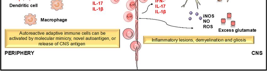
Antioxidants 2019, 8, 35                                                                                                       6 of 30

                                                                                                      Anti-inflammatory,
                           Cherry, Elsberry,      Apigenidin,          ↓MMP-9, MMP-2, ERK,
    Anthocyanidin                                                                                   antioxidant, anticancer,
                            and strawberry         Cyanidin             AP-1, NFKB, MAPK,
                                                                                                       cardioprotective

                                                                                                      antioxidant, anti-
                               Limon,
                                                                       ↓ H2O2, iNOS, COX-2, IL-         inflammatory,
      Flavanonol           aurantium, Milk     Taxifolin, Silibinin
                                                                      1β, TNF-α, NF-κB, IL-8, ROS      neuroprotective,
                               thistle
                                                                                                    antiallergic, antitumor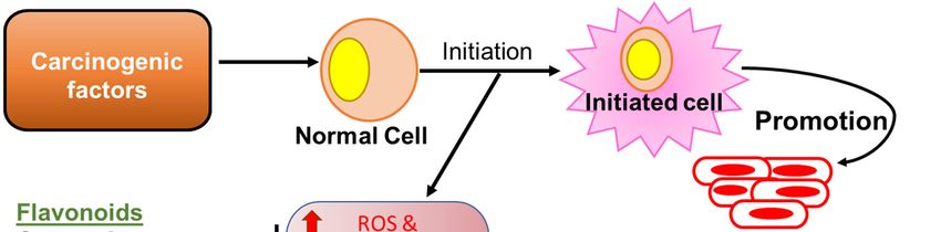
Antioxidants 2019, 8, x FOR PEER REVIEW 7 of 30
2.3. Health Benefits
A wide range of biological activities have been attributed to the flavonoid group of natural
products. These include anti-oxidant, anti-inflammatory, anti-mutagenic, anti-viral, and anti-allergic
properties. Owing to their structure, flavonoids exert various protective effects against several
chronic diseases like cancer, diabetes, and cardiovascular disorders, as well as neurodegenerative
conditions. By complexing with oxidizing species, hydroxyl groups in flavonoids render these
compounds the ability to scavenge and stabilize free radicals, reducing oxidative damage, which is
the hallmark of several chronic diseases [36,41]. Here, we review the effects of flavonoids on various
inflammatory processes that cause or further exacerbate chronic diseases.
2.4. Flavonoids in Diseases of Chronic Inflammation
Flavonoids comprise a wide variety of biologically active compounds, many of which have been
used as components of various medicinal preparations over thousands of years to treat several
human illnesses. Most of the non-infectious diseases develop or are considerably worsened by the
presence of persistent chronic inflammation. Here, we will explore the characteristic functions of
flavonoids that help combat many inflammatory processes underlying several chronic conditions
such as cancer, obesity, and neuroinflammation.
2.4.1. Flavonoids in Cancer
German pathologist, Rudolf Virchow, was the first to find a link between inflammation and
cancer development. Since then, epidemiological studies have established a correlation of at least 20%
of cancers, including lung, prostrate, bladder, pancreatic, esophageal, and melanoma, with long-term
inflammation [42,43]. Chronic unregulated inflammation results in the persistent production of
harmful ROS that can lead to DNA damage and genomic alterations, causing the initiation of tumor
growth. There is also a continuous generation of inflammatory mediators such as IFN-γ, TNF, IL-
1α/β, or IL-6 and proangiogenic growth factors such as cytokines and vascular endothelial growth
factor (VEGF) that promote tumor neovascularization, which brings the much-needed blood supply,
nourishing the growing tumor. Finally, inflammation promotes tumor dissemination through the
production of extracellular matrix degrading enzymes, the matrix metalloproteinases (MMPs) [44].
All these factors are either produced by tumor infiltrating immune cells such as macrophages,
dendritic cells, neutrophils, lymphocytes, and natural killer cells or by the cancer cells themselves to
stimulate their growth and survival. The key inflammatory pathway, NF-B, plays an important role
in cancer cell survival by allowing these cells to escape apoptosis [45]. Consequently, therapeutic
agents that target these inflammatory factors and pathways will serve as a means to treat or prevent
cancer development.
Because flavonoids possess several anti-inflammatory properties, they can serve as potent anti-
cancer phytochemicals (Figure 1) that exert their activity by several mechanisms such as carcinogen
inactivation, triggering cell cycle arrest, induction of apoptosis, and inhibition of angiogenesis [46–
50]. Flavonoids have been shown to inhibit tumor cell proliferation via inhibition of ROS formation,
as well as suppression of xanthine oxidase, COX-2, and 5-LOX, which are the major catalysts for
tumor promotion and progression (reviewed in the work of [51]). Mojzis et al. reported that
flavonoids are able to exert anti-angiogenic effects by regulating the expression of VEGF, MMPs, and
epidermal growth factor receptor (EGFR), as well as by inhibiting the proangiogenic signaling
pathways including the NF-κB, PI3-K/AKt, and ERK1/2 pathways [52]. Accumulating evidence
reported that cyclin dependent kinases (CDKs) are key regulators of cell cycle progression, immune
cell activation, neoangiogenesis, and inflammation [53]. In addition, it has been seen that various
types of cancers are associated with hyper-activation of CDKs, because of mutation of CDK genes or
CDK inhibitor genes. Flavonoids have been shown to induce cell cycle arrest at both checkpoints G1/S
and G2/M through CDK inhibition in human breast cancer and melanoma cells [51]. Flavonoids such
as isoflavones and their metabolites also induce cancer cell apoptosis in cells derived from human
Antioxidants 2019, 8, 35; doi:10.3390/antiox8020035 www.mdpi.com/journal/antioxidants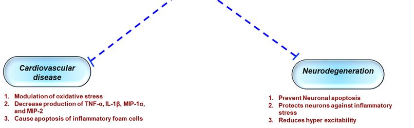
Antioxidants 2019, 8, 35 8 of 30
gastric cancer [50] by inhibiting DNA topoisomerase I/II activity, decreased production of ROS,
regulation of heat shock protein expression, modulation of signaling pathways, suppression of NF-
κB, activation of endonuclease, and suppression of Mcl-1 protein.
Figure 1. Flavonoids in cancer. Flavonoids exert their anti-inflammatory activities by reducing the
production of reactive oxygen species (ROS) and the down-regulation of several inflammatory
mediators through key inhibition of signaling pathways. NF-B—nuclear factor-kappa B; MAPK—
mitogen-activated protein kinase; STAT—signal transducers and activators of transcription.
2.4.2. Flavonoids in Diabetes
Inflammation has long been associated with the promotion of type 1 diabetes, however, more
recently, chronic low-grade inflammation has been deemed responsible for onset and/or exacerbation
of type 2 diabetes, diabetes mellitus [54]. Type 2 diabetes is associated with impaired insulin secretion
and insulin resistance. Nutrient excesses such as hyperglycemia and elevated free fatty acids, as seen
in type 2 diabetes, cause oxidative stress, endoplasmic reticulum stress, amyloid and lipid deposition,
lipotoxicity, and glucotoxicity induced by inflammatory processes. Metabolic dysregulation
associated with diabetes is said to induce a proinflammatory response in macrophages residing in
the adipose tissue, islets, and vasculature, and their infiltration into the adipocytes is directly
proportional to cell size [55]. Cellular stresses are induced by the activation of thioredoxin-interacting
protein and NLRP3 inflammasome, releasing increased amounts of IL-1, contributing to -cell
dysfunction and insulin resistance [56]. Macrophages, endothelial cells, and adipocytes also produce
acute-phase proteins such as C-reactive protein (CRP) and serum amyloid A (SAA) in response to
elevated levels of IL-6. Additionally, proinflammatory cytokine TNF-α released within the adipose
tissue promotes lipolysis and increases free fatty acids [57].
Several studies have reported the beneficial properties of flavonoids that promote their use as a
supplementary treatment for diabetes mellitus [58]. It has been seen that flavonoids can modulate
carbohydrate and lipid metabolism, attenuate hyperglycemia, insulin resistance, alleviate oxidative
stress and stress-sensitive signaling pathways, and inflammatory processes [59]. Flavonoids morin,
hesperidin, rutin, (rats), and chrysin (mice) were effective in reducing inflammatory cytokines IL-1β,
IL-6, and TNF-α in diabetic animals, significantly improving hyperglycemia, glucose intolerance, and
insulin resistance [60–63]. Diabetes mellitus can eventually cause secondary damage to various
organs of the affected individual such as eyes, kidneys, nerves, and heart [64]. Flavonoids not only
help restore glucose homoeostasis attenuating the diabetic condition, but also regulate the secondary
damage to the various peripheral organs. Hesperidin reversed neuropathic pain and improved heart
function in diabetic rats [63,65]. Chrysin improves cognition, prevents the development of diabetic
neuropathy, and improves renal pathology in diabetic rats [66,67].
Peroxisome proliferator-activated receptor gamma (PPAR-γ), a nuclear receptor found in
adipocytes and macrophages, stimulates adipogenesis, lipid uptake, and inulin sensitivity and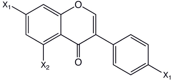
Antioxidants 2019, 8, 35                                                                         9 of 30

regulates inflammation and glucose metabolism through upregulation of adipokines and glucose
transporter, GLUT4, as well as fatty acid binding and transport proteins, respectively [68,69]. Certain
flavonoids such as naringenin, luteolin, and quercetin alter PPAR-γ activation and increase insulin
sensitivity [70]. PPAR-γ also induces various adipokines including leptin, adiponectin, and resistin,
which play crucial roles in glucose homeostasis and regulate inflammation [70]. Adiponectin has
been seen to inhibit the release of pro-inflammatory cytokines such as TNF-α and IL-6. Luteolin,
abundant in vegetables and fruits including celery, parsley, carrots, and apple skins, potentiates
insulin action and increases expression and transcriptional activation of PPAR-γ and expression of
the PPAR-γ target genes, increasing expression of adiponectin, leptin, and GLUT4 in 3T3-L1
adipocytes, as well as in primary mouse adipose cells [71]. Liu et. al. reported that the decrease in
circulating level of inflammatory molecules MCP-1, resistin, and the elevation of adiponectin level in
obese mice may be attributed to luteolin, which, in turn, mediates beneficial effects on metabolic
pathways implicated in insulin resistance and type 2 diabetes pathophysiology [72].

2.4.3. Flavonoids in Inflammatory Bowel Disease
      Inflammatory bowel disease (IBD) is characterized by chronic and uncontrolled gastrointestinal
inflammation, associated with both impaired epithelial mucosal barrier and altered innate and
adaptive immune responses [73,74]. IBD presents as two main clinical and pathological subtypes,
Crohn’s disease (CD) and ulcerative colitis (UC). Both forms of IBD result in poor quality of life and
require prolonged medical and/or surgical interventions. CD can affect any part of the
gastrointestinal tract, from the mouth to the anus, but it is usually, although not always, localized in
the distal small bowel and/or colon. In contrast, UC is restricted to the colon and the rectum. Although
several theories have been put forth to explain IBD pathogenesis, the exact cause of IBD still remains
elusive [73,75]. IBD patients who exhibit a dysfunctional intestinal epithelium barrier with higher
tight junction permeability develop an exaggerated immune response in the gut towards the
intestinal microbiota. Several elements of the mucosal immune system including intestinal epithelial
cells, innate immune cells such as dendritic cells, macrophages, neutrophils, and cells of the adaptive
immune system such as T cells and B cells, along with cytokines and chemokines, have been shown
to participate in the pathogenesis of IBD [75]. Tolerance to commensal flora in IBD is lost as a result
of dendritic cell overactivation and the resultant loss of regulatory T cells and strong induction of
proinflammatory effectors such as Th1, Th17, and natural killer (NK) cells [76,77]. Additionally, a
variety of cell adhesion molecules (CAMs), including intracellular CAM-1 (ICAM-1) and vascular
CAM (VCAM-1) and chemokines such as IL-1β, IL-6, and TNF-α, have been shown to be activated
by endothelial cells, which are responsible for the recruitment of leukocyte and promoting
inflammatory responses [78].
      As the exact etiology of IBD is not well understood, there is no specific treatment available to
cure the disease. Hence, flavonoids, which are known to have a range of biological activities, could
be beneficial in the treatment for IBD [79–81]. Flavonoids such as apigenin and epigallocathechin
gallate have been shown to inhibit the activation of immune cells and the downstream chemokines
and cytokines [82], thereby it may considered as a natural inhibitor and can prevent the activation of
an innate and adaptive immune system [79]. A number of studies have published the anti-
inflammatory impact of flavonoids in several experimental models of colitis (reviewed in the work
of [75]). Flavonoids belonging to different subclasses such as chalcones (cardamonin), isoflavones
(genistein, daidzein, glabridin), anthocyanidins (cyanidin-3-glucoside (C3G)), flavonols (quercetin,
quercitrin, rutin), flavanones (naringenin), flavones (baicalin, chrysin), and catechins
(epigallocatechin-3-gallate) have shown profound intestinal anti-inflammatory activity. Of these,
accumulating evidence has shown that quercetin inhibits bacterial lipopolysaccharide (LPS)-induced
iNOS and TNF-α secretion in macrophages, LPS-induced IL-1β, TNF-α secretion in RAW2647 and
cytokine induced expression of VCAM-1 and ICAM-1, and E-selection in HUVECs (reviewed in the
work of [83]). Glycones have been more effective in curbing intestinal inflammation as they are
metabolized in the colon, where the aglycone form is then released as opposed to the early absorption
of aglycones in the intestines. Quercetin glycosides quercetrin and rutin have more potent effects on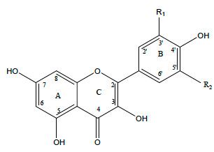
Antioxidants 2019, 8, 35 10 of 30
disease outcomes than the aglycone [84,85]. Quercetrin reduces colonic inflammation in rats through
modulation of the NF-kB pathway and subsequent inhibition of cytokines, as well as iNOS in vivo
[86]. Naringenin (4’,5,7-trihydroxy avanone-7-rhamnoglucoside), which belongs to the flavanone
class, is shown to attenuate the severity of colitis by inhibiting myeloid derived suppressor cells
(MDDCs), pro-inflammatory mediators, and the NF-kB/IL-6/STAT-3 cascade in colorectal tissues
[87].
2.4.4. Flavonoids in Non-Alcoholic Fatty Liver Disease
Non-alcoholic fatty liver disease (NAFLD) is rapidly emerging as the most common etiological
factor for chronic liver disease, largely because of the growing prevalence of insulin resistance,
obesity, and diabetes [88,89]. As the name suggests, NAFLD is characterized by the deposition of free
fatty acids and triglycerides in hepatocytes and can lead to further complications such as liver
fibrosis, cirrhosis, and hepatocellular carcinoma [89,90]. NAFLD comprises a spectrum of disorders
including simple steatosis in the absence of inflammation and hepatocellular damage and the more
severe form of NAFLD, nonalcoholic steatohepatitis (NASH), characterized by lobular inflammation
[91]. Much remains to be elucidated about the pathogenesis of both NAFLD and NASH, however,
attempts to depict a ‘two-hit’ hypothesis of disease pathogenesis have been made. Hepatic steatosis
due to deposition of triglycerides coming from lipolysis of adipose tissue, from de novo synthesis, or
from the diet triggered by insulin resistance constitutes the first ‘hit’ [90,92]. Excessive accumulation
of fat and the presence of circulating free fatty acids contribute to oxidative damage, immune system
activation, and dysregulation of cytokine pathways through recognition by PRRs such as toll-like
receptors (TLR)s, in particular, TLR4 [92]. Lipid accumulation in the liver leads to increased
transcription and the release of IL-6, TNF-, and C-reactive protein, leading to chronic low-grade
inflammation through the reduction of the anti-inflammatory adiponectin, which sensitizes
hepatocytes to insulin [90].
Because the pathogenesis of NAFLD is multifaceted, so far there is no evidence-based treatment
for this disease. In this aspect, bioactive compounds such as flavonoids that can modulate various
pathways are ideal candidates for therapeutic development against NAFLD. Flavonoids have been
shown to have beneficial effects on lipid metabolism, insulin resistance, oxidative stress, and
inflammation, the major causative factors of NAFLD [93]. The anti-inflammatory effects of silymarin,
a flavonoid mix derived from milk thistle, have been documented in animal models of NAFLD [94].
Silibinin, the most active component of silymarin, decreased hepatic NF-B activation and decreased
levels of ROS and iNOS in a mouse model of NASH [95]. Silymarin was also reported to have reduced
the expression of inflammatory TNF-α mRNA in the liver of methionine- and choline-deficient
(MCD) diet induced NASH in insulin-resistant rats [96]. Isoflavones found in soybeans and their
derivatives have also shown beneficial effects on NAFLD in in vivo animal studies. Genistein, a soy
phytoestrogen, has been reported to show anti-inflammatory effects through reduction of TNF-α and
IL-1 mRNA expression in nonalcoholic fatty liver disease db/db mice [97]. Genistein administration
in NASH rats induced by a high fat diet alleviated liver damage through inhibition of inflammatory
processes by reducing serum levels of TNF-α and IL-6 and inhibiting IκB-α phosphorylation, nuclear
translocation of NF-κB p65 subunit, and activation of c-Jun N-terminal kinase (JNK) [98]. C57BL/6
mice on a cholesterol-enriched diet supplemented with the isoflavone, 2-heptyl-formononetin,
demonstrated lowered hepatic inflammation through a reduction in TNF-α levels and macrophage
infiltration [99]. Both quercetin and its glycoside rutin showed reduction in inflammatory markers
TNF-α and IL-6 in NASH mice [100,101]. Other flavonoids such as cyanidin 3-O-β-D-glucoside and
xanthohumol also inhibit inflammatory pathways through ROS inhibition and suppression of NF-κB
and its dependent genes in animal models [102,103].
2.4.5. Flavonoids in Cardiovascular Disorders
Inflammatory mediators are both a predictive and a causative factor in the pathogenesis of
cardiovascular disorders. Acute myocardial infarction (AMI) is the result of rupture of an
atherosclerotic plaque, leading to thrombus formation and loss of blood flow causing ischemia in theAntioxidants 2019, 8, 35 11 of 30
area distal to the occlusion [104]. Prolonged myocardial ischemia leads to cardiomyocyte injury
releasing DAMPS, which activate platelets and leucocytes; recruit neutrophils; and lead to
endothelial cell injury and the production of ROS, proteases, and cytokines. Additionally, the NLRP3
inflammasome is activated in myocardial ischemia, which in turn binds to and activates caspase 1,
which is responsible for the conversion of IL-1 to its active form. Of all the cytokines, TNF-, IL-1,
and IL-6 play central roles in AMI, causing the secretion of other cytokines, chemokines, and adhesion
molecules augmenting further leucocyte infiltration. Similarly, immune cells and released mediators
also play a critical role in the initiation and progression of atherosclerosis [105]. Plaque formation is
initiated by the accumulation of low-density lipoproteins (LDLs) in the subendothelial layers of the
arteries, leading to endothelial injury and dysfunction. This subsequently leads to formation of ROS,
which oxidize the LDLs and contribute to plaque formation. Activated endothelial cells further
release leucocyte adhesion molecules such as vascular cell adhesion molecule-1 (VCAM-1),
intercellular adhesion molecule 1 (ICAM-1), and selectins, which, together with chemokines such as
CCR2, and CCR5, recruit circulating monocytes. Monocytes then differentiate in situ to macrophages
that convert to foam cells via uptake of oxidized LDLs and release a milieu of cytokines such as TNF-
, IL-1, IL-3, IL-8, and IL-18.
Manipulation of these inflammatory events is crucial in the prevention and management of
cardiovascular diseases. Because of the phenol hydroxy groups present in flavonoids, these
compounds possess remarkable anti-oxidant and free-radical scavenging properties. Thus, these
polyphenols are able to show promising effects in the management of cardiovascular injury. In vitro
evidence shows that quercetin reduces LDL oxidation at physiological levels in human umbilical vein
endothelial cells [106]. Similar reduction in macrophage-mediated LDL oxidation was seen after
treatment with fisetin and proanthocyanidins [107]. Quercetin was also shown to decrease the level
of proinflammatory mediators such as TNF-α, IL-6, MIP-1α, and P-selectin in murine RAW264.7
macrophages [108]. Further, quercetin treatment can potentially disrupt atherosclerotic plaques
through the inhibition of matrix metalloproteinase 1 [109]. Soy isoflavone administration reduced the
risk of chronic inflammation-mediated cardiovascular disease by reducing the endothelial
production of TNF-α in a mouse model [110]. Isoflavones have also been reported to protect against
inflammatory vascular disease through the inhibition of monocyte recruitment across the
endothelium. Additionally, several flavonoids such as apigenin, chrysin, and kaempferol have been
shown to inhibit the expression of adhesion molecules on human aortic endothelial cells, thereby
limiting leucocyte infiltration [111].
2.4.6. Flavonoids in Neuroinflammation
Neuroinflammation is the accompanying factor in several neurodegenerative diseases such as
Alzheimer’s disease (AD), Parkinson’s disease (PD), Huntington’s disease (HD), and multiple
sclerosis (MS). These neurodegenerative diseases result in the progressive and irreversible loss of
neurons in the brain [112]. Activation of central nervous system (CNS)-resident microglial cells is one
of the crucial events in the inflammatory cascade, which leads to the progression of
neurodegeneration [113]. Prolonged activation of microglial cells may contribute to
neurodegeneration through the release of pro-inflammatory mediators such as prostaglandins, NO,
TNF-α, IL-6, and IL-1β, resulting in chronic CNS neuroinflammation [114]. These inflammatory
events contribute to the apoptotic cell death of neurons in many neurodegenerative diseases.
Additionally, encephalitogenic inflammatory CD4+ T cells such as Th1, Th17, granulocyte
macrophage colony-stimulating factor (GM-CSF)-producing CD4+ T-cells, and γδT-cells play very
crucial roles in the initiation and propagation of neuroinflammation in autoimmune diseases such as
MS. T cells become activated by antigen presenting cells (APCs) such as dendritic cells (DC) and
macrophages. DCs are the most efficient professional APCs and play an important role in various
autoimmune and neuroinflammatory diseases [115–117]. Our earlier studies have reported that DCs
can migrate into diverse regions of the CNS [118] in response to neuroinflammatory signals (i.e.
chemokine CCL2) both in vitro and in vivo [119]. Additionally, increased frequencies of both
plasmacytoid and myeloid DCs (pDC and mDC) have been reported in the cerebrospinal fluid (CSF)Antioxidants 2019, 8, 35 12 of 30
of MS patients [120]. In fact, CD11c+ DCs were shown to be sufficient to present myelin antigen to
naive T cells, leading to the development of experimental autoimmune encephalomyelitis (EAE) in
mice [121]. Hence, therapeutic agents targeting DC function and their migration across the inflamed
blood–brain barrier (BBB) under neuroinflammatory conditions will be of vital importance in the
treatment of neurodegenerative diseases such as MS. The treatments currently available are rarely
curative and have serious side effects.
Natural flavonoids have been shown to exert neuroprotective properties by inhibiting the
release of pro-inflammatory cytokines. Flavonoids exert an anti-inflammatory effect via interfering
with the development of inflammatory mediators such as IL-6, TNF-α, and IL-1β in several cell lines
through the MAPK signaling pathway [122]. TNF-α and iNOS expression has been seen to be
regulated by inhibiting MAPK signaling cascade molecules such as p38 or ERK1/2. Accumulating
evidence has reported that flavonoids can modulate the activity of various metabolic pathways to
reduce neuronal dysfunction. Moreover, flavonoids have been found to delay or prevent the onset of
neurodegenerative diseases at their effective doses in various animal models [123]. Wogonin,
baicalein, curcumin, apigenin, quercetin, luteolin, and many other flavonoids have been shown to
exhibit neuroprotective effects. Wogonin and baicalein have been shown to exert various anti-
inflammatory effects through the inhibition of inflammatory microglia. It has been seen that wogonin
can inhibit the LPS induced production of NO, inducible NO synthase (iNOS), and NF-B activation
in microglia. LPS induced NF-B activity in BV-2 microglial cells was shown to be inhibited by the
flavonoid baicalein without interfering activation of caspase-11, activator of transcription (STAT-1),
and induction of interferon regulatory factor (IRF-1) [124]. Further, curcumin inhibits the apoptosis
of pre-oligodendrocyte mediated expression of iNOS, NO, and COX-2 in LPS activated microglia
[125]. Luteolin exerts its immunomodulatory effects on peripheral blood mononuclear cells (PBMC)
derived from MS patients. Luteolin has been seen to suppress the PBMC production of several pro-
inflammatory cytokines such as IL-1β, metalloproteinase-9 (MMP-9), and TNF-α, which play very
crucial roles in the pathogenesis of MS [126]. Isoflavones such as daidzein and genistein inhibit TNF-
α, IL-1, IL-6, iNOS, and COX-2 via suppression of ERβ and NF-B, respectively, in primary astrocytes.
Several flavonoids reportedly suppress the inflammatory activity of DCs. Luteolin inhibits LPS-
induced NF-B signaling through the suppression of IB kinase activity in murine bone-marrow
derived DCs. Flavonoids such as silibinin, taxifolin, and epigallocatechin inhibited the
immunostimulatory effects of DCs via different mechanisms such as suppression of MAPK, impaired
p65 translocation, and endocytic ability [127–130]. However, the effects of flavonoids on DC function
in the context of neuroinflammatory diseases are relatively unknown.
Apigenin, a polyphenolic flavonoid, abundant in chamomile plant and also found in other
sources such as parsley, celery, and grape fruit, is a relatively less toxic and non-mutagenic
compound among the various flavones. Apigenin can also cross the blood–brain barrier (BBB) and
has been shown to exert anti-inflammatory effects on BV-2 and primary microglial cells through
inhibition of p38 and JNK. Apigenin prevents neuronal apoptosis by protecting the neurons against
inflammatory stresses [131]. The anti-inflammatory and neuroprotective effects of apigenin have not
yet been extensively characterized. Utilizing EAE models of MS, we recently observed a significant
reduction in disease severity accompanied by an increased retention of immune cells in the periphery
upon treatment with apigenin [132]. These results were supported by decreased immune cell
infiltration and reduced demyelination in the CNS of the apigenin-treated EAE mice. We
hypothesized that the neuroprotective effects of apigenin in EAE were due to its inhibition of DC
phenotypical and functional maturation and its subsequent polarization of CD4 T helper cells
(unpublished data). Because of its relatively long half-life, delayed plasma clearance, and slow
metabolism in the liver [133], apigenin has considerable potential to be developed as a safer, more
cost-effective treatment for neurodegenerative diseases. Here, we shall review the anti-inflammatory
properties of apigenin that have been extensively characterized in various disease systems and debate
its potential as a therapeutic drug candidate for neuroinflammatory conditions like MS.Antioxidants 2019, 8, 35 13 of 30
3. Role of Apigenin as an Anti-Inflammatory Agent
As the health effects of polyphenols depend on intake and bioavailability, the biological
activities of apigenin (4’,5,7-trihydroxyflavone), an abundantly occurring flavone have, been
extensively studied [134,135]. Much like the family to which it belongs, apigenin possesses a wide
array of biological properties including anti-oxidant, anti-cancer, and anti-inflammatory actions
[136–138]. As a result, apigenin has gained a lot of interest in the past few years as a potential
therapeutic agent to treat various diseases such as cancer, diabetes, cardiovascular, and neurological
disorders [139] (Figure 2).
Figure 2. Role of apigenin in chronic inflammatory diseases. Apigenin as an anti-inflammatory
compound acts as a protective agent in several disorders via inhibition of key inflammatory
mediators, signaling pathways, and molecules. COX-2—cyclooxygenase-2; IL—interleukin; TNF—
tumor necrosis factor; NO—nitric oxide.
3.1. Protective Effects of Apigenin Across a Spectrum Of Chronic Diseases
According to the National Cholesterol Education Program’s Adult Treatment Panel III (NCEP:
ATP III), metabolic syndrome is associated with abdominal obesity, dyslipidemia, hyperglycemia,
inflammation, insulin resistance, or diabetes mellitus, as well as an increased risk of developing
cardiovascular disease. Obesity is attributed to adipose tissue dysfunction and expanded adipose
tissue mass, which can lead to the upregulation of proinflammatory cytokines such as TNF-α and IL-
6, resulting in a state of chronic low-grade inflammation, as previously discussed. Apigenin has been
shown to inhibit an important inflammatory biomarker, CD38, in a metabolic syndrome model, as
well as decrease adipose tissue mass and the levels of proinflammatory cytokines [140]. Additionally,
Feng et al. have reportedly shown that apigenin improves obesity and obesity induced inflammation
[141]. Apigenin has also been reported to attenuate inflammation and the resultant pathological
alterations in rats fed with a high fat, fructose diet [142]. In diabetic rats, apigenin reduces metabolic
inflammation by successfully polarizing infiltrating macrophages to an anti-inflammatory M2
phenotype by binding and activating PPAR-γ and the subsequent suppression of the NF-κB pathway
[141]. Apigenin also ameliorated renal dysfunction in diabetic rats by suppressing inflammation
through reduced secretion of TNF-α and IL-6 via MAPK inhibition. Histopathology confirmed
reduced inflammation in the renal tissue along with reduction in collagen deposition and
glomerulosclerosis [143].
Apigenin served as a potent therapy against UC in C57BL/6 mice through the inhibition of
inflammatory cytokines,and COX-2, and through the reduction in immune cell infiltration in colon
tissues [144]. Because NF-κB activation upregulates epithelial cell permeability, promoting colonicAntioxidants 2019, 8, 35 14 of 30
inflammation, testing apigenin effect in vitro on colon carcinoma cells HCT-116 demonstrated NF-
κB downregulation in a dose-dependent manner. Apigenin also reduced levels of matrix
metalloproteinase (MMP-3), which aids in extracellular remodeling, contributing to colonic
inflammation, thereby showing protective effects in a murine DSS (dextran sulphate sodium) colitis
model [145]. The use of a soluble form of apigenin showed amelioration of in colitis models in rats
through the inhibition of various inflammatory markers such as TNF-α, transforming growth factor-
b, IL-6, intercellular adhesion molecule 1, or chemokine (C–C motif) ligand 2 [146].
Inflammation in NAFLD is one of the main causes of insulin resistance with inflammatory
markers such as TNF-α and IL-6 suppressing insulin receptor signaling, thus blocking the action of
insulin in hepatocytes. In NASH mice fed with a high fat diet, apigenin ameliorated inflammation
through reduction of plasma levels of MCP-1, IFN-γ, TNF-α, and IL-6 [147].
Beneficial aspects of apigenin activity help to ameliorate inflammation-mediated cardiac injury,
indicating a role for apigenin as a therapeutic agent against cardiovascular diseases. In an LPS-
induced model of myocardial injury, apigenin relieved injury by modulating both oxidative stress
and inflammatory cytokines such as TNF-α, IL-1β, MIP-1α, and MIP-2 through NF-B regulation
[148]. Macrophages loaded with oxidized LDLs contribute significantly towards the progression of
atherosclerotic plaques. Apigenin was shown to induce apoptosis of murine peritoneal macrophages
through reduction in expression of anti-apoptotic plasminogen activator inhibitor 2 [149]. Apigenin
can promote apoptosis in foam cells through inhibition of autophagy and subsequently reduce the
foam-cell mediated secretion of proinflammatory cytokines during atherogenesis [150]. Additionally,
apigenin helped in cholesterol efflux from macrophages in atherosclerotic lesions in apoE−/− mice
challenged with LPS through the increased expression of ATP binding cassette A1 (ABCA1) and
reduced expression of proinflammatory cytokines, and reduced levels of NF-B and TLR-4 [151].
Several studies have investigated the anti-cancer effects of apigenin and shown its ability to
suppress cancer cell proliferation in various types of tumors, including pancreatic, colorectal, liver,
blood, lung, cervical, prostate, breast, thyroid, skin, head, and neck [152–154]. Because inflammatory
molecules modulate the physiological and pathological states of cancer and its surrounding
microenvironment, and tumor initiation is said to occur as a result of prolonged exposure to
inflammatory conditions, inhibition of inflammatory molecules could be a promising approach to
managing cancer [155]. The NF-B pathway and its associated molecules are key regulators of cancer
cell survival and proliferation through increased expression of cell cycle related VEGF, inflammatory
cytokines, and metastatic genes such as COX-2 [152]. Apigenin reduced prostate tumor volumes in
mouse models through suppression of NF-B activation [156]. In non-small cell lung cancer cell line
A549, apigenin blocks the nuclear translocation of NF-B, thereby suppressing the expression of
tumorigenic genes such as Bcl-2, Mcl-1, and Bcl-xL. Apigenin also inhibits several signaling pathways
including NF-B and MAPK, inducing anti-cancer effects in malignant mesothelioma [157].
Macrophages are the most abundant innate immune cells in the tumor microenvironment that
contribute to chronic low-grade inflammation, leading to tumor growth and metastasis through
tumor neovascularization and matrix remodeling [158]. Apigenin induced apoptosis in mouse ANA-
1 macrophage cell line through regulation of MAPK pathway and suppression of anti-apoptotic gene
Bcl-2 [159]. Exposure to ultraviolet B (UVB) radiation results in acute inflammation due to production
of various cytokines and chemokines via COX-2 expression and the resultant recruitment of
neutrophils, monocytes, and macrophages, leading to acute responses such as skin edema or chronic
inflammation, fibrosis, and cancer. Apigenin suppresses UVB-induced skin carcinogenesis through
inhibition of inflammatory COX-2 and restoration of anti-angiogenic and anti-inflammatory
thrombospondin-1 [160]. TNF-α contributes to breast cancer metastasis through the recruitment of
tumor-infiltrating macrophages, neutrophils, and T cells, leading to immune evasion, tumor growth,
and metastasis. Apigenin was shown to down-modulate TNF-α mediated upregulation of
chemotactic protein, CCL2, granulocyte macrophage colony-stimulating factor (GM-CSF), IL-1α, and
IL-6 in human triple-negative cells (MDA-MB-231 cells) [161].
Apigenin, found abundantly in a variety of plants, herbs, and spices [136,162], has been utilized
for centuries to treat diseases such as asthma, insomnia, Parkinson's, neuralgia, and shinglesAntioxidants 2019, 8, 35 15 of 30
[162,163], suggesting its potential use for both peripheral and CNS disorders. Apigenin has been
shown to exert its neuroprotective effect via suppressing the expression of an inducible form of nitric
oxide synthase (iNOS) and nitric oxide (NO) in microglial cells and macrophages [131]. Also,
through regulation of adhesion molecules such as VCAM-1, ICAM-1, and E-selectin [164], which play
a critical role in controlling leukocyte migration across the endothelial cells of BBB, apigenin might
inhibit immune cells’ entry into the CNS and prevent neuroinflammation. However, it remains
elusive whether apigenin or other flavones could serve as a potential treatment for
neuroinflammatory disorders like multiple sclerosis, which affects approximately 400,000 people in
the United States alone. Very little is known about the neuroprotective effects of apigenin and its
related mechanism of action. In order to assess the therapeutic potential of apigenin in regulating
neuroinflammation, we tested its efficacy in EAE models of relapse-remitting MS wherein apigenin
reduced disease severity through inhibiting immune cell infiltration into the CNS and subsequent
reduction in demyelination [132]. MS is an autoimmune disease with an as yet unknown etiologic
agent mediated by an immunogenic response of auto-inflammatory T cells against the myelin sheath
protecting the neurons. Dysregulation of DC function in MS can result from several possible reasons,
which include, but are not limited to T-cell anergy in response to persistent antigens displayed by
long-lived lymphoid DCs and functional abnormality of DCs (Figure 3). The infiltration of DCs from
the periphery during neuroinflammatory autoimmunity has been studied extensively, particularly
in EAE models of MS wherein DCs infiltrating from the blood increase with the increasing clinical
severity of EAE. In fact, evidence shows that they interact with naive CD4+ T cells, driving Th17
differentiation, a T cell subset involved in chronic inflammatory disease [165,166]. Hence, the
regulation of DC functions and its transmigration into the CNS holds the key to prevent the
detrimental effects of immune infiltration in MS. Current MS therapies such as Natalizumab and
dimethyl fumarate (DMF) that regulate leucocyte entry into the CNS have shown potential in
controlling symptoms and relapse [167]. However, most of these do not control the progressive form
of the MS and are often associated with significant side effects, emphasizing the need for and value
of identifying safer, alternate therapies that could provide clinical level benefits for the debilitating
diseases of the CNS.
Figure 3. Role of dendritic cells (DCs) and T cells in the development and progression of multiple
sclerosis (MS). MS is an immune mediated disease characterized by an initial inflammatory event
consisting of presentation of as yet unknown antigens to CD8 T cells, their entry across the blood–
brain barrier (BBB) into the central nervous system (CNS), and their subsequent reactivation by CNS
resident DCs and microglial cells. This results in an inflammatory cascade involving secretion of
several proinflammatory mediators such as cytokines IL-1, IL-17, and TNF-. The release of these
cytokines initiates the degenerative phase that is characterized by increase in iNOS, NO, glutamate,
and ROS, which brings about formation of inflammatory lesions, gliosis, and demyelination, which
are the hallmarks of MS.Antioxidants 2019, 8, 35 16 of 30
3.2. Apigenin Mediated Modulation in Dendritic Cell Phenotypical and Functional Maturation
Hematopoeitic stem cells in the bone marrow differentiate into plasmacytoid DCs from
lymphoid progenitors in the presence of transcription factors such as like Irf7 and Spi-B [168] (Figure
4). DC progenitors in the bone marrow also give rise to circulating precursors in the presence of other
factors like Batf3 and Irf4 that home to tissues, where they reside as immature cells with high
phagocytic capacity [169]. Following tissue damage, immature DCs capture antigens through PRRs
such as TLRs, and initiate the innate response through the secretion of IL-1 and type I interferons.
Following antigen capture, immature DCs also subsequently migrate to the lymphoid organs, where
they select rare antigen-specific T cells, thereby initiating adaptive immune responses. T cells are also
educated by DCs to recognize and tolerate self-antigens. Sensing of microbial stimuli through PRRs
causes DCs to enter the process of maturation, which involves the upregulation of major
histocompatibility complex (MHC) molecules and co-stimulatory molecules. Peptide-loaded MHC
molecules are recognized by Ag-specific T cells via the T-cell receptor (TCR), constituting signal 1 of
T cell activation. Signal 2 consists of binding of costimulatory molecules on DCs to CD28/CD40L on
T cells. Activated T cells in turn help DCs in terminal maturation through the ligation of CD40 and
CD80/86. The final step in T cell activation is signal 3; the release of inflammatory cytokines and
chemokines promoting the differentiation of naïve antigen-specific T cells into effector cells, as well
as the activation of various other types of immune cells by the dendritic cells. Therapeutic agents
targeting the various steps involved in DC-mediated T cell activation may be critical in the
amelioration of various chronic inflammatory diseases.
Various flavonoids, described earlier in this review, inhibit the inflammatory functions of DCs.
The role of apigenin on DC maturation and function has been investigated in murine bone marrow
derived DCs, where inhibition of p65 translocation has been linked to downmodulation in cell surface
expression of DC co-stimulatory molecules and antigen capture [170]. Apigenin reduced the severity
of arthritis in a collagen-induced arthritis mouse model by reducing proinflammatory cytokine
secretion from serum and supernatants from lymph node DCs. DCs from the apigenin-treated mice
also exhibited low expression of MHC and co-stimulatory molecules [171]. More recently, apigenin
was seen to reduce the expression of co-stimulatory CD80, CD86, and MHC II on murine splenic
CD11c+ DCs. Additionally, LPS-matured splenic DCs pulsed with ovalbumin (OVA)323−339 and
treated with apigenin impaired OVA-specific T cell proliferation [172]. However, the molecular
players involved in the apigenin mediated control of DC function are still mostly unknown. It is also
unclear whether apigenin is able to modulate DC phenotype and functional characteristics to regulate
antigen-specific T cells in neuroinflammatory conditions. Hence, we investigated the effects of
apigenin in EAE mice and reported disease attenuation and reduced demyelination. Amelioration in
the disease phenotype was dictated by reduced CNS infiltration of myeloid immune cells.
Functionality of both Th1 and Th17 cells was impaired and FoxP3+ Treg cell numbers were seen to be
boosted in apigenin-treated EAE mice. To evaluate whether these protective effects of apigenin are
mediated by changes in DC phenotype and function, we investigated the effects of apigenin on
human peripheral blood DCs. Unpublished data from these studies suggest that apigenin reduced
cell surface expression of key antigen presentation and co-stimulatory markers and reduced the
secretion of proinflammatory cytokines in LPS-matured DCs treated with apigenin in a RelB-
dependent manner. It is known that NF-κB activation is required for T-cell activation by DCs,
primarily through the canonical NF-κB pathway [173]. NF-κB consists of a family of five Rel proteins;
namely, c-Rel, RelA/p65, RelB, NF-κB1 (p50 and its precursor, p105), and NF-κB2 (p52 and its
precursor, p100), of which p65 and p50 predominantly compose the canonical pathway. Recent
findings have increasingly suggested a role of NF-κB protein RelB in DC maturation, their antigen
presenting functions, and DC-mediated immunity [173,174]. In mature DCs, RelB is upregulated and
translocated into the nuclei in response to various maturation signals [175]. Additionally, the
apigenin-induced changes in blood DCs lead to T-cell polarization away from Th1 and Th17 cells
towards Treg cells, as was seen in the EAE mice treated with apigenin. Thus, a DC-central anti-
inflammatory agent could be key in resolving CNS inflammation and the resultant pathologies in
various neurodegenerative diseases.You can also read