PARPs and ADP-ribosylation in RNA biology: from RNA expression and processing to protein translation and proteostasis - Genes ...
←
→
Page content transcription
If your browser does not render page correctly, please read the page content below

Downloaded from genesdev.cshlp.org on October 16, 2020 - Published by Cold Spring Harbor Laboratory Press

SPECIAL SECTION: REVIEW

PARPs and ADP-ribosylation in RNA
biology: from RNA expression and
processing to protein translation and
proteostasis
Dae-Seok Kim,1,2,4 Sridevi Challa,1,2,4 Aarin Jones,1,2,3,4 and W. Lee Kraus1,2,3
1
 Laboratory of Signaling and Gene Regulation, Cecil H. and Ida Green Center for Reproductive Biology Sciences, University of
Texas Southwestern Medical Center, Dallas, Texas 75390, USA; 2Division of Basic Research, Department of Obstetrics and
Gynecology, University of Texas Southwestern Medical Center, Dallas, Texas 75390, USA; 3Program in Genetics, Development,
and Disease, Graduate School of Biomedical Sciences, University of Texas Southwestern Medical Center, Dallas, Texas 75390, USA

ADP-ribosylation (ADPRylation) is a posttranslational                       ADP-ribosylation (ADPRylation) is a reversible posttrans-
modification of proteins discovered nearly six decades                      lational modification of proteins resulting in the covalent
ago, but many important questions remain regarding its                      attachment of ADP-ribose (ADPR) units on substrate “ac-
molecular functions and biological roles, as well as the ac-                ceptor” proteins. In this review, we discuss the broad role
tivity of the ADP-ribose (ADPR) transferase enzymes                         of ADPRylation and the poly(ADP-ribose) polymerase
(PARP family members) that catalyze it. Growing evi-                        (PARP) enzymes that catalyze it in RNA-related process-
dence indicates that PARP-mediated ADPRylation events                       es, from rDNA transcription and ribosome biogenesis to
are key regulators of the protein biosynthetic pathway,                     mRNA processing and protein translation (Fig. 1). Howev-
leading from rDNA transcription and ribosome biogenesis                     er, first we provide a brief introduction about PARPs and
to mRNA synthesis, processing, and translation. In this                     ADPRylation, as well as the NAD+ biosynthetic pathways
review we describe the role of PARP proteins and ADPRy-                     that “feed” the PARP enzymes.
lation in all facets of this pathway. PARP-1 and its enzy-
matic activity are key regulators of rDNA transcription,
which is a critical step in ribosome biogenesis. An emerg-                  PARPs and ADP-ribosylation
ing role of PARPs in alternative splicing of mRNAs, as
well as direct ADPRylation of mRNAs, highlight the                          ADPRylation uses β-NAD+ as a donor of ADPR units,
role of PARP members in RNA processing. Furthermore,                        which are covalently linked to a variety of amino acid
PARP activity, stimulated by cellular stresses, such as vi-                 residues (e.g., Glu, Asp, and Ser) in substrate proteins
ral infections and ER stress, leads to the regulation of                    (Schreiber et al. 2006; Gibson and Kraus 2012; Leung
mRNA stability and protein synthesis through posttran-                      2017). The modification may be in the form of a single
scriptional mechanisms. Dysregulation of PARP activity                      ADP-ribose (ADPR) unit [i.e., mono(ADP-ribose), or
in these processes can promote disease states. Collective-                  MAR] or polymers of ADPR units [i.e; poly(ADP-ribose),
ly, these results highlight the importance of PARP family                   or PAR] (Fig. 2). ADPRylation is catalyzed by the PARP
members and ADPRylation in gene regulation, mRNA                            family of enzymes (also known as the ARTD family) (Hot-
processing, and protein abundance. Future studies in                        tiger et al. 2010), consisting of 17 members that have
these areas will yield new insights into the fundamental                    distinct structural domains, activities, subcellular locali-
mechanisms and a broader utility for PARP-targeted ther-                    zations, and functions (Amé et al. 2004; Schreiber et al.
apeutic agents.                                                             2006; Vyas et al. 2013, 2014). PARPs function as ADPR
                                                                            “writers” that covalently attach ADP-ribose units on sub-
                                                                            strate proteins (Hottiger 2015; Gupte et al. 2017). PARP
                                                                            family members can be categorized according to their
[Keywords: ADP-ribosylation (ADPRylation); mono(ADP-ribose) (MAR);          catalytic activities: (1) PARP “polyenzymes” (e.g., PARPs
poly(ADP-ribose) (PAR); poly(ADP-ribose) polymerase (PARP); ribosome
biogenesis; rRNA synthesis; PARP inhibitors (PARPi); DNA damage; RNA
stability; mRNA translation; mRNA processing; mRNA splicing; stress         © 2020 Kim et al. This article is distributed exclusively by Cold Spring
responses]                                                                  Harbor Laboratory Press for the first 6 mo after the full-issue publication
4
 These authors contributed equally to this work.                            date (see http://genesdev.cshlp.org/site/misc/terms.xhtml). After 6 mo,
Corresponding author: lee.kraus@utsouthwestern.edu                          it is available under a Creative Commons License (Attribution-NonCom-
Article published online ahead of print. Article and publication date are   mercial 4.0 International), as described at http://creativecommons.org/li-
online at http://www.genesdev.org/cgi/doi/10.1101/gad.334433.119.           censes/by-nc/4.0/.

                  GENES & DEVELOPMENT 34:1–19 Published by Cold Spring Harbor Laboratory Press; ISSN 0890-9369/20; www.genesdev.org                  1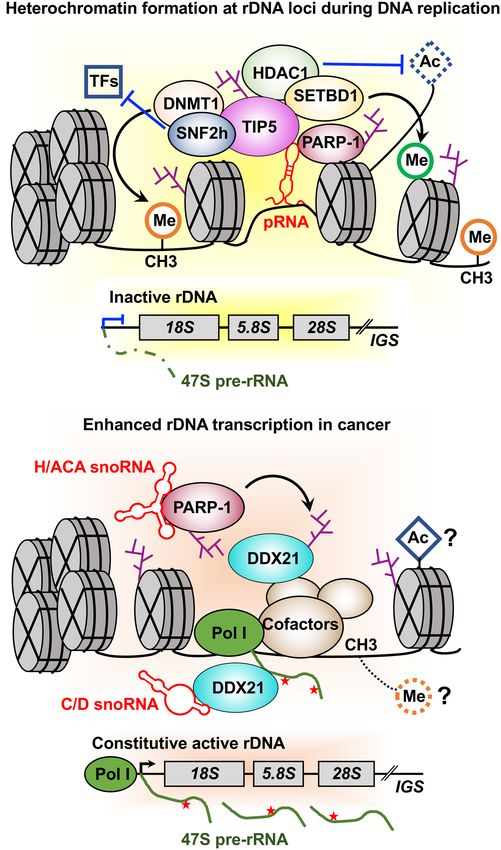
Downloaded from genesdev.cshlp.org on October 16, 2020 - Published by Cold Spring Harbor Laboratory Press

Kim et al.

                                                                    binding transcription factors (e.g., NFAT [Olabisi et al.
                                                                    2008], p53 [Kanai et al. 2007], STAT1α [Iwata et al.
                                                                    2016], and C/EBPβ [Luo et al. 2017]), the chromatin insu-
                                                                    lator protein CTCF (Yu et al. 2004; Farrar et al. 2010), the
                                                                    RNA polymerase II (Pol II) transcription elongation fac-
                                                                    tor NELF (Gibson et al. 2016), and the RNA helicase
                                                                    DDX21 (Kim et al. 2019). However, most of the afore-
                                                                    mentioned substrates are nuclear proteins modified by
                                                                    nuclear PARP polyenzymes. Further systemic identifica-
                                                                    tion and functional analysis of the substrates of cytoplasmic
Figure 1. Role of PARPs and ADP-ribosylation in RNA biology.        PARP monoenzymes and a demonstration of the biologi-
PARPs and ADPRylation play broad roles in RNA-related pro-
                                                                    cal importance of site-specific MARylation is needed.
cesses, from rDNA transcription and ribosome biogenesis to
mRNA processing, protein translation, and proteostasis. See the
text for descriptions.                                              Beyond protein substrates: ADPRylation of DNA
                                                                    and RNA
1 and 2), which catalyze the polymerization of ADPR units           Recent studies have also identified nucleic acids as sub-
in linear or branched chains through a process called PAR-          strates for ADPRylation. ADPRylation of DNA was first
ylation (Gibson and Kraus 2012; Vyas et al. 2014; Gupte et          identified as part of toxin-antitoxin systems in bacterial
al. 2017) and (2) PARP “monoenzymes” [i.e., mono(ADP-               pathogens (e.g., Mycobacterium tuberculosis). DarT is a
ribosyl) transferases or MARTs] (e.g., PARPs 7 and 16),             bacterial enzyme that ADPRylates thymidines on sin-
which catalyze the addition of a single ADPR unit on tar-           gle-stranded DNA in a sequence-specific manner, a mod-
get proteins through a process called MARylation (Gibson            ification that can be removed by another enzyme, DarG
and Kraus 2012; Vyas et al. 2014; Gupte et al. 2017).               (Jankevicius et al. 2016). In mammalian systems, nuclear
                                                                    PARPs 1 and 2 can PARylate and PARP-3 can MARylate
                                                                    phosphorylated DNA termini in vitro (Talhaoui et al.
Functional outcomes of site-specific ADPRylation
                                                                    2016; Munnur and Ahel 2017; Zarkovic et al. 2018), mod-
Historically, PARPs and ADPRylation have been studied               ifications that have been shown in some cases to be re-
in the context of DNA repair, with the primary focus on             moved by ADPR hydrolases (Munnur and Ahel 2017).
PARP-1 and PARylation in cancer (Morales et al. 2014;               Likewise, ADPRylation of RNA by multiple PARP family
Ray Chaudhuri and Nussenzweig 2017). New findings                   members and a highly divergent microbial PARP
on the diverse roles of PARPs in cellular processes beyond
DNA repair (e.g., regulation of chromatin structure and
gene expression, RNA processing, ribosome biogenesis,                A
protein translation) have been complemented by recent
advances that link ADPRylation to metabolism, inflam-
mation, immunity, stress responses, hormonal signaling,
and viral infections (Kim et al. 2005; Luo and Kraus
2012; Vyas and Chang 2014; Gupte et al. 2017). Although
the functions of PARylation are well studied, much less
is known about the functions of MARylation. Recent
studies have begun to reveal novel and interesting func-             B                            C
tions for cytoplasmic PARP monoenzymes, such as
PARP-7, PARP-12, PARP-14, and PARP-16, in molecular
and cellular functions ranging from RNA processing and
translation to stress granule formation and the unfolded
protein response (Leung et al. 2011; Di Paola et al. 2012;
Jwa and Chang 2012; Vyas et al. 2013, 2014; Roper et al.
2014; Ahmed et al. 2015; Bindesbøll et al. 2016; Iwata
et al. 2016).
   The growing understanding of the biological impor-
tance of ADPRylation has spurred interest in identifying            Figure 2. Overview of NAD+-dependent, PARP-mediated
the substrates of specific PARPs and the functional out-            ADPRylation. (A) PARPs use NAD+ as a substrate to catalyze re-
comes of the modification. The identification of specific           versible ADPRylation of substrate proteins. (B) Subcellular local-
                                                                    ization of PARP family members. (C ) NAD+ “feeds” PARP
sites of ADPRylation on a proteome-wide scale has
                                                                    catalytic activity. This enzymatic activity generates nicotin-
lagged behind other PTMs until recently (Daniels et al.             amide (NAM) as a product, which is used by NAMPT in the
2015). Although previous studies demonstrated effects               NAD+ salvage pathway to regenerate NAD+. Several hydrolases
of ADPRylation on target proteins, the sites were usually           that bind to specific structural features of MAR and PAR can hy-
not mapped. Some examples where the sites have been                 drolyze the ADPR, returning the target proteins to their unmod-
mapped and functionally interrogated include DNA-                   ified state.

2       GENES & DEVELOPMENT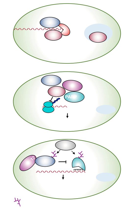
Downloaded from genesdev.cshlp.org on October 16, 2020 - Published by Cold Spring Harbor Laboratory Press

                                                                                                PARPs, ADPRylation, and RNA biology

homolog, TRPT1, as well as its erasure by cellular, viral,          rRNA modification (Boamah et al. 2012; Guetg et al.
and bacterial macrodomain ADPR hydrolases, has been                 2012). In addition, several studies have determined the
demonstrated in a variety of systems (Munir et al. 2018;            physiological functions of PARP-1 in the regulation of ri-
Munnur et al. 2019). Thus, ADPRylation of both DNA                  bosome biogenesis in response to DNA damage (Calkins
and RNA may represent a widespread and physiologically              et al. 2013; Bütepage et al. 2018). In this section, we
relevant modification mediated by both PARP monoen-                 describe in detail the growing awareness of the essential
zymes and polyenzymes.                                              role of PARP-1 in the transcriptional regulation of rDNA
                                                                    and ribosome biogenesis under normal conditions and in
                                                                    various human diseases.
Control of compartment-specific PARP functions
by NAD+ synthases
                                                                    Brief overview of ribosome biogenesis
The activity of the PARP enzymes is intimately tied to the
synthesis of NAD+, which is consumed during ADPRyla-                Ribosome biogenesis, which begins in the nucleolus, is
tion reactions and thus, must be regenerated. While nutri-          the process by which cells generate new ribosomes to
tional supplements such as Vitamin B3 act as dietary                synthesize cellular proteins (Baßler and Hurt 2019). Ribo-
precursors for NAD+ biosynthesis, intracellular salvage             some biogenesis is a tightly controlled, energy-demanding
pathways using nicotinamide (NAM) are the primary                   process, comprising multiple steps including (1) synthesis
source of NAD+ in the cell (Verdin 2015). Nicotinamide              of four different ribosomal RNA (rRNA) molecules (25S/
mononucleotide adenylyltransferases (NMNATs) cata-                  28S, 18S, 5.8S, and 5S) and 79 ribosomal proteins by all
lyze the final step in the NAD+ salvage pathway, combin-            three nuclear RNA polymerases (Pol I, II, and III) in eu-
ing nicotinamide mononucleotide (NMN) and ATP to                    karyotes, (2) rRNA processing and site-specific modifica-
make NAD+ (Ryu et al. 2015). Three different NMNATs,                tions including methylation and pseudouridylation by a
each with a distinct subcellular localization, control              small nucleolar ribonucleoprotein (snoRNP), and (3) as-
the subcellular levels of NAD+ in each compartment:                 sembly with rRNAs and ribosomal proteins to form preri-
NMNAT-1 is localized to the nucleus, NMNAT-2 is local-              bosomal particles. After synthesis and assembly steps
ized to the outer membrane of the golgi, and NMNAT-3 is             in the nucleolus and nucleus, the pre-40S and pre-60S ri-
localized to the mitochondria (Lau et al. 2009; Jayaram             bosomal subunits exit to the cytoplasm to form mature
et al. 2011; Ryu et al. 2015). The specific subcellular local-      ribosomal subunits (Fig. 3). The functional links among
izations of these three NAD+ synthases leads to compart-            PARP-1, ADPRylation, and ribosome biogenesis are ex-
mentalized production of NAD+ (Zhang et al. 2012;                   plored in more detail below.
Cambronne et al. 2016; Ryu et al. 2018), which has impor-
tant functional consequences in the cell. For example, the
                                                                    Role of PARP-1 in the formation of silent rDNA
control of nuclear and cytosolic levels of NAD+ by
                                                                    chromatin and transcriptional silencing
NMNAT-1 and NMNAT-2, respectively, regulate the
PARP-1-dependent gene expression programs that drive                A newly discovered aspect of PARP-1 function is its role in
adipocyte differentiation (Luo et al. 2017; Ryu et al.              modulating ribosome biogenesis in normal physiological
2018). The control of subcellular PARP functions by com-
partmentalized NAD+ synthesis is just beginning to be un-
derstood and requires further study.

Role of PARP-1 and ADPRylation in the regulation
of rDNA transcription and ribosome biogenesis

Ribosome biogenesis is a highly coordinated cellular pro-
cess that involves the synthesis, processing, and modifica-
tion of rRNAs, as well as the proper assembly of the rRNA
with ribosomal proteins (Baßler and Hurt 2019). Emerging
evidence highlights the role of ribosome biogenesis in var-
ious human cancers and its dysregulation promotes cellu-
lar growth and tumorigenesis (Ruggero and Pandolfi 2003;
Aspesi and Ellis 2019). Enrichment of PARP-1 in the nu-
cleolus, the site of ribosome biogenesis, was documented
from the late 1980s to the early 2000s (Fakan et al. 1988;
                                                                    Figure 3. The steps in ribosome biogenesis. rRNA molecules
Desnoyers et al. 1996; Scherl et al. 2002). Subsequent
                                                                    (28S, 18S, 5.8S, and 5S), small and large subunit ribosomal pro-
studies have revealed an important role for PARP-1 in reg-
                                                                    teins, and assembly or processing factors are synthesized by
ulating multiple steps of ribosome biogenesis. A growing            RNA polymerases (Pol I–III), and are subsequently modified and
body of evidence supports the important role of PARP-1              processed. rRNAs assemble with ribosomal proteins to form pre-
in RNA polymerase I (Pol I)-dependent transcriptional reg-          ribosome particles, which are exported to the cytoplasm to form
ulation, preribosomal rRNA (pre-rRNA) processing, and               mature ribosomal subunits and active ribosomes.

                                                                                                    GENES & DEVELOPMENT           3
Downloaded from genesdev.cshlp.org on October 16, 2020 - Published by Cold Spring Harbor Laboratory Press

Kim et al.

conditions through PARP-1-mediated ADPRylation                        with subsequent transcriptional silencing and heterochro-
(Guetg et al. 2012). Tandemly repeated ribosomal RNA                  matin formation. TIP5 is a key regulatory protein in the
genes (rDNA) exist in two distinct epigenetic states: a per-          regulation of silent rDNA chromatin and heterochro-
missive state allowing transcription and repressed state              matin formation. Importantly, pRNA-mediated TIP5-
inhibiting transcription (Li et al. 2005). NoRC, a nucleolar          PARP-1 interactions lead to PARP-1 automodification
chromatin remodeling complex comprising SNF2h and                     and subsequent TIP5 and/or histone ADPRylation. Joint-
TIP5, plays an important role in establishing the silent              ly, the studies described here indicate that PARP-1-medi-
state of rRNA genes by recruiting a DNA methyltrans-                  ated TIP5 and histone ADPRylation are essential for the
ferase and a histone deacetylase to the rDNA promoter                 formation of silent rDNA chromatin and transcriptional
(Santoro et al. 2002). Interestingly, PARP-1 has been im-             silencing by direct interaction with pRNA and the chro-
plicated in NoRC-mediated establishment of transcrip-                 matin-remodeling complex NoRC.
tionally inactive rDNA chromatin during cell division
(Guetg et al. 2012). PARP-1 interacts with NoRC-asso-
                                                                      Role of PARP-1 in Pol I-dependent transcription of rDNA
ciated RNA (pRNA). This interaction is required for re-
cruitment of TIP5, the large subunit of NoRC, to the                  The first evidence of a functional link between rRNA pro-
promoter of silent rDNA after the passage of the rep-                 cessing and PARP-1 was proposed by Boamah et al. (2012)
lication fork. NoRC recruits and interacts with the his-              using Drosophila as a model system. PARP-1 plays an im-
tone deacetylase HDAC1 and DNA methyltransferase                      portant role in the maintenance of nucleolar structure and
(DNMT), leading to epigenetic reprogramming, including                function via the regulation of precursor rRNA processing,
histone modification and DNA methylation (Fig. 4A).                   posttranscriptional modification, and preribosome assem-
   This epigenetic reprogramming leads to impaired bind-              bly (Boamah et al. 2012). PARP-1 and its catalytic activity
ing of various transcription factors to the rDNA promoter,            are required for (1) the maintenance of nucleolar integrity
                                                                      and (2) proper localization of nucleolar-specific proteins,
                                                                      such as fibrillarin, AJ1, nucleolin, and nucleophosmin in
             A                                                        proximity to precursor rRNA in the nucleoli of Droso-
                                                                      phila. Inhibition of PARP-1 enzymatic activity leads to
                                                                      nucleolar fragmentation and aberrant localization of nu-
                                                                      cleolar-specific proteins. PARP-1 deletion mutants exhib-
                                                                      it a delay in rRNA processing and an increase in the levels
                                                                      of rRNA intermediates, such as 47S and 36S rRNA tran-
                                                                      scripts, which represses ribosome biogenesis (Boamah
                                                                      et al. 2012). The role of PARP-1 in regulating ribosome
                                                                      biogenesis will be discussed below.
                                                                         Recent advances in chemical biology and protein engi-
                                                                      neering in the field have led to mass spectrometry-based
                                                                      identification of ADPRylation sites for PARP-1 protein
             B                                                        substrates. This has led to the identification of a set of
                                                                      nucleolar proteins, which are key regulators in ribosome
                                                                      biogenesis, including DDX21, fibrillarin, nucleolar phos-
                                                                      phoproteins numatrin/B23, and nucleolin/C23, as direct
                                                                      targets of PARP-1 enzymatic activity (Gagné et al. 2012;
                                                                      Chiou et al. 2013; Carter-O’Connell et al. 2014; Gibson
                                                                      et al. 2016; Kim et al. 2019). These PARP-1-ADPRylated
                                                                      proteins are known to be involved in rRNA transcription,
                                                                      pre-rRNA processing, and preribosome assembly, suggest-
                                                                      ing that PARP-1 and its enzymatic activity are required
                                                                      in nucleolar functions and, consequently, ribosome
                                                                      biogenesis.
                                                                         In addition to the regulation of rRNA processing, PARP-
                                                                      1 can localize to the nucleolus, and PARP-1 accumulation
Figure 4. Dual roles of PARP-1 in the regulation of rDNA tran-        in nucleoli is altered upon RNA polymerase I inhibition
scription. (A) pRNA-mediates PARP-1 and NoRC complex (TIP5            (Meder et al. 2005). Although this study suggests that
and SNF2h) interactions, leading to PARP-1 automodification           PARP-1 (and PARP-2) does not affect the transcription of
and subsequent TIP5 or histone ADPRylation. TIP5 recruits the         rDNA in murine fibroblasts, other studies have shown a
histone deacetylase HDAC1, DNA methyltransferase DNMT1,
                                                                      role for PARP-1 in the regulation of rDNA transcription
and histone lysine methyltransferase SETBD1 to the establish a
silent state on rRNA genes. (B) snoRNAs interact with PARP-1
                                                                      (Kurl and Jacob 1985; Guetg et al. 2012). Another study
to activate PARP-1 catalytic activity. Subsequently, snoRNA-ac-       also reported that the nucleolar localization of PARP-1
tivated PARP-1 PARylates DDX21 to promote DDX21 associa-              is dependent upon active RNA synthesis (Desnoyers
tion with the rDNA locus and retention in the nucleolus,              et al. 1996). Therefore, it is likely that PARP-1 nucleolar
promoting enhanced rDNA transcription in cancer.                      localization and its enzymatic activity are associated

4       GENES & DEVELOPMENT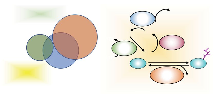
Downloaded from genesdev.cshlp.org on October 16, 2020 - Published by Cold Spring Harbor Laboratory Press

                                                                                                PARPs, ADPRylation, and RNA biology

with the Pol I-dependent transcription of rDNA in                  cellular proliferation and division of cancer cells com-
nucleoli. These studies indicate that PARP-1 plays an              pared with normal cells (Aspesi and Ellis 2019). PARP-1
important role in ribosome biogenesis, including rDNA              and its enzymatic activity are significantly up-regulated
transcription, processing, and ribosome assembly in the            in invasive cancer cells, as well as malignant tissues
nucleolus. Emerging evidence has also implicated                   (Ossovskaya et al. 2010; Domagala et al. 2011). Thus, it
PARP-1 in the regulation of ribosome biogenesis in vari-           is possible that the dual role of PARP-1 is based on the
ous human diseases, as discussed below.                            different amount of ribosomes present in cancer and nor-
                                                                   mal cells to meet their need for protein synthesis and
                                                                   proliferation.
Role of PARP-1 in the regulation of ribosomal biogenesis
                                                                      Interestingly, a recent study reported dispersed and less
in pathological conditions
                                                                   intense nucleolar PARP-1 staining in Alzheimer’s disease
PARP inhibitors (PARPi), such as olaparib, rucaparib, nir-         (AD) compared with the distinct nucleolar localization in
aparib, and talazoparib, are clinically important and have         hippocampal pyramidal neurons in controls (Zeng et al.
been approved by the FDA as monotherapies for treat-               2016). This study proposes that PARP-1 mislocalization
ment of recurrent, high-grade serous ovarian cancers               from the nucleolus in AD (1) leads to hypermethylation
with BRCA1/2 mutations (Bitler et al. 2017; McCann                 of rDNA by DNA methyltransferase 1 (DNMT1), (2) sub-
2019). Olaparib has also been approved for the treatment           sequently reduces rDNA transcription and impairs ribo-
of BRCA-mutated HER2-negative metastatic breast can-               somal biogenesis, and (3) results in disruption of long-
cers (Robson et al. 2017), while niraparib has been shown          term memory formation (Fig. 5A,B). PARP-1-mediated
to be efficacious in patients lacking BRCA mutations or            DNMT1 ADPRylation inhibits the activity of DNMT1,
HR deficiency (Mirza et al. 2016). These PARPi are cur-            subsequently preventing rDNA methylation and up-regu-
rently being evaluated for their therapeutic potential in          lating rRNA expression. While these observations need to
several other cancers. PARPi, acting through nuclear               be confirmed, they suggest an interesting link between
PARPs, are thought to control cancer cell growth primar-           PARP-1 mislocalization and brain pathologies.
ily through inducing synthetic lethality in cancers that
are deficient in homologous recombination (HR)-mediat-
ed DNA repair (e.g., in BRCA1/2 mutant cells) (Bryant
et al. 2005). In the absence of functional BRCA1 or                            A
BRCA2 proteins, PARPi lead to the persistence of DNA le-
sions, resulting in chromosomal instability, subsequent
cell cycle arrest, and apoptosis (Bryant et al. 2005; Farmer
et al. 2005).
   Growing evidence has shown that PARPi have thera-
peutic efficacy in BRCA1/2 wild-type cancers lacking oth-
er known HR or DNA repair defects. Interestingly, a
recent study from our laboratory highlights the patho-
logical significance of PARP-1-mediated site-specific
ADPRylation events that are independent of PARP-1’s
role in DNA repair (Kim et al. 2019). Mechanistically,                         B
this study found that snoRNAs act as critical players
in the activation of PARP-1 enzymatic activity in the nu-
cleolus, which leads to DDX21 ADPRylation. DDX21
ADPRylation results in enhanced rDNA transcription,
as well as breast cancer cell growth (Fig. 4B). Treatment
with PARPi or mutation of the ADPRylation sites in
DDX21 reduces DDX21 nucleolar localization, rDNA
transcription, ribosome biogenesis, and cell growth (Kim
et al. 2019). Thus, this study has uncovered an alternate
molecular pathway for targeting breast cancer with PARPi
irrespective of BRCA1/2 status by attenuating cancer-en-
hanced ribosome biogenesis. As such, this study strength-
ens the rationale for advancing the use of PARPi in clinical       Figure 5. Role for PARP-1 in the regulation of rDNA transcrip-
trials for the treatment of a broader array of cancers, in-        tion in neurons. (A) PARP-1-mediated DNMT1 ADPRylation pre-
cluding those with wild-type BRCA1/2.                              vents rDNA methylation by DNMT1, resulting in the production
                                                                   of rRNAs and subsequent ribosome biogenesis in normal neu-
   Results from Guetg et al. (2012) using HEK-293T cells
                                                                   rons. (B) A substantial reduction in the nucleolar localization of
suggest an alternate mechanism for PARP-1’s role in                PARP-1 leads to hypermethylation of rDNA by DNMT1, result-
rDNA transcription, where PARP-1 and its enzymatic ac-             ing in a reduction of rDNA transcription and ribosome biogene-
tivity promote the formation of silent rDNA chromatin              sis. Impaired ribosome biogenesis causes a disruption of long-
and transcriptional silencing of the rDNA locus. Increased         term memory formation and the development of Alzheimer’s
numbers of ribosomes are required for the uncontrolled             disease.

                                                                                                    GENES & DEVELOPMENT            5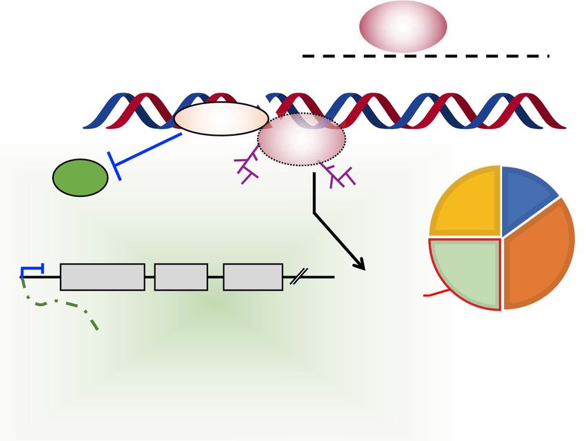
Downloaded from genesdev.cshlp.org on October 16, 2020 - Published by Cold Spring Harbor Laboratory Press

Kim et al.

   Collectively, these studies indicate that PARP-1 and its            Another possible mechanism by which PARP-1 could
enzymatic activity play an important role in the epigenet-          facilitate repression of rRNA synthesis is through direct
ic regulation of rDNA in various aspects of biology. Based          or indirect interaction with target proteins with roles in
on the findings described above, PARP-1 regulates multi-            rDNA transcription. For example, TARG1 localizes to
ple areas of nucleolar function, including (1) establish-           transcriptionally active nucleoli through direct inter-
ment of transcriptionally inactive rDNA chromatin,                  action with rRNA, as well as rRNA processing and ribo-
(2) the maintenance of nucleolar integrity and structure,           somal assembly factors, independent of ADPRylation
and (3) the regulation of Pol I dependent transcription of          (Bütepage et al. 2018). Interestingly, TARG1’s nucleolar
the rDNA.                                                           localization is abrogated by inhibition of rRNA transcrip-
                                                                    tion, indicating that TARG1 may function as a key regula-
                                                                    tor of rRNA synthesis. In addition, TARG1 relocalizes to
Role of PARP-1 in the regulation of ribosomal biogenesis            the nucleoplasm upon DNA damage-induced PARP-1/
during DNA damage                                                   PARP-2-dependent PARylation. TARG1 is mainly local-
                                                                    ized to the nucleolus in the absence of PAR, while it accu-
Recent studies have shown that PARP-1 activation can                mulates in the nucleoplasm in response to DNA damage-
regulate rDNA transcription in response to DNA damage               dependent PAR formation, indicating that TARG1 locali-
(Calkins et al. 2013). DNA replication and rDNA tran-               zation is strongly regulated by PARylation. These findings
scription are inhibited following DNA damage mediated               suggest that DNA damage-induced PARylation might
by UV light, γ radiation (IR), and cross-linking by cisplatin,      serve to sequester TARG1 to the nucleoplasm, resulting
resulting in the accumulation of cells in S phase. Inhibi-          in the loss of nucleolar function of TARG1 in rRNA syn-
tion of the DNA repair proteins, DNA-dependent protein              thesis (Bütepage et al. 2018).
kinase (DNA-PK), or PARP-1 prevents cisplatin-induced
inhibition of rRNA synthesis (Calkins et al. 2013). This
study showed that DNA-PK acts upstream of PARP-1 to                 Role of PARPs and ADPRylation in mRNA processing
recruit PARP-1 to chromatin at sites of DNA damage.
Subsequent activation of PARP-1 leads to the inhibition             Regulation of gene expression extends beyond transcrip-
of rRNA synthesis after DNA damage. Thus, DNA-PK-                   tion to include events that occur cotranscriptionally
dependent PARP-1 activation may result in the mainte-               and posttranscriptionally. From the start of nascent
nance of inherited silencing of rDNA genes and repression           mRNA transcription by RNA polymerase II until degra-
of rRNA synthesis (Fig. 6). However, PARP-1 translocates            dation in the cytoplasm, mRNAs are associated with
from the nucleolus to the nucleoplasm in the first 2 h after        RNA-binding proteins (RBPs). Many PARPs play key
DNA damage and inhibition of rRNA synthesis has been                roles in these processes through various mechanisms, in-
observed between 12 and 24 h after DNA damage (Calkins              cluding (1) binding to RNAs, (2) interacting with, ADPRy-
et al. 2013). Thus, it is possible that nucleolar exit of           lating, and regulating RBPs and RNA processing factors,
PARP-1 after DNA damage, rather than direct silencing               and (3) creating PAR, which can serve as a scaffold for
of rDNA by PARP-1 in nucleolus, affects the inhibition              noncovalent interactions with various RBPs and RNA
of rRNA synthesis in this process. Further studies of nu-           processing factors. In this section, we explore the rela-
cleolar-nucleoplasmic shuttling of PARP-1 after DNA                 tionship among RNA processing events, PARP proteins,
damage are required to elucidate the role of PARP-1 in re-          and ADPRylation.
pression of rRNA synthesis following DNA damage.

                                                                    Brief overview of mRNA processing
                                                                    To reveal its coding potential, a pre-mRNA must undergo
                                                                    processing, including 5′ capping, splicing, and 3′ polyade-
                                                                    nylation, to form mature mRNA before it can be exported
                                                                    to the cytoplasm for translation and degradation (Fig. 7).
                                                                    Nearly every step of this process has been shown to be reg-
                                                                    ulated by PARP proteins. RNA processing events are
                                                                    tightly coordinated by RBPs (Castello et al. 2012), as the
                                                                    ribonucleoprotein (RNP) complex associated with RNA
                                                                    is dynamically remodeled sequentially by loss and gain
                                                                    of RBPs. Thus, RBPs serve a critical role in RNA metabo-
                                                                    lism. The most abundant cellular proteins contributing to
                                                                    RNP complex formation are members of the hnRNP and
                                                                    S/R protein families (Huang and Steitz 2005). The func-
Figure 6. DNA damage-induced formation of silent rDNA chro-
matin. DNA damage leads to the accumulation of cells in S phase,    tions of RBPs are temporally and spatially regulated by
with DNA replication forks stalled at sites of DNA damage.          phosphorylation (Matter et al. 2002), ubiquitylation
DNA-PK acts upstream of PARP-1 to recruit it to chromatin. Sub-     (Bhandari et al. 2011), and ADPRylation (see below).
sequent PARP-1 activation plays an essential role in the forma-     RBPs have specificity for unique RNA targets, which reg-
tion of silent rDNA chromatin at the time of replication.           ulates their splicing, stability, and localization.

6       GENES & DEVELOPMENT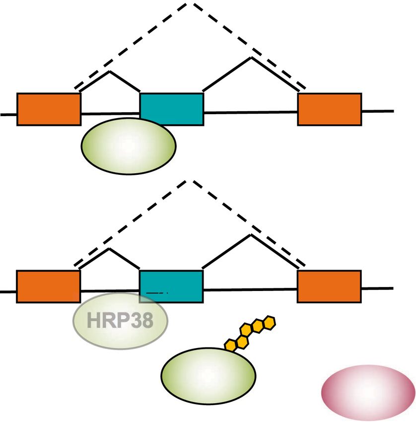
Downloaded from genesdev.cshlp.org on October 16, 2020 - Published by Cold Spring Harbor Laboratory Press

                                                                                                  PARPs, ADPRylation, and RNA biology

                                                                        tein (Krishnakumar and Kraus 2010), and recently the ef-
                                                                        fects of chromatin have been explored in relation to the
                                                                        regulation of splicing. Nucleosome occupancy regulates
                                                                        RNA polymerase (RNAP) kinetics, aiding in the recogni-
                                                                        tion of weak splice sites (Schwartz and Ast 2010). Thus,
                                                                        factors impacting chromatin dynamics may regulate
                                                                        mRNA processing. A survey of the genomic distribution
                                                                        of dPARP-1 using nucleosome-ChIP-sequencing (nuc-
                                                                        ChIP-seq) in Drosophila S2 cells revealed that dPARP-1
                                                                        preferentially binds to the +1 and +2 nucleosomes of ac-
                                                                        tive promoters and at internal exon/intron boundaries
                                                                        (Matveeva et al. 2016). These dPARP-1-bound nucleo-
                                                                        somes have a high GC content; such nucleosomes are
                                                                        thought to mark weak splice sites and promote cotran-
                                                                        scriptional splicing (Amit et al. 2012). Both depletion of
                                                                        dPARP-1 and inhibition of PARylation in S2 cells cause
                                                                        genome-wide changes in alternative splicing events
                                                                        (ASEs) without differential expression changes at consti-
                                                                        tutive exons. These changes in ASEs, however, occur at
                                                                        distinct loci in response to dPARP-1 depletion and
                                                                        dPARP-1 inhibition (Matveeva et al. 2016), highlighting
                                                                        the fact that the physical presence of PARP-1 is important
                                                                        for some molecular pathways, while its catalytic activity
                                                                        is necessary for others.
                                                                           Changes in alternative splicing decisions correlate
                                                                        with PARP-1 nucleosome occupancy (Matveeva et al.
Figure 7. Overview of nuclear RNA processing events facilitat-          2016), suggesting that the PARP-1-mediated chromatin
ing transformation from pre-mRNA to mRNA. Some RNA pro-                 state affects RNAP II kinetics and, thus, cotranscriptional
cessing events occur cotranscriptionally, adding another level of       splicing (Fig. 8A). Deeper investigation of PARP-1 as a
regulation to the RNA processing. mRNAs are modified at their
                                                                        chromatin component revealed that PARP-1-mediated
5′ ends by the addition of a methylguanosine cap by RNA gua-
nine-7 methyltransferase (RNMT) and RNA guanylyltransferase
                                                                        chromatin structure influences transcriptional elongation
and 5′ -phosphatase (RNGTT). RNMT recognizes and physically             by RNAP II (Matveeva et al. 2019). Comparisons of PARP-
interacts with the CTD of RNA Pol II in close proximity to the          1 nuc-ChIP-seq data with PRO-seq data, which delineates
5′ end of the nascent RNA. RNMT (1) recruits RNGTT to add               genomic sites of active transcription, showed that PARP-1
the guanosine to the 5′ end of the mRNA and (2) catalyzes the ad-       is enriched ∼25 bp downstream from RNAP II (Matveeva
dition of a methyl group on the N7 position of the added guano-         et al. 2019). Moreover, in locus-specific examples, PARP-1
sine. As RNA Pol II transcribes the length of the gene body,            depletion led to a corresponding decrease in RNAP II elon-
exon-intron boundaries are transcribed into the nascent pre-            gation, supporting previous results connecting PARP-1
mRNA. The boundaries are identified via specific sequences              and RNAP II elongation (Gibson et al. 2016). These results
that demarcate the exon–intron junctions. As the splice site mo-
                                                                        were supported at the genomic level by results from
tifs are transcribed, the spliceosome proteins snRNP U2 and U1
bind to their respective sequences, recruiting other spliceosome
                                                                        3′ NT-seq and NET-seq, which were used to measure
complex proteins (i.e., U5, U6, and U4). The spliceosome matures        changes in transcript length following PARP-1 depletion
as U4 and U1 exit, followed by catalytic activation of the mature       (Matveeva et al. 2019). Taken together, these results indi-
spliceosome. In a two-step process, the spliceosome forms a lariat      cate PARP-1-bound nucleosomes decrease RNAP II elon-
structure with the exon and intron splice spites, cleaves the lariat,   gation, facilitating increased cotranscriptional splicing.
and ligates the exons together. Upon cleavage, the intron lariat
spliceosome complex is released from the nascent spliced pre-
mRNA. After RNA Pol II progresses into the 3′ UTR, the 3′ end is        Interaction of mRNA processing factors with ADPR
recognized by cleavage and polyadenylation specificity factor           In addition to the role of PARP-1-chromatin interactions,
(CPSF) and cleavage stimulation factor (CSTF). These enzymes
                                                                        PARP family members have long been known to interact
catalyze the cleavage of the pre-mRNA, leaving behind an exposed
adenine, which is extended by polyadenylate polymerase (PAP).
                                                                        and regulate RNA binding and processing proteins
                                                                        through their catalytic activity. Kostka and Schweiger
                                                                        (1982) observed that the radioisotope tracer from [32P]-
                                                                        labeled NAD+ incubated with rat liver nuclei precipitated
PARP-1–chromatin complexes influence
                                                                        in particles carrying heterogeneous nuclear RNA
cotranscriptional mRNA processing
                                                                        (hnRNA), of which the majority is pre-mRNA. The radio-
The nucleic acid-binding function of PARP-1 in its                      active signal was identified in ADPR moieties attached to
N-terminal zinc fingers has been implicated in regulation               RNPs with molecular weights of 36, 39, and 42 kDal. At
of the cotranscriptional aspect of mRNA processing.                     the time, the biological function of ADPRylation was un-
PARP-1 is a well-established chromatin architectural pro-               known. Since then, however, the relationships among

                                                                                                      GENES & DEVELOPMENT          7
Downloaded from genesdev.cshlp.org on October 16, 2020 - Published by Cold Spring Harbor Laboratory Press

Kim et al.

             A                                                        cur in the absence of covalent modification of the
                                                                      proteins.
                                                                        In addition to hnRNP members, serine-arginine-rich (S/
                                                                      R) family members interact with PAR chains as well. S/R
                                                                      factors function in both basal splicing and in the context
                                                                      of sequence-specific alternative splicing (Graveley 2000).
                                                                      Structurally S/R proteins are characterized by the
                                                                      presence of one to two RNA recognition motifs (RRMs)
             B                                                        typically in their N-terminal region and an SR-rich
                                                                      domain (SR domain) in their C-terminal region. Malanga
                                                                      et al. (2008) observed that ASF/SF2, also known as serine-
                                                                      and arginine-rich splicing factor 1 (SRSF1), binds PAR
                                                                      via its RRM and RS domain. S/R proteins are phosphory-
                                                                      lated by SR protein-specific kinases, regulating S/R pro-
                                                                      tein function and localization (Huang and Steitz 2005).
                                                                      When SRSF1 was incubated with [32P]-labeled PAR,
                                                                      SRSF1-PAR high-affinity, salt-resistant complexes were
                                                                      observed (Malanga et al. 2008). The inhibition of SRSF1
                                                                      phosphorylation corresponded to the presence of PAR in
             C                                                        nuclear HeLa extracts. SRSF1 phosphorylation promotes
                                                                      splicing; therefore, the inhibitory action that PAR-bound
                                                                      SRSF1 has on its phosphorylation state could regulate al-
                                                                      ternative splicing or directly impact its RNA binding
                                                                      capacity. The function of PAR as a complex-forming scaf-
                                                                      fold or binding partner likely drives its ability to modulate
                                                                      the behavior of hnRNPs, S/R proteins, and other splicing
Figure 8. Examples of RNA processing events modulated by              factors.
PARP activity. (A) PARP-1 binding to chromatin is enriched at
nucleosomes +1/+2 within active promoters and at intron/exon
boundaries. PARP-1 bound nucleosomes decrease RNA Pol II              Regulation of RNA-binding protein function
processivity, resulting in increased cotranscriptional splicing.      by ADPRylation
(B) RNA-binding protein, HRP38, typically functions to inhibit
                                                                      The interaction between mRNA processing proteins and
splicing events. PARP-1 binds to and ADPRylates HRP38 in its
RNA-binding domain, thereby attenuating HRP38’s ability to
                                                                      ADPR is not solely limited to binding of PAR, but in-
bind RNA. The outcome is increased splicing of target tran-           cludes direct ADPRylation of RBPs. PARylation of
scripts. (C) Both PARP-1 and PARP-2 bind to RNA, resulting in         hnRNPs is conserved in Drosophila, with three hnRNPs
the stimulation of their catalytic activities. Other cytosolic        (Hrp36, Hrp38, Hrp40) shown to be PARylated in vivo (Ji
PARP family members (e.g., PARP-7, PARP-10, PARP-11,                  and Tulin 2009). hnRNPs bind to splice silencing sites
PARP-12, PARP-13, PARP-14, PARP-15) are known to be or are            and are widely accepted as canonical splicing repressors
predicted to bind RNA. Furthermore, PARP-10, PARP-11,                 (Geuens et al. 2016). Immunofluorescent staining showed
PARP-15, and TRPT1 were recently found to ADPRylate single-           that PARylation of Hrp38 inhibited its localization to
stranded RNAs at phosphorylated ends.                                 sites of active transcription. Furthermore, the RNA-bind-
                                                                      ing ability of Hrp38 was negatively impacted by the pres-
PARPs, ADPRylation, and the regulation of mRNA pro-                   ence of PAR (Ji and Tulin 2009), resulting in Hrp38 targets
cessing have been met with growing appreciation.                      having increased production of the spliced isoform in a
   Over a decade after the identification of ADPRylated               PARP-dependent manner. These results highlight the
RNPs, another group (Prasad et al. 1994) confirmed the in-            ability of hnRNP ADPRylation to modulate hnRNP activ-
teraction using similar methods to more specifically iden-            ity (Fig. 8B).
tify heterogeneous ribonucleoprotein particles (hnRNPs)                  RBP interactions with PARP family members or ADPR,
A1 and A2/B1 as major targets of ADPRylation in HeLa                  as well as direct ADPRylation of RBPs, extends beyond
cells. Proteomic approaches have further identified RBPs              the splicing factors and spliceosome components; RBPs
as ADPRylated substrates (Isabelle et al. 2010; Jungmichel            that function in mRNA-processing events besides splicing
et al. 2013; Zhang et al. 2013). Coimmunoprecipitation                are also regulated by PARPs and ADPRylation. Proteomic
with PARP-1 and PARP-2 from human cells followed by                   analysis identified PARP-1 within the 3′ end mRNA pro-
mass spectrometry uncovered a host of hnRNPs (i.e.,                   cessing complex (Shi et al. 2009). 3′ end mRNA processing
A1, A2/B1, C1/C2, G, H, K, E1, A3, L, M, U, and G) and                involves endonucleolytic cleavage of pre-mRNA by
other RNA processing proteins that interact with PARP-                CPSF73, followed by synthesis of a poly(A) tail onto the
1 and PARP-2. The use of PAR-binding domains to                       5′ cleaved products by poly(A) polymerase (PAP) (Fig. 7;
directly pull-down ADPR interacting proteins reinforced               Danckwardt et al. 2008). Key aspects of 3′ processing are
the fact that a large number of hnRNPs and RBPs are in                associated with mRNA stability, export, and translation
complex with ADPR in mammalian cells, which may oc-                   efficiency (Danckwardt et al. 2008). NAD+-dependent

8       GENES & DEVELOPMENT
Downloaded from genesdev.cshlp.org on October 16, 2020 - Published by Cold Spring Harbor Laboratory Press

                                                                                               PARPs, ADPRylation, and RNA biology

PARP-1 catalytic activity in an mRNA polyadenylation               says were mRNAs, other types of RNAs bound by PARP-1
assay significantly decreased poly(A) tail length (Di              have been identified, including noncoding RNAs and
Giammartino et al. 2013). PAP, the enzyme that catalyzes           small nuclear RNAs (Melikishvili et al. 2017; Kim et al.
the creation of the poly(A) tail, directly interacts with          2019), indicating that PARP-1 functions in the regulation
PARP-1 and is ADPRylated in vitro. Activation of                   of other RNA types as well. In contrast to intron-bound
PARP-1 catalytic activity by heat shock decreased both             mRNAs, target mRNAs that were bound uniformly over
PAP binding to mRNA and mRNA polyadenylation levels                the length of the transcript by PARP-1 exhibited a prefer-
in 293T cells. Furthermore, heat shock caused dissocia-            ence for exon boundaries. This favored binding pattern
tion of PAP from the 3′ end of actively transcribing non-          was enriched within a subset of transcripts that are alter-
heat-shock-dependent genes (Di Giammartino et al.                  natively spliced in a PARP-1-dependent manner (Meli-
2013). These results indicate that, in contrast to prior           kishvili et al. 2017).
studies highlighting the binding of PAR by RBPs, direct               Interestingly, the binding of mRNAs stimulates PARP-
PARylation of core processing factors can modulate their           1 catalytic activity (Melikishvili et al. 2017), as shown pre-
activity and, thus, influence pre-mRNA processing.                 viously for PARP-2, which binds rRNA through its SAF/
   Hu antigen R (HuR) is another example of an ADPRy-              SAP domain (Léger et al. 2014). PARP-1 catalytic activity
lated RBP. In lipopolysaccharide (LPS)-treated murine              has also been shown to be stimulated in vitro and in cells
primary peritoneal macrophages, the stability of LPS-re-           by the binding of a selected set of snoRNAs (Kim et al.
sponsive pre-mRNAs decreases upon PARP-1 inhibition                2019). Thus, interactions between nucleic acid-binding
or depletion (Ke et al. 2017). These pre-mRNAs contain re-         PARPs (e.g., PARPs 1 and 2) and their target RNAs can ac-
peats of AUUUA motifs. HuR binds these adenylate–uri-              tivate PARP catalytic activity, promoting additional lev-
dylate-rich elements (AU-rich elements; AREs), which               els of regulation.
are commonly enriched within 3′ UTRs and function as                  In the cytoplasm, further layers of mRNA processing
major mRNA destabilization elements (Zubiaga et al.                modulate mRNA stability and decay. Cytoplasmic
1995). In response to LPS, HuR interacts with PARP-1, re-          PARP family members (i.e., PARP-7, PARP-10, PARP-
sulting in the PARylation of HuR, which directs its                12, PARP-13, and PARP-14) have been predicted to bind
proper subcellular localization and enhances its binding           RNA through RNA-binding CCCH-Zn fingers or RRMs
to LPS-responsive mRNA targets (Ke et al. 2017). PARP-             (Bock et al. 2015). With the exception of PARP-10, these
1-mediated PARylation of HuR illustrates another aspect            predicted RNA-binding PARPs also possess PAR-binding
of RNA metabolism that is PARP regulated, namely                   domains (i.e., WWE domains or macrodomains). PARP-
mRNA stability. These studies demonstrate that PARyla-             13 has been shown to bind to viral RNAs, targeting
tion of RBPs regulates the molecular functions of these            them for decay (Guo et al. 2004; Zhu et al. 2011; Ficarelli
proteins with regard to the formation of RNP complexes             et al. 2019; Meagher et al. 2019). PARP-13 also binds to
and facilitation of pre-mRNA processing.                           cellular mRNAs as well (Todorova et al. 2014; Schwerk
   As illustrated here, PARP family members have been              et al. 2019). PARP-13 recruits the exosome to mRNAs
shown to regulate mRNA processing by (1) catalytic activ-          through protein–protein interactions with exosome
ity directed toward RBPs, (2) direct protein–protein inter-        component hRrp46p, targeting bound mRNAs for degra-
action with RBPs, and (3) changing the chromatin                   dation (Guo et al. 2007). PARP-13 knockout results in
landscape to favor cotranscriptional splicing. The roles           major dysregulation of the transcriptome (Todorova
of interactions between PARP-1 and chromatin or regula-            et al. 2014). PARP-14, acting in the cytoplasm, also regu-
tory proteins in these processes are well established; the         lates mRNA stability. PARP-14 binds the RBP tristetrap-
role of direct interactions between PARP-1 and RNA is              rolin to promote the selective posttranscriptional control
less well understood.                                              of macrophage tissue factor expression by binding the
                                                                   ARE in the 3′ UTR of tissue factor mRNA, promoting
                                                                   its degradation (Iqbal et al. 2014). Tissue factor is required
Function of PARP proteins binding directly to RNA
                                                                   for the control of blood coagulation, and PARP-14-defi-
Recently, after speculation about the possibility, PARPs           cient mice exhibit thrombogenicity or an increased ten-
have been shown to bind directly to RNA. By using photo-           dency of blood clotting (Iqbal et al. 2014). These
activatable-ribonucleoside-enhanced cross-linking and              examples highlight the role of RNA binding by PARP
immunoprecipitation (PAR-CLIP) in S2 Drosophila cells,             proteins, including cytosolic PARP monoenzymes,
dPARP-1 was identified as an RNA-binding protein (Mat-             which allows for another layer of regulation in mRNA
veeva et al. 2016). Furthermore, nucleosome-bound                  processing and metabolism. With these studies exploring
dPARP-1 was able to bind RNA concomitantly with splic-             RNA-mediated PARP activation, the field had yet to ex-
ing factors, forming a bridge that facilitates cotranscrip-        amine whether this catalytic activity could be directed
tional splicing (Matveeva et al. 2016). Using the same             toward RNA as a substrate.
assay in HeLa cells, a greater depth of sequencing allowed
for higher resolution of the breadth and variety of mRNAs
                                                                   ADPRylation of RNAs: a potential role in RNA
bound by PARP-1 (Melikishvili et al. 2017). PARP-1 binds
                                                                   processing?
target transcripts most frequently within the intron, indi-
cating that PARP-1 preferentially binds to pre-mRNAs.              For many years, proteins were assumed to be the only sub-
While the majority of RNAs bound by PARP-1 in these as-            strates for PARP enzymes. However, with recent evidence

                                                                                                  GENES & DEVELOPMENT           9
Downloaded from genesdev.cshlp.org on October 16, 2020 - Published by Cold Spring Harbor Laboratory Press

Kim et al.

that DNA can be ADPRylated by the toxin Pierisin (Taka-             ADPRylation and a translocation domain (B) controlling
mura-Enya et al. 2001), the question of whether RNA can             entry of the toxin into the cell. During infections, the B
be ADPRylated was revisited. Indeed, PARP-like proteins             domain is cleaved by proteases, such as furin, activating
in bacteria and fungi can ADPRylate RNA (Munir et al.               the catalytic A domain and releasing it into the cytosol
2018). In biochemical assays with purified components,              (Fig. 9; Simon et al. 2014). The ADPR transferase activity
PARP-10, PARP-11, PARP-15, and PARP-TRPT1 ADPRy-                    of the A domain can modify a variety of host proteins, in-
late the phosphorylated ends of RNA in a reversible man-            cluding the translation elongation factor EF2 (by diphthe-
ner (Munnur et al. 2019). Using [32P]-labeled NAD+ as an            ria toxin and exotoxin A), G-protein subunits (by cholera
ADPR donor, PARP-10 robustly ADPRylates 5′ and 3′                   toxin and pertussis toxin), and actin cytoskeleton by the
phosphorylated single-stranded RNA (ssRNA). Unexpect-               C2 or C3-like exoenzymes (Deng and Barbieri 2008).
edly, the remaining proposed RNA-binding PARPs, with                   A variety of ADPR acceptor residues in toxin substrates
the exception of PARP-7, were tested in the same manner             have been identified, including Arg, Thr, Asn, Gln, Cys,
and were unable to ADPRylate ssRNA, with or without                 and His (Oppenheimer and Bodley 1981; Manning et al.
phosphorylation (Fig. 8C). ADPRylation of RNA is a novel            1984; West et al. 1985; Vandekerckhove et al. 1987;
biochemical function of PARP family members, but these              Lang et al. 2010, 2017; for detailed review, see Cohen
results need to be confirmed in vivo. In addition, further          and Chang 2018). Of note, diphtheria toxin, a highly po-
studies of RNA ADPRylation are necessary to understand              tent toxin in humans, MARylates residue 715 in the eu-
its biological role and impact. Finally, the mechanism              karyotic elongation factor-2 (eEF2) (Foley et al. 1995),
by which PARPs that lack RNA-binding domains direct                 which is a variant histidine amino acid, termed as diph-
their catalytic activity toward RNA requires further                thamide (Van Ness et al. 1980). Thus far, diphthamide is
investigation.                                                      found only in archaeal or eukaryotic EF2 proteins, with
                                                                    a single residue per molecule of EF2. Diphthamide is

Role of PARPs and ADPRylation in protein synthesis,
protein degradation, and proteostasis

Altered protein homeostasis has been implicated in
numerous pathological disorders, such as cancer and Alz-
heimer’s disease. The maintenance of a homeostatic pro-
teome (i.e., proteostasis) is intimately tied to RNA biology
and is regulated at several stages, including ribosome bio-
genesis, ribosome function, mRNA translation, protein
stability, protein folding, and clearance of misfolded pro-
teins. Various PARPs and ADPRylation have been shown
to regulate each of these processes to support protein ho-
meostasis. In this section, we review current knowledge
about the PARP-mediated regulation of protein synthesis
and proteostasis under normal and pathological states.

Role of ADPRylation in the regulation of mRNA
translation by bacterial toxins
As elaborated below, one of the initial indications of the
role of PARPs as regulators of protein synthesis was
from early studies demonstrating how toxins inhibit
host mRNA translation. In addition, PARPs, especially
cytosolic PARP enzymes, control mRNA translation in
cells when they are subjected to stresses, such as viral in-        Figure 9. Diphtheria toxin-mediated regulation of mRNA trans-
fections. In the next two sections below, we describe the           lation. Diphtheria toxin is internalized in cells by binding to cell
role of ADPRylation and PARPs in the regulation of                  surface receptors and is subsequently activated by cleavage of the
mRNA translation during bacterial and viral infections.             regulatory B domain from the catalytic A domain. The activated
   Protein toxins are secreted by bacteria to disrupt host          A domain MARylates the diphthamide residue in EF2 protein us-
cellular pathways through covalent modifications of                 ing NAD+ as a substrate. Histidine is modified to diphthamide by
                                                                    a multi-step biosynthetic pathway. ADPRylation of EF2 attenu-
host proteins, which supports bacterial virulence. Over
                                                                    ates protein synthesis by inhibiting (1) EF2 incorporation into ri-
the past several decades, a number of bacterial toxins,
                                                                    bosomes, (2) transfer of aminoacyl tRNA from the A site to P site,
such as those produced by Corynebacterium diphtheriae               and (3) reverse translocation and −1 frameshifting of ribosomes
(i.e., diphtheria toxin) and Vibrio cholerae (i.e., cholera         during translational elongation. Loss of the members of the diph-
toxin), have been shown to MARylate host proteins (Si-              thamide biosynthesis pathway results in a loss of sensitivity to
mon et al. 2014). These toxins are A–B-type oligomeric              diphtheria toxin and enhanced sensitivity to TNFα-mediated
molecules comprising a catalytic domain (A) mediating               apoptosis.

10       GENES & DEVELOPMENT
Downloaded from genesdev.cshlp.org on October 16, 2020 - Published by Cold Spring Harbor Laboratory Press
PARPs, ADPRylation, and RNA biology
synthesized in a multi-step biosynthetic pathway. Knock- A
out of genes encoding enzymes in this pathway in cancer
cells results in a loss of EF2 ADPRylation and decreased
sensitivity to diphtheria toxin, while enhancing sensitiv-
ity to TNFα-mediated apoptosis (Stahl et al. 2015).
How does EF2 ADPRylation affect mRNA translation?
EF2 is a GTPase protein essential for mRNA translation,
which catalyzes the transfer of peptidyl-tRNA from the B
ribosome A-site to P-site, moving the ribosome from 5′
to 3′ end of the mRNA template (Jørgensen et al. 2006).
ADPRylation of EF2 has several effects on the function
of ribosomes. Briefly, ADPRylation of EF2 results in (1)
reduced ribosome association in pretranslocation state
(Nygård and Nilsson 1985), (2) blocked translocation of
aminoacyl tRNA from the A-site to P-site of ribosomes
C
(Davydova and Ovchinnikov 1990), (3) errors in −1 frame-
shifting during translation elongation (Liu et al. 2012),
and (4) blocked reverse translocation of ribosome (Fig. 9;
Susorov et al. 2018). While these studies shed light on
the mechanisms by which EF2 ADPRylation by toxins
inhibits host mRNA translation during infections, mam-
malian PARPs also regulate the defense response to
infections.
Figure 10. Role of PARPs in the regulation of mRNA functions.
Role of ADPRylation and PARPs in the regulation Several members of the PARP family control mRNA functions.
of mRNA translation during viral infections These pathways play an important role in the regulation of re-
sponse to viral infections and stress. (A) PARP-7 and PARP-13
Cytokines, such as interferons, which are produced as a bind to viral mRNAs and recruit the exosome complex, resulting
first line of defense against viral infections, induce the ex- in the degradation of the viral mRNAs, thus protecting against vi-
pression of PARP-7, PARP-10, PARP-12, and PARP-13. In ral infections. (B) Interferon-responsive PARPs, such as PARP-7,
addition, PARPs, such as PARP-12, PARP-9, and PARP- PARP-10, PARP-12, and PARP-13, inhibit viral infections by in-
14, were identified as core interferon-stimulated genes hibiting translation of the viral mRNAs. (C) In response to stress,
PARP-13, PARP-12, and PARP-5a are recruited to stress granules,
in 10 different vertebrate species (Shaw et al. 2017). As
where PARP-5a PARylates PARP-13 and Ago2. These PARyla-
mentioned in the previous section, with the exception
tion events inhibit miRNA silencing and increase expression of
of PARP-10, all of the interferon-stimulated PARPs the genes required to elicit an antiviral response.
belong to the CCCH-family, which contain RNA-binding
CCCH-type zinc fingers. Thus, there is a potential role for
these PARPs in mediating antiviral responses by altering Macrophages are key immune cells that regulate in-
the stability or translation of mRNAs (Wang et al. 2010; flammation by secreting cytokines and chemokines that
Atasheva et al. 2014). In the following section, we briefly regulate various pathways important for initiating inflam-
discuss the various mechanisms by which PARPs regulate mation. Multiple PARPs play key roles in this process.
viral replication. Notably, PARP-12 in macrophages localizes to stress gran-
After Sindbis virus (SINV) infection, PARP-7 (also ules, which are aggregates of ribonucleoproteins formed
known as TIPARP) accumulates in the cytoplasm, where on stalled ribosomes under various stress conditions.
its binding to the SINV RNA leads to recruitment of the PARP-12 is also associated with the translational machin-
EXOSC5 complex to degrade the SINV mRNA, hence ery and suppresses mRNA translation (Welsby et al. 2014).
eliminating SINV viral particles from the cell (Fig. 10A). Consistent with this, Atasheva et al. (2014) have shown
Consistent with this observation, Parp7 knockout mice that PARP monoenzymes, such as PARP-7, PARP-10,
are sensitive to SINV infections due to higher replication and PARP-12L (long isoform of PARP-12), inhibit viral
of the SINV virus (Kozaki et al. 2017). Similar to PARP-7, protein synthesis by modulating the cellular translational
PARP-13, a CCCH PARP, binds to mRNAs or viral machinery (Fig. 10B). Although the precise mechanism of
RNAs through its CCCH domain, resulting in the destabi- this regulation is not completely known, a requirement
lization of the mRNAs by recruitment of mRNA degrada- for the RNA-binding domains and the catalytic activity
tion machinery (Fig. 10A; Bick et al. 2003; Guo et al. 2007; of PARPs for this regulation suggests a complex regulation
Zhu et al. 2011; Todorova et al. 2014). Although PARP-13 of translation and protein synthesis by these PARP en-
does not have catalytic activity, it is able to regulate impor- zymes (Atasheva et al. 2014).
tant biological processes, such as miRNA silencing and Considerable evidence suggests that PARPs act as key
stress granule assembly, by binding to and regulating the players in antiviral response. Consistent with this observa-
activity and function of other PARPs, such as PARPs 5a, tion, viruses have developed mechanisms to counteract
12, and 15 (Fig. 10B,C; Leung et al. 2011). PARP-mediated ADPRylation. RNA viruses, such as
GENES & DEVELOPMENT 11You can also read