The Hamstrings: Anatomic and Physiologic Variations and Their Potential Relationships With Injury Risk
←
→
Page content transcription
If your browser does not render page correctly, please read the page content below
REVIEW
                                                                                                                                                      published: 07 July 2021
                                                                                                                                             doi: 10.3389/fphys.2021.694604

                                                The Hamstrings: Anatomic and
                                                Physiologic Variations and Their
                                                Potential Relationships With Injury
                                                Risk
                                                José Afonso 1 , Sílvia Rocha-Rodrigues 2,3,4 , Filipe M. Clemente 2,5 , Michele Aquino 6 ,
                                                Pantelis T. Nikolaidis 7 , Hugo Sarmento 8 , Alberto Fílter 9,10 , Jesús Olivares-Jabalera 9,11
                                                and Rodrigo Ramirez-Campillo 12,13*
                                                1
                                                  Centre for Research, Education, Innovation and Intervention in Sport, Faculty of Sport of the University of Porto, Porto,
                                                Portugal, 2 Escola Superior de Desporto e Lazer, Instituto Politécnico de Viana do Castelo, Viana do Castelo, Portugal,
                                                3
                                                  Research Centre in Sports Sciences, Health Sciences and Human Development, Vila Real, Portugal, 4 Tumor &
                                                Microenvironment Interactions Group, Instituto de Investigação e Inovação em Saúde, Porto, Portugal, 5 Instituto
                                                de Telecomunicações, Delegação da Covilhã, Covilhã, Portugal, 6 Department of Health and Sport Sciences, Adelphi
                                                University, New York, NY, United States, 7 School of Health and Caring Sciences, University of West Attica, Athens, Greece,
                                                8
                                                  Research Unit for Sport and Physical Activity, Faculty of Sport Sciences and Physical Education, University of Coimbra,
                                                Coimbra, Portugal, 9 FSI Sport Research Lab, Football Science Institute, Granada, Spain, 10 Research Group Physical
                             Edited by:
                                                Activity, Health and Sport CTS-948, University of Pablo de Olavide, Seville, Spain, 11 Sport and Health University Research
                         Ricardo Ferraz,
                                                Institute, Department of Physical and Sports Education, University of Granada, Granada, Spain, 12 Department of Physical
   University of Beira Interior, Portugal
                                                Activity Sciences, Universidad de Los Lagos, Santiago, Chile, 13 Centro de Investigación en Fisiología del Ejercicio, Facultad
                        Reviewed by:            de Ciencias, Universidad Mayor, Santiago, Chile
        Barbara St. Pierre Schneider,
     University of Nevada, Las Vegas,
                         United States          The incidence and recurrence of hamstrings injuries are very high in sports, posing
    Pedro Alexandre Duarte-Mendes,
                                                elevated performance and financial-related costs. Attempts to identify the risk factors
      Instituto Politécnico de Castelo
                      Branco, Portugal          involved in predicting vulnerability to hamstrings injury is important for designing
                   Ayako Higashihara,           exercise-based programs that aim to mitigate the rate and severity of hamstrings injuries
                Keio University, Japan
                                                and improve rehabilitation strategies. However, research has shown that non-modifiable
                  *Correspondence:
            Rodrigo Ramirez-Campillo
                                                risk factors may play a greater role than modifiable risk factors. Recognizing non-
                   r.ramirez@ulagos.cl          modifiable risk factors and understanding their implications will afford the prescription
                                                of better suited exercise programs, i.e., that are more respectful of the individual
                   Specialty section:
         This article was submitted to
                                                characteristics. In a nutshell, non-modifiable risk factors can still be acted upon, even
                   Exercise Physiology,         if indirectly. In this context, an underexplored topic is how intra and inter- individual
               a section of the journal
                                                anatomic and physiologic variations in hamstrings (e.g., muscle bellies, fiber types,
               Frontiers in Physiology
                                                tendon length, aponeurosis width, attachment sites, sex- and age-related differences)
             Received: 13 April 2021
            Accepted: 16 June 2021              concur to alter hamstrings injuries risk. Some anatomic and physiologic variations
            Published: 07 July 2021             may be modifiable through exercise interventions (e.g., cross-sectional area), while
                              Citation:         others may not (e.g., supernumerary muscle bellies). This apparent dichotomy may
        Afonso J, Rocha-Rodrigues S,
             Clemente FM, Aquino M,
                                                hide a greater complexity, i.e., there may be risk factors that are partially modifiable.
   Nikolaidis PT, Sarmento H, Fílter A,         Therefore, we explored the available information on the anatomic variations of the
               Olivares-Jabalera J and
                                                hamstrings, providing a deeper insight into the individual risk factors for hamstrings
       Ramirez-Campillo R (2021) The
                Hamstrings: Anatomic            injuries and contributing with better knowledge and potential applications toward a more
  and Physiologic Variations and Their          individualized exercise prescription.
    Potential Relationships With Injury
      Risk. Front. Physiol. 12:694604.          Keywords: hamstrings injuries, interindividual variation, kinesiology, muscle architecture, exercise prescription,
    doi: 10.3389/fphys.2021.694604              hamstrings anatomy

Frontiers in Physiology | www.frontiersin.org                                           1                                              July 2021 | Volume 12 | Article 694604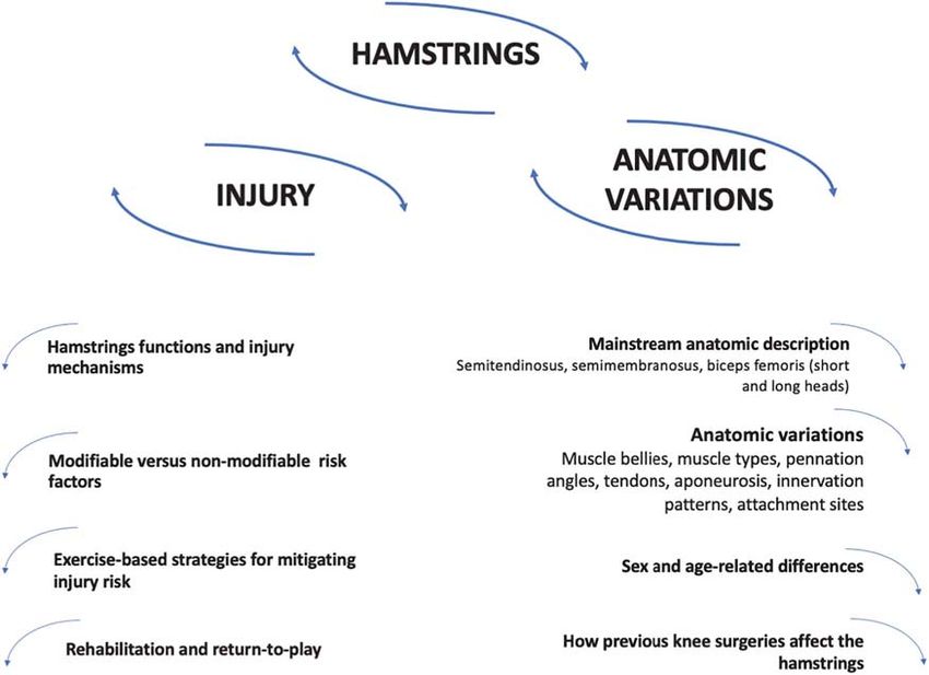
Afonso et al.                                                                                                   Hamstrings Variations and Injury Risk

INTRODUCTION                                                               length, muscle-tendon architecture, muscle fiber type and region-
                                                                           specific innervation have been hypothesized as relevant factors
The hamstrings are a hot topic in Sports Sciences, with a                  for risk of HSI (Timmins, 2017; Huygaerts et al., 2021), and
PubMed search using the terms “hamstring∗ ” and “sport∗ ”                  genetic variations have been explored in response to loading
from the year 2020 to the present showing >500 entries. This               and post-exercise recovery (Pickering and Kiely, 2018). However,
is because hamstrings injuries are common in the athletic                  anatomic and physiologic variations of the hamstrings are more
community (Monajati et al., 2016; Sheean et al., 2021). While              diversified than highlighted here (Tubbs et al., 2016).
hamstrings strain injury (HSI) is a commonly used term, it                     Our goal was to explore the available information on the
does not encompass all hamstrings injuries; therefore, HSI will            anatomic variations of the hamstrings, provide a deeper insight
only be used if the cited authors specifically refer to that subset        into the individual risk factors for hamstrings injuries and
of hamstrings injuries. The number of non-contact training-                contribute evidence-based applications for more individualized
related hamstrings injuries in different sports have increased             intervention regimens. Although acute injury prediction, given
gradually and systematically [e.g., an annual average 2.3%                 its multifactorial nature, will probably never be possible (Bahr,
increase in the total hamstrings injuries rate over the 13-year            2016), some risk factors can be controlled for. In the first
period (2001–2014)] (Ekstrand et al., 2016; Ertelt and Gronwald,           part of this commentary, an overview of hamstrings injuries
2017). Anatomic and functional aspects of the hamstrings,                  is provided, including injury mechanisms, both non- and
including the fact that muscles cross two joints (except the               modifiable risk factors, exercise-based strategies to mitigate risk
short head of the biceps femoris) and that eccentric action                factors, as well as topics of rehabilitation and return to play
during running or stretching carried out to extreme joint                  (RTP). While the literature often dichotomizes risk factors
positions, make it prone to vulnerable to strain-related injuries          into modifiable and non-modifiable, some non-modifiable risk
(Edouard et al., 2016).                                                    factors may be partially modifiable, as will be discussed. In the
    A systematic review including 13 studies with more than                second part, “normal” anatomy of the hamstrings is described,
3,800 athletes and two million sport exposure hours reported an            and the inter- and intraindividual variations of hamstrings
incidence of acute hamstrings injuries ranging from 0.3 to 0.5 per         anatomy and how they might be related to injury risk are
1,000 exposure hours in women, to 0.3–1.9 per 1,000 exposure               explored, considering modifiable and non-modifiable anatomic
hours in men, accounting for 5% to 15% of all soccer-related               variations and how to recognize them to prescribe better adjusted
injuries, with recurrence rates varying from 4 to 68% (Diemer              exercise. Non-modifiable risk factors can still be acted upon,
et al., 2021). The prevalence of hamstrings injuries were reported         by designing exercise interventions that are more respectful
to be more than 60% (unilateral) and more than 30% (bilateral)             of those factors.
in baseball players, with ∼30% recurrence rates (Zachazewski                   A systematic review of the literature was not performed,
et al., 2019), while prevalence rates of 40% were reported for             given the scope and goals of this commentary. However, we
professional soccer players (Ribeiro-Alvares et al., 2020). This           selected literature published in peer-reviewed journals, most of
is challenging in terms of recovery time and financial costs               which indexed in PubMed. The goal was to gather relevant
(Pickering and Kiely, 2018; Shield and Bourne, 2018; Metcalf               information on a wide variety of topics related to our theme.
et al., 2019). In the Australian Football League, data from 10             Where controversy existed, we attempted to provide a balanced
competitive seasons determined that the financial cost of HSI per          discussion, selecting studies with contradictory findings. Where
club increased by 71% from 2003 to 2012 (Hickey et al., 2014).             appropriate, renowned textbooks were used (Standring, 2015;
    A wide body of research explored the implementation of                 Tubbs et al., 2016).
specific exercise-based programs for mitigating hamstrings injury
risk (Al Attar et al., 2017; Chebbi et al., 2020). Due to intra-
and inter-muscular differences in this group of muscles, exercises
                                                                           PART 1: AN OVERVIEW OF
designed for the hamstrings may not provide equal stimuli
for semitendinosus (ST), semimembranosus (SM), and biceps                  HAMSTRINGS INJURIES
femoris (BF) (Kellis, 2018). Relevant works suggested that non-
modifiable factors (i.e., older age, previous injury) may have             Hamstrings Functions and Injury
a greater impact on risk for hamstrings injuries compared to               Mechanisms
modifiable ones (Freckleton and Pizzari, 2013; Green et al., 2020).        The hamstrings are involved in major movements of daily life,
Non-modifiable factors could also extend to aspects of human               such as standing and walking. In the standing position, the line
anatomy which are not possible to modify, such as variations in            of gravity passes behind the hip, producing an extension moment
insertions sites and the presence of supernumerary muscle bellies          that needs to be counterbalanced by the hip flexors (Houglum
(Tubbs et al., 2016).                                                      and Bertoti, 2012). On the contrary, the hamstrings need to act
    A better knowledge of interindividual variation in hamstrings          toward knee flexion, since the line of gravity falls in front of the
anatomy and geometry could allow the design of better-                     knee inducing hyperextension (Smith, 1957). During walking, the
individualized interventions, eventually reducing the likelihood           hamstrings (and gluteus maximus) act on the hip during the final
of suffering (and aid in the recovery from) an injury (Ertelt              leg swing phase by generating an eccentric action to decelerate
and Gronwald, 2017). Interindividual variation in strength                 the rate of hip flexion, and concentrically in the following hip
levels (specifically eccentric strength), hamstrings muscle fascicle       extension (Hogervorst and Vereecke, 2015). During the leg swing

Frontiers in Physiology | www.frontiersin.org                          2                                       July 2021 | Volume 12 | Article 694604
Afonso et al. Hamstrings Variations and Injury Risk
phase, the hamstrings act eccentrically to decelerate the rate of a higher risk of developing an injury (Jones et al., 2015;
knee extension (Houglum and Bertoti, 2012). Buckthorpe et al., 2019; Huygaerts et al., 2020). In the review
The hamstrings are important for sport activities involving of Danielsson et al. (2020), fatigue was associated to HSI,
sprints, jumps, tackles, cutting maneuvers and kicking (Ekstrand underlining that, aside from strength and flexibility, endurance
et al., 2012). The motion of the kicking leg (e.g., soccer was a relevant risk factor (Watson et al., 2017; Farley et al.,
kick) includes a backward and forward motion (Lees and 2020). Eccentric strength endurance of the hamstrings was
Nolan, 1998). During the backward motion, the hamstrings act reported to be significantly lower in male soccer players with
using concentric action in hip extension (together with gluteus previous hamstrings injuries (Schuermans et al., 2014). A study
maximus) and knee flexion and lateral rotation (only the BF) with elite footballers (n = 50) presented contradictory findings
(Fields et al., 2005). During the forward motion, the hamstrings when comparing the previously injured limbs (n = 11) with
act eccentrically to decelerate hip and knee during respective non-injured limbs (n = 89) (Freitas et al., 2021), but the
hip flexion and knee extension motions (Fields et al., 2005). athletes acquired hamstrings injury in the previous 2 years,
The decelerating action of hamstrings occur at a position (hip possibly providing enough time and exercise intervention for the
flexion and knee extension) where these muscles are passively hamstrings to recover their endurance levels. Previous injuries
insufficient (i.e., the muscles reach their maximal length since are non-modifiable risk factors, but improvements in endurance
they are biarticular muscles passing through hip and knee) (Coole and increased tolerance to fatigue can be achieved with exercise
and Gieck, 1987). For these reasons, the hamstrings are prone to training (Delextrat et al., 2018). In this sense, both detraining
injury during high-velocity running or sprinting, due to eccentric and hamstrings damage (secondary to a previous injury) may be
over-loading at the end of the swing phase (Jönhagen et al., 1994). considered partially modifiable factors.
A systematic review with 26 studies explored the mechanisms
behind the hamstrings injuries (Danielsson et al., 2020); the The Warm-Up
authors stratified the mechanisms according to the methods used The role of warm-up in reducing injury risk is not clear
to determine the injury: (i) stretch-related injuries; (ii) kinematic (Fradkin et al., 2006; McCrary et al., 2015), and adherence to
analysis; (iii) electromyography-based kinematic analysis; and warm-up protocols may interfere with its effectiveness (Owoeye
(iv) strength-related injuries. In the first group (i.e., stretch- et al., 2020), i.e., the frequency and degree of compliance to
related injuries), all studies reported that hamstrings injuries the warm-up protocol may largely dictate how effective the
occurred upon extensive hip flexion with hyperextension of the protocol is. It is unclear whether different types of warm-ups
knee (i.e., the hamstrings were strongly stretched at both joints it induce distinct acute effects on modifiable injury risk factors
crosses, which are the hip and knee). In the review (Danielsson (Niederer et al., 2020). A prospective two-season study registered
et al., 2020), hamstrings injuries were associated with running- posterior thigh injuries in 83 Australian rules football players,
based actions and especially to the late swing phase of the of which 62 were confirmed to have hamstrings injury through
running gait cycle, which may imply a powerful eccentric action. magnetic resonance imaging (MRI) (Verrall et al., 2003). In
These powerful actions increase as running velocity increases this study, 15% of hamstrings injuries occurred during the
(Higashihara et al., 2010b). warm-up period and 85% after the warm-up (Verrall et al.,
2003). Considering that the main part of an exercise session
Modifiable Risk Factors is longer than the warm-up, these percentages of distribution
Modifiable risk factors can be modified under certain provide insufficient information. Harking back to the discussion
interventions. In the case of hamstrings risk factors, these include surrounding fatigue (Delextrat et al., 2018; Danielsson et al.,
player load, warm-up preparations, lumbo-pelvic hip stability, 2020), it could be speculated that overly long and/or intense
motor patterns (e.g., “Groucho” position), cardiovascular fitness, warm-ups may generate excessive fatigue for the remainder of
fatigue, mobility, low back pain, recovery strategies, strength, the training session. Warm-up protocols aiming to produce
asymmetry, nutrition and psychosocial factors (Liu et al., 2012; performance potentiation usually generate potentiation and
Mendiguchia et al., 2012; Schuermans et al., 2014; Buckthorpe fatigue, and this balance varies from individual to individual
et al., 2019; Huygaerts et al., 2020). Using proper exercise-based (Afonso et al., 2019; Blazevich and Babault, 2019). Regarding
programs, establishing a balance between load and recovery, hamstrings injury prevention, individually monitoring responses
carefully structuring of the intervention sessions, and providing to warm-up is suggested.
adequate physical preparation, can readily tackle most of these
risk factors, which is valid for nearly all injuries. We do not aim The Role of Flexibility
to provide an in-depth discussion of modifiable risk factors, as A clear relationship between hamstrings flexibility and injury
it would escape the main goals of our work. However, we feel risk has also not been established (Worrell and Perrin,
it is relevant to briefly discuss four topics that should be more 1992; Christopher et al., 2019). de la Motte et al. (2019)
widely acknowledged (i.e., fatigue) or seen under a more critical assessed 27 studies and explicitly stated they considered studies
perspective (i.e., warm-up, flexibility and functional asymmetry). showing association between flexibility and other factors and
musculoskeletal injury in military and civilian populations. The
Fatigue authors showed there was moderate yet conflicting evidence
Fatigue was associated with decreased eccentric hamstrings associating hamstrings flexibility and musculoskeletal injury risk.
strength and an altered neuromuscular coordination, suggesting It is easy for readers to interpret this association using a causative
Frontiers in Physiology | www.frontiersin.org 3 July 2021 | Volume 12 | Article 694604Afonso et al. Hamstrings Variations and Injury Risk
framework, but caution should be used, as previous hamstrings Similar results were reported in another SRMA concerning
injuries reduce flexibility until 20–30 days post-injury (Maniar the NHE (Al Attar et al., 2017), although a reasonable risk of
et al., 2016), but reduced flexibility does not necessarily increase bias has been reported elsewhere (Gérard et al., 2020). These
injury risk. In this context, the role of stretching in reducing two SRMA (Al Attar et al., 2017; Gérard et al., 2020) were
injury risk is controversial (Herbert and Gabriel, 2002; Small alluding to relative risks, reducing the trustworthiness on the
et al., 2008; Behm et al., 2015), specifically in the case of potential beneficial effect of the NHE on injury prevention.
hamstrings injuries (Liu et al., 2012; Rogan et al., 2013). The feasibility of implementing certain protocols into real-world
contexts is questionable since the evidence-based interventions
Functional Asymmetry are not always effective, i.e., translating into practical applications
Some degree of interlimb asymmetries in hamstrings strength is may be problematic (McCall et al., 2020). Beyond the intrinsic
the norm (Boccia et al., 2018; Kulas et al., 2018; Cuthbert et al., characteristics of each exercise-based program, its effectiveness
2021) and, if not excessive, are beneficial for performance of tasks depends on the buy-in, i.e., how well participants adhere to, and
involving change of direction and sprinting, without impaired comply with the program (Buckthorpe et al., 2019). Therefore,
jumping (Coratella et al., 2018). Injured soccer players exhibit a the belief of coaches and athletes in the programs will interfere
more symmetrical recruitment pattern between ST, SM and BF with how well they work. Indeed, the placebo and nocebo
(corresponding to a less economic hamstrings muscle activation), effects have been observed in sports science (Hurst et al., 2020;
as opposed to the more asymmetric pattern of subjects without Raglin et al., 2020).
previous injuries (Schuermans et al., 2014). Athletes with Weekly frequency of exercise-based programs to mitigate
previous hamstrings injuries also demonstrated a decrease of ST injury risk is an important parameter: interventions performed
metabolic activity, compensated by a greater involvement of the ≥2 times per week were more effective in reducing hamstrings
BF (Schuermans et al., 2014). The mechanisms underlying the injury risk than interventions performedAfonso et al. Hamstrings Variations and Injury Risk
or not (Ayuob et al., 2020; Sheean et al., 2021), although there is BOX 1 | Special box – hamstrings’ strength evaluation.
debate regarding the criteria for choosing conservative treatment Measuring muscle strength allows evaluating and comparing muscle
vs. surgical management (Metcalf et al., 2019; Blakeney, 2020). function and performance to obtain a pattern of intramuscular synergistic
recruitment between the BF, ST, and SM muscles (Mjølsnes et al., 2004;
Schuermans et al., 2014; Wiesinger et al., 2020). Improvement of hamstrings
Modifiable Versus Non-modifiable Risk strength is an important strategy to mitigate injury incidence and recurrence,
Factors and one of the RTP criteria (Maniar et al., 2016). Early identification and
management of excessive strength asymmetries and muscular imbalances
Despite the potential of exercise-based interventions for assist in the elaboration of a more effective intervention plan to counteract a
mitigating hamstrings injury risk, non-modifiable risk factors potential injury risk scenario (Schuermans et al., 2014; Wiesinger et al., 2020).
should be acknowledged, such as age and previous injury The stationary isokinetic dynamometry (IKD) is a suitable system for
(Mendiguchia et al., 2012). A SRMA involving 71,324 athletes assessment of torque during concentric and eccentric knee flexor action
from 78 studies analyzed 8,319 HSIs, of which 967 were (Aagaard et al., 1998; van Dyk et al., 2018; Wollin et al., 2018), although its
use is scarce compared to the Nordic hamstrings device (NHD) (Mjølsnes
recurrences (Green et al., 2020). The stronger factors associated et al., 2004). The NHD detects strength deficits or side-to-side imbalances
with an increased risk of HSI were older age, previous history of (Timmins et al., 2016), exercise-related strength progress (Delahunt et al.,
HSI, recent HSI, previous calf strain injury, and previous anterior 2016), and may aid predicting recovery time after injury (McCall et al., 2016).
cruciate ligament (ACL) injury, which are non-modifiable factors However, van Dyk et al. (2018) reported a weak within-subject correlation
(r = 0.35), demonstrating a systematic bias toward lower strength values with
(Green et al., 2020). In contrast, modifiable risk factors, such as
the NHE. Several methodological specificities were not considered when
reductions in strength, strength endurance, power, and motor comparing both methods in previous studies, e.g., IKD and NHD tests were
control, were weakly associated to increased risk for his, and performed under different conditions of joint velocity and hip position
flexibility, mobility or ROM were not associated with risk of HSI (Higashihara et al., 2010a; Guex et al., 2016).
(Green et al., 2020). To understand these issues, Wiesinger et al. (2020) compared the
mechanical output of hamstrings assessed by using both IKD and NHD
Non-modifiable risk factors for HSI, such as age and previous
methods of 25 healthy male athletes in a counterbalanced repeated-measures
injury, were consistently associated with an increased risk of protocol. Higher total eccentric work, peak torque at greater knee extension
injury in a previous SRMA that included 34 articles (Freckleton angles, greater side-to-side strength difference, and lower eccentric peak
and Pizzari, 2013). In this SRMA, the only modifiable factor torque were observed in IKD compared to NHD, whereas bilateral strength
consistently associated with the risk of hamstrings injuries was difference was lower in NHD. The electromyographic analysis showed no
difference in the activation of BF and ST during IKD and NHD (Wiesinger et al.,
the quadriceps concentric peak torque. In a prospective study
2020). These findings suggest that IKD and NHD methods measured distinct
with Australian football league players (n = 125), hamstrings hamstrings muscle activation characteristics. Some aspects were difficult to
injuries were not associated with NHE strength (Smith et al., assess, such as intraindividual strength differences, angle of peak torque and
2021). In this study (Smith et al., 2021), player age greater than side-to-side differences in eccentric knee flexor strength. It should be
25 years and having a previous hamstring, groin or calf injury highlighted that muscles such as the sartorius, gracilis and gastrocnemius
also contribute to knee flexion (Standring, 2015).
increased the risk for hamstrings injury. Attention has been
Surface electromyography (EMG) allow the assessment of muscle
devoted to the modifiable risk factors, but non-modifiable factors activation patterns (Higashihara et al., 2010a). The EMG activity shows that
should be further explored, as well as the mechanisms whereby hamstrings muscles have functional differences, as the SM and BF work
some non-modifiable factors increase injury risk. harder (i.e., greater EMG) during the initial phase of knee flexion, and the ST at
Although it is not possible to erase the existence or previous deep flexion angles to complement the decrease in EMG of the other two
muscles during open kinetic chain exercise (Hirose and Tsuruike, 2018). Miller
injuries, or change the age of the player, a better understanding
et al. (2000) found that during isokinetic movements activation, the BF EMG
of the hamstrings’ architecture, anatomy and mechanisms may increased with increasing movement velocity, while the ST and SM EMG
help to deliver a better-individualized exercise-based approach. activation remained constant during six continuous isokinetic knee extension
It was previously established that previous injuries impair ROM and flexion movements at 60◦ , 180◦ , and 300◦ s−1 . In opposition, functional
(Maniar et al., 2016) and endurance (Farley et al., 2020), MRI evaluates the intramuscular and intermuscular recruitment patterns with a
very high spatial accuracy and detects the magnitude of metabolic activity in
both of which can be improved through well-designed exercise
muscle tissue although with no real-time information about the amount and
intervention protocols. While some architectural features might timing of the underlying muscle activity (Segal, 2007). Using functional MRI,
be modifiable [e.g., cross-sectional area (CSA)], others may not Schuermans et al. (2014) demonstrated that the more symmetrical and less
(e.g., variations in insertions), and an understanding of such dissociated the hamstrings muscles work together, the higher the
features may afford a better-individualized exercise prescription. physiological changes would be inside the recruited muscle fibers. Thus, more
intramuscular variability can be associated with a reduced metabolic turnover
Sex- and age-related risk factors will be explored further in
and more economic muscle functioning. MRI-based methods have high
part 2, as they are closely linked to anatomic and physiologic costs, risk of injury during assessment, lack of portability and long duration of
variations. Even when certain factors are non-modifiable, the assessment. Rapid, non-invasive alternative measures of concentric and
mechanisms that contribute to the increased injury risk could be eccentric hamstrings strength are warranted.
individually targeted with better exercise-based interventions, to An adapted aneroid sphygmomanometer test (Mondin et al., 2018), a rapid
and non-invasive tool, was proposed to assess hamstrings strength. In 14
avoid exacerbating predisposing factors. rugby players, Mondin et al. (2018) found an association between the
sphygmomanometer derived pressures at 30 and 90◦ of knee flexion and
Synopsis of the First Part isokinetic strength measures, suggesting that this method was reliable to
assess hamstrings strength, but not to identify strength asymmetries between
The hamstrings are highly relevant for major movements of dominant and non-dominant legs or knee flexors-to-knee extensors ratios.
daily life and sports (e.g., standing, walking, sprinting, cutting), (Continued)
and play an especially important role in decelerating knee
Frontiers in Physiology | www.frontiersin.org 5 July 2021 | Volume 12 | Article 694604Afonso et al. Hamstrings Variations and Injury Risk
BOX 1 | Continued
nerve, emerging at the level of S1 vertebra, although with
The limitation of knee flexors-to-knee extensors ratios was explained by the interindividual variations.
inability of aneroid sphygmomanometer test to measure quadriceps strength Functionally, the hamstrings play an important role in
at 30◦ knee flexion with consistency, due to many participants exceeding the transferring load between the trunk and the lower limbs, and
readings on the sphygmomanometer scale. This method has a great potential, affect the tension of the thoracolumbar fascia. They affect the
but requires further studies to be useful as a muscle strength assessment and
injury risk screening procedure.
normal lumbar lordosis, as their length and strength interferes
with the position of the innominate bone. When raising the
trunk, the hamstrings act in conjunction with the gluteus
extension and hip flexion in high-velocity actions, common in maximus. It is highlighted that the hamstrings strongly contract
athletic scenarios, such as sprinting. Hamstrings are prone to in actions involving the need to extend or control the rate of
injuries and so it is important to understand the risk factors flexion at the hip, while the gluteus maximus contracts when
involved. Exercise-based interventions for mitigating hamstrings powerful extension of the hip joint is required. The adductor
injury risk have emphasized the role of eccentrically biased magnus also performs hip extension, has attachments to the
strength training, although lumbo-pelvic dynamics, technical ischial tuberosity, and the ischial portion of the adductor magnus
and motor pattern focused work, sprinting, isometric strength shares innervation from the tibial division of the sciatic nerve.
training and general strength and endurance training should The adductor magnus can be considered to have a proper
not be neglected. Weekly frequency and compliance with hamstrings portion (Broski et al., 2016; Jeno and Schindler,
the interventions constitutes a relevant factor mediating the 2020). Pathology or diminished functional capacity of the
effectiveness of exercise-based interventions in mitigating injury adductor magnus and/or the gluteus maximus may result in a
risk. However, research suggests that non-modifiable risk factors, greater demand upon the hamstrings. The coordination of the
namely age and previous injuries, may play a more relevant hamstrings with other muscles of the lumbopelvic region may
role in hamstrings injury risk than modifiable risk factors. In be relevant in understanding hamstrings injuries (Thelen et al.,
this context, relevant anatomic variations may alter the risk 2006; Shield and Bourne, 2018). Figure 1 presents an overview of
of hamstrings injury. Acknowledging the relevant anatomic the hamstrings muscles.
variations may provide relevant information for prescribing
exercise interventions, which will be the focus of the second Semitendinosus
part of this commentary. Box 1 briefly explores hamstrings’ The ST has a posteromedial position in the thigh and has a
strength evaluation. long tendon. Proximally, its tendon is shared with the BFlh,
but then these muscles diverge. In the first ±7.5 cm of their
path, they share an aponeurosis. The belly of the ST usually
PART 2: ANATOMIC AND PHYSIOLOGIC spans only until the mid-thigh, after which its long tendon runs
until its attachment in the upper part of the medial surface of
VARIATIONS OF THE HAMSTRINGS AND
the tibia, behind the attachment of the sartorius muscle and
POTENTIAL IMPLICATIONS FOR INJURY distal to the attachment of gracilis muscle. The terminal portion
RISK the ST tendon is united with the tendon of gracilis (further
reinforcing the functional connections between the hamstrings
In this second part, we address the main variations or variants of and the adductor group), provides an expansion to the deep
the hamstrings’ anatomy and physiology, which imply that there fascia of the leg and to the medial head of the gastrocnemius,
is a “normal” or “usual” state of affairs. We start by presenting the and anatomically (and perhaps functionally) links the ST with
commonly described anatomic features of the hamstrings, before the medial gastrocnemius. It is open to speculation whether
engaging in an exploration of their variations. pathology or dysfunction of the medial gastrocnemius interfere
with the action of the ST. Usually, the midpoint of the ST
Mainstream Anatomic Description of the receives a muscular slip from the BFlh, denoting a role in
Hamstrings lateral force transfer.
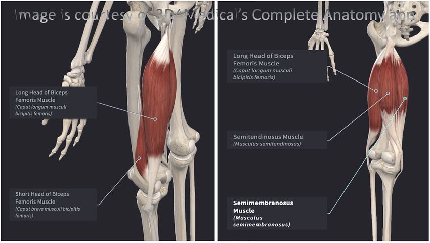
We overview the basic hamstrings anatomy as described in the Innervation to the ST is provided by the sciatic nerve (L5, S1,
41st Edition of Gray’s Anatomy (Standring, 2015). Throughout S2), through its tibial division. This pattern is shared with the SM,
this section, the information derives from this source, unless which lies deep to the ST, and by the BFlh, but not by the BFsh.
otherwise stated. The hamstrings, or ischiocrural muscles, Beyond the actions common to all hamstrings, the ST (as well
comprise the muscles of the posterior compartment of the thigh, as the SM) can medially rotate the knee when this joint is semi-
and include the ST, SM and BF (long head – BFlh; short head – flexed. With the hip extended, ST (and SM) can act to produce
BFsh), which attach proximally to the ischial tuberosity (except medial rotation of the thigh.
the BFsh). The ST, SM and BFlh are biarticular muscles, and
act in extending the coxofemoral joint (hip), as well as flexing Semimembranosus
and rotating (medially and laterally) the knee. Distally, the ST The SM also lies posteromedially in the thigh, deep to the ST.
and SM attach to the tibia, while both heads of the BF attach Proximally, it exhibits a long and flat tendon attached to the
to the fibula. The hamstrings present relevant interindividual ischial tuberosity, and it receives fibrous expansions that flank
variation in length. The hamstrings are innervated by the sciatic the adductor magnus. Distally, the SM has a ramified pattern,
Frontiers in Physiology | www.frontiersin.org 6 July 2021 | Volume 12 | Article 694604Afonso et al. Hamstrings Variations and Injury Risk
FIGURE 1 | (A) Posterolateral view of the left hamstrings. (B) Posteromedial view of the left hamstrings. Both images were elaborated with Complete Anatomy 2021,
version 7.0.0. (desktop version for Mac OS) and reproduced here with permission (3D4Medical, Elsevier).
sharing fibers with both ST and BFlh. Circa the mid-thigh, the SM to the lateral condyle of the tibia. A variation noted in Gray’s
gives off muscle fibers that converge to a second aponeurosis that Anatomy description is the absence of the BFsh.
attaches distally over the terminal tendon. The SM distal tendon While the BFlh shares innervation with the ST and SM, arising
divides into five components: anterior, direct, capsular, inferior from the tibial division of the sciatic nerve (L5, S1, S2), the
and the oblique popliteal ligament (Beltran et al., 2003). The SM BFsh is innervated by nerves emerging from the same levels, but
has a close anatomic relationship with the medial gastrocnemius, traveling in the common fibular division of the sciatic nerve.
usually separated by a bursa. This differential innervation of the two heads of the BF may
Gray’s Anatomy describes that the SM can vary considerably result in asynchrony and impaired coordination (Beltran et al.,
in size and may be absent (which could potentially overload 2012; Huygaerts et al., 2021), potentially placing the BF at greater
the ST) or double (which could potentially underload the ST). risk of injury (Burkett, 1975; Entwisle et al., 2017). Theoretical
Proximally, the SM can arise from the sacrotuberous ligament models have proposed that BF may be at increased injury risk
instead of the ischial tuberosity. It can have muscular slips to in comparison with ST and SM (Dolman et al., 2014), owing to
the femur or to the adductor magnus. Although myotendinous an additional BF compensation induced by a poor ST endurance,
and avulsion injuries are common in the SM, complete tears are placing the BF at higher injury risk (Schuermans et al., 2014).
seldom reported in the literature (Beltran et al., 2003). In a retrospective study with 275 men soccer players who had
sustained hamstrings injuries, the BFlh was the most commonly
Biceps Femoris affected (56.5%), followed by the ST (24.4%), SM (13.7%), and
Unlike the ST and SM, the BF occupies a posterolateral position BFsh (5.6%) (Crema et al., 2016). In Australian rules football
in the thigh. Proximally, the BFlh attaches to the ischial players, injury of the BF represented 78% of all hamstrings
tuberosity, through a common tendon with the ST, but it can injuries (Verrall et al., 2003). Beyond the actions common to all
also insert into the sacrotuberous ligament. The BFlh has a hamstrings, the BF can laterally rotate the knee when this joint is
fusiform belly, and its fibers terminate in an aponeurosis that also semi-flexed. With the hip extended, BF can act to produce lateral
receives fibers from the BFsh. Pennation angle of the BFlh fibers rotation of the thigh.
is non-uniform, being greater in the proximal-middle sections,
in comparison to the distal and extreme proximal sections
(Huygaerts et al., 2021). The BFsh has its proximal attachment in Anatomic and Physiologic Variations of
the lateral lip of the linea aspera and this attachment may extend the Hamstrings
upward to almost the level of gluteus maximus. Distally, these The hamstrings integrate the posterior compartment of the thigh,
muscles share a common tendon inserting into the head of the considered to be the most variable compartment (Tubbs et al.,
fibula and send expansions to the fibular collateral ligament and 2016). We explore known anatomic variations to unfold their
Frontiers in Physiology | www.frontiersin.org 7 July 2021 | Volume 12 | Article 694604Afonso et al. Hamstrings Variations and Injury Risk
implications for injury risk. It is unlikely that any single risk Despite this information, eccentric actions have been emphasized
factor allows establishing a strong relationship with injury risk, by many programs for reducing injury risk of the hamstrings
but recognizing their existence may provide a more thorough (van der Horst et al., 2015; Almeida et al., 2018; Elerian
understanding of individualized injury risk. Technological et al., 2019). Perhaps athletes with greater percentage of type II
developments may translate that enhanced knowledge into fibers should follow a more careful monitoring of the eccentric
practical applications. Table 1 summarizes the articles exploring work they perform.
anatomic variations of the hamstrings. A previous cadaveric study with seven men and three women
(37 to 76 years upon death) showed that type II fibers composed
Muscle Bellies 48.5 to 59.5% of the proximal BFlh, 51.0 to 58% of the distal
Cadaveric analysis showed that the extension of BFsh BFlh, 45.5 to 69.0% of the BFsh, 51.0 to 57.5% of the proximal
attachments to the femur vary substantially between individuals, ST, 50.0 to 69.5% of the distal ST, 47.5 to 54.5% of the proximal
leading to the hypothesis that individuals with more extensive SM, and 44.0 to 55.5% of the distal ST (Garrett et al., 1984). In a
attachments may apply more force, potentially increasing the risk study with 15 sedentary men aged 17 to 40 years old (Dahmane
of a strain injury (Burkett, 1975). The opposite interpretation is et al., 2006), the BF had a mixed fiber composition of ∼50% type
also possible, as attachments that are more extensive would allow 1 and ∼50% type 2 (25.2 ± 1.3% type 2a, 20.7 ± 1.4% type 2x
the BFsh muscle belly to be more firmly attached to the femur, and 5.7 ± 0.7% type 2c), meaning that BF has a considerable
and therefore may be at less risk of a strain injury. The two heads amount of fast-twitch fibers. Biopsies of the BFlh performed in 31
of the BF have different proximal attachments and innervation, healthy, young men, showed a balanced distribution of myosin
possibly representing muscles that were independent, but fused heavy chains (MHC), with 47.1 ± 9.1% MHC-I, 35.5 ± 8.5%
during the human evolutionary process (Tubbs et al., 2016). MHC-IIA, and 17.4 ± 9.1% MHC-IIX (Evangelidis et al., 2017).
In some individuals the BFlh and BFsh remain separate, and For the ST, a composition of 50 ± 13% type 1 fibers, 26 ± 8%
occasionally the BFsh is absent (Tubbs et al., 2016). From 29 type 2A, 23 ± 19% type 2B and 1 ± 1% type 2C was found
cadaveric specimens with a median age of 71.5 (range from 45 to on biopsies from 16 patients 7–11 months after ACL surgery
98), BFlh and BFsh extended approximately 42.0 and 29.8 cm, (Shalabi et al., 2002). In this reported ST composition (Shalabi
respectively (van der Made et al., 2015). et al., 2002), the standard-deviations of the reported ST muscle
The ST can be partly fused with the SM (Tubbs et al., 2016) composition were relevant, especially in type 2B fibers, denoting
or the BFlh (Schmuter et al., 2021), and may present accessory interindividual variation.
slips to the coccyx, sacrotuberous ligament or to the ischial It has been suggested that high-level sprinters are sharply
tuberosity (Fraser et al., 2013). Supernumerary ST bellies have different than the average person, with a percentage of up to
been observed (Gray, 1945). Although ST and BFlh usually ∼70% of type 2 fibers in the lower limb muscles, but this
share a common proximal tendon attachment and aponeurosis, was derived from analysis of the vastus lateralis (Trappe et al.,
complete separation of these two muscles has been reported 2015), and should not be generalized to other lower limb
(Tubbs et al., 2016). Of note, the tendon of the ST may receive muscles. Differences in fiber type composition of the lower
muscle slips from the quadratus femoris (Tubbs et al., 2016). limbs have been observed between endurance runners, power-
The SM can be doubled, absent, or split, and up to four bellies trained individuals, and strength-trained individuals (Methenitis
have been described inserting into the adductor magnus, planum et al., 2019), but again these findings were extrapolated from
popliteum and tibia (Tubbs et al., 2016). The ST, SM and BFlh observations of the vastus lateralis. In vivo knowledge of
may originate from a common tendon, which can be continuous hamstrings muscle composition in humans remains largely
with the intermuscular septum and, occasionally, envelop the unknown (Evangelidis et al., 2017).
piriformis muscle (Tubbs et al., 2016), providing another example As Lievens et al. (2020) pointed out, athletes with
of the connections between the hamstrings and other muscles of distinct muscle typology should train differently, and the
the lumbo-pelvic region. individualization of training on the basis of this information
is important to optimize performance and lowering the risk
Muscle Fiber Type of potential injury. Unfortunately, knowledge of muscle
The muscle fibers vary in their morphology and histochemical fiber composition of the human hamstrings is scarce for
properties (Galpin et al., 2012). Classifications of fiber muscle building a broad picture. Furthermore, feasibility may require
types have evolved through the years and certain classifications the development of more readily available, non-invasive,
are still debated (Talbot and Maves, 2016). Here, we use the non-expensive technologies.
original classification reported by the authors being cited. There
are important functional differences between different muscle Pennation Angles
fiber types, such as distinct metabolic properties (Mishra et al., A study analyzed the pennation angles of BFlh and ST muscles
2015). There may be a bidirectional relationship, whereby from six legs derived from three male cadavers (Kellis et al.,
metabolic environment also influences the characteristics of the 2009). The BFlh and ST exhibited very similar pennation angles
muscle fibers (Julien et al., 2018). Type II muscle fibers are through cadaveric dissection (13.52 ± 2.35◦ versus 13.39 ± 3.31◦ )
more easily fatigued than type I fibers (Lievens et al., 2020), and ultrasound (13.88 ± 2.76◦ versus 13.34 ± 2.61◦ ). The small
and more susceptible to plyometric- (Macaluso et al., 2012) as standard deviation was suggestive of non-relevant intra- and
well as eccentric-induced muscle damage (Fridén et al., 1983). interindividual variations. A posterior study of the same group
Frontiers in Physiology | www.frontiersin.org 8 July 2021 | Volume 12 | Article 694604Afonso et al. Hamstrings Variations and Injury Risk
TABLE 1 | Summary of studies cited in Section “Anatomic and Physiologic Variations of the Hamstrings.”
References Population Anatomic part Assessment method Main findings
assessed
An et al. (2010) 50 lower extremity Innervation patterns Dissection BFsh and SM show one innervation entry, and BFlh and ST show two
cadavers from 15 of HT innervation entries. In those with two entries, for BFlh, the incidence of
males and 12 type I innervation was 82% (18% of type II) while for ST, the incidence of
females (age: type I innervation was 14% (86% of type II).
71 years)
Blazevich et al. 16 women and 15 Architecture of Ultrasound Description of the architecture of the quadriceps. Changes in the
(2006) men physically quadriceps relative activation of individual muscles determines alterations in force,
active (age: velocity movement range and contraction mode. Intramuscular
19.9–20.6 years) activation changes with the movement requirements. Training should
alter the pattern of activations to improve force transmission between
muscles with different architecture, make these variations in activations
efficient, and promote region-specific hypertrophic responses. There
are little differences between sex in these adaptations.
Burkett (1975) Review Anatomical – Extensive biceps femoris-femur attachment coupled with strength
variation of HT imbalances between HT increase the risk of strains.
Dahmane et al. 15 healthy Muscle fiber types Mechanomyography Strong potential for the BF muscle to transform from slow to faster
(2006) sedentary males of BF contracting muscle fibers after long-term sprint training.
(age: 17–40 years)
and 15 male
sprinters (age:
23.2 ± 3.1 years)
Evangelidis et al. 30 healthy Aponeurosis of MRI The proximal aponeurosis size is highly variable between individuals,
(2015) recreationally active BFlh and it is not associated with to muscle size or knee flexor maximal
individuals (age: isometric or eccentric strength. This disproportion may predispose
20.7 ± 2.6 years) those individuals with relatively small aponeurosis to hamstring strain
injuries, as they could be subjected to greater mechanical strain in the
muscle tissue surrounding the aponeurosis.
Evangelidis et al. 31 healthy, Muscle fiber types MRI HT muscles exhibited a balanced myosin heavy chain isoform
(2017) recreationally active of HT distribution comparable to that of vastus lateralis, so that the
participants (age: predominance of fast-twitch fibers in HT muscle increasing the risk of
21 ± 3 years) strains is not supported.
Fiorentino and 12 male track and Musculotendon of MRI A larger muscle and/or narrower proximal aponeurosis of the BFlh
Blemker (2014) field athletes BFlh could predispose an athlete to an increased risk of injury by increasing
peak local muscle tissue strain.
Fraser et al. (2013) 1 female cadaver Anatomical Dissection Two-part origin of the ST, with the variant portion being originated along
(age: 87 years) variation of ST the medial border of the ischial tuberosity. Could predispose to HT
injury, chronic pain and pelvic floor discomfort.
Freitas et al. (2020) 40 male Aponeurosis of MRI There were no significant differences for size aponeurosis of the BFlh
professional football BFlh between players with and without previous BFlh injury.
players (age:
24.5 ± 4.9 years)
Fridén et al. (1983) 12 male physical Muscle fiber types Muscle biopsy Type II fibers were more extensively damaged than type I fibers after
education students of vastus lateralis 30 min of pedaling at a frequency of 60 rpm, at an intensity of 80–100%
(age: 25 ± 7 years) of VO2 max, in a bicycle ergometer modified for use in eccentric work.
Galpin et al. (2012) Description muscle – – –
fiber types
Garrett et al. (1984) 7 male and 3 Muscle fiber types ATPase histochemical Relatively higher percentage of type II fibers in HT compared to other
female cadavers of lower limb reaction thigh and leg muscles.
(age: 60 years) muscles
Gérard et al. (2020) SR: 10 RCTs Architecture of BF MRI and ultrasound Eccentric strength training associated with increased fascicle length
evaluating 346 and muscle thickness, and decreased pennation angle, as well as
healthy adults (age: eccentric strength of the HT.
18.3–29.6 years)
Gray (1945) 2 male cadavers Anatomical Dissection All HT muscles originated from a common tendon in case 1. A muscle
(age: 44 and variation of HT from the linea aspera and passing medially to the capsule of the knee
84 years) joint, homologous with the ST, is presented in case 2.
Huygaerts et al. Critical review Muscle-tendon unit – Fiber-fascicle lengthening is greater, and architectural structure
(2021) of HT non-uniform in the BFlh, with pennation being greater in the
proximal-middle section compared to the distal and extreme proximal
sections, that in addition to the inter-individual differences in BFlh
structural features may predispose to this muscle to higher risk of HT
injury.
(Continued)
Frontiers in Physiology | www.frontiersin.org 9 July 2021 | Volume 12 | Article 694604Afonso et al. Hamstrings Variations and Injury Risk
TABLE 1 | Continued
References Population Anatomic part Assessment method Main findings
assessed
Julien et al. (2018) Review Muscle fiber types – The changing environment could elucidate changes in muscle fiber
characteristics, which could have implications in metabolism-related
muscular atrophies.
Kellis (2018) Narrative review Architecture of HT – BFlh, compared to other HT, may be at higher increase of injury due to
inter-muscular differences in HT architecture. Targeting the specific
muscle-tendon region, instead of HT as a whole, could be beneficial in
rehabilitation programs.
Kellis et al. (2009) 3 cadavers (age: Architectural Ultrasound dissection High level of agreement in BFlh and ST architectural parameters
68.3 years) parameters of BFlh measured through ultrasound compared to direct dissection.
and ST
Kellis et al. (2012) 8 cadavers (age: Architecture of HT Dissection The four hamstring components showed low to moderate architectural
67.8 years) dissimilarity. Pennation angles were similar between BFlh, BFsh and
SM, but higher than for ST.
Lievens et al. (2020) 32 male Muscle fiber types Magnetic resonance Participants with predominantly fast typology fibers fatigues more
recreational of gastrocnemius spectroscopy markedly in repeated Wingate tests than participants with slow typology
athletes fibers.
Macaluso et al. 8 healthy, untrained Muscle fiber types Muscle biopsy An acute bout of plyometric exercise preferentially affects type II fibers
(2012) individuals (age: of vastus lateralis
22 ± 1 years)
Marušič et al. 40 healthy adults Architecture of BFlh Ultrasound A 6-week progressive eccentric HT training in a lengthened position
(2020) (age: showed positive effects for fascicle length (increase) and pennation
23.7 ± 2.5 years) angle (decrease), but not for muscle thickness.
Medeiros et al. 32 adult football Architecture of BFlh Ultrasonography An 8-week Nordic hamstring exercise training program was not
(2020) players (age: effective at elucidating any improvement in muscle thickness, pennation
18–23 years) angle, or fascicle length.
Mendiguchia et al. 32 adult football Architecture of BFlh Ultrasound The sprint training group showed moderate increase in fascicle length,
(2020) players and the Nordic group a small increase. The Nordic group presented a
small increase at pennation angle.
Methenitis et al. 10 sedentary, 9 Muscle fiber Muscle biopsy Muscle fiber composition and rate of force development is affected by
(2019) endurance runners, composition of systematic training among different athletes. Type IIx fibers better
10 power-trained vastus lateralis correlated to rate of force development.
and 9
strength-trained
individuals
Rehorn and 1 healthy male Aponeurosis of MRI and 3D modeling The fact that proximal aponeurosis is narrower than distal aponeurosis
Blemker (2010) individual BFlh in BFlh could explain the prevalence of injuries near the proximal
myotendinous junction in this muscle, and relative aponeurosis
dimensions could also explain the more prevalence of injuries in BFlh
compared to other HT muscles.
Schmuter et al. 1 female cadaver Anatomical Dissection BFlh and ST were fused near their origin at the ischial tuberosity.
(2021) (age: 59 years) variations of BFlh
and ST
Shalabi et al. (2002) 14 males and 2 Muscle fiber types Muscle biopsy Patients following ACL reconstruction show a composition of 50 ± 13%
females (age: of ST (percutaneous type 1 fibers, 26 ± 8% type 2A, 23 ± 19% type 2B and 1 ± 1% type
26 years) ultrasound-guided 2C. Muscle biopsy technique is adequate to identify muscle
biopsy) composition.
Solomon and 1 male (age: Anatomical MRI Case of a bilateral tibial insertion of the BF tendon in a previously
Stevenson (2008) 40 years) variation of BF asymptomatic patient, which could have implications on lateral knee
tendon insertion stability.
Trappe et al. (2015) 1 world champion Muscle fiber types Muscle biopsy Large proportion of vastus lateralis type IIx fibers in the sprinter, and
sprinter of vastus lateralis power output from type IIa and IIx fibers was higher than any human
values reported to date.
van der Made et al. 29 human Anatomical Dissection Overlapping proximal and distal tendons and muscle architecture may
(2015) cadaveric variations of HT lead to a force not in line with the tendon and predispose to muscle
specimens (age: (origin dimensions, injury. Protective effect of the presence of a raphe.
71.5 years) muscle length,
tendon length, MTJ
length and width,
length of tendinous
of ST (raphe).
(Continued)
Frontiers in Physiology | www.frontiersin.org 10 July 2021 | Volume 12 | Article 694604Afonso et al. Hamstrings Variations and Injury Risk
TABLE 1 | Continued
References Population Anatomic part Assessment method Main findings
assessed
Vieira et al. (2007) 41 male and 56 Innervation patterns MRI Description of the normal anatomy of the distal BF and the relationship
female of BF with the peroneal nerve. The peroneal nerve can pass downward
asymptomatic posterior to the BFsh and superficial to the lateral head of the
patients (age: gastrocnemius, but also in a tunnel between the two muscles (23%). An
52.8 years) unusual relationship between the nerve and the distal BF could
predispose to peroneal neuropathy.
Woodley and 3 female and 3 Architecture of HT Dissection ST, SM and BF showed anatomical partitioning defined by architecture
Mercer (2005) male cadavers and/or pattern of innervation. There was high degree of variation from
(age: 69–88 years) subject to subject in most of the architectural features of the HT, such
as, fascicular length, volume, physiological cross-sectional area or
tendon characteristics.
Yang et al. (2018) 93 patients (age: Innervation patterns MRI There were less participants with type I (38.7%) than type II innervation
40.2 years) of BF (61.3%), in which the thickness was lower. The course of common
peroneal neuropathy through the “popliteal tunnel” formed between the
BFsh and the lateral gastrocnemius muscle was approximately 40%.
Yasin et al. (2010) 25 patients Anatomy of Dissection Gracilis and ST tendons showed a variable pattern of accessory bands,
undergoing ACL accessory bands all of them occurring more than 11-cm proximal to the insertion of the
reconstruction with tendons onto the tibial crest.
HT tendon
autograph (age:
28 years)
HT, hamstrings; MTJ, musculotendinous junction; ST, semitendinosus; BFlh, biceps femoris long head; VO2 max, maximal oxygen consumption; ATPase, adenosine
triphosphatase; BF, biceps femoris; MRI, magnetic resonance imaging; ACL, anterior cruciate ligament; BFsh, biceps femoris short head; SM, semimembranosus; RCT,
randomized controlled trial.
dissected eight cadavers (age: 67.8 ± 4.3 years) (Kellis et al., warranted to understand how different exercise modalities affect
2012) and demonstrated that pennation angles of the BFlh, pennation angles.
BFsh and SM were similar (13.46 ± 2.88◦ , 13.17 ± 2.60◦ , and
15.95 ± 2.39◦ , respectively), but substantially different from the Tendons
pennation angle of the ST (9.14 ± 3.54◦ ), which differs from There is variation in the accessory bands of the hamstrings
the results found in their previous work (Kellis et al., 2009). tendons (Yasin et al., 2010), as well as in tendon length. One study
The authors used a combination of mean fiber length, sarcomere showed that the muscle belly of the SM originated at varying
length, physiological CSA, and pennation angle to calculate a distances from the ischial tuberosity in different subjects, ranging
similarity index (δ) between pairs of muscles, with lower values from 8.6 to 14.5 cm (Woodley and Mercer, 2005). Interindividual
denoting greater similarity. While the BFlh and BFsh had a variations in tendon-to-fiber length ratios may play a role in
δ = 0.54, and BFlh and SM had a δ = 0.35, denoting a moderate injury risk, as in the case of tendons of similar structure, tendon
similarity, there was low similarity between SM and ST (δ = 0.98) length critically influences compliance (Huygaerts et al., 2021).
and between BFlh and ST (δ = 1.17). These findings underline If two athletes have different ratios for the ST, it is possible that
that although the hamstrings are usually treated as a muscle one will be at increased risk of injury than the other. It was
group, there are relevant inter-muscular architectural differences hypothesized that bigger tendon-to-fiber length ratios provide a
between its individual muscles (Kellis et al., 2012). greater buffer to the muscle belly, potentially affording increased
A SRMA showed a limited to moderate confidence in protection from injury, but this requires confirmation (Kellis,
evidence that eccentric training performed for a minimum of 2018; Huygaerts et al., 2021).
4 weeks decreased the BFlh pennation angle (Gérard et al.,
2020), which was also shown in a recent study (Marušič Aponeurosis
et al., 2020). However, another study with 32 soccer players The aponeurosis may affect different muscle regions distinctly,
(age, 18–23 years) performing an 8-week NHE program demanding greater elongation of fibers in certain regions and
once a week versus twice a week showed no between-group less so in others (Blazevich et al., 2006; Huygaerts et al.,
differences in BFlh pennation angles (Medeiros et al., 2020). 2021). A modeling study suggested that smaller ratios of
A prospective controlled study with soccer players analyzed aponeurosis to muscle width generated larger maximum peak
the NHE and sprint interventions, showing that only the local strain, postulating those larger muscles and/or narrower
NHE induced small increases in the BFlh pennation angle aponeurosis increased risk of injury (Fiorentino and Blemker,
(Mendiguchia et al., 2020), contradicting the decreases seen 2014). Interindividual variation in anatomic structure may put
in previous studies (Gérard et al., 2020; Marušič et al., individuals with distinct aponeurosis to muscle width ratio at
2020). Results are conflicting, largely limited to the BFlh and different levels of injury risk. However, an MRI study conducted
involve a narrow set of exercise modalities. Further research is in 30 healthy young men performing maximum voluntary actions
Frontiers in Physiology | www.frontiersin.org 11 July 2021 | Volume 12 | Article 694604You can also read