GUIDELINES OSTEOPOROSIS - FOR HEALTH PROFESSIONALS - IRISH OSTEOPOROSIS SOCIETY
←
→
Page content transcription
If your browser does not render page correctly, please read the page content below
OsteOpOrOsis

Guidelines          FOr health prOFessiOnals

     [these are not actors]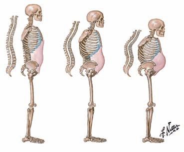
OsteOpOrOsis

Guidelines    FOr health prOFessiOnals

          The Irish Osteoporosis Society wish to acknowledge the support of an
          unrestricted educational grant from Amgen and GSK in producing the booklet.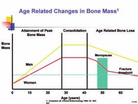
Acknowledgements
The Irish Osteoporosis Society would like to sincerely thank
Professor Moira O’Brien, Michele O’Brien, Genette Owens (NOS) and
Dr John Carey for their help in different area’s of the guidelines.
IrIsh OsteOpOrOsIs sOcIety OsteOpOrOsis Guidelines

Contents

Introduction                                                                              3

Social & Economic Cost of Fractures                                                       4

Extent of Problem                                                                         5

Bone                                                                                      6

Vertebral Fractures                                                                       8

Hip Fractures                                                                             9

Other Fractures                                                                          10

Signs and Symptons of Osteoporosis                                                       11

How is Osteoporosis Diagnosed                                                            13

What to measure                                                                          15

Causes of Osteoporosis                                                                   17

Hormonal Changes                                                                         18

Factors that Predispose to Osteoporosis                                                  19    1
Diagnosis of Osteoporosis by DXA                                                         23

Contraindications to a DXA scan                                                          24

DXA Tips                                                                                 25

What a patient can expect when having a DXA scan                                         26

Why a DXA scan is important                                                              27

DXA rescanning                                                                           28

Who needs a DXA scan?                                                                    29

Clinical indications for bone mass measurements                                         30

Fracture Risk Assessment (FRAX)                                                          31

Results of a DXA scan                                                                    32

If a patients T score has declined                                                       33

Coeliac Disease / gluten sensitivity                                                    34

Investigations for Secondary Osteoporosis                                                35
IrIsh OsteOpOrOsIs sOcIety OsteOpOrOsis Guidelines

                   Vitamin D and Calcium                         36

                   Vitamin D                                     37

                   Vitamin D and the EU                          40

                   Calcium                                       41

                   Calcium and Vitamin D supplements             42

                   Exercise                                      43

                   Prevention of falls                           44

                   Managing pain                                 45

                   Management of Osteoporosis                    46

                   Choosing the Right Treatment                  47

                   Osteoporosis Treatments                       48

                   Approved Osteoporosis Treatments in Ireland   60

                   Life style advice for all age groups          61

                   Prevention                                    62

2                  Children and Adolescences at risk             63

                   Prevention in Children and Teenagers          67

                   Case Studies                                  68

                   DXA List                                      72

                   References                                    74
IrIsh OsteOpOrOsIs sOcIety OsteOpOrOsis Guidelines
Introduction
Osteoporosis is a systemic skeletal disease characterised by low bone mass, micro
architectural deterioration of bone tissue and compromised bone strength, with a
consequent increase in bone fragility and susceptibility to fracture, particularly of
the wrist, hip and spine 1.
Osteoporosis is the commonest bone disease worldwide and is a major Public
Health Hazard, with a high morbidity, mortality and socio-economic costs 2.
1
It is a silent painless disease until a fracture occurs. Fracture is the most
important clinical feature of osteoporosis, many of which are preventable.
Although effective treatments have been available for more than a decade studies
show many persons who fracture, or are at risk of fracture, are never evaluated
or treated for their underlying osteoporosis, who subsequently go on to have
additional fractures and the associated morbidity. 3
Osteoporosis is most common in postmenopausal white women, but it is not
just a disease of old ladies. Osteoporosis can occur at any age in both males
and females, and persons of all races 4. Approximately 25% of all fractures occur
in men. 1 in 5 men (over 50) and 1 in 2 postmenopausal women (over 50) will
develop a fracture during their lifetime. a postmenopausal woman’s annual risk
of fracture is greater than her combined risk of cardiovascular disease and breast
3
cancer.
Osteoporosis is treatable and fractures are preventable. One low trauma fracture
increases the risk of a second in the near future, if not diagnosed and treated. 6, 7IrIsh OsteOpOrOsIs sOcIety OsteOpOrOsis Guidelines
social & economic cost of
Fractures in Ireland
Osteoporotic Fractures Impose a Huge Social Cost in Ireland, approximately €402
Million per annum is spent to treat all falls and fractures which occur in senior
citizens with Osteoporosis in Ireland 9, 10
In addition to Healthcare costs, vertebral fractures can cause back pain, loss of
height, deformity, depression and low esteem. 12
If current trends continue it is estimated that costs will be:
€520 - €551 million by 2010
€922 - €1077 million by 2020
€1587 - €2043 million by 2030
4IrIsh OsteOpOrOsIs sOcIety OsteOpOrOsis Guidelines
extent of problem
Osteoporosis is now a major health problem worldwide and it is increasing due to
increased life expectancy. Approximately 300,000 Irish people aged 50 years and
over may have osteoporosis. 10, 13
■■ The life expectancy in 1995 was 75 years.
■■ In 2030 the predicted life expectancy is 84 years and Ireland has an ageing
population.
■■ Today 11% are aged 65 years or over (468,000).
■■ By 2031 that proportion will increase to 18% i.e. to over one million older
people.
■■ The biggest increase will be among those who are over 80 years of age 10
■■ In 2004 there were 6,113 hospital episodes where a diagnosis of osteoporosis
was recorded but this represents the ‘tip of the iceberg’ 13.
The number of osteoporosis sufferers is increasing at an alarming rate. This is
mainly due to people living longer, exercising less, poor nutrition particularly
inadequate daily intake of calcium and vitamin D.
There are now five times as many fractures each year due to osteoporosis as there
were in the 1960s. The number of people suffering from osteoporosis is set to
5
double in the next 20 years. 13
nOte: More women die from complications of osteoporotic fractures, (mainly
hip and vertebral fractures) than from a combination of all cancers of the ovary,
uterus and cervix, yet only approximately 15% of people with osteoporosis are
diagnosed.
Up to 30% of men will die in the year following a hip fracture which is almost
double the mortality of women. This sentinel event has a greater mortality
in men than heart disease and most cancers, yet men are less likely to be
diagnosed and treated for their osteoporosis than women. The only cancer that
supersedes Osteoporosis is lung cancer.IrIsh OsteOpOrOsIs sOcIety OsteOpOrOsis Guidelines
Bone
Bone is a living tissue that is constantly being removed and replaced. Building
a large bone mass early in life, can help to reduce the risk of developing
osteoporosis in later life. 14 Bone mineral density normally increases steadily from
birth and approaches its peak value by early adult life, depending on the skeletal
site, and remains stable for some years. 5 The greatest increase in bone occurs pre-
pubertal 8-12, depending on the child to the early 20’s, due to the hormones that
are produced around puberty 15. This is a very important period of bone growth,
during these years, the greatest amount of bone is formed and this is known as
“Peak Bone Mass” 15 For instance Peak bone mass occurs at the proximal femur in
women at about 18-20 years of age, spine 20-25 years of age and the skull may
continue to gain bone mass right through the 4th and 5th decades of life. Peak
bone mass occurs at a similar but slightly later age at these sites in men.
6
BMD is on average lower in women than in men, because women have smaller
bones and smaller trabeculae. Women lose more bone on average in their
lifetime than men, as they also go through the menopause, 35-40% in men Vs
50% in women. Muscle contraction increases bone strength 16 and immature
bone responds better to the stimulus of muscle contraction than mature bone.17
Weight bearing exercise is essential in young people, as not only can it reduce
their risk of developing osteoporosis but also many other problems such as:
obesity, hypertension, Type 2 diabetes, heart disease, strokes, low self esteem and
depression.
Bone mass is the result of a dynamic lifetime balance between two processes:
bone formation and bone resorption. Bones require normal levels of sexIrIsh OsteOpOrOsIs sOcIety OsteOpOrOsis Guidelines
hormones, adequate caloric intake, particularly protein, calcium and vitamin D
and regular weight bearing exercise. The rate of bone turnover is determined by
hormonal and local factors, as well as systemic factors, illnesses and genetics.
Up to the age of 20, more bone is laid down than is lost. Following that,
depending on the skeletal site, the amount of bone lost and replaced is
approximately the same, between the late twenties and early forties in healthy
persons. The rate of bone turnover is affected by many factors, including sex
hormones such as oestrogen and testosterone, vitamin D and parathyroid
hormone, and many cytokines and chemokines including tumour necrosis factor
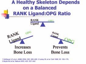
alpha, Receptor Activator of Nuclear Factor Kappa B (RANK ), RANK Ligand, and
its naturally occurring decoy receptor osteoprotegerin (OPG) 18, 19, 20.
Throughout the skeleton there are basic remodelling units where signals from
a resorption pit signals through a variety of factors resulting in activation of
osteoclasts and bone resorption at that site, known as a resorption pit. This results
in a coupled signal to osteoblasts which form new bone. When more bone is
formed there is net bone gain and vice versa as bone is lost. It is estimated that
each human skeleton is remodelled in its entirety several times in the average
adults living into their 8th decade.
In today’s life style, too little or excessive exercise, combined with a low calorie
diet, low intake of calcium and vitamin D and an excessive fibre content, are
counter productive to achieving an adequate peak bone mass. Adolescence is also
the time, when there is an increased risk of eating disorders. 15
7IrIsh OsteOpOrOsIs sOcIety OsteOpOrOsis Guidelines
Vertebral fractures
■■ Vertebral fractures are the commonest osteoporotic fracture, accounting for
almost 50% of all fractures in most epidemiologic studies.
■■ The incidence of vertebral fractures begins to increase in late middle age,
mirroring the age related decrease in bone mass.
■■ Only 1/3 of vertebral fractures are painful, with the remaining 2/3 being
clinically silent. 8
■■ Patients may present with height loss, kyphosis, back pain or restrictive lung
disease. Patients may find it difficult to reach previously accessible shelves due
to loss of height, a dowager’s hump and back pain, and it is the loss of height
that can be a strong indication for possible osteoporosis. These patients should
be sent for a DXA scan to see if they have osteopenia and/or osteoporosis.
When possible a DXA with a lateral view otherwise a lateral thoracic x-ray, to
see if there are spinal fractures present on x-ray.
■■ Similar to hip fractures, vertebral fractures are associated with a higher risk
of future fracture, increased morbidity and mortality. Most studies show
8 that osteoporosis therapies reduce the risk of future vertebral fracture by
approximately 50 to 60% and by as much as 70% in those with a prior fracture.IrIsh OsteOpOrOsIs sOcIety OsteOpOrOsis Guidelines
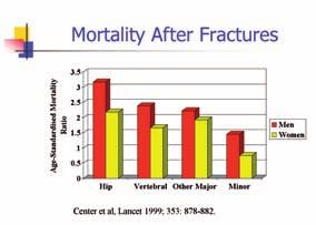
hip Fractures
■■ Hip fractures represent the single most important clinical event in osteoporosis.
They are associated with the greatest cost, highest morbidity and mortality of
any fractures.
■■ Although hip fractures can be missed, they are usually clinically obvious.
■■ In contrast to vertebral fractures and distal radial fractures the incidence of
hip fractures increases exponentially after age 70, so that 90% of hip fractures
occur after this age. One particularly high-risk group for hip fractures is nursing
home residents. The rate of hip fracture among residents of nursing homes is
between 3 and 11 times that of age-matched community-dwellers. 11
■■ 20% of people aged 60+ who fracture their hip will die from complications
within six months to one year. The secondary complications of a hip fracture
are: a blood clot, pneumonia or infection. Men account for approximately 25%
of all fractures and are twice as likely to die as women following such fractures.
■■ 50% aged 60+ who fracture their hip will be unable to wash, dress or walk
across a room unaided. 12
■■ Only 30% aged 60+ who fracture their hip will regain their independence.
9
■■ There is a hip fracture every 30 seconds in the EU, approximately 1700 per day.
This number is expected to double by 2050.
■■ Most studies show that where there is evidence of therapeutic efficacy for
preventing hip fractures such therapy reduces the risk of future hip fracture by
approximately 55%.IrIsh OsteOpOrOsIs sOcIety OsteOpOrOsis Guidelines
Other Fractures
The next most common fracture site is the distal radius accounting for 10-15% of
all fractures in postmenopausal women.
The risk of distal radial, especially Colles’ fracture type, rises after the age of 40 in
women and 50 in men. However this risk appears to plateau at about the age of
60 whereupon it is surpassed by the risk of vertebral and later, hip fracture.
Although not as costly as hip fractures, and without the same mortality, studies
show treatment of such fractures remains costly, and many patients have
significant morbidity following such a fracture.
Other skeletal sites at risk of fracture include the long bones of the skeleton
and pelvic bones. Generally fractures of the fingers and toes are not considered
osteoporotic fractures.
10IrIsh OsteOpOrOsIs sOcIety OsteOpOrOsis Guidelines
signs and symptoms of
Osteoporosis
■■ A fragility fracture is generally agreed to be when someone suffers a broken
bone from a force that is less than or equal to that sustained from a fall from a
standing position, e.g. from a wrist or hip fracture following a trip and fall. With
severe osteoporosis even forces as little as a cough, sneeze, turning over in bed
or lifting a bag of groceries can result in a fracture. nOte: if a person’s bones
are healthy, they should not break from a trip and fall or less as an adult.
■■ Although 50% of children will have broken a bone by adulthood, the
vast majority of these fractures are usually due to an injury, rather than
osteoporosis. However if more than one low trauma break occurs, an
assessment of that person including a careful history and examination is
warranted.
■■ Development of a kyphosis - A persons head is bent forward – may result from
anterior wedge fractures of the spine. In severe cases a hump may develop
on a person’s upper back (Dowager’s hump) which is a strong indication that
osteoporosis should be considered.
■■ Loss in height 2-16cm. It is not normal at any age to suddenly loose height.
11
Height loss of >2 inches is an important sign of an asymptomatic vertebral
fracture and such persons should be evaluated for osteoporosis. A person can
loose height due to wear and tear of vertebrae and/or disc but >2 inches is
unusual in degenerative joint and disc disease. Unfortunately some persons are
measured incorrectly but when the history fits with the measurement, evidence
of vertebral fractures should be screened.
■■ Change in body shape or size is usually associated with loss of height. A
distended abdomen can then develop as there is no place for the stomach
and intestines to go, other than outwards followed by the rib cage ending
up resting on the pelvis. These changes can cause difficulty in breathing,
back pain, depression, loss of functional independence and gastrointestinal
symptoms.
■■ Persons who experience sharp sudden pain in the low, middle or upper back,
especially with height loss should be evaluated for vertebral fractures as this
may be the first presentation of an osteoporotic fracture. When plain films are
normal and symptoms persist, repeat X-rays several weeks later or additional
imaging may show a fracture. Cause of back pain should always be addressed
and vertebral fractures ruled out.Irish Osteoporosis Society Osteoporosis Guidelines 12 The devastating effects of undiagnosed osteoporosis
IrIsh OsteOpOrOsIs sOcIety OsteOpOrOsis Guidelines

how is Osteoporosis
Diagnosed?
Osteoporosis can be diagnosed in the appropriate clinical setting one of 3 ways:
1.   The presence of a fragility fracture
2.   Measurement of bone mineral density (BMD)
3.   Histomorphometric analysis of tetracycline-labelled Bone biopsy.

1. Fragility Fractures
Studies show that fracture risk is highest before the age of 20 years and after the
age of 50 years. In addition they also show the majority of fractures occurring
after 50 years of age are osteoporotic. However not all fractures are low trauma,
e.g. falling off ladders, bicycles, skiing accidents etc. All persons presenting with
a fragility fracture after 50 years of age or menopause should be considered as
possibly osteoporotic. A detailed history of the fracture occurrence, physical
examination, evaluation for other fractures, (note presence of back pain, kyphosis,
and height loss) and additional testing is warranted. Additional testing should
include measurement of bone mineral density where possible and if there is height
loss and/or back pain, imaging of the spine. Blood and urine tests should also be
considered. Remember clinicians diagnose osteoporosis.
                                                                                                 13
2. Measurement of BMd
Studies show BMD accounts for 70% of bone strength in men and women. The
accepted gold standard for non-invasive measurement of BMD today is central
DXA (Dual-energy X-ray Absorptiometry). This method uses very low dose
radiation to measure BMD at the lumbar spine (L1-4) and proximal femur. Criteria
have been developed for diagnosing osteoporosis by measuring BMD at these
sites and only with these devices.

However BMD can be measured in a variety of other ways including ultrasound
of the heel and other small bones, CT scans of the lumbar spine, peripheral DXA
devices and single X-ray absorptiometry. At this time the Irish Osteoporosis
Society only recommends a DXA scan of the spine and hips to diagnose
osteoporosis.

Measurement of BMD remains a critical component of osteoporosis assessment
to establish a diagnosis and monitor therapy. Most currently available therapies
have generally only been evaluated in clinical trials of persons with low BMD. All
guidelines use BMD T-scores and Z-scores as the basis for their recommendations
on who to treat and when. Site specific BMD is a better predictor of fracture
risk and since approximately 70-75% of all osteoporotic fractures occur at the
IrIsh OsteOpOrOsIs sOcIety OsteOpOrOsis Guidelines
spine and hip, and fractures at these sites have the greatest socioeconomic
cost, morbidity and mortality, they remain the most important skeletal sites for
diagnosis and prevention.
3. Bone Biopsy
This last method is not routinely used or available. This should never be
undertaken without consultation with a specialist in osteoporosis and metabolic
bone disease.
14IrIsh OsteOpOrOsIs sOcIety OsteOpOrOsis Guidelines
What to measure?
The currently recommended international standard for bone densitometry is to
measure the lumbar spine (L1-L4) and proximal femur. Although central DXA can
measure BMD of the forearm, such measurement is not routinely recommended.
However in certain circumstances this may be of value e.g. patient too heavy for
the DXA scanner (most have cut-offs in the region of 250-280lbs), if the patient
has hyperparathyroidism, or the patient is unable to get up on a scanner without
assistance, patient has severe disorders of spine and hip, making measurement
impossible to interpret at these sites.
In 1994 the World Health Organisation proposed osteoporosis diagnostic criteria
for BMD measured at the proximal femur in postmenopausal women. 21 These have
been modified somewhat over the years and currently the ISCD recommends that
such criteria may be applied to postmenopausal women and men over 50 years of
age.
these criteria use the t-score for diagnostic classification into three main groups:
1. Normal BMD: T-score > or equal to -1.0.
2. Low bone mass or osteopenia: T-score -1.5 to -2.49
3. Osteoporosis: T-score -2.5 or less. Example: -3.5
4. A footnote to these criteria stated that persons with prevalent fragility
fractures and T-scoresIrIsh OsteOpOrOsIs sOcIety OsteOpOrOsis Guidelines
Standard radiographs of the spine are widely available and may show distinctive
radiographic features of osteoporotic fractures. They are insensitive indicators
of bone loss, since bone density must be decreased by at least 30-50% before
reduction can be appreciated. If osteopenia (that is, low bone mass) is suggested
on an x-ray this is an indication for a DXA scan.
Like all diagnostic tests, DXA is imperfect. BMD is a continuous measure and
threshold values will result in misclassification of some individuals. DXA is a
2-dimensional test which measures bone mineral density of a 3-dimensional
structure. Thus persons with very large or very small bones and those with mineral
disorders may have alterations in their BMD that are not due to osteoporosis.
Thus not all persons with ‘low BMD’ have ‘osteoporosis’ or ‘osteopenia’ and
not everyone with ‘normal BMD’ has normal healthy bone. Thus DXA needs to
be interpreted in the appropriate clinical context and should not be taken as
a panacea for diagnostic and treatment decisions. Note clinicians diagnose
osteoporosis.
New developments in DXA technology enables patients to have a scan of their
spine if appropriate at the same time as they are having their BMD measured.
This technique known as LVA (lateral vertebral assessment) obtains a single view
of the spine (usually T5-L5) where most vertebral fractures occur. Thus clinicians
can evaluate for vertebral fractures in patients on corticosteroids, with height loss
16 or undiagnosed back pain at the time of their DXA scan if required. Studies show
persons with prevalent vertebral fractures and low BMD are at much higher risk of
future fracture than persons with either low BMD or prevalent fracture alone.
note: More details of what to measure and how to interpret bone densitometry
are available from many sources, example: The International Society for Clinical
Densitometry (www.iscd.org) and discussed later in this publication.
note: All devices using ionising radiation in Ireland are governed by the 1991
Radiological Protection Act and also the 2005 Safety, Health and Welfare at
work Act (details available at The Radiological Protection Institute of Ireland
at: www.rpii.ie. The amount of radiation for a central DXA today is similar to
ambient daily exposure.IrIsh OsteOpOrOsIs sOcIety OsteOpOrOsis Guidelines
causes of Osteoporosis
Osteoporosis is multifactorial in origin. Generally it is classified as “primary”
or “idiopathic” and “secondary”. Others have chosen the term ‘involutional’
osteoporosis to reflect the “normal” bone loss and fracture risk increase that is
evident in otherwise healthy persons as they age. The problem with these terms is
that as our understanding of osteoporosis increases, little is idiopathic and most
persons with “primary” or “involutional” have identifiable risks that are amenable
to intervention, making the term problematic. 3, 24 Secondary osteoporosis refers
to the condition when it arises as the result of a specific condition, e.g. rheumatoid
arthritis, or medication, e.g. corticosteroids. The problem with this term is similar
to what has been already stated since not everyone with these conditions may
develop osteoporosis and oversimplification has meant that other disorders such
as osteomalacia are often confused with osteoporosis by clinicians who base their
diagnosis solely on DXA readings.
Current best evidence suggests that the majority of BMD in most populations is
accounted for by genetic factors (approx 70%) being greatest in monozygotic
twins (80%). Genes and lifestyle have their greatest impact on peak bone mass.
Calcium rich balanced diets, adequate vitamin D intake (either dietary or from sun
exposure), regular weight bearing exercise, avoidance of illness or medications
that impair bone growth have all been shown to optimize peak bone mass. Peak
bone mass is achieved at different times in different parts of the skeleton and
17
generally slightly earlier in women than men. Peak bone mass is generally greater
in men than women and lowest in those of Caucasian, and some Asian races.
Bone loss occurs throughout the rest of adult life in healthy individuals but varies
in rate between individuals and skeletal sites. Many factors influence this rate
of loss including lifestyles, certain illnesses and medications and also hormonal
changes such as menopause. In healthy persons little bone loss occurs in the 20s
and 30s in men and premenopausal women. Accelerated bone loss will increase
a person’s fracture risk and result in lower BMD over time than persons with
attenuated bone loss, thus putting persons at greater fracture risk.IrIsh OsteOpOrOsIs sOcIety OsteOpOrOsis Guidelines
hormonal changes
The commonest cause of osteoporosis is the loss of sex hormones, oestrogen
in females and perhaps testosterone in males (oestrogen may actually be more
important!) 5, 15.
The female hormone oestrogen may be lost due to a variety of causes, e.g. the
menopause, stress, irregular periods or no periods for 3 months or longer (not
due to pregnancy) or eating disorders. Loss of oestrogen can result in significant
and accelerated bone loss, particularly in the first 5-10 years following menopause.
Oestrogen deficient bone loss appears to be primarily mediated by tumour
necrosis factor alpha, stimulation of osteoclastogenesis in a dose dependent
manner. Oestrogen deficient bone loss can be attenuated somewhat, though
not completely, by healthy lifestyle including adequate calcium intake, regular
weight-bearing exercise and adequate or supplemental vitamin D. However
pharmacological therapy that reverses or inhibits this bone loss is usually required
if this is to be prevented in the long-term.
Although testosterone deficiency is clearly associated with osteoporosis in men,
unlike women a cause and effect relationship has not been established. Oestrogen
in fact may be more important, at least for bone growth. The relationship is likely
18 more complex than was originally thought. The best evidence of the role of
testosterone comes from prostate cancer therapies where androgen deprivation
therapies have been shown to increase bone loss and consequently fracture risk.
However there is little evidence that testosterone replacement reduces fracture
risk. Signs of low testosterone levels (Hypogonadism) are: loss of sex drive, loss
of erections, depression, and/or fatigue. The leading world experts on male
osteoporosis today generally recommend that osteoporosis and testosterone
deficiency be treated as separate entities. 26
Due to the increase in sedentary life style, particularly in children and teenagers
and the increasing number of senior citizens, the incidence of osteoporosis will
significantly increase.
The good news is that the risk of developing osteoporosis can be reduced, by
taking appropriate preventative measures (such as diet and lifestyle changes), and
through early diagnosis and treatment.
an extensive risk factor questionnaire is available from the charities web site:
www.irishosteoporosis.ieIrIsh OsteOpOrOsIs sOcIety OsteOpOrOsis Guidelines
Factors that predispose to
Osteoporosis
Multiple factors contribute to low bone mass and osteoporotic fractures.
Many medical conditions, or their medications, can increase the risk of
osteoporotic fractures. Not all risk factors have had extensive research, however
all can place a person at risk of developing osteoporosis. All causes should be
found and addressed 5.
■■ Genetic: A family history of osteoporosis is a very strong risk factor,
particularly if it includes a history of hip fracture/s, as approximately 80% of a
persons bone is genetic.
■■ age, senior citizens are more at risk: Senior citizens are more likely to have
low oestrogen and testosterone levels, low vitamin D levels, poor nutrition, take
less exercise and have other medical conditions or be on a medication that can
increase bone loss.
■■ Previous Fracture after minor trauma
■■ Low Bone Mineral Density by DXA of spine and hips
■■ Loss of height – more than 2cm
■■ Undiagnosed upper, middle or low back pain
■■ Undiagnosed hip pain
■■ Low body weight for height
19
■■ endocrine Disorders such as Hypogonadism for any reason, e.g. Surgical
removal of ovaries/s or testes, or infections such as mumps after puberty in
males.
■■ All forms of Turner’s syndrome in females and Klienfelter’s Syndrome in males.
■■ Late menarche, after age 15, prolonged amenorrhea or history of very irregular
menstruation, frequent loss of periods for more than 3 months (not due to
pregnancy).
■■ Endometriosis
■■ Premature menopause (before 45 years)/ Oophorectomy or early menopause,
either natural, surgical or due to radiation or chemotherapy are also at
increased risk.
■■ Depo-Provera contraceptive has been proven to cause bone loss, particularly
high risk if given during adolescence when bone is being laid down.
■■ Eating disorders (Anorexia Nervosa and/or Bulimia – past or present)
■■ Athletic Triad (Amenorrhea, Eating Disorder and Osteoporosis or osteopenia) 15
■■ Osteoporosis of pregnancy or lactation: Osteoporosis of pregnancy may occur
during the third trimester of pregnancy or postpartum. Calcium and vitamin D
should be given, however a DXA scan and treatment should not be initiated till
after the birth of the infant.
■■ Males: Low levels of the male hormoneIrIsh OsteOpOrOsIs sOcIety OsteOpOrOsis Guidelines
■■ Hyperadrenocorticism: endogenous or exogenous, e.g. Cushing’s Syndrome
■■ Hyperthyroidism
■■ Hyperparathyroidism (Primary or secondary due to low vitamin D or poor renal
function)
■■ Acromegaly
■■ Hypopituitarism.
■■ Hyperprolactinaemia
■■ Insulin dependent Diabetes
■■ Haemochromatosis
■■ Hypophosphataemia
■■ Hypercalcuria
renal
■■ Renal Osteodystrophy,
■■ Chronic renal insufficiency,
■■ Renal tubular acidosis
Mobility
■■ Inactivity, or prolonged immobility (especially bed or wheelchair bound) for
more than six weeks or long term, especially in childhood when bone is being
laid down.
20 race
■■ Asian and Caucasians are more at risk, however all races can develop
osteoporosis. nOte: Dark skinned people tend to have larger bones, however
they have decreased ability to absorb vitamin D from the sun.
Vitamin d deficiency
■■ Vitamin D resistant rickets
■■ Low Vitamin D
■■ Osteomalacia
nutritional and lifestyle
■■ Excessive protein increases calcium loss
■■ Excessive fibre, over 40g a day
■■ Excessive caffeine intake
■■ Excessive alcohol intake >7 pints for women (14 units a week) & 11 pints for
men a week (21 units a week)
■■ Smoking
■■ Excessive exercise, particularly with inadequate caloric intake
■■ Excessive psychological stress
■■ Excessive physiological stressIrIsh OsteOpOrOsIs sOcIety OsteOpOrOsis Guidelines
Gastrointestinal disorders
■■ Malabsorption problems; Coeliac or Gluten sensitivity, lactose intolerance or
Cystic Fibrosis
■■ Inflammatory Bowel Disease; Chron’s Disease, Irritable Bowel, Ulcerative Colitis.
■■ Gastrectomy or small bowel resection
■■ Severe liver disease
■■ Chronic obstructive jaundice
■■ Primary Biliary cirrhosis
■■ Amyloidosis
■■ Gaucher’s disease
■■ Severe malnutrition
Bone Marrow disorders
■■ Multiple Myeloma
■■ Systemic Mastocytosis
■■ Lymphoma
■■ Disseminated Carcinomatosis
Collagen disorders and other medical conditions
■■ Rheumatoid Arthritis
■■ Osteogenesis Imperfecta
■■ Childhood Idiopathic Osteoporosis
■■ Ehlers-Danlos Syndrome
21
■■ Marfan’s Syndrome
■■ Homocystinuria
■■ Polymylagia
■■ Sarcoidosis
■■ Psoriatic arthritis
■■ Ankylosing Spondylitis
neurological
■■ Stroke
■■ Dementia
■■ Multiple Sclerosis
■■ Spinal cord lesions
■■ Muscular Dystrophy
■■ Idiopathic Scoliosis
Other conditions
■■ Psychotic patients
■■ Down syndrome or similar with secondary complications
■■ Pernicious Anaemia
■■ ThalassemiaIrIsh OsteOpOrOsIs sOcIety OsteOpOrOsis Guidelines
■■ Haemophilia
■■ Congenital Porphyria
■■ Cancer; Leukaemia, Lymphoma
■■ Severe eczema
■■ COPD
■■ AIDS/ HIV
drug induced
■■ Long-term use of Corticosteroids (e.g. Cortisone, Prednisolone, Delta Cortril,
dexamethasone etc). Corticosteroids, are used for the treatment of many
conditions, and are the most common cause of secondary osteoporosis.
●■ Main bone loss occurs in the first six months of treatment.
●■ Corticosteroids 7.5 mg a day for more than 3 months in a year. Bone loss
may occur at lower doses in some people, particularly if there are other risk
factors5 or if they already have undiagnosed low bone density.
■■ Chemotherapy
■■ Radiation
■■ Thyroxine, if serum levels are high
■■ Post organ transplant
■■ Anticonvulsant therapy, Anti-epileptic medications (phenytoin,
phenobarbitone) can interfere with calcium absorption and the production of
22 vitamin D.
■■ Chronic heparin or Warfarin therapy
■■ Long term lithium therapy
■■ GnRh analogues
■■ LHRH analogues; testosterone suppression ;leuprorelin
■■ Prolactin raising drugs, Antipsychotic medication, e.g. some SSRI
■■ Aromatase inhibitors for the treatment of Prostatic and Breast Cancers:
Arimidex for breast cancer
■■ Diuretics such as Burinex, Lasix.
■■ Proton Pump Inhibitors
■■ Tranquillizers and sedatives may increase the risk of a fallIrIsh OsteOpOrOsIs sOcIety OsteOpOrOsis Guidelines
Diagnosis of Osteoporosis by
DXA
Dual Energy X-ray Absorptiometry (DXA), is a non-invasive method and currently
is the most precise and widely used method of assessing Bone Mineral Density. In
the majority of cases, a DXA is a large non-mobile machine and should be set to
measure the bone density of the spine and both hips.
Bone mineral density measurements are currently the best predictors of fractures,
but are site specific.
For every one standard deviation decrease in BMD, the relative risk of fracture is
significantly increased. Bone mineral density varies at different sites. In a large
majority of cases the spine is the first region to lose a significant amount of bone
mineral density, but in a percentage of women, the loss occurs first in the hip.
Therefore it is essential that both hips are scanned, as fractures of the hip have the
highest mortality and morbidity rates.
It is a painless method for measuring bone mineral density (BMD) and is the Gold
standard for diagnosing osteoporosis, and is recommended by the International
Osteoporosis Foundation.
nOte: if the patient has had a previous DXa scan, the new DXa results should
23
be compared to the previous DXa result.
nOte: the lower the BMD result, the greater the risk of fracture. low bone
density in the hip and vertebrae are more dangerous as they are associated
with a high mortality rate post fracture.
lateral Vertebral assessment: a lateral view of the thoracic and lumbar spine
is available on some DXA machines and shows if there is compression of the
vertebral bodies. This is recommended especially, if there is loss of height or a
kyphosis has developed on the upper back. If not available, a Lateral thoracic
X-ray of the upper back can be done.
Dual (DXA) and Single’ (SXA) Energy X-Ray Absorptiometry both use an x-ray
source with low levels of radiation. The x-ray radiation is 10% of a normal chest
x-ray; hence the level of exposure is much lower.IrIsh OsteOpOrOsIs sOcIety OsteOpOrOsis Guidelines
contraindications to a
DXA scan
■■ Pregnant or possibility of being pregnant
■■ If a patient has had an investigation using contrasts material recently, e.g.,
barium meal or barium enema or an intravenous pyleogram, there needs to be
one week between the tests.
■■ The patient should inform the DXA operator if they have a metal
Implant in the spine or hip, or if they have any metal body piercing.
24IrIsh OsteOpOrOsIs sOcIety OsteOpOrOsis Guidelines
DXA tips
■■ Do not place a DXA machine by a radiator
■■ Both hips should be scanned, there may be a discrepancy between the BMD of
the two hips, one hip may be normal but not the other.
■■ Individual T scores should be looked at, not just the total: L1, L2, L3 & L4
■■ Both areas of the hip should be looked at, as the neck of femur could be lower.
■■ LVA, if there is loss of height or a dowager’s hump, as the software is only
available recently, many machines can not scan the upper back. Lateral
thoracic X-ray should be done if the software is not available.
■■ Repeat DXA should preferable be on the same machine.
25IrIsh OsteOpOrOsIs sOcIety OsteOpOrOsis Guidelines
What a patient can expect
when having a DXA scan
■■ Patient’s height and weight should be measured.
■■ Patients will be asked to remove any metal, such as belts, body piercings and a
bra with an under wire.
■■ There must be no metal in the area that is to be scanned e.g. If a patient has
a hip replacement, the other hip and the lumber vertebrae (L1-L4) can be
scanned.
■■ They will be asked to lie still on the machine during a scan. An electronic arm
will slowly travel over the area of the body to be scanned. It is important for
the patient to be able to remain still, so the images recorded are not distorted.
■■ The patient will lie down on the machine for 5-15 minutes while the moving arm
of the machine passes over them, to take an image of their spine and hips.
■■ The test is not claustrophobic, is not painful and costs approximately €100.
26IrIsh OsteOpOrOsIs sOcIety OsteOpOrOsis Guidelines
Why a DXA scan is important
The disease is silent and since it affects the inside of a person’s bone, a person
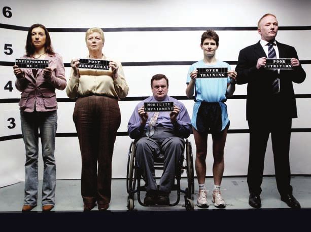
can look perfectly fine on the outside, yet have severe osteoporosis. “Not so usual
suspects” picture on this cover is available from the IOS charity.
The most efficient way to monitor a patient’s response to treatment is with a
repeated DXA scan, as bone markers are not always available.
The bone mineral density result can help to encourage the patient to make a
decision about compliance of treatment and help to encourage them to change
their life style.
A DXA scan can is particularly important in young people with poor diets or
eating disorders, so that they can see if they survive, they could end up being
disfigured. It is also an excellent tool to monitor these patients for compliance.
27IrIsh OsteOpOrOsIs sOcIety OsteOpOrOsis Guidelines
DXA rescanning
It is recommended to be re-scanned on the same machine, when possible, for
greater accuracy in monitoring the response to treatment. Most people are
re-scanned every 2 years, however in certain cases, if compliance is an issue, a
scan could be done after 12 to 18 months to help increase compliance. There is a
world wide compliance problem with osteoporosis patients, which is why it is so
important to monitor a patient.
example: Eating disorders: seeing significant improvement or decline can help
to increase compliance and patient motivation.
example: In the case of someone who does not start or stops their medication, a
decline in their bone health can assist with compliance.
28IrIsh OsteOpOrOsIs sOcIety OsteOpOrOsis Guidelines
Who needs a DXA scan?
If a person has one or more risk factors for Osteoporosis, regardless of age,
male or female, a DXA scan should be considered. Since it is a silent disease,
there are no signs or symptoms prior to fractures. Compliance with taking the
medications along with calcium, vitamin D and weight bearing exercise is much
higher when results show loss of bone and risk of fracture. It is much cheaper to
scan if in doubt, than wait to see if a patient fractures. One fractured hip including
rehabilitation costs approximately €31,000. A DXA scan costs approximately €100.
29IrIsh OsteOpOrOsIs sOcIety OsteOpOrOsis Guidelines
clinical Indications for bone
mass measurements
■■ Postmenopausal women under age 65 with risk factors for fracture
■■ Women during the menopausal transition with clinical risk factors for fracture,
such as low body weight, prior fracture, or high-risk medication use.
■■ Women discontinuing oestrogen or oestrogen deficient
■■ Early menopause, secondary amenorrhoea, athletic triad
■■ Anorexia and/or bulimia
■■ Men aged 70 and older
■■ Men under age 70 with clinical risk factors for fracture: hypogonadism
■■ Adults with a fragility fracture.
■■ Men, women or children with a disease or condition associated with low bone
mass or bone loss.
■■ Men, women or children being considered for pharmacologic therapy that is
known to cause bone loss. e.g. Steroid therapy > 5mg or 7.5 mg Prednisolone
daily
■■ Men, women or children, to monitor treatment effect.
■■ Men, women or children not receiving therapy in who evidence of bone loss
would lead to treatment.
30 ■■ Menopausal women in whom result will influence treatment
■■ Recurrent stress fractures not due to biomechanical causes
■■ Drugs associated with osteoporosis, e.g. Anti-coagulants, Epinutin Aromatase
inhibitors etc
■■ Radiographic indication of vertebral deformity or osteopeniaIrIsh OsteOpOrOsIs sOcIety OsteOpOrOsis Guidelines
Fracture risk
Assessment (FrAX)
The FRAX tool has been developed by WHO to evaluate the fracture risk of
patients.21, 22, 23 FRAX is based on individual patient models, that integrate the risks
associated with clinical risk factors, as well as bone mineral density (BMD) at the
femoral neck. Assessment of fracture risk can be improved by the use of clinical
risk factors, which act independently of bone mineral density to increase the risk
of fracture, and this forms the basis of the WHO approach.
The aims of FRAX are to optimize sensitivity (i.e. detection rate) of fracture risk
prediction, using a case finding strategy in men and, women that can be widely
implemented in primary care. FRAX tool 23, 22, 23 computes the 10-year probability
of a hip fracture or a major osteoporotic fracture (Clinical spine, hip, forearm or
humerus).
However only eight easily identifiable risk factors shown to improve the
prediction of fracture risk are included in FRAX: age, family history of hip fracture,
glucocorticoid (steroid) use, current smoking, alcohol use >2 units/day and,
rheumatoid arthritis. Individually, the presence of these risk factors were shown
to increase the risk of hip fracture at least 1.5 to 2-fold after adjustment for bone
mineral density.3, 22, 23 FRAX has some limitations, as it only accepts one risk factor;
31
while many cases of Osteopenia and/or Osteoporosis have multiple risk factors,
which can significantly increase their risk of fractures, but this will not show up on
FRAX. 23IrIsh OsteOpOrOsIs sOcIety OsteOpOrOsis Guidelines
results of a DXA scan
The results will be presented in the form of a computerised printout that gives the
Bone Mineral Density in Grams/cm2 and a T-score value of the hips and lumbar
Spine.
A T-score compares the patient’s results with the mean peak bone mass (BMD) of
a large number of normal females or males between the ages of 20-40 years. T
scores should only be used in the diagnosis of adults over 21 years of age.
A Z score compares the patient’s results with a large number of normal females or
males of the same age group.
WHO defines a Normal BMD as a T score value greater than -1, which indicates
that the bone mineral density is normal. example: +1.2, -0.5
Osteopenia is a T-score value of between -1 and -2.5, which is the precursor to
osteoporosis, therefore it is essential to put preventative measures in place.
The IOS has broken up the scores in the Osteopenia range, to make it easier
for people to know exactly where they are on the scale. There is a significant
32 difference in fracture risk between a person with mild osteopenia and marked
osteopenia.
Normal T-score = 0 to - 1.0
Mild Osteopenia T-score = -1.0 to -1.49
Mod Osteopenia T-score = -1.5 to -1.9
Marked Osteopenia T-score = -2.0 to -2.49
Osteoporosis T-score = Greater than -2.5
Or
A low trauma fracture (broken bone from a trip and fall from a standing position
or less) is also considered to be osteoporosis unless proved otherwise.
* Research shows that most fractures occur within a T score of -1.5 to -2.49 which
is the moderate to marked osteopenia range. Therefore it is essential that those
in this range are treated preventively, depending on the cause/s, calcium and
vitamin D and life style changes may not be enough.IrIsh OsteOpOrOsIs sOcIety OsteOpOrOsis Guidelines
If a patients t score has
declined
If a decline has occurred, the cause/s should be found and addressed.
Malabsorption is one of the major reasons why a person may have lost BMD.
example: Undiagnosed Coeliac or Undiagnosed gluten sensitivity. Changing a
medication due to a decline in bone density, without finding the cause of this
decline, can place the patient at the risk of possible fracture/s and medical costs
which could possibly be avoided.
example: A patient may have high parathyroid hormone levels due to either
primary hyperparathyroidism, or secondary due to low serum vitamin D levels.
example: A patient may have developed another pathology.
example: The patient may not have taken the medication or may not have taken
it correctly.
example: The patient may not have taken their calcium and vitamin D.
33
example: It could be a combination of several examples listed above.
nOte: Calcium, Vitamin D and weight bearing exercise are essential combined
with the osteoporosis medication for optimal results.
nOte: It is important to explain to the patient their risk of fracture or re-fracture,
to help improve compliance, as with most treatments they will not feel any
different when they take the medication.IrIsh OsteOpOrOsIs sOcIety OsteOpOrOsis Guidelines
coeliac Disease/gluten
sensitivity
Ireland has one of the highest rates of Coeliac disease in the world. However there
are many people who appear not to be “true” Coeliac. Many people think that
bloating of the stomach after food, is because they have eaten too much or eaten
too fast. This is why every patient should be asked if they have any symptoms of
Coeliac disease.
If you have a patient who presents with any of the following symptoms and their
Coeliac test has come back negative, a trial of gluten free food is suggested to see
if they are “gluten sensitive”.
Symptoms – a person can have one or more of these problems:
■■ Bloating of abdomen after food, especially white bread, pasta, cakes, beer:
Foods that contain gluten.
■■ Stomach pain
■■ Diarrhoea (loose stools, stools float in toilet, lighter colour, bad smell)
■■ Constipation
■■ Mouth ulcers
34 ■■ Chronic tiredness
■■ Anaemia
■■ Weight loss
■■ Bone pain
■■ Moodiness
■■ Depression
■■ FlatulenceIrIsh OsteOpOrOsIs sOcIety OsteOpOrOsis Guidelines Investigations for secondary Osteoporosis (depending on history and age) ■■ Full blood count ■■ Erythrocyte sedimentation rate ■■ Serum Ferritin, (Ferritin saturation, if ferritin is high) ■■ Renal function including Urea and electrolytes + Creatinine clearance ■■ Blood sugar ■■ Liver function tests ■■ Serum Calcium, phosphate, and alkaline phosphatase to exclude osteomalacia or primary hyperparathyroidism. ■■ Parathyroid hormone and serum 25(OH) vitamin D ■■ It is advisable to measure the serum PTH (Parathyroid Hormone) prior to prescribing 1-34 PTH (Forsteo) or 1-84 PTH (Preotact) ■■ Thyroid function tests ■■ Serum protein and electrophoresis to exclude multiple myeloma ■■ Coeliac Disease antibodies IGA tissue transglutaminase Antibodies (tTG, less than 1.9U/ML = negative if positive EmA test. Gluten Gliadin There is a possibility of gluten sensitivity even if these tests are negative, if symptoms are present, as many people are not true Coeliacs and a patient going gluten free, 35 may help to eliminate these symptoms. ■■ Prostate specific antigen in men ■■ Cortisol levels ■■ Follicle stimulating hormone, luteinizing hormone, sex hormone binding globulin, testosterone (in men) and oestradiol in males and females and also progesterone in females. Blood tests should be taken in the Luteal phase, after the 21st day of cycle in premenopausal women, to determine progesterone levels. ■■ Prolactin ■■ Insulin Growth Factor I, (IGFI) in anorexics and bulimics ■■ 24 hour urinary Calcium and Protein ■■ Bone markers if available. Serum Osteocalcin (Intact), Serum bone alkaline phoshatase PINP, Serum CTx (Fasting Bloods)
IrIsh OsteOpOrOsIs sOcIety OsteOpOrOsis Guidelines

                    Vitamin D and calcium
                    Calcium and vitamin D are an essential part of the prevention and treatment
                    of osteoporosis, particularly in housebound and nursing home elderly. Bone is
                    a major store of calcium and phosphate. Every cell in the body including those
                    in the heart, nerves and muscles require calcium. Vitamin D helps to regulate
                    cell growth and the immune system.Vitamin D is essential for the absorption of
                    calcium; it increases the body’s ability to absorb calcium by 30-80%. It is the
                    only vitamin you do not have to consume in food or supplements, as it can be
                    manufactured through the skin, when it is exposed to the sun.

36
IrIsh OsteOpOrOsIs sOcIety OsteOpOrOsis Guidelines
Vitamin D
The sun is the most potent source of Vitamin D. When a persons skin is exposed
to ultraviolet B rays, the skin makes vitamin D. Vitamin D is a fat-soluble vitamin
that when consumed or made in the skin, can be stored in the blood and body fat,
for several months. About 15 minutes of sunlight a day, without sun block on the
face and arms during the summer months, will enable the body to store vitamin D.
The amount formed depends on the age of the person and the amount of sun
block and/or make up used. It is very important to avoid over exposure resulting
in sunburn, as we are all aware of the damaging effects of the sun, therefore
sun block should be applied after 15 minutes. Wearing sun block or make up
continuously can inhibit vitamin D absorption.
There may be inadequate amounts of Vitamin D in the diet, and supplementation
is necessary when dietary intake of vitamin D is inadequate. nOte: normal levels
of oestrogen and testosterone are required to form vitamin D. it is important also
to determine whether there is lack of absorption of vitamins D.
Substantial clinical evidence demonstrates that low calcium and vitamin D intake,
or poor absorption are linked to an increased risk of hip fractures in the elderly.
Calcium and Vitamin D supplementation has been shown to reduce the risk of
fracture and falls and improve muscle function in the elderly.
37
In Ireland we have a lack of sunshine and only a few foods naturally contain
vitamin D. Due to Ireland’s northerly latitude, very little UV light is available
between October and March, which can result in low levels of Vitamin D. The
Vitamin D that we store in the summer months has to last through the winter
season.
nOte: We have not had much sun in the summer for years; therefore vitamin D
levels may not be met in our “summer” months.
A growing number of human metabolic, epidemiologic, and animal studies are
indicating that low levels of Vitamin D, appear to be linked to the following
conditions: Immune function diseases such as: Type 1 diabetes, multiple sclerosis
and rheumatoid arthritis. Some cancers (breast, colon and prostate) but further
research is required to prove/understand these links.
Low Vitamin D levels have also been associated with TB and fibromyalgia. A
Vitamin D deficiency is thought to cause aches and pains, which are similar
symptoms to fibromyalgia. A deficiency of Vitamin D can cause rickets in childrenIrIsh OsteOpOrOsIs sOcIety OsteOpOrOsis Guidelines
and osteomalacia in adults. Babies who are just fed breast milk, consume little
vitamin D, unless given a supplement.
nOte:
■■ In Ireland 74% of adults and 88% of primary school children, have less than
half of the recommended daily amount of vitamin D.
■■ Many people do not get the recommended amounts of vitamin D through
food, therefore supplements are usually recommended.
■■ Senior citizen’s ability to produce Vitamin D in their skin from the sun, is
reduced with age and they are less able to convert it into the Vitamin D
hormone that the body needs.
■■ Senior citizens tend to spend very little time outside in the sun, especially
those who have limited mobility or are living in nursing homes.
■■ People who are obese are at risk of low Vitamin D levels, as body fat has a
tendency to hold onto vitamin D, thus reducing its overall availability to the
rest of the body.
■■ Those with darker skin (e.g. Africans) do not absorb Vitamin D from the sun,
as easily as lighter skinned people.
■■ Low levels of vitamin D can result in an increased production of Parathyroid
hormone, which can cause calcium to be taken from bone, to maintain levels
of calcium in the blood, which results in increased bone loss.
38 ■■ Lack of absorption of vitamin D may occur in gastrointestinal disorders
such as Coeliac Disease (Gluten sensitivity), Crohn’s and Ulcerative Colitis or
Primary biliary cirrhosis.
to determine how much vitamin D is needed from food and supplements, the
following should be considered:
■■ Age, as a person ages their ability to produce Vitamin D from the sun is
reduced.
■■ The time of year - Summer or winter
■■ Where a person is living - What latitude
■■ The amount of time they spend outside in the sun
■■ Use/level of sunscreens
■■ Make up can inhibit Vitamin D and many have sun block in them
■■ Skin color - darker skinned people absorb less Vitamin D
■■ Berkas for religious reasons
Everyone from birth throughout life should be taking the daily amounts of calcium
and vitamin D in food, or medically approved supplements.
low levels of vitamin d may be due to:
■■ Low levels of sex hormones: Low levels of Oestrogen
Low levels of Testosterone
■■ Low intake of vitamin DIrIsh OsteOpOrOsIs sOcIety OsteOpOrOsis Guidelines
■■ Poor absorption due to gastrointestinal disorders, particularly, gluten
intolerance.
■■ Serum 25(OH)D levels and Serum parathyroid levels should be carried out to
determine whether there is a primary or secondary hyperparathyroidism.
■■ Poor kidney function
■■ Poor liver function
prolonged low levels of vitamin d
Prolonged Low levels of vitamin D may lead to sub-optimal calcium absorption,
which may increase the levels of Parathyroid Hormone i.e. secondary
hyperparathyroidism, with a high bone turnover and an increased risk of fractures,
especially in older people (≥65 years) and those with osteoporosis. Low levels of
vitamin D will also increase the risk of falls and as a result, the risk of fractures
increases.28-35
Studies indicate that insufficient intake of vitamin D is associated with an
increased risk of fractures, and that vitamin D supplementation may prevent them,
especially when vitamin D is taken in conjunction with calcium. Vitamin D is also
thought to help to increase muscle strength, which in turn helps to prevent falls.
Vitamin D can be found in some foods
■■ Fortified dairy products, margarine and eggs.
■■ Fish oils and species of fish such as salmon, tuna, sardines, mackerel, halibut
and herring.
39
■■ Breakfast cereals, soya milk and rice milk may also be fortified with vitamin D.
Please check individual labels for vitamin D amounts as they can vary.
recommended amounts of Vitamin D
Currently 10ug or 800iu are the current recommended daily dose of vitamin D for
adults 65+.
nOte: The recommended dose of Vitamin D for adults and children may be
increased in the near future, as research has shown that serum Vitamin D levels
above 100nmol/l protects against a much larger variety of diseases.
nOte: Calcitriol is a Vitamin D analogue. It is licensed for the treatment of
established post-menopausal osteoporosis. Patients should have serum calcium
and creatinine monitored for hypercalcaemia.IrIsh OsteOpOrOsIs sOcIety OsteOpOrOsis Guidelines
Vitamin D and the eU
In the last decade, the increasing importance of Vitamin D to health has been
highlighted in several reports. It was part of Recommendation 4 of the European
Osteoporosis Consultation Panel in 1998. In 2004, the US Surgeon General
issued the first-ever report on bone health and osteoporosis and emphasized
the importance of vitamin D, stating that vitamin D is necessary for adequate
absorption of calcium.
“Vitamin D Nutritional Policy in Europe - The Need for Prevention, Education and
Consumer Choice” 23rd March 2010 in the European Parliament, Brussels.
CPME and PA International Foundation: In October 2009 the Comité Permanent
des Médicines Européen, adopted the “Vitamin D nutritional policy in Europe”,
which stated: “It is now also known that the vitamin D endocrine system, is not
only important for bone and muscle health, but also influences many other tissues
such as the immune system, the cardiovascular, metabolic system, cell proliferation
and cancer”. This is based on well documented biochemical, cellular and animal
data generated in many research laboratories around the world. The greatest
risk for bone and several major diseases and preventable health conditions are
associated with 25(OH) D levels below 50 nmol/L.
40IrIsh OsteOpOrOsIs sOcIety OsteOpOrOsis Guidelines
calcium
Calcium is the most abundant mineral found in our bones and helps to give bones
strength and rigidity. Every cell in our body, including those in the heart, nerves
and muscles rely on calcium.
It has been said that osteoporosis is a childhood disease that manifests itself in
adult years. As children, it is necessary to grow a strong healthy skeleton that
will last a lifetime. Typically we reach our peak bone mass by age 25-30, and the
density of our bones will depend in part upon the calcium and vitamin D intake
in childhood and teen years. Calcium is also particularly important at the time
of menopause, because calcium absorption slows down, due to low levels of
oestrogen.
Calcium is best absorbed from dairy products. The best sources of calcium are
milk, cheese and yoghurt. Bread, almonds and tinned fish also contain calcium, as
do some dark green vegetables. Some brands of orange juice and most breakfast
cereals have added calcium.
note: that calcium alone is not enough to treat bone loss and is not a substitute
for drug therapies that treat bone loss.
41
How much do i need?
Adults (Men) 1000 mg per day
Adults (Women) 1000 mg per day
Pregnant Women (2nd half)* 1200 mg per day
Breastfeeding Women (1st 6 months)* 1500 mg per day
Children (1-10 years) 800 mg per day
Teenagers (11-18 years)* 1200 mg per day
* Teenagers & pregnant/breastfeeding mothers may need to increase to 1500 mg calcium
per day if they have Osteopenia or Osteoporosis.
Milk, cheese and yoghurt are some of the best sources of calcium. Low fat options
are available for those with high cholestral. The servings below, each contain
between 250-300mg of calcium. Pregnant women and teenagers require 1200mg/
day of calcium and will need at least 5 of these servings to get the recommended
daily intake.
■■ A glass of milk: ‘Fortified milk’ is fortified with added calcium and vitamin D
and is low fat.
■■ A matchbox-sized piece of cheese
■■ A carton of yoghurtYou can also read