BIOELECTRONICS A Framework for Discovery and Innovation - February 2009 - NIST
←
→
Page content transcription
If your browser does not render page correctly, please read the page content below
A Framework for
    BIOELECTRONICS
Discovery and Innovation

                      February 2009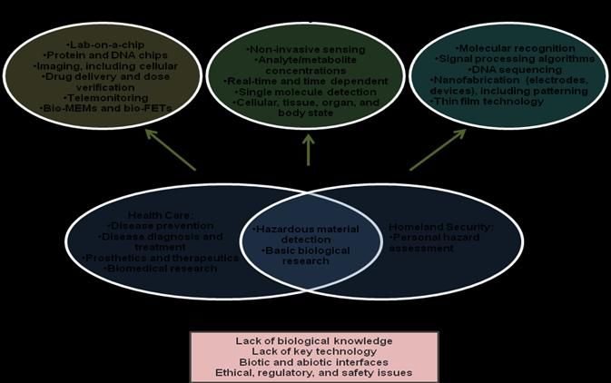
Report prepared by:

Glenn M. Walker
       Joint Department of Biomedical Engineering at the University of North Carolina at Chapel Hill
       and North Carolina State University

J. Michael Ramsey
        Department of Chemistry and the Center for Genome Sciences at the University of North
        Carolina at Chapel Hill and the Joint Department of Biomedical Engineering at the University of
        North Carolina at Chapel Hill and North Carolina State University

Ralph K. Cavin, III
        Semiconductor Research Corporation

Daniel J.C. Herr
         Semiconductor Research Corporation

Celia I. Merzbacher
         Semiconductor Research Corporation

Victor Zhirnov
        Semiconductor Research Corporation

Acknowledgements

The participants of the Bioelectronics Roundtable are thanked for their contributions at the
Bioelectronics Roundtable held November 4, 2008 in Research Triangle Park, North Carolina and their
input to this report. In particular, Madoo Varma of Intel Corporation provided graphic material and
thoughtful feedback that greatly improved the report. Reviews by Dr. William C. Tang, Dr. John
Carruthers, and Dr. Christina Ahn are gratefully acknowledged. Stacey Shirland is thanked for her
support of both the workshop and the preparation of the report.

This study was sponsored by the Semiconductor Electronics Division at the National Institute of
Standards and Technology, an agency of the U.S. Department of Commerce.

    2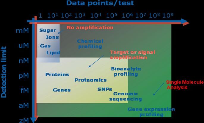
Table of Contents

                                                                                                                                Page

      Executive Summary: The Convergence of Biology and Electronics . . . . . . . . . . . . . . 1
      1. Introduction. . . . . . . . . . . . . . . . . . . . . . . . . . . . . . . . . . . . . . . . . . . . . . . . . . . . . . . 4
      2. Research Activity . . . . . . . . . . . . . . . . . . . . . . . . . . . . . . . . . . . . . . . . . . . . . . . . . . 5
      3. Bioelectronics: A Taxonomy. . . . . . . . . . . . . . . . . . . . . . . . . . . . . . . . . . . . . . . . . . 8
      4. Examples of Technical Areas of Opportunity for Bioelectronics. . . . . . . . . . . . . 10
                  Real-time and Massively Parallel Molecular and Cellular
                  Characterization for Systems Biology
                  Biologically-based Sensors and Fabrication
                  Protection and Restoration of Health
      5. Cross-cutting Challenges. . . . . . . . . . . . . . . . . . . . . . . . . . . . . . . . . . . . . . . . . . . . 11
                  Cellular and Biomolecular Measurements and Analyses
                  Fabrication
                  Device and Material Biocompatibility
                  Power Sources
      6. Current Capabilities and State-of-the-Art. . . . . . . . . . . . . . . . . . . . . . . . . . . . . . . 15
                  Cellular and Biomolecular Measurements and Analyses
                  Fabrication
                  Device Biocompatibility
                  Power sources
                  Implantable devices
      7. Emerging Trends. . . . . . . . . . . . . . . . . . . . . . . . . . . . . . . . . . . . . . . . . . . . . . . . . . . 21
                  Systems biology
                  Forensics
                  Homeland security
                  Medicine
      8. Vision for Ten Years Out. . . . . . . . . . . . . . . . . . . . . . . . . . . . . . . . . . . . . . . . . . . . . 23
      9. Five-year Major Goals. . . . . . . . . . . . . . . . . . . . . . . . . . . . . . . . . . . . . . . . . . . . . . . 25
      10. Framework for Action. . . . . . . . . . . . . . . . . . . . . . . . . . . . . . . . . . . . . . . . . . . . . . . 25
      11. Summary and Recommendations . . . . . . . . . . . . . . . . . . . . . . . . . . . . . . . . . . . . . 26
      References . . . . . . . . . . . . . . . . . . . . . . . . . . . . . . . . . . . . . . . . . . . . . . . . . . . . . . . . . . . 29
      Appendices . . .. . . . . . . . . . . . . . . . . . . . . . . . . . . . . . . . . . . . . . . . . . . . . . . . . . . . . . . . 33
                  Appendix A – Acronyms
                  Appendix B – Bioelectronics in Academia, Industry, and Government
                  Appendix C – Agenda and Attendee Lists from Bioelectronics
                               Roundtable Meeting held November 4, 2008
                  Appendix D – Survey Results Describing the Relative Importance of
                               Topics in Bioelectronics Research Areas
4
Executive Summary: Bringing the Benefits of Moore’s Law to Medicine

Motivation
     There is an opportunity for dramatically increased synergy between electronics and biology,
fostered by the march of electronics technologies to the atomic scale and rapid advances in system, cell,
and molecular biology. In the next decade, it may become possible to restore vision or reverse the
effects of spinal cord injury or disease; for a lab-on-a-chip to allow medical diagnoses without a clinic or
instantaneous biological agent detection. Bioelectronics is the discipline resulting from the convergence
of biology and electronics and it has the potential to significantly impact many areas important to the
nation’s economy and well-being, including healthcare and medicine, homeland security, forensics, and
protecting the environment and the food supply. Not only can advances in electronics impact biology
and medicine, but conversely understanding biology may provide powerful insights into efficient
assembly processes, devices, and architectures for nanoelectronics technologies, as physical limits of
existing technologies are approached. This report develops the thesis that advances in bioelectronics
can offer new and improved methods and tools while simultaneously reducing their costs, due to the
continuing exponential gains in functionality-per-unit-cost in nanoelectronics (aka Moore’s Lawa). These
gains drove the cost per transistor down by a factor of one million between 1970 and 2008 (for
comparison, over the same period, the average cost of a new car rose from $3,900 to $26,000) and
enabled unprecedented increases in productivity.
     In this report, a number of emerging opportunities in bioelectronics are identified. Although, it is
difficult to project the financial benefits at this early stage of research, the following figures for the costs
of just a few diseases that could be impacted by bioelectronics provides a sense of the magnitude of
potential markets and of the benefits to individuals and society in the healthcare sector alone.
     In 2008, an estimated 1.4 million new cases of cancer were diagnosed and over 560,000 cancer
     deaths were reported in the United States, costing nearly $90 billion in direct medical expenses and
     $130 billion more in lost productivity.
     An estimated 22 million Americans suffer from heart disease and about 460,000 die from heart
     attacks each year (about 1 in 5 deaths). In 2008, heart disease cost $172.8 billion in direct medical
     expenses and an additional $114.5 billion in indirect costs.
     An estimated 7.2 million Americans are diagnosed with Type 2 diabetes and millions more are
     undiagnosed. The American Diabetes Association estimates that medical costs associated with
     diabetes were $116 billion in 2007, with an additional $58 billion in indirect costs.
     The overarching technical drivers pushing bioelectronics are the constant advances in
semiconductor technology and in surface chemistry related to the interface of biology and man-made
devices. At the same time, understanding biological systems and processes at the macro- to nano-scale
is growing rapidly.
Research Challenges and Opportunities
     Realizing the promise of bioelectronics requires research that crosses disciplines, such as electrical
engineering, biology, chemistry, physics, and materials science. Challenges and opportunities were
discussed at a roundtable in November 2008 that brought together experts from industry, government,
and academia, including representatives from IBM, Intel, Texas Instruments, Tokyo Electron Ltd.,

     a
       Moore’s Law, which was first observed by the founder of Intel Corporation, Gordon E. Moore, states that the size of integrated circuits
decreases by one half every 18-24 months.

                                                                                                                                                 1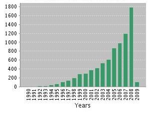
Freescale, and Abbott Laboratories. Also participating were representatives from the National Institute
of Standards and Technology (NIST), National Science Foundation (NSF), and National Institutes for
Health (NIH), and several academic research institutions. Research areas identified at the roundtable
include the following:
     1. Understanding molecule/cell-electronics interfaces;
     2. Understanding cellular responses—and their variabilities—to stimulation (electrical, mechanical,
        chemical, thermal, and the like);
     3. Ability to collect and analyze essential data on the state of biomolecules and cells (chemical,
        physical, structural, functional);
     4. Ability to monitor, in real-time, the biochemistry of a single cell or a population of cells, which
        requires comprehension of interaction between molecules;
     5. Ability to deliver appropriate therapeutic materials and stimuli in real-time; and
     6. Ability to detect, identify, and quantify thousands of different biomarkers simultaneously.
Transitioning the results of this multidisciplinary research to commercial products will be expedited
through collaboration at early stages between the electronics industry and the biomedical device
industry, along with the academic and government research communities. Government, industry, and
academic leaders from different sectors and disciplines who do not necessarily speak the same
‘language’ must be willing to commit to joint efforts where interdisciplinary contributions are necessary
for success.
Observations and Recommendations
    The application of electronics technology to biology and medicine is not new. Examples include
pacemakers and virtually the entire medical imaging industry. Research that enabled these applications
grew out of many disciplines of science and engineering; however, recently, the term “bioelectronics” is
being used more widely to describe this multidisciplinary field. A survey of publications that use the
term in the title or abstract captures on a fraction of the actual research, but suggests that the center of
activity is in Europe (43 percent of publications), followed by Asia (23 percent) and the United States (20
percent). With outstanding research expertise in both biomedicine and semiconductors, the United
States is in a position to become a leader in the field with appropriate and directed investments in the
areas outlined in this report.
    Science and technology experts representing the nano-electronics and biotechnology communities
provided inputs for this report. Collectively, the participants identified a wide range of opportunities and
challenges for the field, which are listed in the table below. The strategic drivers that were most
frequently cited were: disease detection, disease prevention, and prosthetics. The technologies and
devices that will enable applications in these areas will impact other vital areas, such as homeland and
national security, forensics, and the environment. Progress in all of these sectors requires innovation in
crosscutting areas, including measurement and characterization, fabrication, and power sources.
As a next step, stakeholders from government, academic, and industry should jointly develop a detailed
bioelectronics roadmap, which can serve to facilitate effective planning and resource management for
increasing the productivity and commercializaition of bioelectronics research and development. Such an
exercise would define and clarify projected application-specific research metrics and metrology gaps and
needs; timelines for research, development, and prototyping; and emerging market and
commercialization insertion opportunities. The International Technology Roadmap for Semiconductors
(www.itrs.net) , especially its Working Groups on Emerging Research Materials and on Emerging
Research Devices may serve as useful templates for the proposed roadmapping exercise.

    2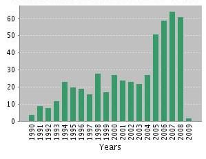
Category            Highest Priority Research Challenges [Priority, where 1.0 = max.]
        Drivers           Prosthetics, including tissue and neural implants, i.e. vision, hearing, etc.
                          [0.91]
                          Disease Prevention, including neural degeneration, cancer, etc. [0.82]
                          Disease Detection, including neural degeneration, cancer, etc. [0.82]
        Devices           Lab on a chip [0.64]
                          Protein and DNA chips [0.64]
                          Imaging, including cellular [0.64]
                          Telemonitoring [0.55]
        Measurements      Noninvasive physical sensing, e.g. vital functions [0.73]
        and Analyses      Concentration of analyte and metabolites, etc. [0.73]
                          Real-time & time dependent measurements [0.64]
                          Single bio-molecule detection, e.g. in Lab-on-Chip environment (including
                          mass, size, chemical, optical, etc.) [0.64]
        Technologies      Molecular recognition [0.73]
                          Signal processing algorithms [0.73]
                          DNA sequencing [0.64]
                          Fabrication (electrodes, devices), including patterning [0.64]
                          Thin film technology [0.64]

   The figure below shows a framework for bioelectronics research, based on input from experts
engaged for this study. This proposed framework is intended to catalyze further analyses and a
comprehensive road-mapping exercise.
     The field of bioelectronics is poised for exponential growth. The Federal government’s expertise in
critical areas of science and technology, including sensors, nanoelectronics, and metrology should be
harnessed and coordinated, along with expertise from academia and industry to firmly establish the
United States as a leader in this high impact area of research and development.

                                                                                                           3
1. Introduction
Electronic devices have been revolutionizing biology and medicine over the past several
generations. The development of the electrocardiograph (i.e., recording the electrical activity of the
heart) approximately 100 years ago was one of the defining moments that helped establish the field of
cardiology and is now an integral part of clinical practice [1]. Today, defibrillators are implanted at a rate
of 160,000 per year in the US alone to restore proper electrical activity to diseased hearts, once again
changing the practice of medicine and creating a new market worth $5 billion per year [2]. Electronic
systems have also been critical to the development of the field of radiology, which has evolved from a
single modality (X-ray) to include magnetic resonance imaging (MRI), computed tomography (CT), and
positron emission tomography (PET), among others. MRI has made possible the imaging of soft tissue to
help treat physical injuries. CT now allows 3D visualization of anatomical features, facilitating surgical
planning. The medical imaging equipment market is expected to be worth $11.4 billion by 2012 [3]. In
short, the application of electronics to medicine has transformed medical practice and will continue to
do so.
In this report, a number of emerging opportunities in bioelectronics are identified. Although, it is
difficult to project the financial benefits at this early stage of research, the following figures for the costs
of just a few diseases that could be impacted by bioelectronics provides a sense of the magnitude of
potential markets and of the benefits to individuals and society in the healthcare sector alone.
1. In 2008, domestic health care spending reached $2.4 trillion, or 17 percent of the gross domestic
product (GDP). This spending is projected to increase to $4.3 trillion in 2016.b
2. In 2008, an estimated 1.4 million new cases of cancer were diagnosed and over 560,000 cancer
deaths were reported in the United States. The National Institutes of Health (NIH) estimates
these cases cost nearly $90 billion in direct medical expenses plus $130 billion more in lost
productivity to the U.S. economy.
3. An estimated 22 million Americans suffer from heart disease and about 460,000 die from heart
attacks each year (about 1 in 5 deaths). NIH estimates the direct cost of heart disease in the
United States in 2008 to be $172.8 billion and the additional indirect cost due to lost
productivity was $114.5 billion.
4. An estimated 7.2 million Americans are diagnosed with Type 2 diabetes and millions more are
undiagnosed. Diabetes leads to a range of debilitating complications, including blindness, nerve
damage and amputation of toes or feet. The American Diabetes Association estimates that
medical costs associated with diabetes were $116 billion in 2007, with an additional $58 billion
in indirect costs.
The study of biology also has been transformed by electronics. In the late 1940s and early 1950s,
understanding the molecular basis of nerve and muscle function was achieved with the use of high-
impedance amplifiers. Those studies led to a new era of quantitative biology and practical clinical
neuroscience. The patch clamp, which allowed researchers to measure the ionic current through single
ion channels gave further insight into nerve action. These studies led directly to three Nobel Prizes and
ultimately seven more. The electron microscope is also an example of applying electronics to biological
problems. First demonstrated in the 1930s and developed over the next decade, the electron
microscope allowed scientists to visualize the miniscule world of cells at an entirely new level of detail
[4]. Much of modern cell biology is built on information captured from these indispensible tools.
b
S. Keehan et al. “Health Spending projections through 2017”, Health Affairs Web Exclusive W146: 21 Feb. 2008
4Given the profound impact electronics has had on medicine and biology in the past, it is easy to
imagine that integrating modern electronics (i.e., semiconductor technology) with biology and medicine
will result in equally profound quantum leaps. The ongoing miniaturization of semiconductor devices is
leading to new opportunities in biomedical research and commercial medical applications. In medicine,
healthcare costs could be drastically reduced and presently untreatable diseases could be cured. The
medical problems of today could have commonplace solutions in the future. Nanoscale bioelectronics
will be an enabler for the development of molecular-based personalized medicine. In particular, the use
of nanoscale electrical measurements will be important in genomics and proteomics for identifying the
function of proteins and their reaction pathways inside cells, as well as on and through cell membranes.
The length scale of features that can reliably be manufactured by semiconductor technology is now
sufficiently small that novel devices can be envisioned to probe cells or biomolecules either in vitro or in
vivo. Advances in integration and packaging mean that circuitry can be integrated with sensors,
actuators, and computers. These enabling technologies will allow the creation of devices and systems
that can intelligently probe biological systems from the molecule to cell to whole organism levels and
thereby open up new areas of basic biological research and new market opportunities for
commercialization. Highly integrated systems also make possible the creation of implantable devices
that can sense their environment and actively choose an appropriate response, as in intelligent drug
delivery chips. Numerous other applications will emerge from the continued integration of electronics
with biology that will result in new revolutionary biomedical advances Moreover, the development of
nanoscale metrologies for the semiconductor industry may well find applications in various biological
and biomedical research areas.
2. Research Activity
The application of electronics technology to biology and medicine is not new, however research
activity in this convergent field is growing rapidly. A proxy for activity, especially in academia, is
publications. In order to assess bioelectronics research activity, an analysis of publications was made
using the Science Citation Index Expanded™ (SCIE), available through the Web of Science®. The SCIE
includes information from more than 10,000 of the world's leading scholarly science and technical
journals in more than 100 disciplines. It also contains papers from over 110,000 conference
proceedings. The database is updated weekly and contains citations dating back to 1900. Publications
that contained the word ‘bioelectronic’ or ‘bioelectronics’ in their title or abstract were identified. The
total number of such papers is 548, published from 1912 through January 2009. It should be noted that
the absolute number of publications on bioelectronic topics (without using the term bioelectronics in
the title or abstract) is undoubtedly much larger; however, this analysis provides a sample that is
believed to be representative of the related activities in the field.
Although publications referencing bioelectronics concepts were published even before 1912, 516 of
the 548 papers that use the term in the title or abstract were published since 1990. Figure 1 shows the
number of publications by year. The number of papers per year increased markedly in 2005 over the
previous year (from about 25 to 50) and by and large has continued to grow. Moreover, the number of
citations to the papers identified has increased exponentially (Figure 2). Because many papers related
to bioelectronics do not use the term in the title or abstract, the absolute number of papers
underestimates the actual level of activity; however, it is clearly increasing over time.
Figure 3 shows the geographical distribution of the location of the authors’ research institutions,
which suggests that, although U.S. authors have published more papers than authors from any other
5country, the center of activity is in Europe (43 percent of publications), followed by Asia (23 percent)
and the United States (20 percent).
The SCIE study also revealed fifteen papers in the bioelectronics area that have received one
hundred or more citations, which is on the order of the number of citations received by Nobel laureates
in the semiconductor sciences. The number of publications each year and the number of citations
received by bioelectronics papers are growing rapidly, indicative of an area of expanding activity. Table
1 lists the fifteen most influential papers in bioelectronics since 1993.
Figure 1: Number of publications with Figure 2: Number of citations to a publication
‘bioelectronic(s)’ in the title or abstract with ‘bioelectronic(s)’ in the title or abstract by
by year. [Source: Science Citation Index year. [Source: Science Citation Index
Expanded™] Expanded™]
Figure 3: Number of publications with ‘bioelectronic(s)’ in the title or abstract by country (left).
Distribution of bioelectronics publications by region (right).
6Table 1. Influential publications in bioelectronicsc
Publication Author(s) Citations
"Integration of layered redox proteins and conductive I. Willner and E. Katz
417
1 supports for bioelectronic applications", Angew. Chem.- Hebrew University
Int. Ed. 39 (2000) 1180 Jerusalem, Israel
"Biological surface science", Surface Science 500 (2002) B. Kasemo
260
2 656 Chalmers Univ. Technology
Gothenburg, Sweden
"Probing biomolecular interactions at conductive and E. Katz and I. Willner
233
semiconductive surfaces by impedance spectroscopy: Hebrew University
3
Routes to impedimetric immunosensors, DNA-Sensors, Jerusalem, Israel
and enzyme biosensors", Electroanalysis 15 (2003) 913
"Control of the structure and functions of biomaterials E. Katz and I. Willner
203
4 by light", Angew. Chem.-Int. Ed. 35 (1996) 367 Hebrew University
Jerusalem, Israel
Supramolecular self-assembly of lipid derivatives on CEA Saclay
200
5 carbon nanotubes, Science 300 (2003) 775 Inst Genet&Biol , Mol&Cell.
Univ Strasbourg, France
Toward bioelectronics: Specific DNA recognition based H. Korri Youssoufi, et al.
192
6 on an oligonucleotide-functionalized polypyrrole, J. Am. CNRS, France
Chem. Soc. 119 (1997) 7388
Preparation and hybridization analysis of DNA/RNA J. Cheng et al.
161
7 from E-coli on microfabricated bioelectronic chips, Nanogen, Inc., San Diego CA
Nature Biotechnology 16 (1998) 541
Dielectrophoretic assembly of electrically functional O. D. Velev et al.
146
8 microwires from nanoparticle suspensions, Science 294 NCSU, Raleigh, NC and
(2001) 5544 Univ. Delaware , Newark, DE
Towards genoelectronics: Electrochemical biosensing of J. Wang
142
9 DNA hybridization, Chemistry- Eur. J. 5 (1999) 1681 NM State University
Albuquerque, NM
Electrical contact of redox enzyme layers associated I. Willner, et al.
126
10 with electrodes: Routes to amperometric biosensors, Hebrew University
Electroanalysis 9 (1997) 965 Jerusalem, Israel
"Biomolecular electronics: Protein-based associative R. R. Birge et al.
120
11 processors and volumetric memories", J. Phys. Chem. Syracuse University
103 (1999) 10746 Syracuse, NY
The application of conducting polymers in biosensors, P. N. Bartlett & P. R. Birkin
12 116
Synthetic Metals 61 (1993) 15 Univ. Southampton, England
Chip and solution detection of DNA hybridization using KPR Nilsson and O. Inganas
106
13 a luminescent zwitterionic polythiophene derivative, Linkoping Univ, Sweden
Nature Materials 2 (2003) 419
Bioelectrocatalyzed amperometric transduction of I. Willner, et al.
105
14 recorded optical signals using monolayer-modified Au- Hebrew University
electrodes, J. Amer. Chem. Soc. 117 (1995) 6581 Jerusalem, Israel
Microchips, microarrays, biochips and nanochips: LJ Kricka
100
15 personal laboratories for the 21st century, Clinica U. Pennsylvania
Chimica Acta 307 (2001) 219 Philadelphia, PA
c
Note the citations given are as of 1/28/2009
7Comparing Table 1 and Figure 3, we note that 1) Israel and the United States each have five of the
fifteen most cited papers; 2) the United States has more than three times the number of papers than
Israel overall; and 3) the nationality of other authors shown in Table 1—England, Israel, France, and
Sweden—are ranked 7th, 8th, 9th, and 11th in Figure 3 . The fact that publication rates and citation
rates are not correlated may be due to the relatively small number of active research groups to date. In
addition to the highly cited papers shown in Table 1, there are many publications that do not use the
term ‘bioelectronics’ in the title or abstract and yet must be considered fundamental to the field.
Examples of such seminal publications include reports on electrical activity of neurons and development
of technologies for characterization of biomaterials, including cells [5-10].
3. Bioelectronics: A Taxonomy
The first reference to bioelectronics, published in 1912, focused on measurement of electrical
signals generated by the body, which is the basis of the electrocardiogram. In the 1960s two new trends
in bioelectronics began to appear. One trend, enabled by the invention of the transistor, centered on
the development of implantable electronic devices and systems to stimulate organs, e.g., the
pacemaker. In the same time frame, fundamental studies were beginning to be reported on electron
transfer in electrochemical reactions. Today, these three areas of endeavor are converging to enable
multi-signal recording and stimulation at the cell level, i.e., there is a kind of physical scaling law that is
moving over time from the organ level toward cellular dimensions. At the same time, studies at the
molecular level are leading to new understanding of cell performance. The analogy with
nanoelectronics is striking; top-down scaling is being abetted by device design from the atomic level.
Bioelectronics encompasses a range of topics at the interface of biology and electronics. One aspect
of bioelectronics is the application of electronics to problems in biology, medicine, and security. This
includes electronics for both detection and characterization of biological materials, such as on the
cellular and subcellular level. Another aspect of bioelectronics is using biological systems in electronic
applications (e.g., processing novel electronic components from DNA, nerves, or cells). Bioelectronics
also focuses on physically interfacing electronic devices with biological systems (e.g., brain-machine,
cell-electrode, or protein-electrode). Applications in this area include assistive technologies for
individuals with brain-related disease or injury, such as paralysis, artificial retinas, and new technologies
for protein structure-function measurements. The identified publications were organized into several
topical areas, as shown in Figure 4.
Examples of research papers in each of the topical areas are given below:
Measurements (35%) – Works on sensors, monitoring systems and metrology. Several distinct categories
were identified:
Sensors (9%) - fabrication and properties of biosensors, biological, and chemical sensors
Biochemical measurements (7%) - application of biosensors
“Bio-electronic Nose” (6%) – Efforts focused on system integration for one targeted application
Neural recording (2%) – Focus on microelectrode arrays and their interfaces with neurons
BioFET (4%) – Field-Effect-Transistor-like devices for bio-sensing
Bio-electronic Instrumentation (7%) – Practical (e.g. clinical) application of bio-electronic devices
Biomaterials (29%) - Materials and fabrication techniques for bio-electronic devices, medical implants,
3D assembly, self-assembly, nano-particles, nano-tubes, nano-wires, etc.
8CMOS
platform Energy
Neural
recording 1%
7%
4%
2%
4% 29%
6%
7%
General
15%
9%
3%
13%
Fundamentals
and Concepts
Figure 4: Distribution of bioelectronics publications by topical area.
Bio-surface science/biochemical reactions (15%) - Research focused on the interaction between bio-
molecules and solid surfaces. Examples include bio-molecule immobilization, electron transfer in
biochemical reactions and between bio-objects (bio-molecules or cells) and the solid surface. The latter
is often referred as “bio-electronic interface”. This definition is narrower than broader concept of bio-
electronic interface as interaction between electronic devices and bio-objects.
General (13%) -Articles including forewords, short abstracts, program descriptions, status reports,
surveys, patent analyses, etc.
CMOS/Semiconductor Platform (4%) – Electronic components of bioelectronic systems and integration
issues. Topics include:
Low-power implantable devices
On-chip integration of sensors
DSP for real-time processing of multi-parametric bioelectronic signals
CMOS IC/microfluidic hybrid systems for cell manipulation and electrochemical analysis
Micro-photodiodes arrays for retinal stimulation
3D chip integration and packaging
Fundamentals and Concepts (3%) – Models, simulation, and new concepts related to bioelectronics.
Energy Sources (1%) – Only three papers were identified and these focused on bio-fuel cells.
The analysis of bioelectronics publications revealed a wide range of research programs spanning
many related thrust areas. Although it is clear that micro- and nanoelectronics is playing an important
role in bioelectronics, via direct application of semiconductor industry products, an evident lack of
semiconductor-related bioelectronic research is very visible. For example, only a small percentage of the
surveyed work dealt with semiconductor platforms for biological applications. Moreover, the critical
area of autonomous (e. g. implantable) energy sources doesn’t appear to have received much attention
9in the context of bioelectronics. There is an opportunity, therefore, for a concerted research effort
directed toward the utilization of the semiconductor industry’s capabilities to provide bioelectronic-
specific tools and systems.
4. Examples of Technical Areas of Opportunity for Bioelectronics
The overarching technical drivers for bioelectronics are the constant advances in semiconductor
technology coupled with advances in surface chemistry and their application to life sciences research.
Much of the work that has been done in the bioelectronics field since the 1990s has been focused on
creating better biosensors by integrating biomolecules with semiconductors [11]. For decades,
semiconductor technology has advanced at an exponential rate and is described by Moore’s law, which
states that the number of features in a given area of substrate doubles every 18–24 months. A result of
Moore’s law is that the computing power and capabilities increase with each generation of device while
the cost per function decreases.
As Moore’s law has progressed, so have the number of semiconductor applications in the life
sciences. In particular, significant effort has gone into developing surface chemistries that can be used to
attach biological molecules to semiconductor substrates [12]. Two examples are biosensors that use
enzymes covalently linked to electrode surfaces [13] and DNA recognition based on surface-bound DNA-
functionalized polypyrrole molecules [14]. Although advances have been made in binding biological
molecules to substrates, no truly biomimetic synthetic surfaces yet exist (e.g., for use in implants). The
following are examples of technical areas or opportunities that will impact bioelectronics:
Real-Time and Massively Parallel Molecular and Cellular Characterization for Systems Biology
The nascent field of systems biology—using systems engineering approaches to analyze cellular
function—is driving the development of new technology that can monitor multiple aspects of cellular
behavior over many timepoints. Systems biology embodies a new perspective from which to view
biological systems and knowledge culled from its approaches could lead to advances in medicine and
security. However, significant investment is needed to develop tools and associated standards and
metrology that can characterize and continuously monitor the states of cells at sub-cellular resolutions
[15]. Advances in micro- and nano-fluidic systems that can monitor many cell populations in parallel are
beginning to address this need [16, 17].
Biologically-based Sensors and Fabrication
Another driver of bioelectronics is that cells and their components can be used as biological
transducers for measurements or as components in building novel materials or circuits. In other words,
components of cells have been used for novel applications outside of their originally observed purpose.
Cells are sensitive to many environmental cues. For example, identifying the response of a cell to a
known toxin could allow it to be used as a “canary in a coal mine” to detect toxic substances in captured
air samples [18]. Biomolecules, in particular antibodies, can also be used as transducers, via their
exquisite specificity for complementary molecules. Coupling antibodies with emerging nano-scale
technologies could result in ultra-sensitive detection methods [19]. Bio-inspired fabrication shows
promise for constructing nanoscale assemblies, which could lead to significant advances in sensor design
and materials technology [20].
Protection and Restoration of Health
The development of new technology that can protect lives (e.g., by preventing bioterrorism) and
restore health (e.g., by developing novel implantable therapeutic devices) is also driving the
10development of bioelectronics. National security demands new technology for monitoring chemical or
biological threats, and advances in lab-on-a-chip technology have been developed to address this need
[21]. Further miniaturization of these technologies will likely result in smaller units with increasing
capabilities. Advances in miniaturization and power transmission, storage, and generation have allowed
implantable medical devices to emerge [22]. The artificial retina, which is a first step toward restoring
sight in people with degenerative diseases of the retina, is representative of these advancements [23].
Implantable drug delivery devices, based on micro-electro-mechanical systems (MEMS), are beginning
to emerge, and the next generation of drug delivery devices are likely to be “smart” and to have
embedded transceivers (i.e., able to sense and respond to their environment and transmit and receive
data) instead of passively delivering drugs at pre-defined intervals [24].
The health and safety issues associated with the synthesis and use of nano-materials in
semiconductor manufacturing will be informed by the understanding that will come from using
bioelectronics to study toxicity effects at the cellular and molecular levels. These data will allow
exposure limits to be determined and guide standards for environmental monitoring.
5. Cross-cutting Challenges
Moving bioelectronics forward requires innovation among the broad areas of measurements and
analyses, fabrication, biocompatibility, and power sources. In general, these cross-cutting challenges
either stem from a lack of technology, biological understanding, or a combination of both. Expertise that
is resident across government agencies, academic research institutions, and industry will need to be
coordinated and brought to bear in order to to achieve the necessary innovations.
Cellular and Biomolecular Measurements and Analyses
New methods are needed for the detection, identification, and quantification of DNA, proteins, and
other biomolecules. These methods would also provide a more accurate measurement of cell
phenotype (e.g., DNA sequence, surface markers, etc.) and enable high throughput real-time
information about cellular behavior.
Biomolecular measurements are increasingly focused on higher bandwidth measurements (i.e.,
multiple analytes measured at increasingly high frequency). The biomolecules of interest usually have
very low concentrations, on the order of nanomolar to femtomolar. When considering protein analysis
of single cells, the concentration of even highly expressed molecules becomes very small. As more
cellular and biomolecular measurements are needed to paint a comprehensive picture of cell behavior
in situ, larger numbers of measurements per observation will be needed at increasingly smaller
concentrations per molecule. Figure 5 summarizes the parameter space. Metabolic measurements of
cells occupy the upper-left portion of the graph, while the protein and DNA characterization of cells
occupy the lower-right corner.
The fundamental challenge facing DNA analysis is the ability to rapidly and accurately determine the
sequence. Sequencing technologies have advanced significantly, with three generations of disruptive
innovations already productized. But an ultimate goal is a method that can rapidly and inexpensively
sequence DNA from a small and complex sample without need for sample manipulation. Single molecule
and nanopore sequencing are promising approaches that may provide a solution [25]. A secondary
challenge of DNA analysis is determining which genes are active. Gene expression can be quantified by
measuring messenger RNA (mRNA) expression as a surrogate using DNA arrays or gene chips. While
gene chips are currently the best way to provide a comprehensive picture of gene expression, DNA
hybridization arrays require sample amplification, which adds time to the assay and can lead to
11Figure 5: Trade-offs between measurement sensitivity and measurement time are
due to the increasing need for information-rich data.
increased levels of noise, due to PCR errors introduced into the DNA [26]. Furthermore, collecting gene
expression data over many time points (e.g., minutes or hours) is tedious. An ideal technique
forquantifying gene expression would require minimal human intervention (i.e., sample manipulation)
and allow multiple time point measurements within the same inexpensive, reusable device.
Electrochemical methods show promise for creating highly sensitive and integrated (e.g., in a lab-on-a-
chip device) DNA arrays [27].
The challenges for cellular and molecular measurements are based on a lack of adequate
metrological tools. To address these challenges, new devices in combination with other techniques
(multi-modal metrologies) need to be developed that can:
Rapidly determine whole genome and RNA sequences;
Quantify the up or down regulation of multiple genes over many timepoints;
Identify biomarkers for disease states (e.g., DNA, RNA, proteins, polysaccharides, metabolites) in
both serum and actual blood and tissue samples;
Identify and quantify biomarkers in real-time at sensitivity levels equal to or better than existing lab
techniques such as enzyme linked immunoassays (ELISA) and also be reusable;
Monitor multiple intracellular events in real-time;
Simultaneously determine membrane protein structure and function in an in-vivo-like environment;
Identify the chemical and mechanical cues that drive stem cell differentiation;
Recapitulate physicochemical cellular microenvironments; and
Implement ion channels, ion channel-like devices, as sensors for improved molecular recognition
and sensitivity.
12Figure 6: Increasing the density of semiconductor features will ultimately allow
arrays of single-molecule measurements. Courtesy of Madoo Varma,
Intel Corporation.
Fabrication
Fabrication challenges include creating better sensors and developing novel fabrication techniques.
Integrating multiple sensing technologies with integrated circuit (IC) technologies also presents a
challenge. Biosensors will play a key role in meeting future bioelectronics demands and improvements
to increase bandwidth and lower detection limits are needed. Massive parallelization of sensors is
necessary to see the same benefits from future bioelectronics devices that now exist in ICs because of
Moore’s law. Figure 6 illustrates how the miniaturization of semiconductor technology has led to
acommensurate decrease in the size of liquid volumes used for biological assays. As technology
continues to advance, high throughput nanofabricated arrays capable of quantifying single molecules in
solution will become commonplace.
The challenges faced in realizing single molecule biosensor arrays are based in part on a lack of
fabrication technologies. New methods are needed to fabricate structures reliably at the nanoscale, and
new metrology and standards are needed to characterize these structures. To date, the vast majority of
nano-fluidic devices have been characterized by planar surfaces and simple features defined by one,
two, or at most several device depths [28]. As the functionality of a nano-fluidic device is determined by
its dimensionality and complexity, the development of more intricate three dimensional structures
would lead to enhanced control over nanometer scale fluidic environments and analytes, which could
result in new or improved device utility. Progress in this regard has been limited by conventional
nanofabrication processes, which are inherently planar and become increasingly restrictive, when many
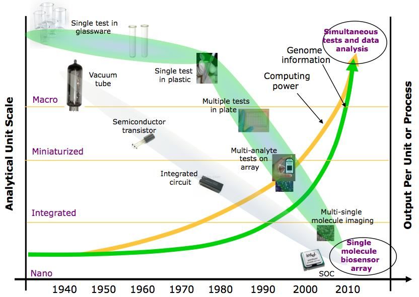
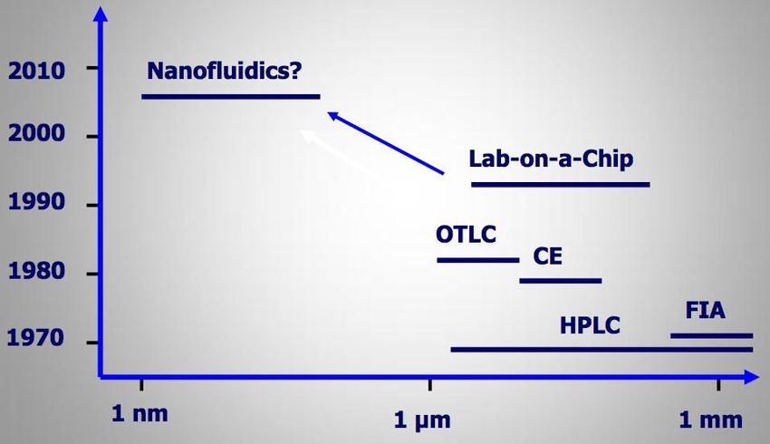
layers of high resolution lithography are performed in a research facility. Figure 7 shows how nano-
fluidic devices relate to some of the more popular methods for molecular analysis.
13Figure 7: New fabrication technologies will be required to fabricate nano-fluidic
devices that have single-molecule sensitivity. Current technologies
include lab-on-a-chip, open tubular liquid chromatography (OTLC),
capillary electrophoresis (CE), high-pressure liquid chromatography
(HPLC), and flow injection analysis (FIA).
The fabrication of nano-materials and nanoscale devices raises health and safety concerns that will
need to be addressed [29]. The technologies developed in response to challenges in the previous section
can be used to address the concerns associated with the synthesis and use of nano-materials and nano-
devices in semiconductor manufacturing.
Fabrication challenges that need to be overcome include:
Improving fabrication techniques for on-chip integrated optical excitation and detection in
nanoliter-femtoliter volumes;
Endowing electrochemical sensors with sub-zeptomole sensitivity;
Developing electronics with a sensitivity of < 1 pA and a bandwidth of hundreds of MHz;
Integrating high-density photodiode arrays into substrates;
Harnessing biological assemblies for nanoscale fabrication;
Fabrication of complex physical features into substrates with critical dimensions ≥ 1 nm;
Developing metrology tools for test and measurement of nanoscale features. Developing surfaces
for control of antibody and antigen binding;
Immobilization of electrode surfaces for control of antibody and antigen binding. This requires
extensive development of the needed biochemistries.
Device and Material Biocompatibility
Accurate bioelectronic measurements and safe use in patients demand that the device surface not
inhibit or cause deleterious effects on the biological system. One of the most critical issues in realizing
the potential of bioelectronic devices is to understand how they perturb the local environment and
14cellular response, which includes but is not limited to physical contact and radiation effects. Improving
biocompatibility, via surface chemistry, is critical for enabling future implantable bioelectronic devices,
such as insulin delivery systems or chronic neurological implants [12]. The optimization of the material
biocompatibility, however, cannot come at the loss of device or measurement functionality. The surface
modification or coating must allow for adequate environmental sampling for timely and accurate
electronic measurements.
Key challenges include:
Implementing biocompatible materials that do not kill tissue or cells and maintain the native
structure of biomolecules, while not compromising device operation.
Limiting implant rejection by maintaining and stimulating tissue integrity.
Minimizing the amount of tissue heating that occurs from electromagnetic radiation (e.g., during the
transmission of power).
Power Sources
New methods for harvesting and storing energy to power mobile and implantable devices are
required [30]. Next-generation devices will need power sources that offer prolonged device life with
minimal intervention (e.g., through multiple surgeries) and minimal increase in device size. Methods for
producing energy from biological or bio-inspired sources will also be needed to meet future domestic
energy demands. One example is using microbial fuel cells to generate electricity [31].
High-level challenges include:
Improving the efficiency of RF power transmission to devices within the body while minimizing
tissue damage;
Improving battery energy density;
Developing alternative energy generation strategies for devices such as kinetic power production
(e.g., power harvested from body motion);
Developing alternative energy strategies that use biological or bio-inspired approaches, such as
synthetic photosynthesis (light harvesting) or microbial energy generation.
6. Current Capabilities and State-of-the-Art
Current technologies for cell and biomolecular measurements, fabrication, device biocompatibility,
power sources, and implantable devices are briefly reviewed. Where applicable, drawbacks of the
current approaches are pointed out.
Cellular and Biomolecular Measurements and Analyses
DNA and proteins can be studied either in their native state within a living organism (in vivo) or
extracted from their native environment (in vitro). Cellular measurements are typically conducted in
vitro in artificial microenvironments to identify a particular cellular state, through the characterization of
DNA, proteins, and other components. However, a growing number of researchers are realizing that
studying cells in these artificial environments can skew experimental results [32]. A common drawback
to all current capabilities is that they do not allow the simultaneous measurement of biological function
(DNA, proteins, cells, etc.) over multiple timepoints in an in-vivo-like environment.
15DNA
DNA has been studied for decades but the three largest areas of emphases have been to develop
rapid methods for (1) sequencing DNA, (2) recognizing short strands, and (3) detecting single nucleotide
variations. Current methods include slab gel electrophoresis, capillary gel electrophoresis, and DNA
microarrays.
The ability to amplify the number of DNA copies via the polymerase chain reaction (PCR) has been
critical to the growth of genetics research. The traditional method for DNA sequencing involves
polymerase amplification of DNA fragments followed by sizing, using electrophoretic separation (Sanger
method). Separation of DNA using electrophoresis in agarose slabs is the traditional approach for sizing
biopolymers and is used to size polymerase-generated DNA fragments, which allows determination of
DNA sequence. The small capillary diameters (< 100 µm) used in capillary gel electrophoresis provide
improved thermal transport, allowing the use of higher electric field strengths to correspondingly
reduce separation times.
Although sensitive and reliable, high-end setups are large, expensive, and require optical (laser)
systems to accurately read data. The classic approach to DNA sequencing also requires a large time
investment for sequencing whole genomes and large amounts of sample. Current methods of DNA
analysis lack the speed and ease of use that is desired for personalized medicine and systems biology,
among other applications. DNA detection methods used in biomedical research are continually
improving, but are still slow and cumbersome to use for monitoring gene expression over multiple time
points with multiple cell types. For example, real-time PCR can be used to monitor gene expression over
time, and looking at a single cell line expressing a single gene or protein is a routine process. Looking at
multiple cell lines (e.g., as in a co-culture) expressing more than a handful of genes or proteins over time
is currently impractical.
Next generation DNA sequencing tools have become available in the past couple of years that have
reduced whole genome sequencing costs by several orders of magnitude. Some of these new
sequencing techniques are classified as sequencing by synthesis methods, which generally use DNA
polymerases that allow incorporation of recognizable deoxynucleotide triphosphates, one at a time [33].
Synthesis of complementary strands of template DNA fragments is done in a massively parallel (105)
fashion to increase speed and reduce costs.
Current methods for direct sequencing of DNA eliminate the need for PCR and, thus, significantly
reduce the time needed for genomic analysis. However, these methods, such as electrical detection of
sequence pairs when DNA molecules are translocated through nanopores, need major engineering
development.
Proteins
Useful characteristics of proteins include their structure, function, sequence, concentration, and
binding characteristics. A significant number of proteins, especially those of interest to the
pharmaceutical industry, are embedded in the cellular membrane, which confers on them a particular
three-dimensional structure [34]. To date, no method exists that can determine both the structure and
function of membrane proteins in their in vivo conformation. Common methods of protein
characterization include slab gel separations, mass spectrometry, fluorescence imaging, x-ray
crystallography, and NMR spectroscopy.
While 2D slab gel separations reveal size and charge information for proteins, and have historically
been the lab workhorse for analyzing proteins (e.g., Western blots), they do not provide any information
about polypeptide sequence, which is needed for identifying unknown proteins. Mass spectrometry is a
16powerful tool, but high concentrations of common biological proteins that may also be present in a
sample, such as albumin, can mask signals from less concentrated proteins important to cellular activity.
Fluorescence methods can reveal useful information, but preparing fluorescent markers is often time-
consuming, tedious, and expensive. Because a large number of proteins cannot be monitored easily with
these techniques, the throughput of discovery and monitoring of complex intracellular interactions is
limited.
Mass spectrometry is a rapidly evolving field in which proteins are characterized wholly or by
digesting them into smaller fragments (peptides), further fragmenting the peptides in the gas phase, and
determining the molecular weight of the gas phase components. The gas phase fragmentation of
peptides generates a mixture of peptides that differ by one amino acid residue and yields the peptide
sequence. Comparing this peptide sequence information with genomic sequence information often
reveals the identity of a unique protein. Information on expression levels (concentration) of the proteins
is simultaneously determined. A recent development in size (or mass) spectrometry of individual
molecules in the aqueous phase may prove useful for protein fragment analysis [35].
Genetic engineering can be used to incorporate fluorescent protein (e.g., GFP) sequence
information next to unidentified target proteins so that their production within a cell can be monitored.
Optical fluorescence techniques, such as fluorescence recovery after photobleaching (FRAP), can be
used to monitor protein production within a cell. Fluorescence resonance energy transfer (FRET) can be
used to monitor protein-protein interactions. However, these altered proteins can take months to
prepare and are limited in their throughput once they have been incorporated into the cell(s) being
studied.
X-ray crystallography and NMR spectroscopy are used to infer the structure of proteins, but cannot
provide information on function. Furthermore, neither method can capture the conformation of the
protein in its native (i.e., within a cell or on a cellular membrane) state. NMR does offer the advantage
that protein structure can be observed within an aqueous solution.
Cell Phenotype
Cell phenotype is determined through the interaction of the genome with the environment. The
phenotype can be quantified by measuring the genome, proteome, secretome, metabolic state,
electrical characteristics, and mechanical characteristics of the cell. Techniques commonly used to
quantify cells include:
Optical and fluorescence microscopy,
DNA analysis,
Protein analysis,
Patch clamps,
Cell deformation
Optical and fluorescence microscopy are the most common method of observing cell phenotype.
Cells can be visually inspected with optical microscopy to reveal distinguishing physical characteristics,
such as unique structural features, and to observe cell movement. Cell migration studies, which are key
to many fields, including cancer metastasis, are performed with temporal snapshots of cell position on a
substrate. Fluorescence microscopy provides more detailed cellular information in that dyes are used
that can highlight particular features of cells. For instance, the presence of metabolic activity within a
cell can be observed using the proper dye, which in some cases offers better spatial resolution and
easier implementation than electrochemical methods. Cell viability is also routinely measured using
fluorescent live/dead assays. Fluorescent probes exist for hundreds of cellular characteristics, but they
17suffer from three main drawbacks. First, most dyes cannot be used over multiple timepoints—cells often
need to be fixed in order to be stained. Second, fluorescent dyes are subject to photobleaching. Third,
probes for proteins that are expressed in very small quantities may not give a detectable signal.
Semiconductor quantum dots with protein and fluorescent tags address some of these issues but their
toxicity remains an issue for deployment in vivo.
Cell phenotype can also be determined by DNA array chips through the identification of mRNA.
Proteins are synthesized from translation of the mRNA sequence into a specific amino acid polymer.
Measurement of mRNA expression levels in cells is an indirect way to determine probable protein
expression. DNA array chips can rapidly provide a global snapshot of cell activity. However, the
sequences of interest must be known to some degree (to provide a complimentary binding site). DNA
chips also provide only a single snapshot of which genes are up- or down-regulated. Multiple timepoints
of cell state, which would be more valuable to biologists, are tedious to measure with current
techniques.
The proteome, the protein catalog expressed by a cell, is quantified by lysing the cell and detecting
the complement of expressed proteins. Mass spectrometry combined with various chemical separation
techniques is used to determine the partial proteome of cells, although larger volumes are required to
identify low-concentration proteins. Comprehensive proteome analysis strategies still remain elusive.
Fluorescently tagging of proteins is a way to identify protein expression and interaction within cells but
this method suffers from the drawbacks mentioned previously.
The secretome is the catalog of all proteins secreted into the extracellular environment. There is a
growing realization among researchers of its importance in determining and regulating cell behavior
[36]. Also important is the uptake of proteins and other small molecules by cells. With the exception of
well-known proteins like insulin or certain neurotransmitters, no widely accessible techniques exist to
readily identify or characterize the secretome of cells.
Electrochemical methods have historically been used to determine the metabolic state of a cell by
measuring the uptake or secretion of metabolites and waste such as O2, CO2, glucose, and NH4. The state
of electrically active cells, such as neurons, is measured by electrochemically detecting intracellular ion
concentrations. Transport of small molecules across the cell membrane is an active area of research that
impacts both basic cell biology and the development of pharmaceuticals. Broadly speaking,
electrochemical measurements rely on electron transfer at an electrode surface to generate a current
that can then be recorded. Electrochemical measurements are attractive because electrodes can be
miniaturized and integrated with semiconductor processes.
Disadvantages of electrochemical measurements include the impact of electrode degradation (e.g.,
through chemical reactions or adsorption of proteins) on signal quality and limitations on the types of
molecules that can be monitored [37]. The types of molecules that can be electrochemically measured
depend on the technique used and the electroactivity of the molecule. These issues will be addressed
with further advances in surface chemistry.
Mechanical characteristics of cells include cell deformation, cell adhesion, and cell migration. Cell
deformation studies are performed by aspirating cells into a micropipette tip and observing the
deformation at a given pressure. Cell deformation is increasingly being viewed as a viable marker for cell
behavior and has recently been studied as a metric for identifying differentiated stem cells and cancer
cells [38, 39]. Cell deformation is also increasingly being used as a way to drive stem cells down specific
differentiation pathways [40]. Cell adhesion studies seek to characterize how strongly a cell is attached
to a substrate. Common methods for measuring adhesion include pulling on cells with optical tweezers
or an atomic force microscope tip or even quantifying interference patterns on a flexible substrate [41].
18You can also read