RSC Chemical Biology rsc.li/rsc-chembio - RSC Publishing
←
→
Page content transcription
If your browser does not render page correctly, please read the page content below
Volume 2
                                                                                  Number 1
                                                                                  February 2021
                                                                                  Pages 1–276

RSC
Chemical Biology
rsc.li/rsc-chembio

                     ISSN 2633-0679

                     REVIEW ARTICLE
                     Rob C. Oslund, Olugbeminiyi O. Fadeyi, Andrew Emili et al.
                     The chemical biology of coronavirus host–cell interactions
RSC
Chemical Biology
This article is licensed under a Creative Commons Attribution-NonCommercial 3.0 Unported Licence.
View Article Online
REVIEW View Journal | View Issue
The chemical biology of coronavirus
host–cell interactions
Open Access Article. Published on 23 December 2020. Downloaded on 4/1/2021 3:41:39 AM.
Cite this: RSC Chem. Biol., 2021,
2, 30 Suprama Datta,a Erik C. Hett,b Kalpit A. Vora,c Daria J. Hazuda,bc
Rob C. Oslund, *b Olugbeminiyi O. Fadeyi *b and Andrew Emili*a
Severe acute respiratory syndrome coronavirus 2 (SARS-CoV-2) is responsible for the current coronavirus
disease 2019 (COVID-19) pandemic that has led to a global economic disruption and collapse. With several
ongoing efforts to develop vaccines and treatments for COVID-19, understanding the molecular interaction
between the coronavirus, host cells, and the immune system is critical for effective therapeutic
Received 4th November 2020, interventions. Greater insight into these mechanisms will require the contribution and combination of
Accepted 6th December 2020 multiple scientific disciplines including the techniques and strategies that have been successfully deployed
DOI: 10.1039/d0cb00197j by chemical biology to tease apart complex biological pathways. We highlight in this review well-established
strategies and methods to study coronavirus–host biophysical interactions and discuss the impact chemical
rsc.li/rsc-chembio biology will have on understanding these interactions at the molecular level.
Introduction some genetic changes, particularly the D614G mutation in the
spike protein has been associated with higher infectivity but
The severe acute respiratory syndrome coronavirus 2 (SARS-CoV-2) probably lower mortality.5 Mutations at position 614 are unlikely
strain that causes coronavirus disease 2019 (COVID-19) has to affect antibodies that bind to the neutralizing epitopes. The
infected over 60 million people with nearly 1.5 million deaths immunological sequalae during and after infection remain
worldwide as of late November 2020 leading to not only severe incompletely understood.6 The ensuing immune response can
impact on human health, but widespread global economic and cause a variety of outcomes. These range from severe disease
social disruption.1 While no vaccines are currently approved to initially in some patients, exemplified by acute respiratory distress
prevent SARS-CoV-2 infection or spread of COVID-19, more syndrome (ARDS), especially those at older age or with com-
than 100 coronavirus vaccine candidates based on various plat- orbidities, to recovery and resolution in most others with the
form technologies are currently in preclinical and clinical potential for establishing immunity to subsequent exposure.
development.2 Furthermore, several clinical trials are currently The portal of entry for SARS-CoV-2 is through nose, mouth,
under investigation to evaluate potential therapeutics to alleviate and eyes establishing an early infection in the upper respiratory
COVID-19 symptoms and disease progression.3 Thus far, tract (Fig. 1). In the majority of individuals this virus replication
remdesivir4 (a nucleoside-analogue RNA-dependent RNA poly- in the upper respiratory tract does not progress to severe
merase (RdRP) inhibitor), is the only approved antiviral shown to disease and is cleared quickly.7 It is postulated that a robust
be beneficial to patients who don’t require oxygen ventilation. innate immune response and/or trained immunity, and likely
With the surging number of cases this Fall, normalcy will likely pre-existing cellular immunity (T-cells generated to prior expo-
not return until safe and efficacious vaccines and therapeutics sures to common cold coronaviruses) could account for the
become widely available. limited infection in the upper respiratory tract.8 Subsequently if
SARS-CoV-2 originated in the Wuhan province in China after the virus is not cleared from the upper respiratory tract, the
the summer of 2019, rapidly spread across the globe, and was virus will travel to the lower respiratory tract and establish the
eventually declared a pandemic by the World Health Organiza- infection in lung airway or bronchiole cells leading to moderate
tion (WHO) in early 2020. The virus has subsequently shown or severe COVID-19 symptoms (Fig. 1). It is also hypothesized
that the ensuing immune response in the lung to clear the
infection causes pathogenic inflammation and, in some instances,
a
Center for Network Systems Biology, Department of Biochemistry, Boston manifests into ARDS.9
University School of Medicine, Boston, MA, USA. E-mail: aemili@bu.edu
b
Immunity to several respiratory viral pathogens, including
Exploratory Science Center, Merck & Co., Inc., Cambridge, Massachusetts, USA.
E-mail: rob.oslund@merck.com, olugbeminiyi.fadeyi@merck.com
respiratory syncytial virus (RSV), human metapneumovirus
c
Infectious Diseases and Vaccine Research, Merck & Co., Inc., West Point, (hMPV), parainfluenza virus type 3 (PIV3) and Rhino viruses
Pennsylvania, USA in the upper respiratory tract is considered short-lived and
30 | RSC Chem. Biol., 2021, 2, 3046 2021 The Author(s). Published by the Royal Society of Chemistry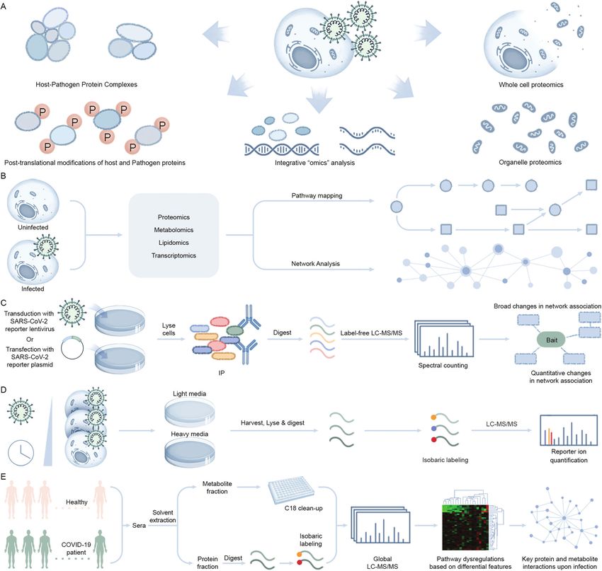
View Article Online

                                                                                                                Review                                                                                                                    RSC Chemical Biology
            This article is licensed under a Creative Commons Attribution-NonCommercial 3.0 Unported Licence.
Open Access Article. Published on 23 December 2020. Downloaded on 4/1/2021 3:41:39 AM.

                                                                                                                Fig. 1 SARS-CoV-2 infection lifecycle. Following entry into host cells by engagement of the severe acute respiratory syndrome coronavirus
                                                                                                                2 (SARS-CoV-2) viral envelope glycoprotein spike (S) with receptors on the surface of epithelial cells located in distal lung, e.g. angiotensin-
                                                                                                                converting enzyme 2 (ACE2) on alveolar cells, and establishment of upper respiratory tract infection via nose, eyes and mouth, the viral lifecycle
                                                                                                                proceeds with the fusion of viral envelope with cell membrane and subsequent release of viral RNA into the cell. The RNA is then translated to produce
                                                                                                                viral pre-proteins that are processed by its own proteases to release over two dozen effector proteins which eventually participate in viral replication,
                                                                                                                hijacking of host pathways, and assembly of virions followed by release.

                                                                                                                partial.7 Likewise, protective immunity to coronavirus infection             has several epitopes that can elicit CD4-T cell responses and some
                                                                                                                may also be short-lived, as has been observed with common                    CD8-T cells.13 Additional viral proteins like nucleocapsid and
                                                                                                                cold corona, severe acute respiratory syndrome (SARS) and                    proteases are more conserved than the spike protein and therefore
                                                                                                                Middle East respiratory syndrome (MERS) viruses.10 Protection                are candidate antigens to elicit additional T-cell responses or
                                                                                                                from severe, lower respiratory tract disease may be more robust              boost pre-existing T cell responses.14
                                                                                                                and durable but is unlikely to be complete, especially in at-risk               The goal of the SARS-CoV-2 vaccine is to raise robust spike
                                                                                                                populations. Any form of prior specific or non-specific immune               specific humoral responses that prevent viral infection. Addition of
                                                                                                                education by way of vaccination or exposure may slow down the                CD4 T-cell responses can also help the process, but on their own
                                                                                                                march of the virus to the lower respiratory tract. Therefore,                may not afford protection. Requirement of CD8 T-cell responses is
                                                                                                                vaccination against SARS-CoV-2 is highly desirable to avoid                  controversial as the potential for collateral damage to the lung by
                                                                                                                serious disease.11 Most prophylactic vaccines block viral entry by           inducing cytotoxicity, re-modeling and fibrosis while clearing
                                                                                                                eliciting antibodies to the viral surface glycoproteins responsible          infected cells is a concern. Additionally, any vaccine developed will
                                                                                                                for viral entry into the host cells and hence the spike protein of           have to deal with the perceived risk of enhanced disease by vaccina-
                                                                                                                SARS-CoV-2 has become an attractive vaccine candidate antigen.12             tions that raise suboptimal antibody and/or Th2 T-cell responses.
                                                                                                                In addition to eliciting neutralizing antibodies, the spike protein          Therefore, understanding the molecular interaction between the

                                                                                                                  2021 The Author(s). Published by the Royal Society of Chemistry                                                RSC Chem. Biol., 2021, 2, 3046 | 31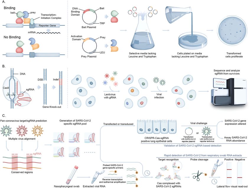
View Article Online
RSC Chemical Biology Review
virus, host cells and the immune system is of paramount importance druggable targets to counter infection. Chemical probes targeting
for developing prophylactic and therapeutic interventions. key host pathways co-opted during viral replication, or other
With a history deeply rooted in understanding how bio- potential ligand binders involved in antiviral immune response,
molecules engage within the complexity of biological systems, could leverage metabolic responses and pathways that are com-
This article is licensed under a Creative Commons Attribution-NonCommercial 3.0 Unported Licence.
chemical biology is well-suited for interrogating the dynamic prised of actionable proteins (e.g. enzymes and transcription
repertoire of SARS-CoV-2–host molecular interactions. factors) and small molecules metabolites (glutamine, citrate,
The successful integration of chemical biology within the drug palmitate, etc.) that natively engage with bioactive compounds.
discovery pipeline through the development and application of The dynamic interplay between host proteins and metabolites is
chemical probe technologies enables the elucidation of ligand– therefore key to the metabolic reprogramming that occurs upon
target pairs and enhances exploration of biological pathway infection. For example, pattern recognition receptors such as
Open Access Article. Published on 23 December 2020. Downloaded on 4/1/2021 3:41:39 AM.
interactions through targeted protein modulation.15,16 The Toll-like receptors (TLRs), retinoic-acid-inducible protein I (RIG-I),
potential utility of chemical biology-based approaches for RIG-1-like receptors (RLRs) and cytosolic sensors (cGAS) sense
understanding viral pathobiology have been further enhanced pathogen-associated molecular patterns, such as small molecules,
through the development of a portfolio of protein activity-based and dimerize with their cognate adaptor molecules to activate IKK
probes and labeling methods that allow for high-resolution and TBK-1/IKK.29 These kinases in turn activate the transcription
investigation of cellular enzyme function and proximal protein factors IRF3 and NF-kB to promote the production of cytokines.
communities in cellular environments, respectively.17 Mass Energy precursors like citrates, succinates and other glyco-
spectrometry-based proteomic endeavors to identify viral–host lysis and TCA cycle intermediates also modulate production of
protein engagement within cells have also been improved inflammatory cytokines during host immune response.30
through chemical biology-based affinity tags, enrichment stra- Lipids and fatty acids play active roles in protein modifications
tegies, and selective labeling methods.18 and the formation of the viral envelope.31 Amino acids like
The successful demonstration of these chemical biology- glutamine can serve as an alternative carbon source for viral
based strategies and technologies have propelled expansion infected cells.32 Endogenous nucleotides like cGMP and cAMP
into other functional characterization initiatives such as the act as second messengers that signal interferon gene
elucidation of metabolite–protein interactions,19 understanding pro- expression.33 Specialized organelles such as mitochondria
tein community environments within different cellular regions,20 and lysosomes serve as important subcellular hubs that inte-
and detailing mechanisms behind cell–cell engagement.21–24 The grate diverse players at the interface of immune response and
field of chemical biology is ideally suited to address these and other metabolism.34,35 Conversely, the metabolic-reprogramming
fundamental questions that are central to unlocking a more detailed activity of the cytokine response can originate from immune
understanding of coronavirus–host interactions at all phases of a signaling components whereby metabolites, such as succinate
viral replication life cycle. In this review, we highlight previous efforts and citrate, can directly or indirectly regulate the canonical
to study coronavirus host molecular interactions and discuss the NF-kB pathway (Fig. 2). Studies suggest that itaconate, a struc-
impact chemical biology will have on understanding these interac- turally similar small molecule metabolite to malate, is trans-
tions at the molecular level. ported across the mitochondrial inner membrane by the
TCA cycle intermediate carriers (e.g. citrate) and activate the
anti-inflammatory transcription factor nuclear factor erythroid
Rationale behind studying virus–host 2-related factor 2 (Nrf2) following lipopolysaccharide (LPS)
small molecule interactions stimulation.36 A recent study by Olagnier et al. reported the
antiviral effects of a chemically synthesized cell permeable
Viruses are obligatory intracellular parasites that hijack key derivative of itaconate and fumarate, 4-octyl itaconate (4-OI)
biochemical processes and regulatory pathways of infected host and dimethyl fumarate (DMF) respectively, in SARS-CoV-2
cells to favor their replication. Since the number of genes infection37 and will be discussed further in the strategy section
encoded by the host genome is exponentially larger than that of this review. The antiviral effects of metabolic sensing
of a viral genome, upon infection, a relatively few different enzymes (e.g., mammalian target of rapamycin (mTOR)38 and
types of viral components interact with a complex pool of host adenosine monophosphate-activated protein kinase (AMPK)39)
factors to take advantage of the host–cell machinery. Perturba- play major regulatory roles in both metabolism and immune
tion of host pathways results in rewired intracellular signaling, responses with the onset of proinflammatory signals.
transcription, translation, metabolism, and dysregulated With the havoc created by the COVID-19 pandemic coupled
immunity and stress responses.25–27 with the recent history of other zoonotic coronavirus infections,
Central metabolic pathways, including glycolysis, tricarboxylic MERS and SARS-CoV, it has become imperative to understand
acid (TCA) cycle and lipid metabolism are important targets for the pathophysiology of host–SARS-CoV-2 and coronavirus infec-
energy resources for viral replication (Fig. 2).28 Mapping the host– tions more generally with the goal of uncovering the mechanisms
virus interaction interface impacting metabolic systems is crucial associated with host cell invasion, replication and persistence.
to understanding the: (1) cellular pathways co-opted to support Although most of the structural and non-structural coronaviral
viral replication and infection, (2) function of uncharacterized proteins have been assigned viral replication and cell/immune
viral components (guilt-by-association), and (3) reveal potentially modulatory functions, respectively, the functions of the accessory
32 | RSC Chem. Biol., 2021, 2, 3046 2021 The Author(s). Published by the Royal Society of Chemistry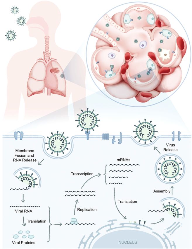
View Article Online

                                                                                                                Review                                                                                                                     RSC Chemical Biology
            This article is licensed under a Creative Commons Attribution-NonCommercial 3.0 Unported Licence.
Open Access Article. Published on 23 December 2020. Downloaded on 4/1/2021 3:41:39 AM.

                                                                                                                Fig. 2 Host signaling and metabolic responses to SARS-CoV-2 infection predicted based on strong similarities with two previous highly pathogenic
                                                                                                                human b-coronaviruses SARS-CoV and MERS-CoV. After viral entry, the single-stranded RNA genome (gRNA) of SARS-CoV-2 is released in the host cell
                                                                                                                cytoplasm. ORF1a and ORF1ab mRNAs are translated to produce two large polyproteins, pp1a and pp1ab, which are proteolytically cleaved into
                                                                                                                16 non-structural proteins (nsps), including papain-like protease (PLpro), 3C-like protease (3CLpro), RNA-dependent RNA polymerase (RdRp), helicase
                                                                                                                (Hel), exonuclease (ExoN), and 4 structural proteins. The envelope glycoprotein spike (S) forms a layer of glycoproteins that protrude from the envelope.
                                                                                                                Two additional transmembrane glycoproteins are incorporated in the virion: envelope (E) and membrane (M). Inside the viral envelope resides the helical
                                                                                                                nucleocapsid, which consists of the viral positive-sense RNA ((+)RNA) genome encapsidated by protein nucleocapsid (N). An additional 9–12 ORFs are
                                                                                                                encoded through the transcription of a nested set of sub-genomic RNAs. Functions of these truncated or untruncated sub-genomic RNAs are still
                                                                                                                unknown. The viral elements (ssRNA or sub-genomic RNAs) are recognized by pattern recognition receptors such as Toll-like receptors (TLRs), retinoic-
                                                                                                                acid-inducible protein I (RIG-I), RIG-I like receptor, and melanoma differentiation-associated protein 5 (MDA-5) that recruit adapter mitochondrial
                                                                                                                antiviral-signaling protein (MAVS) and leads to activation of interferon regulatory factor 3 (IRF3) and NF-kB and the subsequent production of type I
                                                                                                                Interferons (IFNs) and pro-inflammatory cytokines (e.g. IL-1b and IL-6), respectively. Itaconate, an anti-inflammatory metabolite transported by TCA cycle
                                                                                                                intermediates (e.g. citrate), results in activation of pathways involved in proinflammatory cytokines. Proinflammatory cytokines activate the mammalian
                                                                                                                target of rapamycin (mTOR) signaling in addition to the increased energy metabolism through aerobic glycolysis. The increased production of
                                                                                                                tricarboxylic acid cycle (TCA) intermediates e.g. succinate and citrate activate NF-kB downstream signaling resulting in the production of proin-
                                                                                                                flammatory cytokines. The antiviral activity of type I IFNs limit viral replication and modulate the innate and adaptive immune responses. They bind to
                                                                                                                interferon-a/b receptors (IFNARs), which are expressed on a number of different cells including macrophages and activate the JAK/STAT signaling
                                                                                                                pathway. This signaling leads to the formation of the STAT1/2/IRF9 complex and the induction of a plethora of IFN-stimulated genes (ISGs). Cytokines
                                                                                                                released by infected cells modulate the adaptive immune response by recruiting and activating immune cells such as macrophages, B-cells, and T-cells
                                                                                                                to orchestrate the elimination of the virus. However, an unbalanced immune response has been reported to cause hyper-inflammation, a condition
                                                                                                                termed as ‘cytokine storm’ in cases of severe clinical symptoms of COVID-19. Many, but not all, aspects of this model have been directly verified in the
                                                                                                                rapidly emerging SARS-CoV-2 experimental literature.

                                                                                                                proteins (7b, 8a, 8b and 9b) and truncated/untruncated sub-                   metabolic players like nuclear hormone receptors (e.g. PPARs)
                                                                                                                genomic mRNAs are still unknown (Fig. 2). We also have limited                and TCA cycle metabolites (e.g. succinate, citrate) activate the
                                                                                                                knowledge of the immune players involved in SARS-CoV-2 infec-                 NF-kB signaling cascade which release proinflammatory cytokines
                                                                                                                tion. In a conventional viral infection model, innate immune                  (IL-1, IL-6, TNF-a) and interferons (IFNs) (Fig. 2).40,41 These

                                                                                                                  2021 The Author(s). Published by the Royal Society of Chemistry                                                 RSC Chem. Biol., 2021, 2, 3046 | 33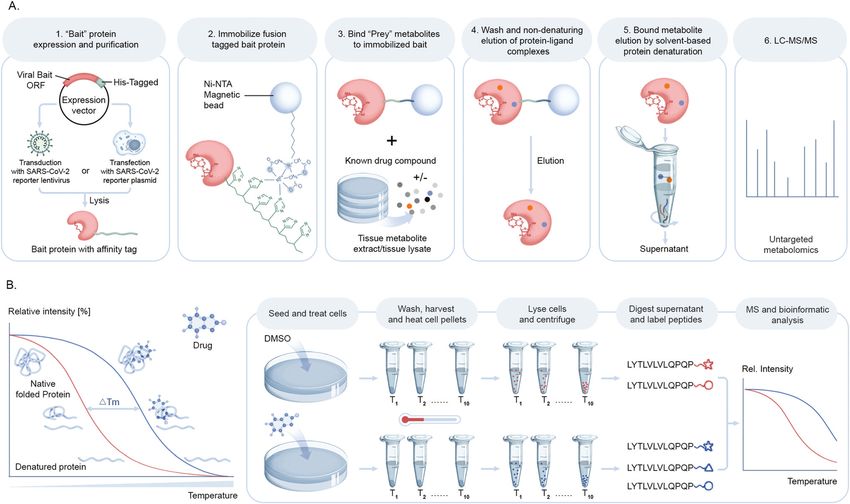
View Article Online
RSC Chemical Biology Review
proinflammatory cytokines in turn activate the mTOR signaling immunofluorescence analysis (IFA) and the utilization of fluor-
pathway of adjacent host cells (Fig. 2).38,42 IFNs are critical to both escent proteins. IFA utilizes fixed cells or tissues to stain a
innate and adaptive immunity, and function as the primary protein of interest with fluorescently marked antibodies, while
activator of macrophages, in addition to stimulating natural killer fluorescent proteins can be expressed in vivo for utilization in
This article is licensed under a Creative Commons Attribution-NonCommercial 3.0 Unported Licence.
cells and neutrophils.43 IFNs also take part in activating the live-cell imaging (Fig. 3). One of the examples in the context of
JAK-STAT signaling pathway upon binding to its receptor on the virology is to image reporter viruses expressing fluorescent
adjacent host cell membrane.43 While all these aspects are still to proteins as described in the next section.
be figured out for SARS-CoV-2 infection, their elucidation will be Notably, recent IFA studies report tracking membranous struc-
possible upon employing suitable strategies to map these virus– tures colocalizing with coronaviral replication/transcriptional
host interactions. The following section describes some of the complexes. Müller et al. studied the influence of cellular lipid
Open Access Article. Published on 23 December 2020. Downloaded on 4/1/2021 3:41:39 AM.
well-established methods and strategies to address coronavirus– metabolism on human coronavirus replication in culture using
host interactions. both confocal and transmission electron microscopy.50 Specifi-
cally, the group was interested in understanding the colocalization
Strategies to profile coronavirus–host of viral-mediated dsRNA production and lysophospholipids gen-
interactions erated by cytosolic phospholipase A2a (cPLA2a) activity. They
infected Huh-7 cells with HCov-229E or MERS-CoV, and stained
Since identification and characterization of key host–viral pro- these cells with antibodies against dsRNA, the coronavirus Nucleo-
tein interactions is crucial towards understanding how inter/ capsid (N) and NSP8 protein. To monitor lysophospholipid gen-
intra-cellular signaling cascades and biochemical processes are eration and localization, they employed the use of a fluorogenic
initiated to discover new therapeutic targets, a number of cPLA2a active probe. In this cell system, viral replication/transcrip-
chemical biology strategies have been developed and employed tion complexes (RTC) were observed to co-localize with lysopho-
over the years to address this challenge. The advancement of spholipids. Notably, when cells were treated with the cPLA2a
experimental strategies to map these interactions using mono- inhibitor pyrrolidine-2 to disrupt lysophospholipid production, a
clonal and anti-idiotypic antibodies, solid-phase assays, and corresponding decrease in viral RNA and protein accumulation
affinity purification with receptor antibodies44–48 to more recent was detected in coronavirus infected cells.
high-throughput screening (HTS) technologies based on gene Another study by Poppe et al. investigated the influence of
targeting methods and new generation high resolution mass HCoV-229E infection on cellular NF-kB signaling and the
spectrometry49 has brought the flexibility of choosing workflows cellular transcriptome landscape.51 The authors used IFA to
based on the desired output for mapping these interactions. In track coronavirus N protein expression to monitor viral infection
this section, these strategies are discussed in detail with respect to and its spread in an A549 lung-derived epithelial cell line. Other
the principle of the workflows and the impact each is contributing microscopic methods have involved the detection of viral RNA or
towards unravelling the mystery around the pathophysiological DNA by fluorescence in situ hybridization (FISH) whereby a
and chemical biology of coronavirus–host interactions. fluorescent probe is used to target viral sequences.52
A recent imaging technique for visualizing SARS-CoV-2
1. Imaging-based interaction analysis infected nasopharyngeal epithelial cells was developed by Rut
Microscopy is an important tool in virology and infection et al. using activity-based probes (ABPs) for the SARS-CoV-2 main
biology. Different microscopic imaging strategies and work- protease (Mpro) that were developed from a hybrid combinatorial
flows are employed to understand the underlying principles of substrate library.53 An ABP from this library screen was able to
receptor binding, genome release, replication, assembly, and detect SARS-CoV-2 Mpro at 5 nM enzyme concentration when
virus budding, as well as the response of the host immune incubated in the presence of 2.5 mM probe for 5 min. This study
system. Virologists deploy a broad range of light microscopy also resulted in the development of a potent SARS-CoV-2 Mpro
techniques, with the primary choice being fluorescence micro- inhibitor with half-maximal effective concentration (EC50) of
scopy. Fluorescence microscopy can be roughly subdivided into 3.7 mM in a human cell line-based viral infection assay.
Fig. 3 Imaging-based interaction analysis using viral reporter strains for monitoring virus–host interactions. Workflow illustrates fluorescence imaging
using immunolabeling of fixed cells or tissues to stain a protein of interest, or reporter viruses stained with antibodies labeled with fluorophores.
34 | RSC Chem. Biol., 2021, 2, 3046 2021 The Author(s). Published by the Royal Society of Chemistry
View Article Online
Review RSC Chemical Biology
2. Viral reporter strains for virus–host interaction analysis virus expressing fluorescently tagged structural proteins. One
Recombinant reporter viruses are vital tools for studying the disadvantage of using GFP as a reporter gene in the context of
pathogenicity and transmission of viruses in animal models. an animal model is that some tissues have an endogenous
They are generated to incorporate a marker, such as a fluor- autofluorescence that makes the detection of the reporter
This article is licensed under a Creative Commons Attribution-NonCommercial 3.0 Unported Licence.
escent protein whose expression can be readily followed, into a difficult relative to the background. The liver in particular can
specific gene/locus (or multiple loci) of the viral genome. The be very problematic due high autofluorescence background.
Antibodies to GFP and other fluorescent proteins partly over-
simplest recombinants are those generated by insertion of a
come this problem via the use of immunohistochemical staining
reporter gene into a nonessential site on the viral genome,
of tissue.
which can either cause a disruption of a particular gene that is
A potential alternative for in vivo detection of viruses is the
Open Access Article. Published on 23 December 2020. Downloaded on 4/1/2021 3:41:39 AM.
not required for viral replication in tissue culture (and/or
use of bioluminescence imaging (BLI). This particular tech-
in vivo) or alternatively an insertion of an intergenic site such
nique is extremely sensitive and enables the detection of virus
that the repertoire of viral genes is unaffected. Disruption of a
in real time in the same animal over days to weeks post-
specific gene enables the ability to characterize the functional
infection. BLI requires the introduction of the firefly (Photinus
significance of the perturbed viral gene. The presence of the
pyralis) luciferase reporter gene into the viral genome of interest.
reporter allows for facile tracking of viral infection to provide a
After interaction with their substrate, firefly luciferase (FLuc)
better understanding of pathogenesis. However, the genetic
enzymes emit photons; the specific wavelength (400–615 nm) is
manipulations required to achieve reporter gene expression
dependent upon the enzyme. A fairly recent introduction of a
comes with the caveats of altering the viral genome and
smaller variant of the 62 kDa FLuc called nanoLuciferase (nLuc),
potential downstream function as well as creating potential
a 19 kDa enzyme from deep sea shrimp, now comprises a
instability of reporter expression and detection depending on
popular reporter assay system which produces a luminescent
the tissue type.54 However, despite these limitations, this
signal 4100-fold brighter than FLuc. The key feature that makes
technology has proved to be instrumental in understanding
nLuc an ideal reporter is that its luminescent signal exhibits a
viral infections in both living cells and animals.
long lifetime, resulting in glow kinetics rather than a rapid flash
Classically, one of the most commonly used reporter genes
signal typically produced by other small, bright luciferases such
has been lacZ (encoding beta-galactosidase), which can either
as Gaussia luciferase. nLuc produces blue light with maximum
be placed under the control of a viral promoter specific to the emission at 460 nm, is thermally stable, active over a broad pH
virus being studied or under a strong universal promoter such range, and requires no post-translational modifications or matura-
as HCMV immediate early enhancer element or the SV40 tion after translation to make an active enzyme. Agostini et al.
promoter.55 The choice of a promoter is dependent on the used nLuc fusions to study the susceptibility of b-coronaviruses
intended application for the recombinant virus. Although lacZ is such as mouse heptatitis virus, MERS-CoV, and SARS-CoV to
an effective reporter, the infected cells/tissues require downstream remdesivir in cell-based assays and observed RNA polymerase-
processing/staining to enable the detection of beta-galactosidase, mediated inhibition of viral replication.57
and this can add an additional 6–18 h to the protocol for detection
of virus. An additional concern is the large size of the lacZ gene, 3. Bioinformatic-based interaction analysis
which might be an important consideration for viruses with A thorough analysis of virus–host protein interactomes can
limited genome oversizing potential because of packaging con- identify potential drug targets while deciphering the molecular
straints. This could potentially result in second site mutations etiology underlying viral replication and pathogenesis. With the
(e.g. gene deletion) unrelated to the reporter gene insertion locus, application of high-throughput methods, described below,
which would also alter the phenotype of the virus. or literature mining, researchers have collected reasonably
Fluorescent proteins have become extremely popular for complete and high quality virus–host protein–protein interac-
incorporation into recombinant viruses based on their sensitive tomes (PPIs), generating invaluable virus–host PPI databases58,59
detection in live cells in real time via fluorescent microscopy. while providing a global view of human cellular processes
The most common fluorescent reporter is green fluorescent controlled by viruses. These assays are typically performed using
protein (GFP) from the jelly fish (Aequorea victoria) and engi- individual viral proteins expressed in isolation in transformed
neered variants that produce high levels of fluorescent signal cell lines that may be quite dissimilar phenotypically from native
and consequently easy detection and real-time tracking. For host target cells, such as lung epithelia.
example, Sims and co-workers generated recombinant SARS- While most recent efforts have pivoted towards more targeted
CoV constructs by deletion of open reading frame 7a/7b proteomic approaches to study viral–host protein interactions in
(ORF7a/7b) and insertion of GFP to study the infectivity of a cellular context, as described further below, structure guided
SARS-CoV virus in human angiotensin 1 converting enzyme computational approaches aim to predict the conserved set of
2 (hACE2) mediated invasion of an in vitro model of human putative interactions of virus and host (Fig. 4). This has dee-
ciliated airway epithelia (HAE) derived from nasal and tracheo- pened understanding of virus-specific cellular targets that lead to
bronchial airway regions.56 rewiring of host pathways and are illuminating promising new
An additional advantage of fluorescent proteins is that their aspects of disease intervention. These computational inferences
small size enables viral protein tagging and construction of involve data mining from literature-curated binary PPIs from
2021 The Author(s). Published by the Royal Society of Chemistry RSC Chem. Biol., 2021, 2, 3046 | 35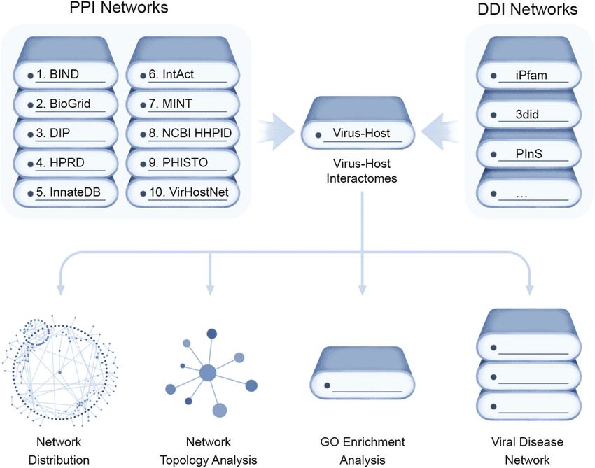
View Article Online

                                                                                                                RSC Chemical Biology                                                                                                                            Review
            This article is licensed under a Creative Commons Attribution-NonCommercial 3.0 Unported Licence.
Open Access Article. Published on 23 December 2020. Downloaded on 4/1/2021 3:41:39 AM.

                                                                                                                Fig. 4 Computational approaches for virus–host interaction analysis. Bioinformatics pipeline for identifying virus–host interactions by data parsing from
                                                                                                                high quality virus–host protein–protein interactomes (PPIs) databases and mapped to form putative domain–domain interaction networks (DDIs) for
                                                                                                                conserved domains of both virus and host proteins that can be assessed for their network distribution properties, such as node topology, as well as
                                                                                                                enrichment for gene ontology (GO) functional annotations and disease associations.

                                                                                                                public databases e.g. Biomolecular Interaction Network Database               processes involved in SARS-CoV-2 entry, replication, and host–
                                                                                                                (BIND),60 the Database of Interaction Proteins (DIP),61 Human                 pathogen interactions, as well as elucidated immune response,
                                                                                                                Protein Reference Database (HPRD),62 IntAct,63 the Molecular                  host cell recovery, and repair mechanisms.
                                                                                                                INTeraction database (MINT),64 Virus–Host Network (VirHost-
                                                                                                                Net),65 Biological General Repository for Interaction Datasets                4. Molecular biology-based interaction analysis
                                                                                                                (BioGrid),66 InnateDB,67 and the Pathogen–Host Interaction                    Advances in functional genomics and proteomics have allowed
                                                                                                                Search Tool (PHISTO).68 These PPI datasets can be scanned for                 for the unbiased identification of cellular factors involved in
                                                                                                                conserved Pfam-A domains present in both virus and host                       viral infection. Diverse screening techniques have been applied
                                                                                                                encoded proteins, which are mapped to form putative domain–                   to this field, including loss-of-function and gain-of-function
                                                                                                                domain interaction networks (DDIs) that can be assessed for                   screens. When applied proteome-wide, these techniques allow
                                                                                                                the topological parameters using R packages like igraph or                    for the interrogation of cellular requirements for viral infection,
                                                                                                                Cytoscape.                                                                    generating information on those factors that are most important
                                                                                                                   To analyze the functional impact of predicted targeting of                 for viral infection.
                                                                                                                host protein domains by viruses, enrichment analysis of GO                       The yeast two-hybrid assay (Y2H), devised by Fields and
                                                                                                                functional annotation terms can be carried out using R and the                Song for mapping binary protein–protein interactions (PPI)72
                                                                                                                Bioconductor topGO package. Finally, the DDIs, along with their               has been used in multiple high throughput viral–host PPI
                                                                                                                assigned functionality, can be assessed for disease relevance                 screens for unbiased viral genome-wide interactome explora-
                                                                                                                from public databases (e.g. Online Mendelian Inheritance in                   tion or focusing on a subset of viral proteins.73 These studies
                                                                                                                Man (OMIM), Human Gene Mutation Database (HGMD Public)69                      are based on an initial construction of a viral ORFeome,
                                                                                                                and Catalogue of Somatic Mutations in Cancer (COSMIC)70) to                   comprising clone ORFs encoding distinct viral proteins fused
                                                                                                                make a comprehensive list of disease-associated viral host                    to a DNA binding domain (Fig. 5A). Interactions with human
                                                                                                                targets. For example, Ostaszewski et al. built a comprehensive                proteins expressed as activation domain fusions can reconsti-
                                                                                                                COVID-19 disease map as a standardized knowledge repository                   tute a functional transcriptional factor, driving expression of a
                                                                                                                of all putative SARS-CoV-2 virus–host interactions.71 This pro-               reporter gene(s). This genetic assay is prone to false positives
                                                                                                                ject was an open collaboration between scientists across the                  (and negatives), and so putative interactions discovered using
                                                                                                                globe curating SARS-CoV-2 infection related information in one                Y2H screens must be confirmed by a secondary, biochemical
                                                                                                                platform to support the research community in understanding                   method to generate high confidence data.
                                                                                                                SARS-CoV-2 pathophysiology and aid the development of effi-                      Using the Y2H assay, Xiao et al. reported the interaction of
                                                                                                                cient diagnostics and therapies. This platform has enabled the                the N-terminal region of SARS-CoV Spike (S) protein with eIF3f,
                                                                                                                visual exploration and computational analyses of molecular                    a subunit of the eukaryotic initiation factor 3 (eIF3), which they

                                                                                                                36 | RSC Chem. Biol., 2021, 2, 3046                                                 2021 The Author(s). Published by the Royal Society of Chemistry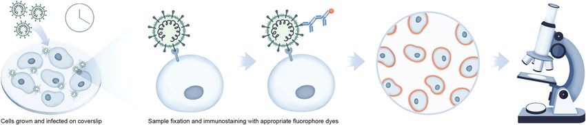
View Article Online

                                                                                                                Review                                                                                                                     RSC Chemical Biology
            This article is licensed under a Creative Commons Attribution-NonCommercial 3.0 Unported Licence.
Open Access Article. Published on 23 December 2020. Downloaded on 4/1/2021 3:41:39 AM.

                                                                                                                Fig. 5 Molecular biology-based investigations of coronavirus–host interactions. (A) The yeast 2-hybrid methods of screening PPIs involve mating two
                                                                                                                haploid complementary yeast strains each expressing a distinct expression plasmid. The first strain expresses a protein bait fused to a DNA-binding
                                                                                                                domain (DBD) that binds to its cognate binding site, usually upstream of a reporter gene. The second strain expresses a protein prey fused to a
                                                                                                                transcription activation domain (AD). If there is interaction between bait and prey, the AD is brought into the proximity of the DBD which causes
                                                                                                                transcriptional activation of the reporter gene leading to selection. (B) Engineered CRISPR systems contain two components, a single guide RNA (sgRNA)
                                                                                                                and a CRISPR-associated endonuclease (Cas9 protein) to edit (insert or delete) DNA sequences by generating double stranded breaks (DSB) and
                                                                                                                introducing indels (insertions/deletions) for generation of gene knock-out libraries. In CRISPR–Cas9-pooled screen methods, single-guide RNA (sgRNA)
                                                                                                                libraries are initially synthesized as lentiviral vectors and amplified and packaged into lentiviruses. Lentivirus pools are then used to transduce Cas9
                                                                                                                positive target cells at low multiplicity of infection (MOI). The genomic DNA is extracted from input versus selected cell pools, and the integrated sgRNA
                                                                                                                sequences are PCR-amplified. The sgRNA abundance is then determined by next-generation DNA sequencing. (C) Workflow showing the application of
                                                                                                                the CRISPR–Cas screening platform in detection of SARS-CoV-2 infection. A SARS-CoV-2 gene specific CRISPR–Cas sgRNA library is created from
                                                                                                                identifying the conserved gene sequences of pan-coronaviral genome and validated for detection of SARS-CoV-2 RNAs (PAC-MAN method). This library
                                                                                                                is then used to generate lateral flow strip-based assays to detect the viral RNAs in nasopharyngeal swabs from COVID-19 patients.

                                                                                                                subsequently confirmed by co-immunoprecipitation (co-IP) and                  that are specific to target mRNA sequences, into cells allow for
                                                                                                                IFA.74 Another systems biology study employing a proteome-                    sequence-specific degradation of the mRNA. This method is a
                                                                                                                wide Y2H screen to identify immunophilins as interaction                      relatively fast, simple, and robust approach to specifically
                                                                                                                partners of the SARS-CoV non-structural protein 1 (Nsp1) was                  downregulate protein expression and study resulting cellular
                                                                                                                performed by Pfefferle et al.75 Immunophilins act as receptors of              function. Expression knockdown has been successfully used in
                                                                                                                immunosuppressive drugs such as cyclosporin A (CsA) and tacro-                virology to study the role of specific host factors in the infec-
                                                                                                                limus and thus this study suggests non-immunosuppressive                      tious life cycle and replication of viruses. Millet et al. used this
                                                                                                                derivatives of CsA may serve as potential broad-range inhibitors              platform to successfully validate the functional impact of ezrin,
                                                                                                                of SARS-CoV infection.                                                        an actin binding protein, that they identified by Y2H screens as
                                                                                                                   Another powerful tool to investigate the biological function               an interactor with the SARS-CoV spike (S) protein.76
                                                                                                                of specific proteins either in vitro or in vivo is the RNA inter-                Recently, clustered regularly interspaced short palindromic
                                                                                                                ference (RNAi) platform. The introduction of small interfering                repeats (CRISPR/Cas) technology has greatly expanded the
                                                                                                                RNAs (siRNA), 20–25 nucleotide short double-stranded RNAs                     ability to genetically probe virus–host functional dependencies

                                                                                                                  2021 The Author(s). Published by the Royal Society of Chemistry                                                 RSC Chem. Biol., 2021, 2, 3046 | 37
View Article Online
RSC Chemical Biology Review
by means of initial screening and loss/gain-of-function based and compared it to the interface for SARS-CoV. This study
confirmation of targets (Fig. 5B).77 Exploiting the characteris- found the ACE2-binding ridge in SARS-CoV-2 RBD assumes a
tics of virus–host interactions and the basic rules of nucleic more compact conformation compared to SARS-CoV RBD along
acid cleavage or, most recently, gene regulation via the CRISPR– with several residue changes that stabilize binding pockets
This article is licensed under a Creative Commons Attribution-NonCommercial 3.0 Unported Licence.
Cas system, it can be used to target components associated with at the RBD–hACE2 interface, thereby increasing the binding
both the virus genome and host factors to define their roles in affinity for ACE2.
virus infection or replication. These include CRISPR interfer-
ence (CRISPRi) technology that can inhibit expression of 6. Omic-based interaction analysis methods
target genes, or CRISPR activation (CRISPRa) that can increase During the last decade, a multitude of additional omic
target gene expression to test contrasting impact on viral approaches have emerged as powerful tools in basic, transla-
Open Access Article. Published on 23 December 2020. Downloaded on 4/1/2021 3:41:39 AM.
proliferation. tional, and clinical research for the study of biological pathways
In a notable recent study, Abbot et al. reported a CRISPR– involved in pathogen replication, host response, and disease
Cas13-based antiviral strategy, PAC-MAN (prophylactic antiviral progression. Advancement of proteomic technologies offering
CRISPR in human cells), for viral inhibition that can effectively sensitive protein detection and quantification and integration
degrade SARS-CoV-2 RNA sequences in human lung epithelial of proteomics with other biochemical and molecular biology
cells.78 They designed and screened multiple single guide RNAs methods, as well as with other omic approaches, has expanded
(sgRNAs) targeting conserved viral genomic regions to identify the repertoire of tools to study pathogen infections (Fig. 6A).
functional sgRNAs potently targeting SARS-CoV-2 (Fig. 5C). Proteomics, the study of the protein component of biological
Another study by Broughton et al. developed a CRISPR–Cas12- systems, has emerged to the frontline for the discovery and
based lateral flow assay for the rapid detection of SARS-CoV-2 understanding of host–pathogen interactions in SARS-CoV-2.49
from respiratory swab RNA extracts (Fig. 5C).79 Daly et al. Genomic, transcriptomic, and metabolomic profiling studies
reported the interaction of host cell receptor Neuropilin-1 provide orthogonal information that complements the data
(NRP-1) with CendR motif of SARS-CoV-2 spike (S), a polybasic generated by proteomic analyses to achieve a global, systems-
Arg-Arg-Ala-Arg C-terminal sequence on the S1 subunit upon level understanding of the infection process (Fig. 6B). For
cleavage by host protease furin. NRP-1 is a cell-surface receptor example, Olagnier et al. reported suppression of Nrf2 signaling
that plays an essential role in angiogenesis, regulation of events upon SARS-CoV-2 infection by transcriptomic analysis of
vascular permeability, and the development of the nervous COVID-19 patient lung biopsies. To further reinforce the ther-
system. This group probed the functional relevance of this apeutic potential of modulating this pathway as an antiviral
interaction through CRISPR/Cas-mediated knockout of NRP-1.80 approach, they performed in vitro assays of cell lines treated
with Nrf2 agonists, 4-OI and DMF, and reported significant
5. Biochemical-based interaction assays reduction in the SARS-CoV-2 RNA abundance in the treated cell
Surface plasmon resonance (SPR) is an established, label-free lines when compared with untreated infected ones.37 Also,
optical technique used to study the strength of binary bio- given that the majority of proteins do not function in isolation
molecular interactions in real time by detecting changes in the and their interactions with other proteins define their cellular
reflection of light from proteins affixed to a prism–gold film functions, a detailed understanding of PPIs and larger protein
interface. SPR facilitates investigation of the biophysical nature interaction environments are key for deciphering regulation of
of binding interactions, and provides detailed binding kinetic cellular networks and pathways.
information across a wide range of molecular weights, includ- The method that has seen the widest implementation in
ing small molecule ligands, all without the use of labels. host–pathogen interaction studies is immunoaffinity or epitope
Automated instrument platforms for SPR analysis are commer- tag-based affinity purification coupled to mass spectrometry
cially available that can be used for a wide variety of protein (Fig. 6C). In immunoprecipitation mass spectrometry (IP-MS), a
interaction assays, including specificity, active concentration target of interest is isolated using an antibody raised against
measurement, and profiling thermodynamic parameters. SPR the endogenous protein whereas in the case of affinity purifica-
is increasingly used in the field of virology, spanning from the tion coupled to mass spectrometry (AP-MS) the target protein of
study of biological interactions to the identification of putative interest is co-expressed with an epitope tag which is isolated
antiviral drugs. using antibodies raised against the epitope tag. This immuno-
The receptor recognition mechanism of SARS-CoV spike (S) precipitated or affinity-purified protein of interest, along with
protein towards hACE2, which defines host cell infectivity, co-isolated interacting proteins, is then identified by MS. IP-MS
pathogenesis and host range, was assessed using SPR by Wu and AP-MS studies can be performed from both the pathogen
et al. (2012).81 They measured the binding affinities between and host perspective. For example, selective enrichment of a
the receptor binding domain (RBD) of S and hACE2 variants by viral protein can facilitate understanding of the host factors it
immobilizing serial dilutions of the proteins on a sensor chip interacts with to promote replication or suppress host defense
through covalent coupling via amine groups. An alternate pathways. Alternatively, enrichment of host cellular protein can
biophysical approach was likewise employed to explore the be performed to identify interactions with surrounding protein
RBD–hACE2 interface by Shang et al. (2020).82 They generated partners during viral infection to characterize possible changes
a crystal structure of SARS-CoV-2 RBD complexed with hACE2 in the hijacked host protein function(s). Both IP-MS and AP-MS
38 | RSC Chem. Biol., 2021, 2, 3046 2021 The Author(s). Published by the Royal Society of ChemistryView Article Online
Review RSC Chemical Biology
This article is licensed under a Creative Commons Attribution-NonCommercial 3.0 Unported Licence.
Open Access Article. Published on 23 December 2020. Downloaded on 4/1/2021 3:41:39 AM.
Fig. 6 Mass spectrometry-based techniques used to identify virus–host interaction networks. (A) Overview of proteomic tools utilized in the of study
host–pathogen interactions and their integration with omics approaches. (B) Quantitative multi-omic analysis workflow to define the host response to an
infection. The resulting datasets are mapped to known metabolic pathways to measure the up- or downregulation upon infection and integrated with
phenotype data to construct correlation networks. (C) Schematic of immunoaffinity or epitope tag-based affinity purification coupled to mass
spectrometry (IP-MS/AP-MS) workflow. Components include immunoaffinity purification of protein complexes, enzymatic digestion of proteins,
nano-liquid chromatography coupled to mass spectrometry (nLC-MS/MS), and bioinformatic analysis to identify proteins. Label-free protein
quantification may be performed by MS/MS spectral counting or precursor ion intensity (MS1) integration. (D) Global proteomic approaches can be
used to study alterations throughout infection in protein abundance. These changes can be quantified at the MS level, by pulse labeling via Stable Isotope
Labeling in Cell Culture (SILAC) and/or isobaric tagging (such as tandem mass tagging, TMT) of samples after proteolysis and comparing the ion intensities
to define proteome alterations at multiple time points of infection. In this workflow, cells are harvested at different infection times after pulse labeling and
digested peptides from each sample are labeled with isobaric tags consisting of unique reporter masses. The samples are mixed together for MS analysis,
and peptide quantification is assessed at the MS/MS level using the reporter ion intensities. Peptide quantitative values derived from sequences assigned
to the same protein are used to calculate the overall relative protein abundance. (E) Workflow showing differential analysis of global proteomic and
metabolomic profiles of COVID-19 patient cohorts vs. healthy individuals via an untargeted LC-MS/MS platform. (A–D) adapted from ref. 95.
studies that capture differential interactions are often per- the temporal cascade of cellular events that occur during a
formed in conjunction with fluorescent tagging and micro- pathogen infection. One advantage of IP-MS over AP-MS is that
scopy. These studies provide complementary spatial–temporal experiments can be performed in a native cellular context to
information about host–pathogen interactions to follow through enable unbiased detection of PPIs whereas AP-MS requires
2021 The Author(s). Published by the Royal Society of Chemistry RSC Chem. Biol., 2021, 2, 3046 | 39View Article Online
RSC Chemical Biology Review
overexpression of the protein of interest to capture interacting cells cultured as an air–liquid interface during infection with
proteins and can result in background binding artifacts. Further- SARS-CoV-2 (Hekman et al., in press). The resulting time-course
more, in both methods, weakly bound protein interactors or profiles revealed rapid remodeling of diverse host cell systems,
localized, but non-interacting, by-stander proteins cannot be including signal transduction machinery, RNA processing, trans-
This article is licensed under a Creative Commons Attribution-NonCommercial 3.0 Unported Licence.
captured. lation, metabolism, nuclear integrity, protein trafficking,
A recent example by Gordon et al. used AP-MS to investigate cytoskeletal-microtubule organization, leading to cell cycle arrest,
the SARS-CoV-2 virus–host protein interactions. The authors genotoxic stress, and innate immunity. Comparison with other
devised a viral–host SARS-CoV-2 protein interaction map by proteomic studies of undifferentiated transformed cell lines high-
cloning, tagging, and expressing 26 of the 29 proteins encoded lighted convergent and divergent responses, reflected by differen-
by the SARS-CoV-2 genome in HEK293T cell lines and identified tial sensitivity to antiviral compounds, providing a rich perspective
Open Access Article. Published on 23 December 2020. Downloaded on 4/1/2021 3:41:39 AM.
the human proteins that physically associate with each viral for targeting respiratory processes hijacked by SARS-CoV-2 as
protein bait.83 They identified 332 putative viral–host PPIs potential therapeutic avenues.
encompassing 66 druggable human proteins targeted by Global proteomic and metabolomic profiling of COVID-19
69 small molecule compounds (including 29 drugs approved patient sera is another approach adopted to address the differ-
by the US Food and Drug Administration, 12 in clinical trials, ential expression of proteins and metabolites upon infection
and 28 in preclinical studies), and established several antiviral when compared to healthy cohorts (Fig. 6E). Shen et al. found
candidates potentially suitable for repurposing as antiviral 105 differentially expressed proteins in the sera of COVID-19
therapeutics. This group also did a comparative coronavirus – patients.89 They correlated expression profiles with clinical
human PPI study to understand the conservation of target pro- disease severity that showed 93 proteins to be specifically
teins and cellular processes between SARS-CoV-2, SARS-CoV, and modulated in severe patients. Activation of the complement
MERS-CoV.84 system, macrophage function, and platelet degranulation were
Global proteomics approaches that quantify changes in the major pathways correlated to 50 out of the 93 differentially
protein abundance and post-translational modifications expressed proteins as shown by their network enrichment
(PTMs) (e.g. phosphorylation) are powerful tools to elucidate analyses. 373 metabolites were significantly changed in
mechanisms of viral pathogenesis by providing a snapshot of COVID-19 patients and 204 metabolites were correlated with
how cellular pathways are rewired upon infection. Importantly, disease severity. Activation of the complement system, macro-
the functional outcomes of many protein and PTM event phage function, and platelet degranulation were the three
changes are well annotated, especially for kinases as drug major pathways correlated to the key dysregulated proteins
targets where phosphorylation directly regulates their activity. and metabolites from the pathway and network enrichment
These approaches employ mass-spectrometry analysis coupled analyses in this study.
with bioinformatics-based tools to quantitatively assess
changes to protein and/or PTM levels. Bojkova et al. performed
a global quantitative proteomic analysis of SARS-CoV-2 infected 7. Emerging chemical proteomic-based interaction
Caco-2 cell lines and revealed that the virus rewires host technologies
cellular pathways such as translation, splicing, carbon metabo- Mapping protein–metabolite interactions (PMI) and identifying
lism, protein homeostasis (proteostasis) and nucleic acid the endogenous host metabolites that complex with proteins is
metabolism.85 The authors used a previously developed of utmost importance to address the gap that still lies in
method known as multiplexed enhanced protein dynamics understanding functional association networks. Small mole-
(mePROD) proteomics that combined both Stable Isotope cule metabolites are key to allosteric regulations of enzymes
Labeling in Cell Culture (SILAC) and Tandem Mass Tags and signaling molecules for receptors involved in nuclear or
(TMT) labeling methodologies to enhance quantification of cellular transport. In the context of viral infection, the discovery
protein level changes with high temporal resolution of small molecule ligands of viral proteins upon infection or
(Fig. 6D).86 A quantitative profiling of the global phosphoryla- viral–host protein complexes will not only shed light on struc-
tion and protein abundance landscape of SARS-CoV-2 infection tural and functional configuration of binding sites to facilitate
was also reported by Bouhaddou et al. where they mapped the design of novel inhibitors for these complexes, but will also
phosphorylation changes to disrupted kinases and pathways led to identification of pathways and cellular events involved in
and used this information to identify potential anti-viral small these interactions. While these chemical proteomic approaches
molecules.87 Collectively, the combination of these quantitative are challenging in terms of confident identification of small
methods with other chemogenomic screening efforts88 have the molecules due to the diverse structural variation, they can be
potential to rapidly prioritize proteins, PTMs, and associated overcome with stringent filters for false discoveries.
drugs and compounds for treating SARS-CoV-2 infection. Our group has been building a ligand discovery pipeline
Since SARS-CoV-2 infects pneumocytes, leading to acute lung employing the AP-MS approach for screening host endogenous
injury and impaired gas exchange, the molecular mechanisms metabolite ligands of diverse protein baits, including SARS-
driving infection and pathology remain unclear. To address this CoV-2 proteases (Fig. 7A). We believe that this study will
gap, our team performed a quantitative phosphoproteomic survey contribute to basic understanding of novel aspects of the
of pluripotent stem cell-derived human alveolar epithelial type 2 chemical biology of SARS-CoV-2–host interactions and possibly
40 | RSC Chem. Biol., 2021, 2, 3046 2021 The Author(s). Published by the Royal Society of ChemistryYou can also read