Computational Studies of Snake Venom Toxins - MDPI
←
→
Page content transcription
If your browser does not render page correctly, please read the page content below
toxins
Review
Computational Studies of Snake Venom Toxins
Paola G. Ojeda 1,2, *, David Ramírez 1,2 ID , Jans Alzate-Morales 1      ID
                                                                              , Julio Caballero 1 ,
Quentin Kaas 3 and Wendy González 1,4, *
 1    Center for Bioinformatics and Molecular Simulations (CBSM), Universidad de Talca, 3460000 Talca, Chile;
      david.ramirez@uautonoma.cl (D.R.); jalzate@utalca.cl (J.A.-M.); jcaballero@utalca.cl (J.C.)
 2    Facultad de Ciencias de la Salud, Instituto de Ciencias Biomedicas, Universidad Autonoma de Chile,
      3460000 Talca, Chile
 3    Institute for Molecular Bioscience, The University of Queensland, Brisbane, Queensland 4072, Australia;
      q.kaas@imb.uq.edu.au
 4    Millennium Nucleus of Ion Channels-Associated Diseases (MiNICAD), Universidad de Talca, 3460000 Talca,
      Chile
 *    Correspondence: paola.ojeda@uautonoma.cl (P.G.O.); wgonzalez@utalca.cl (W.G.);
      Tel.: +56-712-201-674 (W.G.)

 Received: 6 November 2017; Accepted: 18 December 2017; Published: 22 December 2017

 Abstract: Most snake venom toxins are proteins, and participate to envenomation through a diverse
 array of bioactivities, such as bleeding, inflammation, and pain, cytotoxic, cardiotoxic or neurotoxic
 effects. The venom of a single snake species contains hundreds of toxins, and the venoms of the
 725 species of venomous snakes represent a large pool of potentially bioactive proteins. Despite
 considerable discovery efforts, most of the snake venom toxins are still uncharacterized. Modern
 bioinformatics tools have been recently developed to mine snake venoms, helping focus experimental
 research on the most potentially interesting toxins. Some computational techniques predict toxin
 molecular targets, and the binding mode to these targets. This review gives an overview of current
 knowledge on the ~2200 sequences, and more than 400 three-dimensional structures of snake toxins
 deposited in public repositories, as well as of molecular modeling studies of the interaction between
 these toxins and their molecular targets. We also describe how modern bioinformatics have been used
 to study the snake venom protein phospholipase A2, the small basic myotoxin Crotamine, and the
 three-finger peptide Mambalgin.

 Keywords: molecular dynamics simulations; databases; snake peptides; proteomics; molecular
 modeling

1. Introduction
       Snake venom is a complex mixture of proteins and peptides, and presents several medical and
pharmaceutical applications [1–3]. Since the Greek antiquity, substances extracted from snake have
been recognized for their medicinal properties, and the rod of Asclepius, a snake coiled around
a staff, is the most commonly used symbol of medicine and health. In modern times, a number
of notable molecules derived from snake toxins are used in the clinic or are in various stages of
clinical development [4]. The most famous example of snake-derived medicine is captopril (Capoten),
which was developed by Bristol-Myers Squibb, and is now used as a generic medicine for treating
hypertension and congestive heart failure [5,6]. It is a small molecule inhibitor of the angiotensin
converting enzyme (ACE), and is derived from bradykinin potentiating peptides found in the venom of
the South American snake Bothrops jararaca [5]. Another snake-derived compound potentially used for
heart failure is cenderitide (CD-NP, Mayo Clinic/Capricor Therapeutics, Beverly Hills, CA, USA) [7].
It is a chimera between the green mamba Dendroaspis Natriuretic Peptide DNP and the human C-type

Toxins 2018, 10, 8; doi:10.3390/toxins10010008                                     www.mdpi.com/journal/toxins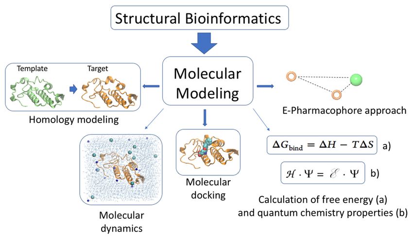
Toxins 2018, 10, 8                                                                                  2 of 24

natriuretic peptides, and activates guanylyl cyclases. It completed Phase I/II clinical trial for chronic
heart failures.
      Eptifibatide (Integrilin, Millennium Pharmaceuticals/Merck, Cambridge, MA, USA), a RGD-motif
cyclic heptapeptide that acts on glycoprotein IIb/IIIa integrin receptors of the blood platelets, is a
medicine used in the clinic to prevent platelet aggregation and thrombus formation in acute coronary
syndromes. It was derived from a much larger protein from the pygmy rattlesnake (Sistrurus miliarius
barbouri). In the same category, anfibatide (Declotana, Lee’s Pharmaceutical, Hong Kong, China) is a
snake venom-derived from platelet aggregation inhibitor peptide that antagonizes platelet glycoprotein
Ib receptor. It is investigated for the treatment of acute thrombotic thrombocytopenic purpura, a fatal
blood clot disorder [8]. It completed phase Ib–IIa, and will undergo phase II according to Lee’s website.
It also has potential for treatment of acute experimental ischaemic stroke and reperfusion injury.
      A “detoxified” variant of the α-cobratoxin (RPI-78M, ReceptoPharm/Nutri Pharm, Coral Springs,
FL, USA), stimulates the production of cytokines. It has application in autoimmune diseases,
myasthenia gravis (MG), muscular dystrophy (MD), and amyotrophic lateral sclerosis (ALS). It recently
received, from the U.S. Food and Drug Administration (FDA), an orphan drug designation for the
treatment of pediatric multiple sclerosis. Gyroxin is a serine protease from Crotalus durissus terrificus,
and has applications aiming at tissue regeneration, repairing nervous system traumas and bone
marrow. Phase I clinical trial found the protein to be safe and preclinical study showed promising
results for treating chronic venous ulcers [9].
      Counterintuitively, some snake toxins can inhibit pain, and have, therefore, potential application
as analgesics. For example, crotalphine is a 14 amino acid peptide with one disulfide bond,
and has analgesic properties through TRPA1 desensitization [10]. It was reported to be developed
by the Brazilian firm Biolab Farmaceutica, but the compound is not mentioned on their website.
The mambalgins are three-finger toxins with antagonist activity on Acid-sensing ion channel (ASIC)
channels, and are active on a range of pain models [11]. They were to be developed by Theralpha,
a French startup company, which has ceased activity.
      The examples mentioned above stress why snake venom peptides attract scientific and medical
interest [3], and considerable efforts are made to mine snake venoms for interesting new compounds.
The venom of a single snake species (from the roughly 725 species of venomous snakes) contains
hundreds of potentially pharmacologically active and useful molecules [12]. In recent years,
important technological progress in transcriptomics and proteomics has resulted in a rapidly increasing
knowledge of peptides and proteins in animal venoms, include those of snakes. These technologies
enable rapid discovery of the nearly complete set of toxins of a snake venom [13–15], which was
hitherto not achievable using previous methods based only on reversed phase-high performance
liquid chromatography (RP-HPLC) and mass spectrometry [16]. The number of toxins unraveled by
modern “omics” approaches is very large, efficient computational methods are required to mine this
massive amount of data. This review will first focus on databases that allow searching and retrieving
information on snake toxins. Computational approaches that have been developed to help study snake
toxin activity, such as the prediction of toxin three-dimensional structures and of their interactions
with molecular targets, will be described. The successes and challenges of predicting binding affinities
and specificities using molecular modeling will be discussed, with the future perspective to integrate
the biology of venoms and prediction of toxin biological targets. Finally, we will describe how modern
bioinformatics have been used to study the activity and biological targets of the snake venom protein
PLA2, the small basic myotoxin Crotamine and the three-finger peptide Mambalgin.

2. Extent of Our Knowledge on Snake Toxins
     General databases, such as UniProt [17,18], NCBI Genbank/GenPept [19], and the Protein
Data Bank (PDB) [20,21], play essential roles in simplifying access to information regarding protein
sequences and three-dimensional structures [22]. However, the information about peptides and
proteins from snake toxins is not standardized in these resources, especially the naming of toxins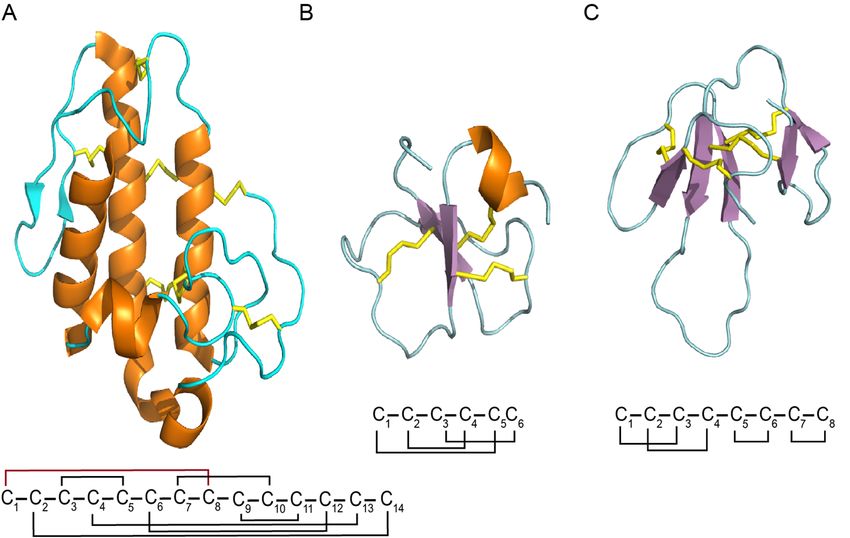
Toxins 2018, 10, 8 3 of 24
and pharmacological activities, and mining for snake venom peptides is difficult. Moreover,
Toxins 2017,
the database 10, 8 on depositions of information from authors to feed the data, leading to numerous
relies 3 of 23
duplications of entries, and a large body of work is being published in peer-reviewed articles,
pharmacological activities, and mining for snake venom peptides is difficult. Moreover, the database
but never submitted to the general databases. Specialized databases, from venomous animals,
relies on depositions of information from authors to feed the data, leading to numerous duplications
are slowly emerging.
of entries, and a large Conoserver
body of work [23], Arachnoserver
is being [24], and ISOB
published in peer-reviewed (Indigenous
articles, but never snake species
submitted
of Bangladesh)
to the general[25] databases.
provide information
Specialized on venomsfrom
databases, fromvenomous
cone snail,animals,
spider, areandslowly
snakes,emerging.
respectively.
A recently developed
Conoserver resource, VenomZone,
[23], Arachnoserver is provided
[24], and ISOB by the
(Indigenous snakeSwiss Institute
species of Bioinformatics
of Bangladesh) [25]
provide information on venoms from cone snail, spider, and snakes,
(SIB), and has information about the venoms from six types of organisms, including snakes. Access respectively. A recently
to thedeveloped
information resource, VenomZone,
is divided is provided
in taxonomy, by the Swiss
activity, and Institute of Bioinformatics
venom protein families,(SIB), and has
making it easy
information
to search throughabout the venoms
the website. from six types
Furthermore, of organisms,
all the information including
is linked snakes.
to theAccess
venom to protein
the
information is divided in taxonomy, activity, and venom protein families, making it easy to search
information from the UniProtKB/Swiss-Prot database (manually annotated and reviewed) and
through the website. Furthermore, all the information is linked to the venom protein information
UniProtKB/Trembl (automatically annotated). The knowledge on the activity of peptides that have
from the UniProtKB/Swiss-Prot database (manually annotated and reviewed) and
been fully characterized helps to predict the possible function of uncharacterized peptides and proteins.
UniProtKB/Trembl (automatically annotated). The knowledge on the activity of peptides that have
In thisbeen
context,
fully specialized
characterized databases
helps to play an the
predict essential role
possible in providing
function access to data,
of uncharacterized predicting
peptides and the
three-dimensional structures and functions of toxins, and identifying outstanding
proteins. In this context, specialized databases play an essential role in providing access to data, toxins with potential
new characteristics (Figure 1). However,
predicting the three-dimensional thereand
structures is presently
functions no commonly
of toxins, established
and identifying and standard
outstanding
way fortoxins with annotation
practical potential new characteristics
of toxins from the(Figure 1). However,
data sources mentionedthereabove,
is presently
leadingnosometimes
commonlyto an
erraticestablished
estimate for andthestandard
numberway for practical
of toxins annotation
in the venom of toxins
of one animal.from the data
Machine sources mentioned
learning-based classifiers
above, leading sometimes to an erratic estimate for the number of toxins in the venom
could help to solve this problem. ToxClassifier [26] is a machine learning tool that allows a consistent of one animal.
Machine learning-based classifiers could help to solve this problem. ToxClassifier [26] is a machine
differentiation of toxins from non-toxin sequences, and reports the best-hit annotation, permitting
learning tool that allows a consistent differentiation of toxins from non-toxin sequences, and reports
assignment of a toxin into the most correct toxin protein family, providing increased curation of these
the best-hit annotation, permitting assignment of a toxin into the most correct toxin protein family,
existing databases [27].
providing increased curation of these existing databases [27].
Figure 1. Integration of proteomics, transcriptomics, and databases in the study of venoms. (A)
Figure 1. Integration of proteomics, transcriptomics, and databases in the study of venoms.
Classical peptidomics analysis of toxins; (B) nucleotide discovery from transcript to precursor
(A) Classical peptidomics analysis of toxins; (B) nucleotide discovery from transcript to
sequence.
precursor sequence.
2.1. Transcriptomic Analyses of Peptides and Proteins from Snake Venom Glands
2.1. Transcriptomic Analyses of Peptides and Proteins from Snake Venom Glands
Genome sequencing of venom gland is still highly expensive, and the assembly of genomic
information
Genome and their analysis
sequencing of venomrequires
gland substantial bioinformatics
is still highly commitment
expensive, and the [28,29].
assemblyBy contrast,
of genomic
sequencing cDNA libraries created from venom gland mRNA using next generation sequencing
information and their analysis requires substantial bioinformatics commitment [28,29]. By contrast,
(NGS) and their assembly became mainstream in research. The transcriptomes of several snake
sequencing cDNA libraries created from venom gland mRNA using next generation sequencing (NGS)
Toxins 2018, 10, 8                                                                                4 of 24

and their assembly became mainstream in research. The transcriptomes of several snake venoms have
been reported [30–33]. NGS platforms produce near comprehensive sequence transcript information
coding for venom peptides and proteins, complementing the traditional PCR techniques, which could
only discover toxin transcripts related to those already discovered in other species or from proteomic
study. The most common NGS technologies used for venom transcriptomics, i.e., 454 GS FLX Titanium
and Illumina, proceed by fragmenting the cDNA and sequencing all or part of these fragments [34].
The 454 technology, which is losing momentum and will be discontinued, produces longer stretch DNA
sequence (reads) than the Illumina technology (limited to read of 150 bp), but of lower quality [34,35].
The fragments can be assembled back into full length or partial transcripts (the assembled fragments
being called contigs) using different software, such as Trinity, Trans-Abyss or SoapdeNovo [36].
     An older technology still in use today consists in using the classical Sanger sequencing of
expressed sequence tags (ESTs), which are short DNA sequences obtained by sequencing the ends
of transcript fragments. Given that most snake venom peptides are around 100 residues long,
the sequencing of ESTs often yields the complete DNA sequence of a peptide. The EST approach
has been successfully applied to venom from snakes, noting that the number of retrieved sequences
are much smaller than generated by NGS, and typically only the most expressed transcripts will be
identified using Sanger/ESTs [32,37]. The ESTs can be assembled back into the original contigs using
several algorithms, such as CAP3 [38], Phrap [39], SeqMan [39,40], or MIRA [41].
     Transcriptomic analysis generally provides insight into the peptide/protein profile of snake
venom, and can be used for discovering putative new peptides and their isoforms [15,42–45],
or peptides that are lowly expressed, and consequently hard to identify by proteomic analysis [46].
Finally, with advances in bioinformatics—which is no more than “the application of information
science to biology” [47]—venom gland transcriptomic data is an excellent tool for studying
peptides evolution [45,48], exploring antivenom and therapeutic agents [32,49], and understanding
structure–function relationships [50].

2.2. Proteomic Analyses of Peptides and Proteins from Snake Venom Glands
     Snake venom proteomes are highly complex mixtures of peptides and proteins [51]. Proteomic
approaches to investigate snake venoms were recently reviewed [2,52]. These approaches generally use
a combination of electrophoresis, liquid chromatography, Edman degradation sequencing, amino acid
analysis, enzymatic digestion, and mass spectrometry, among other techniques. The methods more
widely used are a combination of high performance liquid chromatography, Edman degradation,
MALDI-TOF/MS of proteins, 1D or 2D PAGE and ESI/MS/MS sequencing of digested proteins [40].
The first step to study the proteomes of a snake venom is the venom extraction, which is performed
by “milking” the living snake. Snake milking is achieved by forcing the snake to bite into a
proper container. After venom collection, the proteins are separated using high performance liquid
chromatography (RP-HPLC), ultra-high performance liquid chromatography (UHPLC), and exchange
chromatography [53]. Once the crude venom is fractionated, the sequences of the peptides are
determined using a combination of mass spectrometry and Edman degradation. Prior to the MS/MS
analysis, the peptides are usually reduced, alkylated, and enzymatically digested (usually with trypsin
or chemotryspin) [53]. Finally, the tertiary structure is studied using nuclear magnetic resonance
spectroscopy [53,54].
     Peptides and proteins from snake venoms have a high content of cysteine residues in their
primary sequences, and most of these cysteines form crosslinking disulfide bridges. The stabilization
underpinned by the creation of disulfide bridges has been linked to several important features of
snake toxins: enhancing activity, higher resistance to proteases, improving selectivity, and stabilizing
secondary structure elements [55–58]. Furthermore, the number of disulfide bridges in snake peptides
varies (Figure 2); for instance, natriuretic peptides present one disulfide bond, sarafotoxines present
two disulfide bonds, and more complex toxins, such as omwarpin, have four disulfide bonds. In the
widely-studied phospholipase A2 family, the acidic phospholipase A2 subfamily, with 142 proteins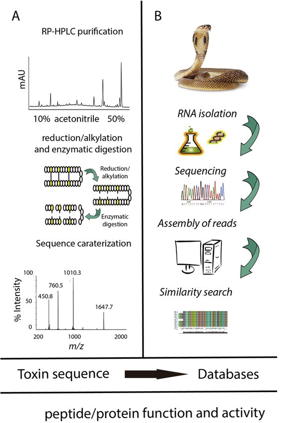
Toxins 2018, 10, 8 5 of 24
Toxins 2017, 10, 8 5 of 23
from snake venoms deposited in UniProt (search criteria: taxonomy: “Serpentes (snakes) (8570)” protein
“acidicAND
phospholipase a2” (keyword:
reviewed: yes) toxin OR annotation:
share a characteristic (type: “tissue
disulfide bridge specificity”
connectivity, wherevenom))
14 Cys AND reviewed:
residues form yes)
seven disulfide bonds.
share a characteristic disulfide bridge connectivity, where 14 Cys residues form seven disulfide bonds.
C C C C C C
Natriuretic peptide Sarafotoxins
C C C C C C
Crotamine
C C C C C C C C C C C C C C
Omwaprin Textilinin-1
C C C C C C C C C C C C C C
Acidic phospholipase A2
FigureFigure 2. Cys
2. Cys patterns
patterns of some
of some toxinsfrom
toxins fromsnake
snake venoms.
venoms.Cartoon
Cartoon representation of aof
representation disulfide bridge
a disulfide bridge
(top left).
(top left). The The disulfide
disulfide bond
bond arrangementsare
arrangements areshown
shown as
as black
blacklines.
lines.
3. Snake Toxin Structures and Activities
3. Snake Toxin Structures and Activities
To comprehend the structure and function of snake toxins will provide a better understanding
To comprehend
of their the structure
role in venom and function
toxicity. Elucidation of snake
of their toxins
structures willwill provide
further a better
help us understanding
to better understand of
their the
roleprotein–protein
in venom toxicity. Elucidation of their structures will further help us
interactions in snake venom, as well as their target receptor/ion channels to better understand
[59].
the protein–protein
Toxins in generalinteractions
seem to adoptina snake
limitedvenom,
number as well as their
of structural target
scaffolds. receptor/ion
It was channels
initially proposed in [59].
Toxinsthein1970s that seem
general the 57 to snake
adoptvenom toxins described
a limited number of at structural
the time as scaffolds.
being neurotoxic
It wasorinitially
cytotoxicproposed
had
in thesimilar
1970ssecondary
that the 57 structure
snake content
venom [60]. Thedescribed
toxins first snakeat venom 3D structure
the time as beingwas also solved
neurotoxic orby X-
cytotoxic
ray diffraction in the 1970s (reported on 1978) [61]. It was released in the
had similar secondary structure content [60]. The first snake venom 3D structure was also solvedProtein DataBank (PDB) in by
1981 under the PDB ID: 1NXB (snake venom curarimimetic neurotoxins) [61]. In the past 20 years,
X-ray diffraction in the 1970s (reported on 1978) [61]. It was released in the Protein DataBank (PDB)
the discovery and unraveling of snake venoms has largely paralleled the technological development
in 1981 under the PDB ID: 1NXB (snake venom curarimimetic neurotoxins) [61]. In the past 20 years,
in proteomics and transcriptomic sciences. Additionally, the number of 3D structures has increased,
the discovery
due to theand unraveling
amazing progressofofsnake venomstechniques,
spectroscopic has largelysuchparalleled
as X-raythe technological
crystallography development
and nuclear
in proteomics and transcriptomic sciences. Additionally, the number of 3D
magnetic resonance (NMR) spectroscopy [62]. Structural genomics appeared early 2000s, and had structures has increased,
a
due to the amazing
dramatic influenceprogress of spectroscopic
on the structural study of techniques,
snake venoms such as X-ray
[63,64]. crystallography
The increased and 3D
pace at which nuclear
magnetic resonance
structure of snake(NMR) spectroscopy
toxins are deposited in[62]. Structural
the PDB in recentgenomics appeared
years is striking, withearly 2000s,
101 and 409and
3D had
structures deposited before and after 2000, respectively.
a dramatic influence on the structural study of snake venoms [63,64]. The increased pace at which
3D structureThe of
specific
snakestructure
toxins are fordeposited
a given toxin is important
in the to understand
PDB in recent years is the molecular
striking, withevents at the
101 and 409 3D
origin of toxin activity. Based on these experimental structures, molecular modeling has been used
structures deposited before and after 2000, respectively.
to understand the molecular interactions related with toxin affinity and specificity.
The specific structure for a given toxin is important to understand the molecular events at the
origin3.1.
of Classification
toxin activity. Based
of Snake on these
Venom Toxins experimental structures, molecular modeling has been used to
understand the molecular interactions related with toxin affinity and specificity.
The majority of snake venom proteins, i.e., 2224 proteins and peptides, could be categorized into
3.1. Classification[65]
30 families (TableVenom
of Snake 1); whereas
Toxins12 proteins are not classified yet. To this day, 410 and 100 3D
structures of snake toxins have been solved by X-ray crystallography or NMR spectroscopic
The majority
techniques, of snake and
respectively; venom
37 3Dproteins,
structuresi.e.,
have 2224
beenproteins
modeledand peptides,
and reported in could be categorized
the Protein Model
into 30 families
Portal [65] (TableStructural
of the PSI-Nature 1); whereas 12 proteins
Biology are not[66].
Knowledgebase classified yet. To was
This information this obtained
day, 410from
and 100
UniProtKBof[67,68]
3D structures snakeby usinghave
toxins the following
been solvedsearchby criteria: taxonomy: “Serpentes
X-ray crystallography (snakes)
or NMR (8570)”
spectroscopic
(keyword:
techniques, toxin OR annotation:
respectively; and 37 (type: “tissue specificity”
3D structures have been venom)) AND reviewed:
modeled yes. in the Protein Model
and reported
Portal of the PSI-Nature Structural Biology Knowledgebase [66]. This information was obtained from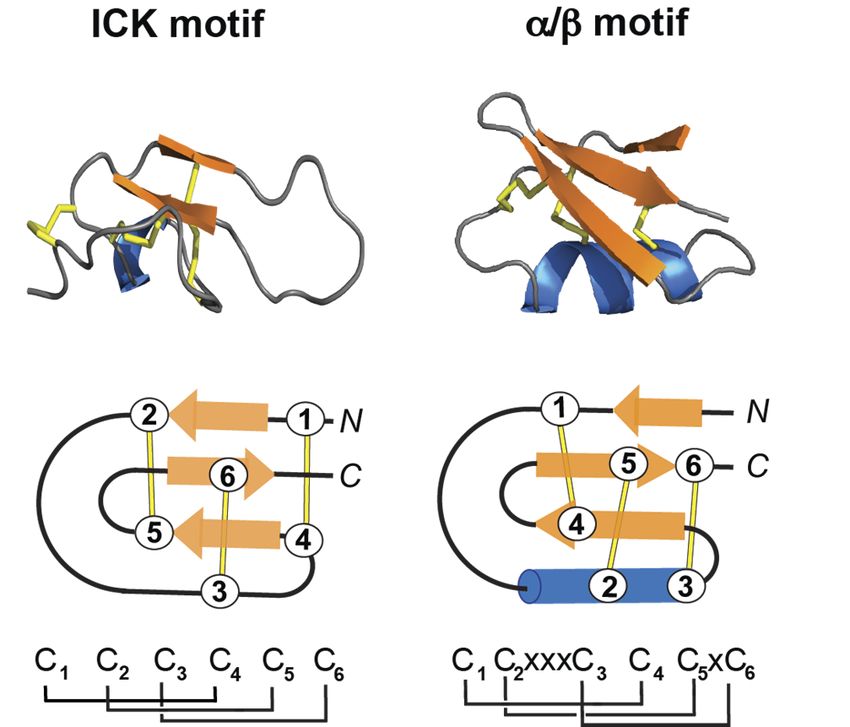
Toxins 2018, 10, 8                                                                                                     6 of 24

UniProtKB [67,68] by using the following search criteria: taxonomy: “Serpentes (snakes) (8570)” (keyword:
toxin OR annotation: (type: “tissue specificity” venom)) AND reviewed: yes.

                                                  Table 1. Snake venoms.

                                                                                                  3D Structure
                     Snake Venom Protein Family                          Entries
                                                                                         X-ray           NMR     Model
                           50 -nucleotidase                                  3
            AB hydrolase superfamily, lipase family                          2
                      AVIT (prokineticin)                                    1                            1
               Bradykinin-potentiating peptide                              65              2
                         Cathelicidin                                       10                            1
                   Complement C3 homolog                                      5             5
                            CRISP                                           66              9
                    Crotamine-myotoxin *                                    17              1             2
                           Cystatin                                          17
                         Desintegrin *                                      33              5             5
                    Endothelin/sarafotoxin                                    6                           3
       Flavin monoamine oxidase (L-amino acid oxidase)                      51              9
             Glycosyl hydrolase 56 (hyaluronidase)                           9
                     Multicopper oxidase                                      3             1
                      Natriuretic peptide                                   55              2             1
                          NGF-beta                                          38              1
       Nucleotide pyrophosphatase/phosphodiesterase                           2
                       Ohanin/vespryn                                        4
                  PDGF/VEGF growth factor                                    20            2
                 Peptidase S1 (serine protease)                             202           11                       4
                       Phospholipase A2                                    469            187                      2
                     Phospholipase B-like                                     2
              pHpG (metalloprotease inhibitor) *                             8
                           Snaclec *                                        172            60                     2
                   Snake three-finger toxin *                              505             65            65       18
        TCTP (translationally controlled tumor protein)                      3
               True venom lectin (C-type lectin)                            44              2                      8
                Type-B carboxylesterase/lipase                                2            1
                      Venom, Kunitz-type                                    143            8              6
               Venom metalloproteinase (M12B)                              255             39            16        3
                        Not in a family                                      12
                             Total                                         2224           410            100      37
                         * Those entries that are specific to venomous taxa are marked by an asterisk.

3.2. Structures of Snake Venom Toxins
     There are several structural differences between snake venom families, starting with the size
of the peptides: snake peptides can be classified as being very short (228 snake peptides are under
25 amino acids) and as being longer (1996 snake peptides and proteins are more than 26 amino acids).
For instance, the bradykinin-potentiating peptide 7a snake venom from Bothrops jararaca has only
seven amino acids, and the Austrelaps superbus venom factor 1 is the largest identified snake venom
proteins, with 1652 amino acids). A second structural feature used to easily classify snake toxins is the
presence of disulfide bridges. Disulfide bonds confer rigidity, stability, and resistance to denaturation,
but also give the molecule some flexible domains that are important for target recognition, and more
recently, for engineering purposes [69,70].
     There are several toxins folds, and they can be classified according to the ion channel they are
active on, or the type of fold resulting after peptide oxidation [71]. The peptides and proteins found in
snake venoms with high content of disulfide bonds and the different resulting frameworks, structures,
and biological functions, were recently reviewed by Reeks et al. [1].
Toxins 2018, 10, 8 7 of 24
3.2.1. ICKToxins
Fold 2017, 10, 8 7 of 23
The 3.2.1.
inhibitor cysteine knot (ICK) motif is a structural fold displayed by a large number of
ICK Fold
peptides with diverse sequences, length, and activities, and present in all kingdom of life [72].
The inhibitor cysteine knot (ICK) motif is a structural fold displayed by a large number of
The ICK peptides
containswith
a ring made
diverse by twolength,
sequences, disulfide bonds (Cys
and activities, and I-IV, Cys
present II-V),
in all the third
kingdom of lifedisulfide
[72]. The bond
(Cys III-VI)
ICK penetrates themade
contains a ring ringbytotwo
form the “knot”
disulfide (Figure
bonds (Cys 3) [73].
I-IV, Cys II-V), Peptides containing
the third disulfide bond ICK
(Cys motif
are 26–50III-VI)
residues long,the
penetrates and
ringpresent
to form different
the “knot” activities, including
(Figure 3) [73]. Peptides ion channel
containing ICKblockers, hemolytic,
motif are 26–50
antiviral,residues long, and present
and antibacterial different
peptides activities,
[74]. including are
ICK peptides ion channel
also veryblockers,
stablehemolytic, antiviral,
to chemical, thermal,
and antibacterial peptides [74]. ICK peptides are also very stable to chemical, thermal, and biological
and biological denaturation. Several reviews describe, in detail, their structural characteristics and
denaturation. Several reviews describe, in detail, their structural characteristics and biomedical
biomedical applications [71,73–76].
applications. [71,73–76].
Figure 3. 3D structure of the inhibitor cystine knot (ICK) and the CSα/β motif. The disulfide bonds
3Dshown
Figure 3. are structure of the
in stick inhibitor
format or linescystine
(yellow),knot
the (ICK)
β-sheetand the CSα/β
in arrow motif. The
format (orange), anddisulfide bonds
the α-helix in are
shown inblue.
stick(Top)
format or lines (yellow), the β-sheet in arrow format (orange), and
3D structure of maurocalcin (PDB: 1C6W) and charybdotoxin (PDB: 2CRD). (Bottom) the α-helix in blue.
(Top) 3D Schematic
structurerepresentation
of maurocalcin (PDB:
of the 1C6W)
ICK motif, and
and the charybdotoxin
CSα/β motif. The (PDB: 2CRD).
disulfide (Bottom) Schematic
bond connectivities for
each motif
representation of theareICK
shown at the
motif, andbottom of the motif.
the CSα/β panel, where “C” means
The disulfide cysteine
bond and “x” shows
connectivities the motif
for each
are shown conserved spacingof
at the bottom between cysteine
the panel, whereresidues. The cysteine
“C” means residues
cysteine andare labeled
“x” shows 1 to 6. conserved spacing
the
between cysteine residues. The cysteine residues are labeled 1 to 6.
3.2.2. α/β Fold
A structural motif also found among toxins present in snake venoms is the CSα/β motif
3.2.2. α/β Fold
(cysteine-stabilized α/β) (Figure 3). The CSα/β motif is composed of an α-helix and an antiparallel
triple-stranded
A structural motifβ-sheet
alsostabilized by three or
found among four disulfide
toxins present bonds [77]. Peptides
in snake venoms containing
is the the CSα/β motif
CSα/β
motif are more abundant in scorpions, and include sodium, potassium and chloride
(cysteine-stabilized α/β) (Figure 3). The CSα/β motif is composed of an α-helix and an antiparallel channels
modulators [78]. Crotamine has the overall fold of a prototypical alpha/beta toxin, and it will be
triple-stranded β-sheet stabilized by three or four disulfide bonds [77]. Peptides containing the
described in Section 3.4.
CSα/β motif are more abundant in scorpions, and include sodium, potassium and chloride channels
modulators [78]. Crotamine
3.3. Molecular Modeling ofhas the
Snake overall
Toxin fold of a prototypical alpha/beta toxin, and it will be
Structures
described in Section
Molecular3.4.
modeling of snake toxins aims at providing atomistic explanations of their biological
activity in terms of structure, dynamics, and molecular interactions. Structure-based molecular
3.3. Molecular Modeling
modeling of Snake
methods, such Toxin Structures
as docking and molecular dynamics (MD) simulations, require a 3D
structure of the toxin as a starting point. The 3D structures of 510 snake toxins have been solved by
Molecular modeling of snake toxins aims at providing atomistic explanations of their biological
X-ray crystallography and NMR spectroscopy. These structures serve as templates to build homology
activity in terms of structure, dynamics, and molecular interactions. Structure-based molecular
models from structurally uncharacterized snake toxins. In the absence of an experimentally resolved
modelingstructure,
methods, such
this as docking
technique and
can give molecular
a 3D model fordynamics (MD)
a toxin that simulations,
is evolutionary require
linked to at a 3D one
least structure
of the toxin
identified protein structure. Homology modeling predicts, then, the 3D structure of a certain toxin X-ray
as a starting point. The 3D structures of 510 snake toxins have been solved by
sequence and
crystallography (target)
NMRbasedspectroscopy.
on its alignment to one orstructures
These more proteins of known
serve structures (templates)
as templates [79].
to build homology
Most homology models have been built using software such as Modeller [80]. Other
models from structurally uncharacterized snake toxins. In the absence of an experimentally resolved programs used
structure, this technique can give a 3D model for a toxin that is evolutionary linked to at least one
identified protein structure. Homology modeling predicts, then, the 3D structure of a certain toxin
sequence (target) based on its alignment to one or more proteins of known structures (templates) [79].
Most homology models have been built using software such as Modeller [80]. Other programs used toToxins 2018, 10, 8 8 of 24
perform homology models are ICM [81], module Prime in Schödinger suite [82], as well as web servers
such as SWISS-Model [83] and I-TASSER [84]. Using the force field that has been given to the atoms in
the system, it is possible to find a stable conformation or a minimum on the potential energy surface
in order to start MD. There will be more than one local minimum for a toxin. In principle there may
be a global minimum, but this will not likely be found without an extensive conformational search.
The initial energy minimized structure is usually subjected to molecular dynamics to study the motion
of molecules with respect to time. MD represents an option to study the structure and dynamics of
snake toxins at atomistic resolution simultaneously. The growing significance of MD simulations for
structural prediction has been highlighted by the critical assessment of structure prediction (CASP)
experiments, where MD turned out to improve the model refinement notably [85].
MD simulation [86] is based on the numerical integration of the classical Newtonian equations of
motions for all the atoms in a system. The interactions between atoms are described by physic-based
force fields, such as AMBER [7], CHARMM [8], Gromos [9], and OPLS [10], among others. The force
fields have been fitted to reproduce values from experiments or gas phase quantum mechanical
calculations [87]. Short MD simulations are frequently employed to refine the conformation of
homology models of snake toxins. MD simulations are also employed to suggest the molecular
interaction between toxins and their target, and for the rational design of novel inhibitors using an
initial pose, often resulting from docking [88,89].
3.4. Molecular Modeling of Snake Toxin—Target Complexes
Molecular modeling can provide structural information and theoretical understanding that is
not easily derivable from experimental results. Molecular modeling comprises the ways to simulate
the behavior of molecules and molecular systems. Nowadays, this definition is ever associated
with computer modeling [90], and in consequence, is a branch of the structural bioinformatics.
Various molecular modeling techniques (Figure 4) [91,92] have been used to understand the molecular
interactions at the origin of toxin affinity and specificity. Docking approaches use heuristic algorithm
to produce a large number of docked “poses”, which are then clustered and ranked using knowledge
from experiments, or on the basis of a scoring function. Despite that they have given valuable insights
about protein-ligand binding modes, docking methods are not reliable for predicting binding energies,
due to the simple scoring functions they use [93]. An effort to improve affinity prediction with
docking is typically performed using a rescoring process with other simple functions or solvated-based
scoring functions. The poses generated by the docking program are taken, and methods such
as MM/PBSA (molecular mechanics/Poisson–Boltzmann surface area) or MM/GBSA (molecular
mechanics/generalized Born surface area) [64,94–97] can be used to improve docking accuracy [98].
Another strategy is the use of MD simulations to sample the conformations of the complexes obtained
using docking, and subsequent calculation of the binding energy by averaging the score values for
different poses extracted from the trajectory [99,100]. Under this approach, the receptor flexibility and
the presence of water molecules contribute to a more realistic description of the complex, which could
have an influence in binding energy calculations.
The molecular foundation of the bioactivity of most snake toxins relies on the recognition by an
interface ligand region toward the complementary surface of the receptor. On the ligand side, the atoms
involved in the interaction are usually defined as the pharmacophore. When chemical knowledge
of numerous active toxins in a receptor is available, one can detect a common pharmacophore
between them. A novel method to develop energetically optimized, structure-based pharmacophores
for use in rapid in silico screening to detect similar ligands (potentially active) was developed by
Salam et al. [101]. This approach has been used to identify potential specific inhibitors of snake
proteins, such as PLA2.
The present major bottleneck in snake toxin investigation is the determination of the activity of
individual toxins, and several molecular modeling approaches could potentially help to solve this
problem. In the following section, we will describe how homology modeling, molecular dynamics,Toxins 2018, 10, 8 9 of 24
Toxins 2017, 10, 8 9 of 23
molecular docking, free energy, quantum chemical calculation, and e-pharmacophore approach have
(Figure 4) been
(Figure used
4) been to study
used thethe
to study activity
activityof
ofthe
the snake venomprotein
snake venom protein PLA2,
PLA2, thethe small
small basicbasic myotoxin
myotoxin
crotamine and and
crotamine the the
three-finger
three-fingerpeptide
peptidemambalgin.
mambalgin.
Figure 4. Molecular modeling is a branch of structural bioinformatics. Molecular modeling
Figure 4. Molecular modeling is a branch of structural bioinformatics. Molecular modeling comprises
comprises
severalseveral
methodsmethods to the
to simulate simulate theofbehavior
behavior molecules of
andmolecules
molecular and molecular
systems. Some ofsystems.
them, suchSome
as of
them,homology
such as homology modeling, molecular dynamics, molecular docking, free energy,
modeling, molecular dynamics, molecular docking, free energy, quantum chemical quantum
chemical calculation,
calculation, and e-pharmacophore
and e-pharmacophore approach,
approach, have beenhave
usedbeen usedthe
to study to activity
study the activity
of snake of snake
venom
venom proteins.
proteins.
3.5. Molecular
3.5. Molecular Modeling
Modeling Applied
Applied to to
thetheStudy
StudyofofPLA2,
PLA2, Crotamine
Crotamineand
andMambalgin
Mambalgin
3.5.1.3.5.1.
PLA2 PLA2
Phospholipases A2 (PLA2; EC 3.1.1.4) are proteins present in snake venoms with a digestive role
Phospholipases A2 (PLA2; EC 3.1.1.4) are proteins present in snake venoms with a digestive role
in phospholipid hydrolysis [102]. They specifically hydrolyze the sn-2 ester bond of phospholipids,
in phospholipid
releasing fattyhydrolysis
acids from the[102]. They
second specifically
carbon group ofhydrolyze
glycerol, and the sn-2 ester
display enhancedbond of phospholipids,
catalytic activity
releasing fatty acids
in micellar from theaggregates,
and lamellar second carbon group
both in of glycerol,
membranes and atand display
other enhanced
lipid–water catalytic
interfaces activity
[103].
in micellar
When aand lamellar
snakebite aggregates,
occurs, PLA2 toxins both in amembranes
exhibit wide varietyand at other lipid–water
of pharmacological effects oninterfaces
the normal[103].
Whenphysiological
a snakebiteprocesses
occurs, ofPLA2 toxins
victims, such exhibit a wideneurotoxicity,
as myotoxicity, variety of pharmacological
and edema-inducing effects on the
activity
[104,105].
normal Due to their
physiological toxic pathophysiological
processes of victims, suchrole, there is a considerable
as myotoxicity, pharmacological
neurotoxicity, interest
and edema-inducing
towards
activity the design
[104,105]. Due and discovery
to their of PLA2 specific inhibitors
toxic pathophysiological role, for antivenom
there therapies inpharmacological
is a considerable humans.
There are the
interest towards several reports
design andwhere computational
discovery of PLA2molecular modeling
specific inhibitorsmethodsfor have been used
antivenom for
therapies
characterizing some functional aspects of PLA2s, or the development of PLA2 inhibitors that
in humans.
contribute to the weakening or annihilation of snake venom toxicity. These applications use the X-
There are several reports where computational molecular modeling methods have been used
ray crystallographic 3D structural information generated in the last decades, and methods such as
for characterizing
molecular dynamicssome(MD)functional aspects
simulations of PLA2s, or the development of PLA2 inhibitors that
and docking.
contributeStructural
to the weakening or annihilation
architecture of snake venom PLA2s of snake venominto
is divided toxicity.
classesThese
I and II,applications
based on their use the
X-rayamino
crystallographic 3D structural information generated in the last decades,
acid sequence and disulfide bonding pattern [106]. However, they have a conserved structure and methods such as
molecular
whichdynamics
contains an(MD) simulations
N-terminal α-helixand docking.
(H1), a Ca -binding loop, two antiparallel α-helices (H2 and
2+
H3), a two-stranded
Structural architecture antiparallel
of snakesheet
venom (β-wing),
PLA2s and a long into
is divided C-terminal
classesloop.
I andInII,general,
based on folding
their is
amino
stabilized by seven disulfide bonds (with different pattern in classes I and
acid sequence and disulfide bonding pattern [106]. However, they have a conserved structure which II) (Figure 5A). Some
PLA2s
contains an undergo
N-terminalaggregation
α-helix in a concentration-dependent
(H1), a Ca2+ -binding loop, manner. Crystal structures
two antiparallel α-helicesavailable
(H2 andforH3), a
several PLA2s confirm that they can form associations in dimer, and more units with physiological
two-stranded antiparallel sheet (β-wing), and a long C-terminal loop. In general, folding is stabilized
implications.
by seven disulfide bonds (with different pattern in classes I and II) (Figure 5A). Some PLA2s undergo
aggregation in a concentration-dependent manner. Crystal structures available for several PLA2s
confirm that they can form associations in dimer, and more units with physiological implications.Toxins 2018, 10, 8 10 of 24
Toxins 2017, 10, 8 10 of 23
Toxins 2017, 10, 8 10 of 23
Figure 5. Cartoon structure of snake toxins. (A) Structure of PLA2 (PDB ID: 1PPA); (B) structure of
Figure 5. Cartoon structure of snake toxins. (A) Structure of PLA2 (PDB ID: 1PPA); (B) structure of
crotamine (PDB ID 15HO); (C) structure of mambalgin 2 (PDB ID: 2MFA). Disulfide bond
crotamine
Figure(PDB ID are
connectivities 15HO);
5. Cartoon shown(C)asstructure
structure ofyellow of mambalgin
snake sticks,
toxins. 2 (PDB
(A) β-sheets
the Structure ID: 2MFA).
of shown
are PLA2 (PDB Disulfide
ID: 1PPA);
as arrows, bond connectivities
(B) α-helices
and the structure of
are
are shown as
crotamine yellow
(PDB ID
shown in orange. sticks, the
The15HO); β-sheets
(C)bond
disulfide are
structureshown as arrows,
of mambalgin
connectivity shown in 2redand
(PDB the α-helices
ID: 2MFA).
for PLA2 are shown
Disulfide
represents in orange.
bond
the difference
The disulfide
between bond
connectivities Iconnectivity
classare
andshown shownsticks,
II. as yellow in redthe
forβ-sheets
PLA2 represents
are shownthe difference
as arrows, andbetween class are
the α-helices I and II.
shown in orange. The disulfide bond connectivity shown in red for PLA2 represents the difference
The conserved
between residue,
class I and II. Asp49, plays a key role coordinating Ca2+2+
in the catalytic site of PLA2s,
The conserved residue, Asp49, plays a key role coordinating Ca in the catalytic site of PLA2s,
thus assisting the stabilization of the intermediary transition state in catalysis; however, there are
thus assisting
The the stabilization
conserved residue, of the intermediary transition state inCacatalysis; however, sitethere are PLA2
PLA2 homologues in whichAsp49,
Asp49 plays a key to
is changed role coordinating
lysine. These K49 2+ in the catalytic
PLA2s are catalytically of inactive,
PLA2s,
homologues
thus in which
assisting
but retain the Asp49
cytolytic is changed
stabilization
activity, and the to
of destroy lysine.
intermediary These
the integrity K49
transition PLA2s
state are
of synthetic in catalytically
catalysis;
liposome inactive,
however,
membranes abut
there
by Ca 2+retain
are -
cytolytic activity, and destroy the integrity of synthetic liposome membranes by a Ca 2+ -independent
PLA2 homologues in which Asp49 is changed to lysine. These K49 PLA2s
independent process. Crystal structures of K49 PLA2s reveal that the Nε atom of K49 occupies the are catalytically inactive,
but
process. retainofcytolytic
Crystal
position 2+ in activity,
structures
Ca the of K49andPLA2s
destroy
catalytically theAsp49
reveal
active integritytheofNε
thatPLA2s synthetic ofliposome
atomRecently,
[107]. ourmembranes
K49 occupiesgroupthe by a Catwo
position
published
2+-
of Ca2+
in theindependent
catalytically
homology process.
active
models, Crystal
Asp49
their structures
PLA2s
respective of model
K49
[107].
careful PLA2s
Recently, reveal
our group
validation, thatmolecular
and the Nε atom
published of K49
two
dynamics occupies the
homology
simulations models,
of
position
their respective of
a) one Asp49 Ca 2+ in the catalytically active Asp49 PLA2s [107]. Recently, our group published two
PLA2model
careful purified from Agkistrodon
validation, piscivorusdynamics
and molecular leucostoma simulations
snake venomof (AplTx-I) and b) PLA2
a) one Asp49 a
homology
K49from models,
PLA2Agkistrodon theirpurified
(CoaTx-II) respective careful model validation, and (Figure
molecular dynamics simulations of
purified piscivorusfrom Crotalus
leucostoma oreganus
snake abyssus
venom (AplTx-I) and 6A). AplTx-I
b) a K49 andPLA2 CoaTx-II
(CoaTx-II)
a) one Asp49
exhibit PLA2 purified from Agkistrodon piscivorusandleucostoma snake venom (AplTx-I)thatand b) a
purified fromanCrotalus
expected commonabyssus
oreganus molecular architecture
(Figure 6A). AplTx-I secondary
and CoaTx-II structure similar
exhibit an toexpected of other
common
K49 PLA2 (CoaTx-II) purified from Crotalus
PLA2s (Figure 6B), except the residue 49 (Figure 6C). oreganus abyssus (Figure 6A). AplTx-I and CoaTx-II
molecular
exhibitarchitecture
an expected and common secondary
molecular structure similar
architecture andtosecondary
that of other PLA2s
structure (Figure
similar 6B),
to that of except
other the
residue 49 (Figure
PLA2s 6C).except the residue 49 (Figure 6C).
(Figure 6B),
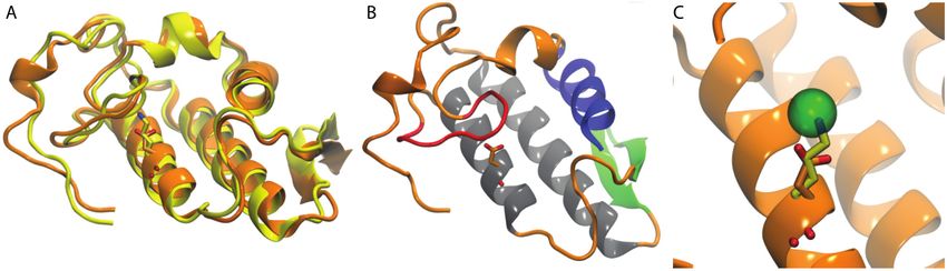
Figure 6. Acidic and basic PLA2s share a common molecular architecture. (A) Homology models of
the acidic PLA2 AplTx-I (orange) and the basic PLA2 CoaTx-II (yellow). Their different amino acids
Figure 6. Acidic areand basicinPLA2s share a common molecular architecture. (A) Homology models the
of
Figureat6.position
Acidic49and shown
basic licorice;
PLA2s share(B)athe architecture
common of PLA2s
molecular is shown, using
architecture. (A)for this purpose,
Homology models of
the acidic PLA2 AplTx-I (orange) and the basic PLA2 CoaTx-II (yellow). Their different
homology model of AplTx-I. N-terminal α-helix (blue), Ca -binding loop (red), antiparallel α-helices
2+ amino acids
the acidic PLA2 AplTx-I (orange) and the basic PLA2 CoaTx-II (yellow). Their different amino acids
at(gray),
position 49 are shown
two-stranded in licorice; (B) the
antiparallel-sheet architecture
(green); (C) Theofanalysis
PLA2s is ofshown,
positionusing
49 atfor this purpose,
AplTx-I the
and CoaTx-
at position 49 are shown in licorice; (B) the architecture of PLA2s is shown, using for this purpose,
homology model of AplTx-I. N-terminal α-helix (blue), Ca 2+-binding loop (red), antiparallel α-helices
II, superimposing the structures with the monomeric PLA2 from2+Agkistrodon halys pallas (PDB ID:
the homology
(gray), model of AplTx-I. N-terminal α-helix (blue), Caposition
-binding
49 at loop (red), antiparallel
1M8R)two-stranded
show that Nεantiparallel-sheet
atom of K49 occupies (green); (C) The analysis
the position of Ca2+ inofthe catalyticallyAplTx-I and CoaTx-
active Asp49 PLA2.
α-helices
II, (gray), two-stranded
superimposing
Cadmium the in
ion which
antiparallel-sheet
structures with the
1M8R occupies
(green);
themonomeric
(C)
PLA2
site of Ca2+ is shown
The
from analysis
Agkistrodon halys pallas (PDB ID: and
in licorice.
of position 49 at AplTx-I
CoaTx-II,
1M8R) superimposing
show that Nε atom the of
structures withthe
K49 occupies the monomeric
position of Ca2+ PLA2 from Agkistrodon
in the catalytically halys pallas
active Asp49 PLA2.(PDB
ID: 1M8R) show 2+ in the catalytically active Asp49
Cadmium ionthat
whichNεinatom
1M8Rofoccupies
K49 occupies
the site oftheCaposition
2+ is shown of in
Calicorice.
PLA2. Cadmium ion which in 1M8R occupies the site of Ca2+ is shown in licorice.Toxins 2018, 10, 8 11 of 24
The majority of molecular modeling applications in literature for studying PLA2s are oriented to
rational design of novel inhibitors for the treatment of different Viperidae snakebites. Some examples
are cited here:
Most examples have been applied to PLA2 of Daboia russelii. Recently, Nargotra et al. [108]
evaluated a library of natural products and synthetic molecules through docking studies on D. russelii
PLA2 to identify possible inhibitors. Their study lead to in silico identification of several molecules as
PLA2 inhibitors, with most of them belonging to phenolic and substituted benzaldehydic compounds.
It is important to note that the selection in this work was performed by considering docking energy
scores, which is a reliable criterion, according to literature [60]. The same authors proposed the docking
poses inside PLA2 of D. russelii for synthetic phenolic compounds effective against snake venom [109].
They found that phenolic compounds having hydroxyl and methoxyl groups in their benzene ring
showed maximum inhibitory potency, as they form hydrogen bonds with the residues Asp49 and
Gly30 in the binding site of D. russelii PLA2 (these residues configurate together with Gly30 and
Tyr28 a Ca2+ coordination site and are involved in the binding of several ligands reported in Protein
Data Bank).
Other works applied to D. russelii PLA2 are listed below: Anilkumar et al. [110] docked
imidazopyridine derivatives inside the binding site of D. russelii PLA2, and they found that compounds
form π–π stacking interactions with Trp31, and extend towards Gly32, potentially adding further
amide–π stacking contributions. Yadava et al. [111] docked nine pyrazolo[3,4-d]pyrimidines inside
D. russelii PLA2 for describing their binding modes. They found that the studied compounds have
better docking binding energies than indomethacin, however, it is necessary to prevent, again, the use
of docking binding energies for comparing the affinities of two molecules [93]. In other work,
Ramakrishnan et al. [112] generated pharmacophore models based on the interaction of different
types of inhibitors (peptides, vitamin E, indole derivatives, and nonsteroidal anti-inflammatory drugs)
with their preferred subsites in the active site of the subunit A of D. russelli PLA2. Authors validated
the final model and subjected it for screening a library of drug-like compounds. They identified eight
compounds and subjected them to molecular docking and MD simulation, to assess their binding
mode with both subunits. After analyzing these computational experiments, they selected four
compounds for further biochemical assays, and found that two compounds can bind both the subunits
of PLA2 of D. russelli venom, in spite of its aggregated form. Sivaramakrishnan et al. [113] reported an
integrated approach involving homology modeling, MD, and molecular docking studies on D. russelli
venom PLA2 fraction V belonging to Group IIB secretory PLA2 from D. russelli, in order to study the
structure-based inhibitor design (3D structure of D. russelli PLA2 fraction IIIa was used as template,
with >93% of identity). Authors also constructed a pharmacophore model, and identified potential
specific inhibitors. Additionally, they highlighted the role of His47 and Asp48 within the PLA2 binding
pocket as key residues for HB interactions with ligands.
Finally, Ramakrishnan et al. [114] performed comparative MD simulations of free and
inhibitor-bound form of secretory D. russelli PLA2. This enzyme dimerizes asymmetrically with
different orientation of Trp31 at the gateway of the active site of both the subunits A and B. Hence,
the active site of subunit A is open, and that of subunit B is inaccessible to monodispersed inhibitors.
Authors performed MD simulations for monomer and dimer forms of PLA2s in both native and
complex forms (the bovine pancreatic PLA2 was selected as the monomeric form). They reported a
comparison of trajectories with respect to fluctuation and deviation, which discloses the dynamics
of surface and calcium-binding loops, as well as the difference in dynamics of active site residues.
Their study discloses the sort of restrictions in D. russelli PLA2 active site for inhibitor binding, and
implies suitable sites for further design of inhibitors based on active site scaffold.
Other recent works were applied to study the interactions between drugs and other PLA2s with
X-ray structures available in PDB. For instance, in a recent work, Pereañez et al. [115] studied the mode
of action of morelloflavone with PLA2 of Crotalus durissus terrificus, using docking. Authors found
that morelloflavone occupies part of the substrate binding cleft of C. durissus PLA2, forming hydrogenToxins 2018, 10, 8 12 of 24
bonds (HBs) with the residues Gly33, Asp49, Gly53, and Thr68 of the enzyme, and π–π stacking with
the residue Tyr52. The same authors used docking to investigate the interactions between C. durissus
PLA2 and bile acids, such as cholic acid (CA) and ursodeoxycholic acid (UDCA) [115]. Authors found
that bile acids interact with the binding active site of PLA2 through different interactions, CA showed
HBs with His48, whereas, UDCA showed HBs with Asp49 and Tyr28. In other work, Zhang et al. [116]
docked structural elements of the persimmon tannin PT40 (a highly galloylated condensed tannin with
an unusual flavonol terminal unit) inside Chinese cobra (Naja atra) PLA2 binding site, to understand
the inhibitory mechanism of this natural product. They found that the residues Trp18, Try27, Gly29,
His47, and Tyr63 are involved in the interactions. Finally, Chavan and Deobagkar [117] applied
docking and MD simulation techniques to propose the putative interactions of LT10 peptide (small
synthetic peptide derived from N-terminal of the lethal toxin neutralizing factor) with Naja naja PLA2.
MD was performed to analyze the stability of the complex obtained by docking method.
Other applications used available structures in PDB for creating comparative models. For instance,
Chinnasamy et al. [98] modeled the 3D structure of PLA2 of Naja sputatrix (Malayan spitting cobra)
using the structure of N. naja PLA2 as template, applied 10 ns MD to the get stable conformations
of the studied protein, and used the final structure to perform high throughput virtual screening by
performing massive docking of compounds from different databases. After applying this protocol,
authors selected seven compounds based on the docking score and free energy binding calculations.
In other work, Chavanayarn et al. [118] studied the binding of the antibodies VH H-P3-1, VH H-P3-3,
and VH P3-7 to PLA2 of Naja kaouthia (monocled cobra), using docking methods. They developed a
homology model of the N. kaouthia PLA2 using N. atra PLA2 as template, and found that the antibodies
covered the areas around the PLA2 catalytic groove and inserted their complementarity determining
regions (CDRs) into the enzymatic cleft. Finally, Hage-Melim et al. [119] constructed a homology
model of PLA2 of Bothrops jararacussu using a survey of complexes of PLA2 deposited in PDB. Authors
carried out the pairwise alignment through involving eight sequences selected by crystallographic
criteria, followed by a multiple alignment with the sequence of B. jararacussu PLA2. Authors claimed
that X-ray structures of B. jararacussu PLA2 are in PDB, but no structure in complex with any inhibitor
is available. Therefore, they performed the homology modeling to get a correct description of the
binding site. They performed virtual screening in a large database, yielding a set of potential bioactive
inhibitors, and confirmed the important role of Lys49 for binding ligands.
Fewer applications have been focused to study functional characteristics of PLA2s; however,
there are some reports with interesting, more specific purposes. For instance, Murakami et al. [120]
performed MD simulations of bothropstoxin-I (a K49 PLA2 of Bothrops jararacuss with myotoxic and
neurotoxic activities) to study its complex with suramin (a polysulphonated naphthyl urea derivative).
Instead, another report uses molecular modeling for studying the interactions between one PLA2
and one lipid. In this report, Abiram and Kolandaivel [91] studied the interaction of myristic fatty
acid with acutohaemolysin and piratoxin-II (K49 PLA2s from Bothrops pirajai and Agkistrodon acutus
respectively) using the hybrid two layered ONIOM (B3LYP/6-31G*: UFF) method [121]. Specifically,
authors performed quantum chemical calculations on the tripeptides AFA and AVA present in
acutohaemolysin and piratoxin-II. They found that the mode of interaction of the fatty acid with
protein is electrostatic, confirmed further through molecular electrostatic potential maps, and the
AFA shows stronger interaction than AVA, validating the impact of mutation on catalytic activity.
The preferred secondary structural configuration and conformational properties of AVA and AFA
validated the strong interaction of fatty acid with phenylalanine.
Other report tried to explain the higher activity of PLA2s at solvent–lipid interface. In this
report, De Oliveira et al. [122] performed MD simulations of PLA2 of Agkistrodon halys pallas in water,
methanol, and octanol. Authors used these simulations to propose an interfacial activation model for
PLA2 in atomic detail. When the enzyme is in a more hydrophobic environment, they noted that a
series of conformational changes occurs: (a) increase of solvent accessible surface area; (b) side chain
reorientation of Asp49 residue that allows Ca2+ coordination; (c) reduction of the distance betweenYou can also read