Marine Toxins Targeting Kv1 Channels: Pharmacological Tools and Therapeutic Scaffolds - MDPI
←
→
Page content transcription
If your browser does not render page correctly, please read the page content below
marine drugs
Review
Marine Toxins Targeting Kv1 Channels:
Pharmacological Tools and Therapeutic Scaffolds
Rocio K. Finol-Urdaneta 1,2, * , Aleksandra Belovanovic 3,† , Milica Micic-Vicovac 3,† ,
Gemma K. Kinsella 4 , Jeffrey R. McArthur 1 and Ahmed Al-Sabi 3, *
 1   Illawarra Health and Medical Research Institute, University of Wollongong, Wollongong, NSW 2522,
     Australia; jeffreym@uow.edu.au
 2   Electrophysiology Facility for Cell Phenotyping and Drug Discovery, Wollongong, NSW 2522, Australia
 3   College of Engineering and Technology, American University of the Middle East, Kuwait;
     Aleksandra.B@aum.edu.kw (A.B.); Milica.Micic-vicovac@aum.edu.kw (M.M.-V.)
 4   School of Food Science and Environmental Health, College of Sciences and Health, Technological University
     Dublin, D07 ADY7 Dublin, Ireland; Gemma.Kinsella@tudublin.ie
 *   Correspondence: rfinolu@uow.edu.au (R.K.F.-U.); Ahmed.Al-Sabi@aum.edu.kw (A.A.-S.);
     Tel.: +965-22251400 (ext.1798) (A.A.-S.)
 †   These authors contributed equally to this review.

 Received: 3 February 2020; Accepted: 16 March 2020; Published: 20 March 2020

 Abstract: Toxins from marine animals provide molecular tools for the study of many ion channels,
 including mammalian voltage-gated potassium channels of the Kv1 family. Selectivity profiling
 and molecular investigation of these toxins have contributed to the development of novel drug
 leads with therapeutic potential for the treatment of ion channel-related diseases or channelopathies.
 Here, we review specific peptide and small-molecule marine toxins modulating Kv1 channels and
 thus cover recent findings of bioactives found in the venoms of marine Gastropod (cone snails),
 Cnidarian (sea anemones), and small compounds from cyanobacteria. Furthermore, we discuss
 pivotal advancements at exploiting the interaction of κM-conotoxin RIIIJ and heteromeric Kv1.1/1.2
 channels as prevalent neuronal Kv complex. RIIIJ’s exquisite Kv1 subtype selectivity underpins a
 novel and facile functional classification of large-diameter dorsal root ganglion neurons. The vast
 potential of marine toxins warrants further collaborative efforts and high-throughput approaches
 aimed at the discovery and profiling of Kv1-targeted bioactives, which will greatly accelerate the
 development of a thorough molecular toolbox and much-needed therapeutics.

 Keywords: bioactives; conotoxins 2; Kv1; marine toxins; modulators; potassium channels; sea
 anemone toxins

1. Introduction

1.1. Kv1 Channels
     Voltage-gated K+ channels (Kv) are intrinsic plasma membrane proteins mediating the selective
flow of K+ ions down their electrochemical gradient in response to a depolarization in the
transmembrane electric field [1]. The selectivity and voltage dependence of Kv channels make
them central players in virtually all physiological functions, including the maintenance and modulation
of neuronal [2–4] and muscular (both cardiac and skeletal) excitability [5–7], regulation of calcium
signalling cascades (reviewed by Reference [8]), control of cell volume [9,10], immune response [11],
hormonal secretion [12], and others.
     The Kv channel α-subunit belongs to the six transmembrane (6-TM) family of ion channels
(Figure 1a,b) in which the voltage-sensing domain (VSD) formed by transmembrane segments S1–S4

Mar. Drugs 2020, 18, 173; doi:10.3390/md18030173                            www.mdpi.com/journal/marinedrugs
Mar.
Mar. Drugs
Drugs 2019,
2020, 17,
18, x173
FOR PEER REVIEW 22 of
of 31
28
The Kv channel α-subunit belongs to the six transmembrane (6-TM) family of ion channels
controls1a,b)
(Figure poreinopening
which the viavoltage-sensing
the S4–S5 intracellular
domain (VSD) loop that
formedis connected to the pore
by transmembrane domainS1–S4
segments (PD).
The PD is formed by transmembrane segments S5–S6 including a
controls pore opening via the S4–S5 intracellular loop that is connected to the pore domain (PD). There-entrant pore loop bearing the
potassium
PD is formed selectivity sequence TVGYG
by transmembrane segments [13].S5–S6
Depolarization
including aofre-entrant
the transmembrane
pore loop electric
bearingfieldthe
induces a conformational
potassium selectivity sequence changeTVGYG in the VSD that
[13]. leads to channel
Depolarization of activation, leading to opening
the transmembrane electric of the
field
water-filled permeation pathway permitting K + to flow down their electrochemical gradient. Upon
induces a conformational change in the VSD that leads to channel activation, leading to opening of
repolarization,
the water-filled the VSD returns
permeation to its permitting
pathway resting state, K+closing
to flowthe channel
down theirgate and terminating
electrochemical ionic
gradient.
flow in a process called deactivation. Immediately after deactivation,
Upon repolarization, the VSD returns to its resting state, closing the channel gate and terminating channels can be reactivated;
however,
ionic flowif depolarization-induced
in a process called deactivation. channel activation extends beyond
Immediately a few milliseconds,
after deactivation, channels inactivation
can be
ensues, ceasing
reactivated; K+ permeability.
however, Kv channels recoverchannel
if depolarization-induced from inactivation
activationonly after spending
extends beyond enough
a few
time at a hyperpolarized potential [14]. The molecular underpinnings
milliseconds, inactivation ensues, ceasing K permeability. Kv channels recover from inactivation
+ of the inactivation processes
haveafter
only beenspending
thoroughly examined
enough time functionally and structurally,
at a hyperpolarized potential identifying various inactivation
[14]. The molecular underpinnings typesof
involving
the distinct
inactivation and complex
processes have molecular mechanisms.
been thoroughly examined Voltage-gated
functionally ionand
channels can inactivate
structurally, from
identifying
pre-openinactivation
various closed-states (closed-state
types involvinginactivation,
distinct andCSI) or from
complex the openmechanisms.
molecular state(s) (open-state inactivation,
Voltage-gated ion
OSI) [15].can
channels Inactivation
inactivate can fromalso be categorized
pre-open closed-states depending on theinactivation,
(closed-state speed of its onset CSI) orupon
fromactivation.
the open
In some(open-state
state(s) Kv channels, fast inactivation
inactivation, or N-type
OSI) [15]. inactivation
Inactivation can alsooccurs
be soon after thedepending
categorized channel activates
on the
and it is
speed ofmainly
its onset dueupon
to an activation.
intracellularInblock somebyKv thechannels,
channel’s fastintracellular
inactivation N-terminus
or N-type hence known as
inactivation
the inactivation
occurs soon afterparticle
the channel [16]. activates
This processand ithas been directly
is mainly due to observed by cryo-electron
an intracellular block by the microscopy
channel’s
(cryo-EM) in a related prokaryotic K channel [17]. In addition
intracellular N-terminus hence known as the inactivation particle [16]. In addition to N-type to N-type inactivation, a common but
relatively slower
inactivation, process
a common buthappens
relatively after tensprocess
slower or hundreds
happens of after
milliseconds from channel
tens or hundreds activation
of milliseconds
that is
from termedactivation
channel C-type (or thatslow) inactivation
is termed C-type [18].
(or slow)Even though the[17].
inactivation extent
Even and complexity
though the extentof slow
and
inactivation of
complexity remain
slow the subjects ofremain
inactivation investigation, the pore
the subjects of structure and the
investigation, thepermeating ions appear
pore structure and the to
play a vital role
permeating ions[19]. Recent
appear structural
to play a vitalandrolefunctional
[18]. Recent studies support
structural anda mechanism
functional through which the
studies support a
redistribution of structural water molecules accompanies
mechanism through which the redistribution of structural water molecules accompanies thethe rearrangement of amino acids within
the channel’s inner
rearrangement cavityacids
of amino and outer
within vestibule, ultimately
the channel’s innerleading
cavity to theouter
and collapse of the permeation
vestibule, ultimately
pathwaytointhe
leading C-type inactivation
collapse (reviewedpathway
of the permeation in Reference [20]).inactivation
in C-type Modulation(reviewed
of the inactivation
in Reference process
[19]).is
a powerful strategy to control the cellular availability of Kv channel-mediated
Modulation of the inactivation process is a powerful strategy to control the cellular availability of Kv currents; thus, both N-
and C-type inactivation
channel-mediated currents;are responsive
thus, both N- to and
the cellular redox environment
C-type inactivation [21]. Fortoinstance,
are responsive structural
the cellular redox
motifs within[20].
environment the KvForchannel’s
instance,N-terminus/inactivation
structural motifs withinparticle the Kv serve as sensors
channel’s of the cytoplasmic
N-terminus/inactivation
redox potential
particle serve as[22]. sensors of the cytoplasmic redox potential [21].
Figure 1.
Figure (a)Schematic
1. (a) Schematicillustration
illustration of
of K
KVVchannel
channelmembrane
membranetopology
topologydepicting
depictingthethe66transmembrane
transmembrane
subunits including
subunits including the
the voltage
voltage sensing
sensing domain
domain (voltage-sensing
(voltage-sensing domain
domain (VSD):
(VSD): S1–S4)
S1–S4) and
and the
the pore
pore
domain (PD)
domain (PD) between
between S5S5 and
and S6S6 segments.
segments. (b)
(b)Top
Top and
and side
side views
views of of representative
representative homomeric
homomeric and and
heteromeric Kv1
heteromeric Kv1channels
channelsbasedbasedononthethe
crystal structure
crystal of Kv1.2
structure channels
of Kv1.2 [(Protein
channels Data Bank
[(Protein Data number,
Bank
PDB: 2A79)]
number, PDB:[13]. (c) [13].
2A79)] Current trancestrances
(c) Current of homomeric Kv1.1 Kv1.1
of homomeric (left) and
(left)1.2
and(right) channels
1.2 (right) and their
channels and
heteromeric combination (middle) revealing distinct sensitivity to the classical pharmacological
their heteromeric combination (middle) revealing distinct sensitivity to the classical pharmacological tool
tetraethylammonium
tool tetraethylammonium (TEA) [23,24].
(TEA) [22,23].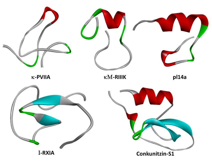
Mar. Drugs 2020, 18, 173                                                                             3 of 28

      K+ channels are the most diverse family of ion channels in excitable and nonexcitable tissues,
encompassing 40 Kv members allocated into 12 subfamilies: voltage-gated Kv subfamilies, the
Ether-à-go go (EAG) subfamily, and the Ca2+ -activated subfamilies [1]. As such, they are implicated in
many neurological, cardiac, and autoimmune disorders, which position them as important therapeutic
targets [25]. The identified genes for Kv channel α-subunits are classified into twelve subfamilies:
Kv1 (Shaker); Kv2 (Shab); Kv3 (Shaw); Kv4 (Shal); Kv7 (KvLQT); Kv10 (HERG); Kv11 (EAG);
Kv12 (ELK); and the modulatory “electrically silent” Kv5, Kv6, Kv8, and Kv9 subfamilies (https:
//doi.org/10.2218/gtopdb/F81/2019.4). The Shaker-related Kv1 family is comprised of eight members
(Kv1.1–Kv1.8) encoded by the corresponding KCNA1–KCNA8 genes. Several Kv1 channels have been
identified and functionally characterized within their native tissues, exploiting selective blockers
(reviewed by References [2,26,27]). The first Kv1 complexes were purified from mammalian brain using
the snake venom toxins called dendrotoxins (DTX). These studies indicated that the functional Kv1
channel is a large (Mr ~400 kDa) sialoglycoprotein complex consisting of four pore-forming α-subunits
and four cytoplasmically associated auxiliary β-proteins [28] that modulate K+ channel activation and
inactivation kinetics (for a thorough review, refer to Reference [29]).
      The Kv1 channels are expressed in a variety of tissues as homo- or heterotetrameric complexes
(Figure 1a,b) [30]. These complexes are formed in the endoplasmic reticulum [31], where monomers
are randomly recruited, assembled, and inserted in the plasma membrane [31]. The four cytoplasmic
N-terminal domains interact with one another in a strictly subfamily-specific manner, thus providing the
molecular basis for the selective formation of heteromultimeric channels in vivo [32,33]. The predominant
pathway in tetramer formation involves dimerization of subunit dimers, thereby creating interaction
sites different from those involved in the monomer–monomer association during the oligomerization
process [34].
      In heterologous expression systems, all Potassium Voltage-gated channel subfamily A Member
gene (KCNA) transcripts encoding Kv1 α-subunits yield functional homo-tetrameric complexes with
distinct biophysical and pharmacological profiles [35], (Figure 1c). While theoretically, the combination
of different Kv1 subunits could afford impressive functional diversity, only a subset of oligomeric
combinations has been elucidated [36–40], suggesting their synthesis and/or assembly are carefully
orchestrated. Amongst the KV 1 channels, Kv1.2 is the most prevalent isoform in neuronal membranes
where only a small fraction occurs as a homo-tetramer, while the majority are a hetero-tetramer with
other Kv1 α-subunits [36,40]. In these preparations, the less abundant Kv1.1 subunit is consistently
identified in oligomers containing Kv1.2 channels.

1.2. Mechanisms of Kv Channel Inhibition by Marine Toxins
      The diversity of Kv1 channels and their wide distribution, including their specific expression
pattern in the central and peripheral nervous systems (CNS and PNS, respectively), together with their
vital function in the excitability of nerve and muscle, make them strategic targets of marine toxins.
These natural products are synthesized by marine organisms to deter competitors and to aid predation
or for self-defense [41]. Many venomous organisms block Kv channel-mediated currents, crippling
membrane repolarization, yielding enhanced excitability, and ultimately engendering paralysis in pray
or foe [42].
      Marine toxins exploit different Kv channel traits to exert their modulatory actions. A commonly
used strategy relies on direct occlusion of the narrow potassium permeation pathway from the
extracellular side of the channel protein (Figure 2a,b). Toxins inhibiting ionic current via this mechanism
are referred to as “pore blockers”. Many structurally and phylogenetically unrelated pore-blocking
toxins of Kv channels share a dyad motif composed of a lysine (positive) and a tyrosine/phenylalanine
(hydrophobic) [43–45]. The lysine residue fits snugly in the Kv channel selectivity filter, sterically
occluding K+ ion flow, whilst the hydrophobic amino acid in the dyad aids docking and consolidation
of the toxin binding. This dyad motif has been proposed to be the minimal core domain of the Kv
channel-binding pharmacophore (Figure 2b) [46–49].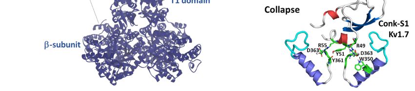
Mar. Drugs 2020, 18, 173 4 of 28
Mutational
Mar. Drugs 2019, 17, xanalysis
FOR PEERand docking
REVIEW calculations have demonstrated that some marine toxins do 4 ofnot
31
possess the canonical functional dyad or do not seem to use one in the classic “pore blocker” fashion to
fashion
prevent to prevent permeation.
potassium potassium permeation. In these
In these toxins, toxins, charged
a positively a positively
ring charged
of aminoring
acidsofparticipates
amino acids in
participates in electrostatic interactions with the Kv1 outer vestibule. These residues provide
electrostatic interactions with the Kv1 outer vestibule. These residues provide surface recognition surface
and
recognition
anchoring, andandconcurrently,
anchoring, aand concurrently,
network a network
of hydrogen bonds andof hydrophobic
hydrogen bonds and hydrophobic
interactions consolidate
interactions consolidate “capping” of the channel vestibule, with the peptide toxin acting
“capping” of the channel vestibule, with the peptide toxin acting as a lid over the Kv channel as a lid over
pore
the Kv channel
(Figure 2b) [50].pore (Figure 2b) [49].
Figure 2.2.(a)(a)Schematic
Figure Schematic presentation
presentation of of a side
a side viewviewKV1Kchannel
V 1 channel showing
showing theofsite
the site of interaction
interaction with
with representative
representative pore-blocking
pore-blocking peptide
peptide toxinstoxins
fromfromConeCone snail
snail (κM-RIIIK,
(κM-RIIIK, [51]and
[50] andConK-S1,
ConK-S1,PDB:
PDB:
2CA7, [51])
2CA7, [52]) and
and seasea anemone
anemone ShK ShK (PDB:
(PDB: 1ROO,
1ROO,[52])[53]) and
and gating
gatingmodifier
modifiertoxin
toxinfrom
fromspider
spider(HaTx;
(HaTx;
PDB: 1D1H,
PDB: 1D1H, [53]).
[54]). (b)
(b)The
Themodes
modesof ofpore
poreblocking
blocking(plug,
(plug,lid,
lid,ororcollapse)
collapse)illustrated
illustratedby bymarine
marinepeptide
peptide
blockers as revealed by the docking models. The outer turret regions (residues 348–359 for Kv1.1,Kv1.1,
blockers as revealed by the docking models. The outer turret regions (residues 348–359 for 350–
350–359
359 for Kv1.2,
for Kv1.2, and and 334–343
334–343 for Kv1.7)
for Kv1.7) are are in cyan,
in cyan, andand
thethe inner
inner turretregions
turret regions(residues
(residues377–386
377–386for
for
Kv1.1, 377–386
Kv1.1, 377–386 for for Kv1.2,
Kv1.2, andand 462–469
462–469 forfor Kv1.7)
Kv1.7) are
are indicated
indicated in in green.
green. Only
Onlytwotwosubunits
subunitsofofthe
theKv1
Kv1
channels are
channels are shown,
shown, forfor simplicity.
simplicity. Docking
Docking was was performed
performed using using thetheHaddock
Haddock webserver
webserver [54,55]
[55,56]and
and
the docking model image were generated using Pymol (The PyMOL
the docking model image were generated using Pymol (The PyMOL Molecular Graphics System, Molecular Graphics System, [57]).
[56]).
A distinct mechanism from pore block is achieved by interacting with the gating mechanism
of KvA channels. Toxins acting
distinct mechanism from in this
pore fashion
block are known
is achieved as “gatingwith
by interacting modifiers”.
the gatingThe VSD in Kv
mechanism of
channels controls pore opening; hence, toxins binding to the extracellularly exposed
Kv channels. Toxins acting in this fashion are known as “gating modifiers”. The VSD in Kv channels linker between
transmembrane
controls segmentshence,
pore opening; S3 andtoxins
S4, the binding
S3–S4 linker paddle
to the motif withinexposed
extracellularly the VSD,linker
inhibitbetween
channel
function by increasing the energy required to open the channel’s gate by shifting the
transmembrane segments S3 and S4, the S3–S4 linker paddle motif within the VSD, inhibit channel voltage dependence
of activation
function to more depolarized
by increasing the energypotentials.
required Alternatively, some toxins
to open the channel’s destabilize
gate the the
by shifting Kv channel
voltage
open state reflected as enhanced entry into a nonconductive inactivated state at
dependence of activation to more depolarized potentials. Alternatively, some toxins destabilize potentials wheretheKv
activity would normally be favored [58]. This modulatory mechanism was first shown
Kv channel open state reflected as enhanced entry into a nonconductive inactivated state at potentials for Hanatoxin,
a gating
where Kvmodifier peptidenormally
activity would component of the Chilean
be favored rose-hair
[57]. This modulatorytarantula venomwas
mechanism thatfirst
inhibits
shownKv2.1
for
channels (Figure 2a) [59].
Hanatoxin, a gating modifier peptide component of the Chilean rose-hair tarantula venom that
A recently
inhibits proposed
Kv2.1 channels inhibitory
(Figure mechanism appears as a hybrid strategy between pore blockade
2a) [58].
and Agating
recentlymodification. A toxin sitting
proposed inhibitory mechanismin theappears
channel’s
as aouter
hybrid vestibule
strategy blocks
between thepore
extracellular
blockade
and gating modification. A toxin sitting in the channel’s outer vestibule blocks the extracellular side
of the permeation pathway and modifies the permeation of water molecules into proteinaceous
peripheral cavities in the channel. This creates asymmetries in the distribution of water molecules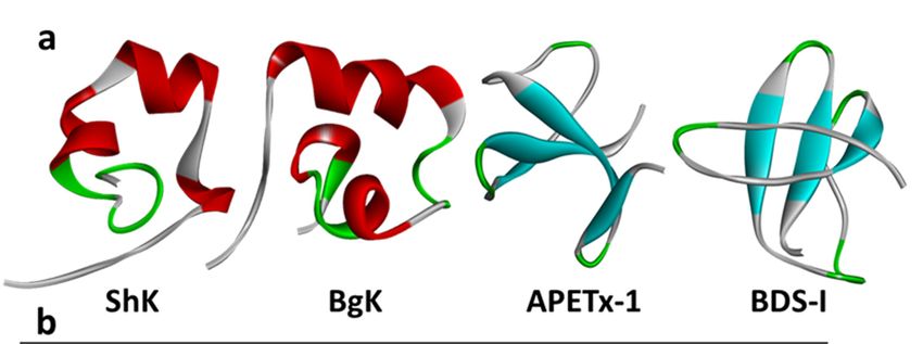
Mar. Drugs 2020, 18, 173                                                                               5 of 28

side of the permeation pathway and modifies the permeation of water molecules into proteinaceous
peripheral cavities in the channel. This creates asymmetries in the distribution of water molecules
around the selectivity filter, triggering a local collapse of the channel pore akin to Kv C-type inactivation
(Figure 2b) [60].

2. Molluscan Peptides that Inhibit Kv1 Channels
     Conotoxins constitute a family of small peptide toxins found in the venom glands of cone snails [61].
These marine gastropods of the genus Conus are represented by ~800 predatory mollusks [62]. It is
believed that the large arsenal of conotoxins within a single venom is used for fast pray immobilization
in hunting cone snails [63].
     Conotoxins are typically 8–60 amino acid peptides that potently interact with a wide range of
voltage- and ligand-gated ion channels and receptors [64]. The cone snail venom peptides evolved
to capture their prey (worms, fish, and other mollusks), and their venom is known to interact and
modulate several mammalian ion channels with great selectivity [65]. The pharmacological properties
of conotoxins have been exploited as molecular tools for the study of mammalian targets [66], and
their scaffolds are employed for drug development and potential treatment of human diseases [67].
     Mature conotoxins are structurally diverse, including disulfide-free and mono- and poly-disulfide-
bonded peptides (several reviews deal with the structural diversity of conotoxins; see References [64,68]).
Peptides lacking disulfide bonds are flexible, whereas the presence of multiple disulfide linkages
provides structural rigidity and provides different three-dimensional conformations depending
on the cysteine disulfide framework within the toxin sequence [69]. Cone snail VDPs are
often post-translationally modified, including C-terminal amidation, bromination, γ-carboxylation,
hydroxylation, O-glycosylation, N-terminal pyroglutamylation, and sulfation [70].
     Pharmacological classification of the structurally diverse (i.e., cysteine framework/connectivity,
loop length, and fold) conotoxins is based on the target type and mechanism of action of the peptides.
Twelve pharmacological families are currently recognized (ConoServer [71]). Due to the variable
nature of conotoxins, a consensus classification-linking pharmacology to structure has not been agreed
upon. Given the nature of this review, we will focus on the pharmacological family classification of the
kappa- or κ-conotoxins, which are defined by modulatory activity over potassium-selective channels.
The founding member of the κ-conotoxins was identified in the venom of the piscivorous snail Conus
purpurascens κ-PVIIA by its potent block of Drosophila voltage-gated Shaker channels [72].
     Up to now, nine conotoxins are listed as mammalian Kv1 channel blockers in the Kalium
database [73]. From those, the activity of Contryphan-Vn from Conus ventricosus against Kv1.1 and
Kv1.2 was tested by displacement of radiolabeled Bunodosoma granulifera Kv1 blocker (BgK), showing
weak activity at 600 µM [74]. Therefore, Contryphan-Vn modulatory activity against Kv1 channels
remains to be verified.
     The other κ-conotoxins listed belong to various structural families of disulfide-rich peptides (A, I,
J, M, O, and the Conkunitzins; Figure 3 and Table 1). Disulfide-rich κ-conotoxins have been shown
to act as pore blockers using canonical interactions through the “functional dyad” and the “ring of
basic residues” as molecular determinants of κ-conotoxin modulation of Kv1 channel conductance.
Such mechanisms of action have been described in scorpion and cnidarian VDP toxins blocking Kv1
channels; hence, κ-conotoxins share important features that enable Kv1 channel inhibition in a similar
way to other animal VDP blockers.
Mar.
Mar. Drugs 2020,
2019, 18,
17, 173
x FOR PEER REVIEW 6 of 28
31
Figure 3. Structures of representative cone snail venom-derived peptide toxins κ-PVIIA (PDB:
Figure 3. Structures of representative cone snail venom-derived peptide toxins κ-PVIIA (PDB: 1AV3,
1AV3, [75]), κM-RIIIK [51], pl14a (PDB: 2FQC, [76]), I-RXIA (PDB: 2JTU, http://www.rcsb.org/structure/
[74]), κM-RIIIK [50], pl14a (PDB: 2FQC, [75]), I-RXIA (PDB: 2JTU,
2JTU), and Conkunitzin-S1 (PDB: 2CA7, [52]): β-sheets are in cyan, and α-helices are in red.2.1.
http://www.rcsb.org/structure/2JTU), and Conkunitzin-S1 (PDB: 2CA7, [51]): β-sheets are in cyan, and
κM-RIIIK.
α-helices are in red.2.1. κM-RIIIK.
Despite the great abundance of Conus peptides characterized to date, relatively few have been
Despite the great abundance of Conus peptides characterized to date, relatively few have been
shown to interact with Kv channels. κM-RIIIK from Conus radiatus [77] (Figures 2 and 3) is 24 residues
shown to interact with Kv channels. κM-RIIIK from Conus radiatus [76] (Figure 2 and Figure 3) is 24
long, and it is structurally homologous to the well-known voltage-gated sodium channel blocker
residues long, and it is structurally homologous to the well-known voltage-gated sodium channel
µ-GIIIA [78]. RIIIK was originally identified as a Shaker (Drosophila) and TSha1 (trout) Kv1 orthologue
blocker µ-GIIIA [77]. RIIIK was originally identified as a Shaker (Drosophila) and TSha1 (trout) Kv1
channel blocker [79]. Later, RIIIK became the first conotoxin described to modulate human Kv1
orthologue channel blocker [78]. Later, RIIIK became the first conotoxin described to modulate
channels, selectively blocking homomeric Kv1.2 without apparent effects over Navs or mammalian
human Kv1 channels, selectively blocking homomeric Kv1.2 without apparent effects over Navs or
homologs Kv1.1, Kv1.3, Kv1.4, Kv1.5, and Kv1.6 recorded by Two Electrode Voltage Clamp recording
mammalian homologs Kv1.1, Kv1.3, Kv1.4, Kv1.5, and Kv1.6 recorded by Two Electrode Voltage
(TEVC) in Xenopus oocytes [77]. Interestingly, heteromerization with Kv1.2 α-subunits suffices to render
Clamp recording (TEVC) in Xenopus oocytes [76]. Interestingly, heteromerization with Kv1.2 α-
Kv1.1, Kv1.5, and Kv1.7, containing heterodimeric channels sensitive to low micromolar RIIIK [80].
subunits suffices to render Kv1.1, Kv1.5, and Kv1.7, containing heterodimeric channels sensitive to
Binding of κM-RIIIK to closed (deactivated) Kv1.2 channels is ~2-fold stronger than to the open
low micromolar RIIIK [79].
state, hinting towards state-dependent interactions between this peptide and the Kv1s. Importantly,
Binding of κM-RIIIK to closed (deactivated) Kv1.2 channels is ~2-fold stronger than to the open
RIIIK blocks its Kv1 channel targets, through a pharmacophore comprised of a ring of positive charges
state, hinting towards state-dependent interactions between this peptide and the Kv1s. Importantly,
and not via the classical “dyad motif” [51,81].
RIIIK blocks its Kv1 channel targets, through a pharmacophore comprised of a ring of positive
2.1. κM-RIIIJ
charges and not via the classical “dyad motif” [50,80].
M-RIIIJ analyses of the venom of Conus radiatus revealed a second, closely related peptide, named
2.2. κFurther
κM-RIIIJ, that displayed 10-fold higher potency (~30 nM) blocking homomeric Kv1.2-mediated currents.
Further of
Comparison analyses
RIIIK andof the
RIIIJvenom
activityofinConus radiatus
an animal modelrevealed a second, closely revealed
of ischemia/reperfusion related peptide,
that the
named κM-RIIIJ, that displayed 10-fold higher potency (~30 nM) blocking homomeric
latter was cardioprotective, an effect adjudicated to RIIIJ’s higher potency at inhibiting heterodimeric Kv1.2-
mediated currents.
Kv1-mediated currents [80].Comparison of RIIIK and RIIIJ activity in an animal model of
ischemia/reperfusion revealedofthat
An in-depth evaluation RIIIJthe
was latter was cardioprotective,
performed an effect
against heteromeric adjudicated
channels to RIIIJ’s
generated by a
higher potency at inhibiting heterodimeric Kv1-mediated currents [79].
covalent linkage composed of Kv1.2 and all other Kv1s subunits (except for Kv1.8) at different
An in-depth
stoichiometries andevaluation of RIIIJ
arrangements [82]. was
Thisperformed against
work revealed heteromeric
that RIIIJ channels
exquisitely targetsgenerated
asymmetric byKva
covalent composed
channels linkage composed of Kv1.2
of three Kv1.2 and all
subunits andother Kv1s or
one Kv1.1 subunits (except RIIIJ’s
Kv1.6 subunit. for Kv1.8) at different
apparent affinity
stoichiometries and arrangements [81]. This work revealed that RIIIJ exquisitely
for the asymmetric complex is ~100-fold higher than for the homomeric Kv1.2 complex. targets asymmetric
Kv channels composed of three Kv1.2 subunits and one Kv1.1 or Kv1.6 subunit. RIIIJ’s apparent
affinity for the asymmetric complex is ~100-fold higher than for the homomeric Kv1.2 complex.Mar. Drugs 2020, 18, 173 7 of 28
Recently, the discerning sensitivity of RIIIJ to its heteromeric Kv1 channel target was exploited
to comprehensively classify and characterize individual somatosensory neuronal subclasses within
heterogenous populations of dorsal root ganglion (DRG) neurons [83]. RIIIJ’s selectivity was used to
distinguish two functional Kv1 complexes in mouse dorsal root ganglion (DRG) neurons. One being
RIIIJ’s high-affinity target (3 × Kv1.2 + Kv1.1 or Kv1.6), and the second component characterized by
inhibition at higher RIIIJ concentrations arguably composed of homo-tetrameric Kv1.2 subunits [82].
The functional behavior of large DRG (L-DRG) neurons exposed to RIIIJ was used to classify L-DRGs
in six discrete neuronal subpopulations (L1–L6). Interestingly, this peptide’s block of heteromeric
Kv1 channels in subclass L3 and L5 neurons lead to enhanced calcium signals consistent with
their contribution to repolarization after a depolarizing stimulus, whilst in subclass L1 and L2
neurons, exposure to RIIIJ decreased the threshold for action potential firing. The integration of
constellation pharmacology [66], electrophysiology, and transcriptomic profiling using RIIIJ as a
pharmacological tool served to functionally assess three biological levels spanning the molecular target
Kv1 channel (Kv1.2/Kv1.1 heteromer), the functional characteristics of specific neuronal subclasses and
the physiological system (i.e., proprioception) in which they participate.
Table 1. Some characteristics of known conotoxins targeting the Kv1 channel.
Conopeptide Source Family Target Channel(s) (IC50 ) References
CPY-Pl1 C. planorbis CPY Kv1.2 (2 µM); Kv1.6 (170 nM) [84]
CPY-Fe1 C. ferruginesus CPY Kv1.2 (30 µM); Kv1.6 (8.8 µM) [84]
κM-RIIIJ C. radiatus M hKv1.2 (33 nM) [80]
hKv1.2 (300 nM)
κM-RIIIK C. radiatus M [79]
rKv1.2 (335 nM)
Pl14a
C. planorbis J hKv1.6 (1.6 µM) [76]
(κJ-PlXIVA)
rKv1.1 (1.6 µM)
κ-ViTx C. vigro I2 [85]
rKv1.3
Conkunitzin-S1 C. Striatus Conkunitzins Kv1.7 (< nM) [12]
2.2. Conk-S1
Conk-S1 (Conkunitzin-S1; Figures 2 and 3) was the first reported member of a novel family
of marine toxins characterized by the Kunitz structural fold [52]. Conk-S1 is a 60-residue marine
toxin from the venom of Conus striatus that blocks Shaker [52] and mammalian Kv1 channels [12].
The crystal structure of Conk-S1 displays a Kunitz-type fold in which an NH2 - terminal 3-10 helix,
2-stranded β-sheet, and the COOH- terminal α-helix are stabilized by 2 disulfide bridges and a network
of non-covalent interactions [86]. Despite having only two of the three highly conserved cysteine
bridges present in canonical Kunitz peptides, such as bovine pancreatic trypsin inhibitor (BPTI) and
Dendrotoxin-κ, structural analyses using NMR spectroscopy verified the presence of the Kunitz-type
fold not only in Conk-S1 but also in its homolog Conk-S2 [87].
A comprehensive selectivity profile amongst Kv1, Kv2, Kv3, Kv4, BK and EAG channels is
available for Conk-S1 [12]. With such information in hand, it was possible to utilize Conk-S1 as a
pharmacological tool to identify the role of Kv1.7 channels in glucose-stimulated insulin secretion
(GSIS) in pancreatic β cells [12]. Conk-S1 not only was useful as a molecular tool but also was
shown to enhance insulin secretion ex vivo in a glucose-dependent manner in islets of Langerhans as
well as in vivo in anesthetized rats. Rats treated with Conk-S1 did not evidence any adverse effects,
highlighting the potential of Conk-S1 as a therapeutic scaffold for the treatment of hyperglycemia
related disorders.
Mutation of the Shaker K+ channel residue K427 to aspartate enhances Conk-S1 potency of block
>2000-fold, suggesting Conk-S1 interactions with the Kv channel vestibule (see Figure 2b) [52]. Recent
structural, functional, and computational work proposes a novel mechanism of the block for K+ channel
blockers [60]. Conk-S1 does not seem to directly block the ion conduction pathway, but instead, itsMar. Drugs 2020, 18, 173 8 of 28
binding causes disruptions in the structural water network responsible for the stabilization of the Kv
channel activated state [88], causing the collapse of the permeation and the consequent hindrance of
K+ ion flow, similar to what has been described to occur during slow inactivation [60]. This systematic
and elegant work was performed on Kv1.2 channels (Conk-S1, IC50 : 3.4 ± 1.3 µM) instead of the
previously described highest affinity mammalian target Kv1.7. The affinity of Conk-S1 for human Kv1.7
channels is 37 ± 5 nM (Xenopus oocytes [89]) and 439 ± 82 nM for the mouse orthologue determined in
mammalian cells [12]. Interestingly, comparison of the pore sequences of Kv1.1–Kv1.6 and Shaker
channels lead to the conclusion that Conk-S1’s preferential toxin action against the Drosophila channel
(502 ± 140 nM, Xenopus oocytes) was dominated by aromatic interactions mediated by a phenylalanine
in Shaker position 425, whilst in hKv1.7, a histidine (341) is present in the equivalent position.
The observation that heteromerization with Kv1.7 enhances Conk-S1 affinity towards Kv1.2
containing hetero-multimeric Kv channels [12] suggests that such a mechanism of block would extend
to Kv1.7-mediated current inhibition as well as to other homo and hetero-tetrameric Kv1 channels.
2.3. κ-PVIIA
The venom of Conus purpurascens is a source of the founding member of the kappa conotoxins
κ-PVIIA (or CGX-1051). PVIIA is a 27-amino-acid-long peptide that potently blocks Shaker potassium
channels [72,75]. PVIIA was reported to reduce myocardial lesions in rabbit, rats, and dogs, exhibiting
protective effects relevant to ischemia/reperfusion-induced cardiomyocyte damage [90]. In these
animal models, acute intravenous administration of PVIIA substantially reduces myocardium infarct
size without adverse alterations in cardiovascular hemodynamics [91]. However, attempts to identify
a mammalian target of PVIIA have been unsuccessful with 2 µM PVIIA failing to inhibit Kv1.1 or
Kv1.4-mediated currents expressed in Xenopus laevis oocytes and recorded by two-electrode voltage
clamp [92].
While the mechanism underlying the cardioprotective efficacy of κ-PVIIA is unclear, the reported
preclinical results in animal models of ischemia/reperfusion suggest that κ-PVIIA may represent a
valuable adjunct therapy in the management of acute myocardial infarction [93].
2.4. κ-ViTx
The structural superfamily I2 of Conus peptides was established with the discovery of κ-conotoxin
ViTx in the venom of Conus virgo. In ViTx, four disulfide bridges crosslink a chain of 35 amino acids.
TEVC recordings in Xenopus oocytes showed that ViTx inhibits voltage-gated K+ channels rKv1.1 (IC50 :
1.59±0.14 µM) and hKv1.3 (IC50 : 2.09±0.11 µM) but not Kv1.2 (up to 4 µM). Activity on other Kv1
channels has not been reported [85].
2.5. SrXIa
SrXIa was purified from the venom of vermivorous Conus spurius and was found to inhibit Kv1.2
and Kv1.6 without apparent effects over Kv1.3 channels. SrXIa does not contain lysine residues and
thus is considered to lack a functional dyad to support Kv1 channel blockade. Moreover, a ring of
arginine including R17 and R29 were shown to be important for its biological activity [94]. Activity on
other Kv1 channels is missing.
2.6. Promiscuous Conotoxins Interacting with Kv1 Channels
2.6.1. pl14a
The J-conotoxin pI14a isolated from the vermivorous cone snail Conus planorbis is 25 amino acids
long, from which six residues form an elongated NH2 - terminus and four cysteines are bonded into the
“1-3, 2-4” connectivity, and is decorated with a C-terminal amide group (Figure 3). NMR structure
determination revealed one α-helix and two 3(10 )-helices stabilized by two disulfide bridges [76]. pl14a
inhibits Kv1.6 channels (IC50 = 1.59 µM) as well as neuromuscular α1β1εδ and neuronal α3β4 nicotinicMar. Drugs 2020, 18, 173 9 of 28
acetylcholine receptors (IC50 s = 0.54 µM and 8.7 µM, respectively). Importantly, 1-5 µM pl14a had
negligible effects over Kv1.1−Kv1.5, Kv2.1, Kv3.4, Nav1.2, or N-type presynaptic Cav channels [76].
An interesting feature of pl14a is that it contains a putative Kv channel blocking “dyad” formed by
residues K18 and T19 as well as a ring of basic residues consisting of R3, R5, R12, and R25. In silico
predictions suggest that pI14a inhibition of Kv1.6-mediated currents is mainly supported by the basic
ring of amino acids [95]; however, this awaits experimental verification.
2.6.2. Tyrosine-Rich Conopeptides CPY-Pl1 and CPY-Fe1
The conopeptide family Y (CPY) was defined by the discovery of two 30-amino-acid-long peptides,
named CPY-Pl1 and CPY-Fe1, found in the venoms of vermivorous marine snails Conus planorbis
and Conus ferrugineus. VDPs belonging to this family do not contain disulfide bridges and appear
unstructured in solution. Nevertheless, the NMR analysis of CPY-PI1 revealed a helical region around
residues 12–18 [84].
Functionally, both peptides are more active against Kv1.6 and Kv1.2 than Kv1.1, Kv1.3, Kv1.4,
and Kv1.5, with CPY-PI1 displaying ~50-fold more potency against Kv1.6 than CPY-Fe1 (IC50 0.17 µM
and 8.8 µM, respectively), being ∼18-fold more potent for pl14a. At 1 µM, these peptides also inhibit
currents mediated by N-methyl-D-Aspartate (NMDA) receptors (NR1–3b/NR2A and NR1–3b/NR2B)
and Nav1.2 channels. Anecdotally, devitellinized oocytes exposed to hydrophobic CPY peptides
become “leaky”, suggesting that these peptides could intercalate into the plasma membrane either to
destabilize it or to perhaps display pore-forming activity [84].
2.6.3. µ-PIIIA
Conotoxin µ-PIIIA and µ-SIIIA inhibit mammalian Nav1.2 and Nav1.7 channels with nanomolar
potency [96] and bacterial sodium channels NaChBac and NavSp1 in the picomolar range [97]. It has
been recently shown that these µ-conopeptides can also selectively inhibit Kv1.1 and Kv1.6 channels
with nanomolar affinity while sparing other Kv1 and Kv2 family members [96]. Functional evaluation
of chimeras between µ-PIIIA sensitive and insensitive isoforms revealed that these toxins interact with
the Kv pore region with subtype specificity largely determined by the extracellular loop connecting the
channel pore and transmembrane helix S5 (turret).
In contrast to all other pore-blocking κ-conotoxins, the binding of µ-PIIIA to Kv1.6 channels
reaches equilibrium after several tens of minutes, pointing towards an alternative Kv1 mechanism of
inhibition for µ-PIIIA. Docking and molecular dynamics simulations were used to assess the interaction
between µ-conotoxins and Kv1 channels [98]. This work proposed similar binding modes of µ-PIIIA to
Kv1.6 and Kv1.1 homomeric channels supported by hydrogen bonding between R and K residues from
µ-PIIIA’s α-helical core and the central pore residues of the Kv channel. In such circumstances, effective
pore blockage would occur by dual interaction of the µ-conotoxin with both inner and outer pore
loops of the Kv channel. This implies that the composition of the channel inner pore loop determines
the orientation of the µ-PIIIA, which is further consolidated by hydrogen bonding with the Kv1
extracellular pore loops. The subtype specificity of µ-PIIIA among the Kv1 family members was then
rationalized by unfavorable electrostatic interactions between charged residues in the pore loops of the
µ-PIIIA-resistant Kv1s.
The apparent binding kinetics of µ-PIIIA to Kv1.6 channels were too slow to allow estimation
of potency from concertation response curves as it is customary for potassium channels blocking
peptides. This poses the question of whether common peptide screenings on Kv channels performed
by relatively short (~5 min) exposures to the toxins are consistently missing positive hits. Alternatively,
binding equilibrium determinations in experiments that extend over tens of minutes may be providing
an overestimation of potency due to intrinsic confounding factors (such as current rundown and cell
viability) inherent to the biological system and experimental conditions used.Mar. Drugs 2020, 18, 173 10 of 28
2.6.4. κP-Crassipeptides
From Crassispirine snails, a group of venomous marine gastropods, κP-crassipeptides were
isolated [99]. Three peptides were characterized to be Kv1 channel blockers, CceIXa, CceIXb, and
IqiIXa. The same study showed that, among the tested neuronal hKv1 channels, CceIXb was selective
for Kv1.1 with IC50 ~ 3 µM. In 1 mM concentration, the other two toxins did not elicit any detectable
effects when tested on these Kv1 targets [99]. However, CceIXa and b peptides elicited an excitatory
phenotype in a subset of small-diameter capsaicin-sensitive mouse DRG neurons that were affected
by the Kv1.6 blocker κJ conotoxin pl14a [94,99]. Since κJ conotoxin pl14a is broader in selectivity
among Kv1 channels expressed in DRGs, CceIXa might be more selective for particular combinations
of heteromeric Kv1 channels.
3. Cnidarian Peptides that Inhibit Kv1 Channels
Sea anemones (phylum Cnidarian) produce various classes of peptide toxins targeting a diverse
array of ion channels that serve the functions of defense from predators and immobilization of
potential prey [100]. Some marine toxins found in sea anemones target Kv1 channels in which block
leads to neuronal hyperexcitability and muscle spasms. These marine toxins have been shown to
have important therapeutic applications in the treatment of autoimmune diseases including multiple
sclerosis, rheumatoid arthritis, and diabetes [101,102].
Due to the large diversity of toxins produced from sea anemones and both their functional
convergence and promiscuity, classification of sea anemone toxins has proven difficult. A recent review
has attempted to circumvent this by classifying sea anemone proteinaceous toxins into three major
groups: (1) enzymes, (2) nonenzymatic cytotoxins, or (3) nonenzymatic peptide neurotoxins [103]. The
Kv channel targeting sea anemone toxins all fall into the third group, peptide neurotoxins, which can
be further classified into 9 structural families. To date, of these subfamilies, only six have a Kv-selective
toxin representative (ShK, Kv type 1; Kunitz-Domain, Kv type 2; B-Defensin-like, Kv type 3; Boundless
β-hairpin (BBH), Kv type 4; Inhibitor Cystine-Knot (ICK), Kv type 5; and Proline-hinged asymmetric
β-hairpin (PHAB), Kv type 6; see Table 2).
The anemone VDP toxins interact with Kv1 channels, are typically 17–66 amino acids long, and are
cross-linked by 2–4 disulfide bridges [104,105]. Up to 21 sea anemone toxins are listed as mammalian
Kv1 channel blockers in the kalium database, which populate all Kv types, except Kv type 5. Each of
these types of cnidarian toxins are examined in detail below.
Table 2. Sea anemone peptides directed against Kv1 channel.
Toxin Source Inhibited Kv1 Channels References
Type 1
ShK Stichodactyla helianthus Kv1.1, Kv1.3, Kv1.4, 1.6 [106,107]
125 I α-DTX binding
AeK Actinia equina [108]
to synaptosomal membranes (IC50 22 nM)
125 I α-dendrotoxinDTX binding
AETX K Anemonia erythraea [109]
to synaptosomal membranes (IC50 91 nM)
AsKS Anemonia sulcata Kv1.2 [110,111]
BcsTx1 Kv1.2, Kv1.6
BcsTX1/2 Bunodosoma caissarum BcsTx2 Kv1.1, Kv1.2, Kv 1.3, Kv1.6, Shaker
IR with nM IC50
BgK Bunodosoma granulifera Kv1.1, Kv1.2, Kv1.3, Kv1.6 [112,113]
Heteractis (Radianthus)
HmK Kv1.2, Kv1.3 [114,115]
magnifica
Type 2
AsKC1 Anemonia sulcata Kv1.2 [111]
AsKC2 Anemonia sulcata Kv1.2 [116]
AsKC3 Anemonia sulcata Kv1.2 [116]
APEXTx1 Anthopleura elegantissima Kv1.1
125 I α-DTXdendrotoxin binding
SHTXIII Stichodactyla haddoni [117]
to synaptosomal membranes (IC50 270 nM)Mar. Drugs 2020, 18, 173 11 of 28
Table 2. Cont.
Toxin Source Inhibited Kv1 Channels References
Type 3
BDS-I Anemonia sulcata Kv1.1–5 < 20% inhibition at 10 µM [116]
APETx1/2/4 Anthopleura elegantissima Kv1.1-6 < 30% inhibition at 100 nM
Slight inhibition on DRG Kv currents at µM
PhcrTx2 Phymanthus crucifer [118,119]
concentrations
Type 4
SHTX I/II Stichodactyla haddoni None
Type 5
BcsTx3 Bunodosoma caissarum Kv1.1, Kv1.2, Kv 1.3, Kv1.6, Shaker IR [110]
Slight inhibition on DRG Kv currents at µM
PhcrTx1 Phymanthus crucifer [120]
concentrations
Type 6
AbeTx1 Actinia bermudensis Kv1.1, Kv1.2, Kv1.6, Shaker IR [121]
3.1. Kv Type 1 Anemone Toxins
Kv type 1 toxins are toxins that include an ShK motif identified from stichodactylatoxin ShK
extracted from Stichodactyla helianthus. Other VDPs that fall in this family include AeK (Actinia equina),
AETX K (Anemonia erythraea), Kaliseptine AsKS (Anemonia sulcata), BcsTXI/II (Bunodosoma caissarum),
BgK (Bunodosoma granulifera), and HmK (Heteractis magnifica). They are composed of 34–38 amino12
Mar. Drugs 2019, 17, x FOR PEER REVIEW
acids
of 31
and cross-linked by three disulfide bridges (3–35, 12–28, and 17–32) [100]; see Figure 4.
Figure4.4. (a)
Figure (a) Structures
Structures ofof sea
sea anemone
anemone peptide
peptide toxins
toxins ShK
ShK (PDB:
(PDB: 1ROO,
1ROO, [53]),
[52]), BgK
BgK (PDB:
(PDB: 1BGK,
1BGK, [46]),
[45]),
APETx-1
APETx-1 (PDB: 1WQK, [121]), and BDS-I (PDB: 2BDS, [122]): The location of the disulfide linkagesare
(PDB: 1WQK, [122]), and BDS-I (PDB: 2BDS, [123]): The location of the disulfide linkages are
shown
shownin ingreen,
green,beta-sheets
beta-sheetsareareininblue,
blue,and
andalpha-helices
alpha-helicesare inin
are red. (b)(b)
red. Sequence
Sequence alignment
alignment of of
type 1
type
sea anemone K -toxins according to their cysteine framework with the pairings indicated
1 sea anemoneV KV-toxins according to their cysteine framework with the pairings indicated by the by the lines
linking them: them:
lines linking Amino acid identity
Amino (dark (dark
acid identity shade)shade)
and similarities (light (light
and similarities shade)shade)
are shown [110]. [109].
are shown
3.1.1. ShK
3.1.1. ShK
One of the first Kv1 channel blockers characterized was ShK (Stichodactyla helianthus K+ channel
toxin;One of the2first
Figures andKv1 channel blockers
4a [124,125]. characterized
ShK potently blockswas ShKand
Kv1.3 (Stichodactyla
Kv1.1 overhelianthus K+ channel
Kv1.4 and Kv1.6
toxin; Figure 2 and Figure 4a [123,124]. ShK potently blocks Kv1.3 and Kv1.1 over Kv1.4 and Kv1.6
channels [105,106]. The amount of ShK found in the Stichodactyla helianthus body is relatively small,
yet chemical synthesis of the wild-type peptide and its analogs allowed its in-depth study. ShK is a
35-amino-acid peptide with a molecular mass of 4055 Da containing three disulfide-bonded cysteine
pairs (C3–C35, C12–C28, and C17–C32) [124] [52]. Surface residues of ShK bind at the entrance of theMar. Drugs 2020, 18, 173 12 of 28
channels [106,107]. The amount of ShK found in the Stichodactyla helianthus body is relatively small,
yet chemical synthesis of the wild-type peptide and its analogs allowed its in-depth study. ShK is a
35-amino-acid peptide with a molecular mass of 4055 Da containing three disulfide-bonded cysteine
pairs (C3–C35, C12–C28, and C17–C32) [53,125]. Surface residues of ShK bind at the entrance of the Kv1
channel and block ion conduction by plugging the pore using Lys22 (Figure 2b). The position of the two
key binding residues (K22 and Y23) in ShK is conserved in related K+ channel blocking peptides from
other sea anemones (Figure 4b) [46]. Alanine scanning experiments also identified three other amino
acids, S20, K22, and Y23, as essential for the binding of ShK to rat brain potassium channels [107]. In T
lymphocytes, Kv1.3 channel activity seems to dominate the membrane potential, where high potency
block of this current by ShK highlights its potential use as an immunosuppressant [106,107,126].
However, this peptide has strong binding affinity for neuronal Kv1.1 as well as for its bona fide
target Kv1.3 in effector-memory T cells [127]. Thus, the identification of ShK analogs that are highly
selective for Kv1.3 over Kv1.1 would enable their use in the treatment of autoimmune diseases such
as rheumatoid arthritis and diabetes [102]. To address this, much effort has been dedicated to the
optimization of ShK’s sequence to bias selectivity towards Kv1.3. For example, the N-terminal extension
on ShK (EWSS) is 158-fold more selective to Kv1.3 over Kv1.1 [127]. Non-peptide-based modifications
of ShK include the addition of a 20 kDa poly(ethylene glycol) in ShK-PEG, which increased ShK
selectivity 1000-fold, reaching picomolar potency in whole-blood T cell assays and improved the
peptide’s half-life in vivo [128].
Albeit with lower potency, ShK also blocks Kv3.2 channel (IC50 ~0.3 nM, [129]). Hence, the ShK
therapeutic scaffold is being exploited for the development of analogs with improved selectivity
profiles [130]. More selective analogs for Kv1.1 and Kv1.3 were developed by amino acid replacements
with differently charged or non-natural amino acids (ShK-Dap22 , IC50 23 pM), analogs containing
phospho-tyrosine (ShK-186, IC50 69 pM), and phosphono-phenylalanine (ShK-192, IC50 140 pM), which
contain non-protein adducts and hydrolysable phosphorylated residues [115,131]. Such work suggests
that selective Kv1.3 antagonists such as ShK-Dap22 , for which structural and functional data are
available, might represent promising immunosuppressant leads [106,114].
ShK-K-amide is an ShK analog in which an amidated lysine residue has been added to the
C-terminus, resulting in potent and selective block of Kv1.3 [132]. ShK inhibits Kv1.3 and Kv1.1
channels with similar potencies (IC50 of 9 ± 2 pM and 23 ± 3 pM, respectively). While retaining potency
(IC50 26 ± 3 pM) against Kv1.3 channels, ShK-K-amide’s affinity for Kv1.1 is greatly reduced (IC50
942 ± 120 pM), thus being 36-fold more selective between these two Kv1 isoforms. It is reasoned
that addition of a C-terminal-amidated positive charge by the extra lysine changes the electrostatic
interaction between the peptide’s C and N-termini, resulting in more favorable interaction with Kv1.3
by allowing arginine 1 to engage with the channel vestibule as well as the previously reported strongly
coupled pair R29-S379 in Kv1.3-ShK [127]. However, the extra C-terminal lysine (in ShK-K-amide)
disrupts binding with Kv1.1 by apparently altering K18 and R29 interactions with negatively charged
residues in the channel.
3.1.2. BgK
BgK is a 37-amino-acid peptide isolated from the sea anemone Bunodosoma granulifera, which
blocks Kv1.1, Kv1.2, and Kv1.3 channels [112]. BgK is a 37 amino acid peptide crosslinked by three
disulfide bridges (C2–C37, C11–C30, and C20–C34), and free C-terminal carboxylate. Both natural and
synthetic BgK inhibit binding of 125 I-α-DTX to rat brain synaptosomal membranes with nanomolar
potency [112]. Corresponding BgK residues (S23, K25, and Y26) are involved in binding to rat brain
potassium channels Kv1.1, Kv1.2, Kv1.3, and Kv1.6 [133]. BgK does not select between Kv1.1, Kv1.2,
and Kv1.3 channels expressed in Xenopus oocytes, displaying quite similar dissociating constants (Kd
= 6 nM, 15 nM, and 10 nM, respectively [113]). BgK and ShK share 13 residues and present similar but
not exact topologies (Figure 2a) ([46,126] Figure 4b). It has been shown that shortening of K25 side
chain by removal of the four methylene groups dramatically decreases the affinity of BgK towards allMar. Drugs 2020, 18, 173 13 of 28
Kv1 channels [134]. Mutations at position F6 in BgK reduce potency towards both Kv1.2 and Kv1.3
while not affecting Kv1.1, making BgK-F6A selective for Kv1.1 [135]. BgK-F6A increased miniature
excitatory postsynaptic current in neurons while not affecting T-cell activation. This suggests that the
Kv1.1 blockade has potential in neuro-inflammatory diseases including multiple sclerosis and stroke
and BgK-F6A as a scaffold for drug design.
3.1.3. BcsTx1/2
Two toxins from the venom of Bunodosoma caissarum were isolated and named, BcsTX1 and
BcsTx2 [110]. These peptides contained the classical three disulfide bonding pattern of Kv type 1 toxins
and were screened against a panel of Kv channels, displaying no affinity towards channels outside of
the Kv1 subfamily. Both toxins showed differences in their Kv1 selectivity, with BcsTX1 being 10-fold
selective for Kv1.2 (~30 nM) over Kv1.6 (~1.6 µM), which in turn was >10-fold selective over other Kv1
channels examined, while BcsTX1 was less selective, displaying the highest affinity for Kv1.6 but less
than 10-fold greater than Kv1.1, Kv1.2, and Kv1.3 [110].
3.1.4. Other Kv Type 1 Toxins
Other Kv type 1 sea anemone toxins are known to interact with Kv1 channels; however, little
follow has been completed looking at their selectivity or therapeutic potential in depth. The sea
anemone Heteractis magnifica venom contains the VDP, HmK. It is 35 amino acids long, having an
identical molecular weight (MW 4055) to ShK, with 60% homology. HmK is approximately 40%
identical to BgK and AsKS (Figure 4b). Partial reduction at acidic pH and rapid alkylation allowed
the full assignment of the disulfide linkages (C3–C35, C12–C28, and C17–C32). HmK inhibits the
binding of 125 I-α-DTX to rat brain synaptosomal membranes with a ~1 nM Ki and block Kv1.2 channels
and facilitates neuromuscular junction acetylcholine [114]. Alanine scanning analyses proved that six
amino acids (D5, S20, and the dipeptides KY22–23 and KT30–31) are crucial for binding to rat brain Kv
channels and perfectly conserved between BgK, ShK AsKS, and HmK [114].
AeK, isolated from Actinia equine, is a Kv1 channel toxin that inhibits the binding of 125 I-α-DTX
rat synaptosomal membranes in a dose-dependent manner with an IC50 of 22 nM [108]. The complete
amino acid sequence of AeK is composed of 36 amino acid and six cysteine residues. AeK’s three
disulfides are located between C2–C36, C11–C29, and C20–C33. AeK contains the canonical dyad for
Kv channel block formed by K22 and Y23. AeK is similar to AsKS structurally with which it shares
86% sequence homology, 53% with BgK, and 36% with ShK (Figure 4b). However, the selectivity of
this peptide has not been addressed functionally.
AsKS, or kaliseptine, is a 36-amino-acid peptide isolated from the sea anemone Anemonia sulcata that
blocks Kv1 channels and impedes the binding of 125 I-α-DTX to receptors in rat brain membrane [111].
AsKS shares 49% sequence homology with BgK toxin ([134] Figure 4b) but differs in two of its cysteine
residues (C33 and C36) and the C-terminus. Dendrotoxin I (DTX-I) is a potent blocker of Kv1.1-,
Kv1.2-, and Kv1.6-mediated currents in Xenopus oocytes. Despite being structurally dissimilar, AsKS
appears to share a receptor site in Kv1 channels with the kalicludines (AsKC) and DTX-I. The simple
comparison on the capacity of AsKS inhibition for Kv1.2 channel with inhibition 125 I-α-dendrotoxin
binding to neuronal membranes should be followed with a more in-depth investigation.
The mature AETxK peptide from Anemonia erythraea is 34 residues long; six cysteines are paired
to form three disulfide bridges (C2–C34, C11–C27, and C16–C31) and presents a canonical Kv
channel-blocking dyad comprised of K21 and Y22. AETxK is 59% and 65% homologous to ShK and
HmK respectively, whereas it shares 41–44% sequence homology to all other type 1 anemone toxins
(Figure 4b). AETxK blocks 125 I-α-DTX binding to rat synaptosomal membranes with an estimated IC50
of 91 nM [109]. No electrophysiological or related functional data has been reported for this peptide;
therefore, its selectivity is unknown [109].
These VDP are all similar to the well-studied ShK; thus, in-depth selectivity profiling is required
for these toxins on both homomeric and heteromeric Kv1 channels. These toxins have the potentialYou can also read