A Microbial Nitrogen Engine Modulated by Bacteriosyncytia in Hexactinellid Sponges: Ecological Implications for Deep-Sea Communities - OceanRep
←
→
Page content transcription
If your browser does not render page correctly, please read the page content below
ORIGINAL RESEARCH
                                                                                                                                             published: 17 March 2021
                                                                                                                                      doi: 10.3389/fmars.2021.638505

                                             A Microbial Nitrogen Engine
                                             Modulated by Bacteriosyncytia in
                                             Hexactinellid Sponges: Ecological
                                             Implications for Deep-Sea
                                             Communities
                                             Manuel Maldonado 1*, María López-Acosta 1 , Kathrin Busch 2 , Beate M. Slaby 2 ,
                                             Kristina Bayer 2 , Lindsay Beazley 3 , Ute Hentschel 2,4 , Ellen Kenchington 3 and
                                             Hans Tore Rapp 5†
                                             1
                                              Department of Marine Ecology, Center for Advanced Studies of Blanes (CEAB-CSIC), Girona, Spain, 2 GEOMAR Helmholtz
                                             Centre for Ocean Research Kiel, Kiel, Germany, 3 Department of Fisheries and Oceans, Bedford Institute of Oceanography,
                            Edited by:       Dartmouth, NS, Canada, 4 Unit of Marine Symbioses, Christian-Albrechts University of Kiel, Kiel, Germany, 5 Department of
                     Lorenzo Angeletti,      Biological Sciences, University of Bergen, Bergen, Norway
Institute of Marine Science (CNR), Italy

                       Reviewed by:          Hexactinellid sponges are common in the deep sea, but their functional integration
                  Robert W. Thacker,
 Stony Brook University, United States       into those ecosystems remains poorly understood. The phylogenetically related
                     Cole G. Easson,         species Schaudinnia rosea and Vazella pourtalesii were herein incubated for nitrogen
   Middle Tennessee State University,
                         United States
                                             and phosphorous, returning markedly different nutrient fluxes. Transmission electron
                         Fengli Zhang,       microscopy (TEM) revealed S. rosea to host a low abundance of extracellular microbes,
 Shanghai Jiao Tong University, China        while Vazella pourtalesii showed higher microbial abundance and hosted most microbes
                 Giorgio Bavestrello,
            University of Genoa, Italy       within bacteriosyncytia, a novel feature for Hexactinellida. Amplicon sequences of the
                  *Correspondence:           microbiome corroborated large between-species differences, also between the sponges
                   Manuel Maldonado          and the seawater of their habitats. Metagenome-assembled genome of the V. pourtalesii
             maldonado@ceab.csic.es
                                             microbiota revealed genes coding for enzymes operating in nitrification, denitrification,
                            † Deceased
                                             dissimilatory nitrate reduction to ammonium, nitrogen fixation, and ammonia/ammonium
                                             assimilation. In the nitrification and denitrification pathways some enzymes were missing,
                 Specialty section:
       This article was submitted to         but alternative bridging routes allow the microbiota to close a N cycle in the holobiont.
Deep-Sea Environments and Ecology,           Interconnections between aerobic and anaerobic pathways may facilitate the sponges
             a section of the journal
                                             to withstand the low-oxygen conditions of deep-sea habitats. Importantly, various
         Frontiers in Marine Science
                                             N pathways coupled to generate ammonium, which, through assimilation, fosters
       Received: 06 December 2020
        Accepted: 16 February 2021           the growth of the sponge microbiota. TEM showed that the farmed microbiota is
         Published: 17 March 2021            digested by the sponge cells, becoming an internal food source. This microbial farming
                              Citation:      demands more ammonium that can be provided internally by the host sponges and
     Maldonado M, López-Acosta M,
        Busch K, Slaby BM, Bayer K,
                                             some 2.6 million kg of ammonium from the seawater become annually consumed
               Beazley L, Hentschel U,       by the aggregations of V. pourtalesii. Such ammonium removal is likely impairing the
Kenchington E and Rapp HT (2021) A
                                             development of the free-living bacterioplankton and the survival chances of other sponge
 Microbial Nitrogen Engine Modulated
  by Bacteriosyncytia in Hexactinellid       species that feed on bacterioplankton. Such nutritional competitive exclusion would favor
 Sponges: Ecological Implications for        the monospecific character of the V. pourtalesii aggregations. These aggregations also
              Deep-Sea Communities.
            Front. Mar. Sci. 8:638505.
                                             affect the surrounding environment through an annual release of 27.3 million kg of nitrite
    doi: 10.3389/fmars.2021.638505           and, in smaller quantities, of nitrate and phosphate. The complex metabolic integration

Frontiers in Marine Science | www.frontiersin.org                                  1                                            March 2021 | Volume 8 | Article 638505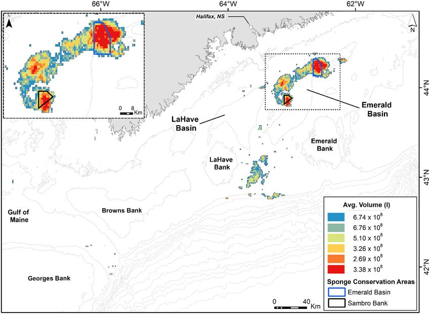
Maldonado et al.                                                                                                      A Microbial N Engine in Hexactinellida

                                             among the microbiota and the sponge suggests that the holobiont depends critically on
                                             the correct functioning of its N-driven microbial engine. The metabolic intertwining is so
                                             delicate that it changed after moving the sponges out of their habitat for a few days, a
                                             serious warning on the conservation needs of these sponge aggregations.

                                             Keywords: sponge physiology, sponge microbiota, nitrogen cycling, phosphorous cycling, functional deep-sea
                                             ecology, benthic-pelagic coupling, sponge aggregations, nutritional competitive exclusion

INTRODUCTION                                                                      Often the microbial communities hosted by demosponges are
                                                                                  complex and may include simultaneously archaea and bacteria,
Glass sponges (phylum Porifera, class Hexactinellida) are marine                  including cyanobacteria. Demosponges with high abundance of
organisms markedly restricted to the deep sea (Tabachnick,                        microbes in their tissues (i.e., HMA sponges) often —but not
1994), with only a handful of species that can be found at                        always (Bayer et al., 2008)— take up ammonium from seawater,
SCUBA-diving depths (Mackie and Singla, 1983; Vacelet et al.,                     which is a N source for chemo- and phototrophic bacteria and
1994; Cook et al., 2008). Such bathymetric confinement has                        an energy source for ammonia-oxidizing bacteria (AOB) and
hindered empirical investigations on this large sponge lineage,                   archaea (AOA). In contrast, the HMA demosponges typically
the physiology and functional ecology of which remains poorly                     release nitrate and nitrite, as the probable result of the metabolic
understood. During the last two decades, the advent of high-                      activity of nitrifying symbiotic bacteria (Corredor et al., 1988).
capacity remote operated vehicles (ROVs) has opened deep-sea                      The demosponges with low abundance of associated microbes
communities to experimental and manipulative research, making                     (LMA sponges) are typically net sources of both nitrate and
it possible to approach the physiology of the organisms and their                 nitrite as well, and, in many cases, also release ammonium
functional integration into the deep-water ecosystems.                            (Jiménez and Ribes, 2007; Southwell et al., 2008a,b; Morganti
    At bathyal depths, hexactinellid sponges can occur forming                    et al., 2017). Most investigated demosponges —but not all— are
dense and extensive aggregations (reviewed in Maldonado et al.,                   net sources of phosphate, which is released at very low rates
2017). While the physical presence of the sponges increases the                   (Maldonado et al., 2012; Ribes et al., 2012; López-Acosta et al.,
3D-complexity of the habitats (Beazley et al., 2013; Dunham                       2019).
et al., 2018; Hawkes et al., 2019), their physiological activity also                In this regard, there is no comprehensive information
carries an exchange of organic and inorganic nutrients with the                   for hexactinellids about the sign and magnitude of the net
surrounding environment (Pile and Young, 2006; Yahel et al.,                      flux of ammonium, nitrite, nitrate and phosphate. Given the
2007; Kahn et al., 2018). Such nutrient exchange is foreseen to                   idiosyncrasy of the syncytial tissues of hexactinellids, it is not
affect the surrounding deep-sea system in a multiplicity of ways,                 even clear whether these sponges can be objectively identified
but the exact mechanisms behind it, as well as the magnitude of                   as belonging to either the HMA or LMA models established
the net fluxes, remain poorly investigated, at least in comparison                for demosponges and, if so, it remains to be described where
with the information available for shallow-water demosponges                      the abundance of microbes would be located within their body,
(reviewed in Maldonado et al., 2012 and Zhang et al., 2019).                      typically consisting of large internal aquiferous spaces and only
The implications of the hexactinellid aggregations on the cycling                 very thin threads of tissue (i.e., epithelia + mesohyl) attached to
of silicon have been demonstrated at regional oceanographic                       the siliceous skeleton. A recent study of South Pacific deep-sea
levels (Chu et al., 2011; Maldonado et al., 2021), but their role                 sponges, including demosponges and hexactinellids, has reported
in respect to the flux of nitrogen (N) and phosphorous (P)                        microbial communities in hexactinellids that appear to show
nutrients remains less understood. Traditionally, hexactinellid                   patterns equivalent to the LMA-HMA dichotomy known for
sponges have been interpreted as long-lived organisms with                        shallow-water demosponges (Steinert et al., 2020). Likewise, a
low rates of physiological activity, so that the net flux rates of                recent pioneering study (Tian et al., 2016) on the hexactinellid
nutrient exchange with the environment were a priori expected                     Lophophysema eversa has reported the microbial community to
to be modest. However, while the rates of silicon utilization                     be dominated by chemoautotrophic bacteria, mostly ammonia-
depend directly on the metabolic activity of the sponge cells,                    oxidizing archaea (AOA), nitrite-oxidizing bacteria (NOB), and
the net flux of N and P inorganic nutrients of the sponges                        sulfur-oxidizing bacteria (SOB). These three functional groups
depend on the metabolic activity not only of the sponge cells                     of microbes were interpreted as playing roles in the cycling of
but also of the microbial populations (the microbiota) that the                   carbon, nitrogen and sulfur in the micro-environment inside
sponges host. From studies on shallow-water demosponges, it                       the sponge body, being scavengers of toxic ammonia, nitrite
is well-known that the “holobiont” constituted by a sponge and                    and sulfide waste produced by the metabolism of the sponge
its microbiota is able to either incorporate or release a variety                 cells in the conditions of the deep-sea environment. The NOB
of dissolved inorganic nutrients such as ammonium, nitrate,                       groups also appear to participate in pathways that lead to
nitrite, and phosphate, depending on the sponge species (Diaz                     production of vitamin B12 (Tian et al., 2016). In a recent study
and Ward, 1997; Maldonado et al., 2012; Ribes et al., 2012;                       on the hexactinellid Vazella pourtalesii, the microbiome has
Fiore et al., 2013; Keesing et al., 2013; Hoer et al., 2018; Pita                 been well-characterized through genomic approach, as well as
et al., 2018; López-Acosta et al., 2019; Zhang et al., 2019).                     the functional traits that allow the microbes to survive and

Frontiers in Marine Science | www.frontiersin.org                             2                                      March 2021 | Volume 8 | Article 638505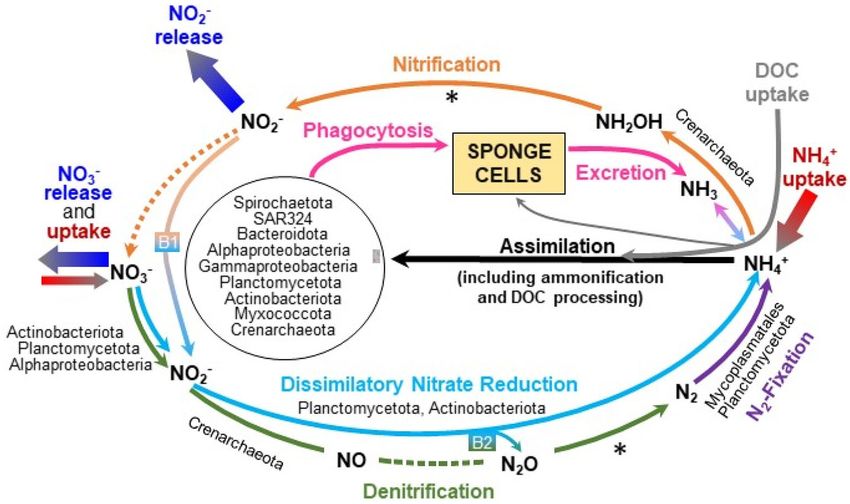
Maldonado et al. A Microbial N Engine in Hexactinellida
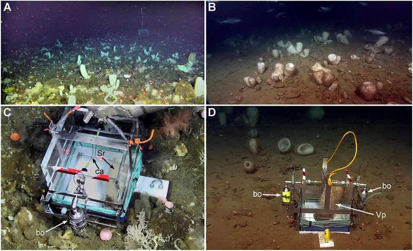
FIGURE 1 | (A) View of a multi-species sponge community at the Schulz Bank seamount (located between the Greenland and Norwegian Seas; 73.8 ◦ N; 7.5 ◦ E),
where the whitish tubes of the hexactinellid Schaudinnia rosea co-dominate in abundance along with a variety of demosponges and small cnidarians. (B) View of the
monospecific aggregations of the hexactinellid Vazella pourtalesii that extend on the deep region of the continental shelf in Nova Scotia (Canada). (C,D) In-situ
incubation of an individual of Schaudinnia rosea (Sr) and a Vazella pourtalesii (Vp) in their respective natural communities at depths of 580 and 180 m, respectively. Note
that the incubation chambers incorporate two external steel bottles (bo) for water sampling within the camera by means of capillaries (cp) that pierce the chamber wall.
proliferate within the sponge tissue (Bayer et al., 2020). In the have also been unveiled. Therefore, this study seeks to contribute
current study, we attempt to integrate the genomic microbial to a better understanding of (1) the mechanisms behind the
information in a physiological and ecological context to better nutrient flux rates of hexactinellid sponges and (2) the ecological
understand the nitrogen and phosphorus fluxes of the sponges, significance of those fluxes to the deep-sea ecosystems.
what sponge capabilities may rely on the metabolic integration
with their microbes, and what the implications are for the
MATERIALS AND METHODS
deep-sea ecosystem where aggregations of these hexactinellid
sponges occur. Sponge Species
To this aim, in the frame of the EU H2020 project “SponGES,” The studied hexactinellid sponges, S. rosea and V. pourtalesii, are
we have investigated two ecologically important deep-sea phylogenetically closely related, both belonging to the subfamily
hexactinellid sponge species, Schaudinnia rosea (Fristedt, Rossellinae in the family Rossellidae. The species S. rosea is
1887) and Vazella pourtalesii (Schmidt, 1870). In both cases, distributed along the Arctic Ocean from 90 to 3110 m depth.
the sponges form dense aggregations, representing relevant This species becomes co-dominant in abundance at the Schulz
community components at both the physical and the functional Bank, a seamount located on the Arctic Mid-Ocean Ridge (73.8
levels. We have quantified in situ the net flux rates of ammonium, ◦ N; 7.5 ◦ E) between the Greenland and Norwegian Seas. There
nitrate, nitrite, and phosphate, using benthic incubation it forms, along with several demosponges, a sponge-dominated
chambers manipulated by high-capacity ROVs. By collecting benthic community that extends over the top of the seamount
live animals, net flux rates were also measured in laboratory at depths of 550–600 m (Figure 1A, Supplementary Video 1).
experiments for one of the species, namely V. pourtalesii. The species V. pourtalesii occurs in the northwest Atlantic, from
Through metagenomic analysis, the main microbial lineages 100 to 935 m depth. This species is mostly known from dense,
occurring within the sponges have been revealed along with their monospecific aggregations (Figure 1B, Supplementary Video 2)
functional implication in a variety of relevant biochemical routes extending on the deep continental shelf of Nova Scotia (Canada)
in the N and P metabolism. Through ultrastructural research, at 150–200 m deep (Beazley et al., 2018; Maldonado et al.,
the particular locations of the microbes within the sponge body 2021). At those depths, the areas of the Scotian continental
Frontiers in Marine Science | www.frontiersin.org 3 March 2021 | Volume 8 | Article 638505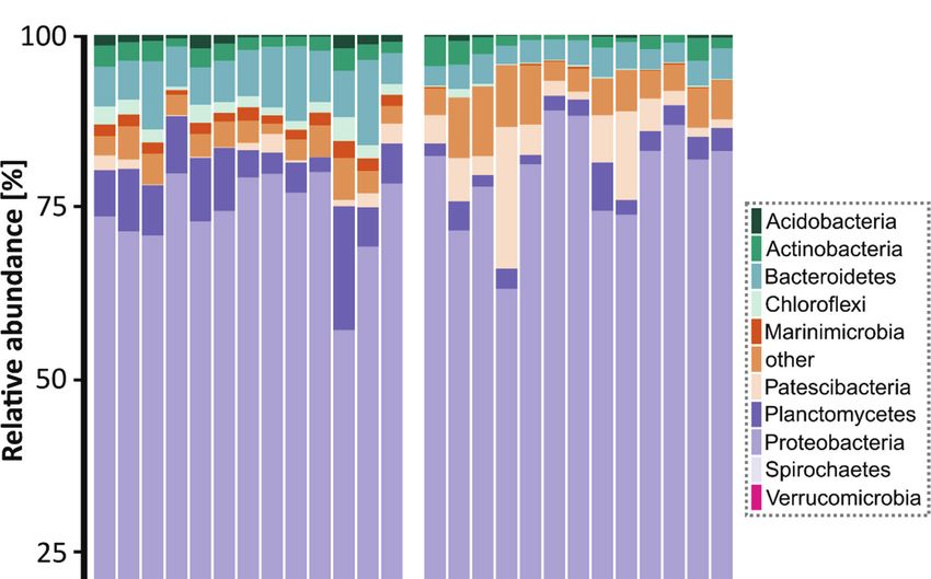
Maldonado et al.                                                                                             A Microbial N Engine in Hexactinellida

shelf where the sponge aggregations occur are an aphotic                 Laboratory Incubations of Sponges
environment with bottom water temperatures below the 10◦ C               To examine whether differences occur in the magnitude
permanent thermocline, features favored by the high latitude.            and/or the sign between in-situ and laboratory sponge
Those conditions, according to a functional definition of the            incubations, we collected individuals of V. pourtalesii (at
deep-sea (Gage and Tyler, 1991), qualify the Vazella grounds as          Sambro Bank Sponge Conservation Area and LaHave Basin),
a deep-sea habitat despite being topographically located on the          took them to the aquarium room of the Bedford Institute of
continental shelf.                                                       Oceanography (Dartmouth, Canada) and conducted seven
                                                                         laboratory incubations during a period of 2 weeks, as detailed in
Field Incubations of Sponges                                             Maldonado et al. (2020). The enormous distance from Schulz
We built 6 benthic incubation chambers in methyl methacrylate            Bank to any land laboratory made it logistically impossible to
and inox steel, with an incubation volume of either 17.3 or              repeat the laboratory experiments with the species S. rosea.
13.3 L (as described in Maldonado et al., 2020). Chambers                   In short, 11 individuals of V. pourtalesii were collected along
incorporated a floor piece of Delrin acetal resin, which allowed         with the small rock on which it was attached. Sponges were
for the incubation of sponges in isolation from the external             maintained alive on board for 5 days in a 700 L tank filled with
environment. This approach avoided interference by nutrient              seawater refrigerated to 9 ± 1◦ C. Upon arrival to the laboratory,
fluxes from the resuspended sediments that occurred during               sponges were transferred to a 360 L tank and left there 2 days
deployments and ROV activity on the sea bottom. The chambers             for acclimation to a refrigerated (9 ± 0.5◦ C) seawater system
incorporated two external sampling bottles (120 mL) made                 with recirculation, fed with seawater from the Bedford Basin at
of steel and internally folded with polytetrafluoroethylene              the head of Halifax Bay. The seawater was filtered on a 1 µm
(Figures 1C,D). Through a steel capillary (20 cm long and                mesh, a pore size preventing phytoplankton but allowing in part
0.6 mm wide) that pierced the wall of the chamber, each bottle           of the natural sponge food (i.e., most of the bacterioplankton).
was designed to collect a water sample (under negative pressure          After acclimation, each sponge was incubated separately in a
conditions) from inside the incubation chamber while it was              polypropylene 16 L container for 24 h and then transferred, along
opened for 5 min and then closed using the ROV manipulator               with all other incubated individuals, to a common 300 L water
arms (Figures 1C,D).                                                     tank for resting during the following 24 h, until starting a new
    In-situ incubations were conducted during two oceanographic          24 h period of individual incubations in the 16 L containers. The
cruises. In August 2018, on board of R/V GO Sars and using               sponges were subjected to a total of seven successive incubations
the ROV ÆGIR 6000, we incubated seven individuals of S. rosea            intercalated with their corresponding 24 h resting periods. The
(Supplementary Video 3) and a control on the Schultz Massif              idea for this design was to examine whether a midterm exposure
seamount, at depths of 577–580 m. In September 2017, on board            to the coastal water in laboratory conditions would induce
of CCGS Martha L Black and using the ROV ROPOS, a total                  detectable changes in the sign and magnitude of the net flux of the
of four individuals of V. pourtalesii and a control (see below)          various nutrients and relative to the field incubations. From each
were incubated in situ on the Scotian Shelf at depths of 160–            individual incubating container, seawater was sampled at the
180 m (Supplementary Video 4), as described in Maldonado                 beginning and at the end of each incubation period to determine
et al. (2020). The number of incubations was limited by inclement        the magnitude and the sign of the change in the respective
weather in the area during the entire cruise. For the incubations,       nutrient concentrations (i.e., ammonium, nitrate, nitrite, and
each sponge was grabbed by the manipulator arm from the                  phosphate), following the methods explained in the below section
small rock on which it was attached, then placed on the floor            of “Nutrient analyses.”
piece of the incubation chamber, and covered with the methyl
methacrylate box, which rested into a groove in the floor piece          Nutrient Analyses
designed to prevent leakages from the incubation unit. Once              Immediately after recovering the sampling bottles from
the sponge was inside the chamber and the chamber properly               deployment, seawater samples were passed through 0.22
sealed, one of the sampling bottles was opened to collect water          µm-pore, syringe filters (Millex-GS Millipore) and frozen at
for 5 min and then closed again to avoid water exchange with             −20◦ C, until they were analyzed using an Auto-Analyzer AA3
the surrounding seawater. As a control, we used a rock selected          (Bran+Luebbe) following the standard colorimetric method
from the sponge grounds but without an attached sponge. After            (Strickland and Parsons, 1972), with a determination accuracy
an incubation period of 19–28 h (incubation time varied due to           of 1%. The nutrient flux was finally calculated as the value of
weather and the logistics of the cruises), the ROV returned to           its concentration at the beginning of the incubation minus
the chamber position and triggered the second sampling bottle.           that at the end of incubation, being normalized by duration of
After this second water collection, the sponge and its attachment        incubation (h), sponge size (mL), and seawater volume in the
rock were collected to estimate volume and biomass, needed to            incubation unit (L) after discounting sponge and rock volume.
normalize N and P flux rates by sponge volume (mL); sponges              Flux rates were also corrected by the control flux. Therefore,
were later combusted for flux rates to be also normalized by ash-        negative flux values represent nutrient release by the sponges and
free dry weight (AFDW; g). Seawater samples were processed               positive values incorporation. We preferentially expressed data
for determination of nutrient concentrations to further derive           normalized to sponge volume because it facilitates their future
net flux rates of ammonium, nitrate, nitrite, and phosphate (see         applicability to field sponge populations using ROV images
method section on “Nutrient analyses”).                                  without the need of collecting individuals. However, we have also

Frontiers in Marine Science | www.frontiersin.org                    4                                     March 2021 | Volume 8 | Article 638505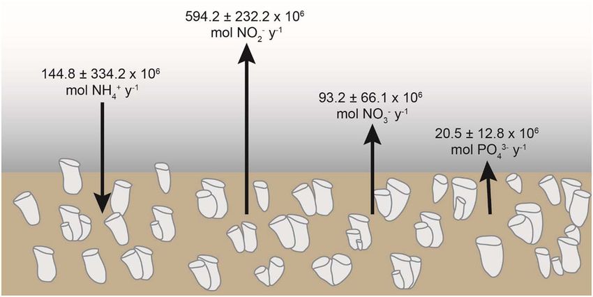
Maldonado et al.                                                                                               A Microbial N Engine in Hexactinellida

expressed data as AFDW-normalized, for more correct between-                Microbial Functional Gene Repertoire in V. pourtalesii
species physiological comparisons. Finally, we examined the                 To assess the involvement of microbial symbionts in nitrogen
potential relationships between flux rates by nutrient pairs and            and phosphorous cycling, we focused on the V. pourtalesii
between the flux rate of a nutrient and its ambient concentration           holobiont. The functional gene repertoire of metagenome-
at the beginning of the incubation, using linear and non-linear             assembled genomes (MAGs) that were used for this objective
regression analysis.                                                        has been published recently (Bayer et al., 2020) and is available
                                                                            on NCBI under BioProject PRJNA613976. Briefly, metagenomes
                                                                            were sequenced from seven sponge samples and five seawater
                                                                            controls by Illumina Next Generation Sequencing (HiSeq 4000,
Diversity and Functional Gene Analysis of                                   2 × 150 bp paired-end) at the Institute of Clinical Molecular
Associated Microbiota                                                       Biology (IKMB) of Kiel University. The raw reads were trimmed
Microbial Diversity From Amplicon Sequencing                                with Trimmomatic v0.36 and co-assembled with Megahit v1.1.3
In order to examine the microbial diversity in the studied                  (Li et al., 2016). The metagenomic assembly was binned with
sponges, 13 individuals of V. pourtalesii were collected from               the metaWRAP pipeline v1.0.2 (Uritskiy et al., 2018), and
Sambro Bank and 13 of S. rosea from Schulz Bank. The sample                 functionally annotated by Interproscan v5.30-69.0 including GO
processing procedure is described in detail elsewhere (Busch                term and pathway annotations (Jones et al., 2014; Sangrador-
et al., 2020, 2021; see also these references for data availability).       Vegas et al., 2016). Interpro annotations are available via
Briefly, from each sponge individual chunks of tissue were                  DOI 10.6084/m9.figshare.12280313. Taxonomy was determined
subsampled, rinsed and frozen at −80◦ C until DNA extraction.               with GTDB-Tk (Chaumeil et al., 2019). V. pourtalesii-enriched
Seawater reference samples were also collected from each of the             MAGs were identified by linear discriminant analysis (LDA)
two sponge habitats. At Sambro Bank, three samples of bottoms               scores using LEfSe v1.0 (Segata et al., 2011). For enzymes or
seawater (3 m from bottom) were collected with a Niskin bottle              functions of specific pathways whose genes were not found in
deployed on the ROV. At Schulz Bank, six seawater samples                   MAGs of identified microbial lineages, we still searched the
were collected, three of which by Niskin bottles deployed on                unbinned metagenomic data (i.e., all contigs not assigned to a
the ROV (3 m from bottom) and three by a CTD rosette water                  specific MAG) to determine whether the genes for the missing
sampler. All seawater samples (2 L) were filtered onto PVDF filter          metabolic step could be present somehow within the sponge
membranes (Merck Millipore) with a pore size of 0.22 µm and a               microbial community.
diameter of 47 mm. Until DNA extraction the filters were stored
at −80◦ C.
    Approximately, 0.25 g of sponge tissue and half of a seawater           Histology and Ultrastructure of Deep-Sea
filter were used for DNA extraction with the DNeasy Power                   Hexactinellids
Soil Kit (Qiagen). Quality and quantity of extracted DNA was                To understand and document where the various microbes may
checked and a one-step PCR conducted to amplify the V3 and V4               physically occur within the sponge body, we conducted both light
variable regions of the 16S rRNA gene (primer pair 341F-806R).              microscopy and transmission electron microscopy (TEM) on V.
Afterwards, PCR-products were examined by gel electrophoresis,              pourtalesii and S. rosea.
normalized and pooled. Sequencing was performed on a MiSeq                     For light microscopy, tissue pieces of 0.5 cm3 were fixed
platform (MiSeqFGx, Ilumina) using v3 chemistry (producing 2                in 4% formalin in saline dibasic phosphate buffer for 3 weeks,
× 300 bp).                                                                  until arriving to the laboratory. Samples were then rinsed
    Adapters were removed and raw sequences (forward reads)                 in distilled water, desilicified in 5% hydrofluoric acid for
were truncated to a length of 270 nt. Quality of sequences                  5 h, rinsed in distilled water, dehydrated through an ethanol
was evaluated in QIIME2 version 2018.11 (Bolyen et al.,                     series of increasing concentration (50–100%) and finally xylene,
2019) before applying the DADA2 algorithm (Callahan et al.,                 embedded in paraffin, and sectioned in a Leica RM2125 RTS
2016); one million reads were used to train the error                       manual microtome. The obtained 5 µm-thick sections were
model. Several denoising steps were conducted to remove                     extended on glass slides, stained with hematoxylin and eosin,
chimeras, chloroplasts, unassigned and mitochondrial sequences.             and a cover-slide attached using DPX mounting medium for
A minimum sequencing depth of 13000 was applied. Taxonomic                  observation through an IX50 Olympus microscope connected to
classification of Amplicon Sequence Variants (ASVs) was                     a ProgRes C7 digital camera.
performed using a Bayes classifier (Bokulich et al., 2018)                     For TEM, tissue pieces of about 2 mm3 were immersed for 3 h
trained on the Silva 132.99% OTUs 16S database (Quast et al.,               in a fixative cocktail consisting of 2% glutaraldehyde, 2% osmium
2013). Between-sample Weighted UniFrac distances (Lozupone                  tetroxide, 65% sodium acetate buffer, 11% sucrose, and 20%
et al., 2011) were calculated based on a phylogeny produced                 distilled water (Maldonado, 2015). Initial dehydration steps took
with FastTree2 (Price et al., 2010). To assess the effect on                place in 50% ethanol and then 70% ethanol, in which samples
Weighted UniFrac distances on the spatial distribution of                   were preserved for 1 month, until their arrival to the laboratory.
samples in an ordination space, a non-metric multidimensional               Some of these samples were then rehydrated and desilicified in
scaling (nMDS) was performed, followed by PERMANOVA tests                   hydrofluoric acid for 5h prior to dehydration; the rest of the
to examine the statistical significance of the between-group                samples were subjected to dehydration without desilicification.
differences identified by the nMDS.                                         Dehydration was then resumed in 70% (10 min), 80% (10 min),

Frontiers in Marine Science | www.frontiersin.org                       5                                     March 2021 | Volume 8 | Article 638505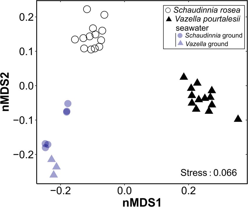
Maldonado et al.                                                                                                                 A Microbial N Engine in Hexactinellida

 FIGURE 2 | Spatial distribution of Vazella pourtalesii biomass (by volume in liters) across the Central Scotian Shelf, as calculated in Maldonado et al. (2021). The
 sponge grounds correspond to areas predicted to have > 70% presence probability of the species, according to Beazley et al. (2018). The areas of densest sponge
 aggregation are protected from any bottom fishing activity through the creation of two Sponge Conservation Areas (SCA): Emerald Basin SCA and Sambro Bank SCA.

90% (3 × 10 min), 96% (3 × 10 min), and 100% ethanol (3 ×                             probability outputs for this species, as indicated in Beazley et al.
10 min), followed by propylene oxide (2 × 10 min). Embedding                          (2018). The presence probability surface was generated from
in Spur resin required five immersion steps with gentle shaking                       random forest modeling using presence/absence data. Using
during each one: 6 h in a 3:1 propylene-oxide/resin solution, 12 h                    ArcMap version 10.6.1., the 1 × 1 km raster grid containing all
in 2:2 propylene-oxide/resin solution, 7 h in a 1:3 propylene-                        predicted presence probabilities of V. pourtalesii from > 70 to
oxide/resin solution and two 6-h steps in pure resin. Resin was                       100% and displayed in 5% equal presence probability intervals
hardened at 60◦ C for 2 days. Ultrathin sections were obtained                        in Beazley et al. (2018), was converted to a polygon layer of
with an Ultracut Reichert-Jung ultramicrotome, mounted on                             1 × 1 km cells, each representing a given presence probability.
gold grids and stained with 2% uranyl acetate for 30 min, then                        The total area encompassed by all cells with probability values
with lead citrate for 10 min. Observations were conducted with a                      within each 5% probability interval (six intervals from > 70 to
JEOL 1010 TEM operating at 80 kv and provided with an external                        100%) was calculated at 2,105 km2 (Figure 2). The two densest
Gatan module for acquisition of digital images.                                       areas of the sponge aggregation were closed by Fisheries and
                                                                                      Oceans Canada to all bottom fishing activities, creating in 2013
                                                                                      the Emerald Basin Sponge Conservation Area (195 km2 ) and the
Nutrient Fluxes at the Deep-Sea                                                       Sambro Bank Sponge Conservation Area (62 km2 ), as indicated
Community Level                                                                       in Figure 2. ROV-driven, transect and quadrat surveys in the
The nutrient utilization rates measured during the in-situ and                        two Sponge Conservation Areas additionally characterized the
laboratory incubations were used to estimate the net annual flux                      spatial distribution of both the individuals (i.e., density) and
of ammonium, nitrite, nitrate, and phosphate at the aggregation                       the individual biomass (in volume) in cells of highest presence
of V. pourtalesii, which is well-characterized in terms of the                        probability, as detailed in Maldonado et al. (2021). It was
spatial distribution of the sponge biomass. The area occupied by                      found that, at the densest areas of the aggregations (cells of
the Vazella sponge grounds on the Scotian Shelf was estimated                         0.975 presence probability), the sponge density was 3.78 ± 3.22
by applying a threshold criterion (70%) to the modeled presence                       individuals m−2 , with a biomass of 1.6 ± 2.8 L of sponge tissue

Frontiers in Marine Science | www.frontiersin.org                                 6                                            March 2021 | Volume 8 | Article 638505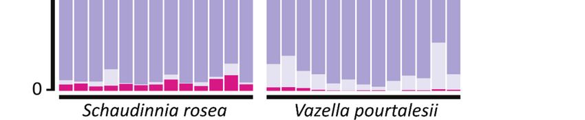
Maldonado et al. A Microbial N Engine in Hexactinellida
FIGURE 3 | and Vazella pourtalesii. Note that positive values indicate
incorporation of a nutrient from the seawater into the sponge, while negative
values indicate nutrient release from the sponge. (A) Global average (±SD) flux
rate (µmol mL−1 h−1 ) for each nutrient and sponge species when all assayed
individuals are considered in the calculations (S. rosea, n = 7; V. pourtalesii, n
= 15). Note that individual measurements for in-situ incubations (crosses) and
the first laboratory incubation of V. pourtalesii (open circles) are also plotted to
show between individual variability. (B) Average (±SD) flux rates (µmol mL−1
h−1 ) considering only individuals with the same flux sign in the averaging
process. The number of individuals (n) considered in these averages by flux
sign for each nutrient and species are indicated in the graph. (C) Ammonium
flux rate in the V. pourtalesii individuals over the seven successive laboratory
incubations. See Supplementary Table 2 for ammonium concentration at the
onset of incubations.
m−2 . Finally, in order to convert presence probability into density
of sponges and sponge biomass, as required for successively
estimating the flux rate of the respective dissolved inorganic
nutrient across the Vazella grounds, the average sponge density
and volume of sponge tissue were respectively multiplied by the
mid-point of each 5% presence probability bin. This procedure
revealed that the aggregation consists of some 6,479 ± 5,518
million sponges, with a total sponge biomass (in volume) of
2,791 ± 3,832 million L (Maldonado et al., 2021). The biomass
is known to be spatially distributed on the Nova Scotia Central
Shelf, as it is indicated in Figure 2. The resulting spatial cells of
sponge biomass were then multiplied by the global average value
of the net flux rate obtained for each nutrient after combining
all individuals from in-situ and laboratory incubations. For the
laboratory net flux, the average values considered only the first
incubation of the individuals. By considering only the first
laboratory incubation, we had more chances of avoiding the
effects that the coastal seawater feeding the laboratory could have
on the microbiota of V. pourtalesii, minimizing physiological
artifacts in the sponge response. Indeed, to test whether the
results of “in-situ” and “first-day laboratory” incubations could
be pooled safely, we examined potential differences in the net
flux rate between in-situ incubations and the first-day laboratory
incubations for each nutrient, using either the t-test or its non-
parametric equivalent Mann-Whitney U-test, whenever required
to deal with non-normal and/or heteroscedastic data sets. In all
cases, there were no statistically significant differences between
in-situ and laboratory flux rates (see Supplementary Table 1)
and, consequently, data were suitable for pooling. The result of
the subsequent calculations provided a mean (± SD) estimate of
the annual net flux rate of each nutrient through the V. pourtalesii
aggregations on the Nova Scotia Continental Shelf.
It must be noted that the large standard deviations associated
to the average values of number of sponges per square meter,
volume of sponge per square meter, and total number of
individuals in the aggregation obtained from Maldonado et al.
(2021), when propagated through the scaling up of data to the
aggregation level, will lead to a large SD value associated to the
average of the global fluxes. As explained in Maldonado et al.
FIGURE 3 | (A,B) Magnitude (average ±SD) and sign of the flux (µmol mL−1 (2021), the large SD values of sponge density and volume initially
3−
h−1 ) of ammonium (NH+ − −
4 ), nitrite (NO2 ), nitrate (NO3 ), and phosphate (PO4 ) derive from the largest individuals of V. pourtalesii being often
measured for individuals of the hexactinellid sponges Schaudinnia rosea
(Continued)
connected by their bases, forming small clumps of individuals.
As a result, even within the most densely populated areas of the
Frontiers in Marine Science | www.frontiersin.org 7 March 2021 | Volume 8 | Article 638505Maldonado et al. A Microbial N Engine in Hexactinellida aggregation, some square meters of bottom are devoid of sponges Supplementary Table 2). This shift suggests that changes in while others contain many. Such spatial pattern, aggregated at the population of associated microbes responsible for the a scale
Maldonado et al.                                                                                                                                 A Microbial N Engine in Hexactinellida

TABLE 2 | Summary of nutrient fluxes and associated parameters in the incubations of Vazella pourtalesii.

Code          Size                              Initial nutrient concentration                                                           Nutrient flux rate
              (mL)                                             (µM)                                                                   (10−3 µmol mL−1 h−1 )

                                          NH+
                                            4                         NO−
                                                                        2         NO−
                                                                                    3           PO3−
                                                                                                  4                          NH+
                                                                                                                               4                         NO−
                                                                                                                                                           2          NO−
                                                                                                                                                                        3          PO3−
                                                                                                                                                                                     4

                            1st        1st-3th         4–7th                                                   1st         1st-3th         4–7th

1            492.4         0.04            −             −            0.14        15.66         1.08            5.3           −              −          −25.5         −4.0         −0.9
2              64.5        0.12            −             −            0.05        17.35         1.14           41.6           −              −          −33.8          17.0        −0.4
3            126.4         0.08            −             −            0.09        16.51         1.11           11.7           −              −          −20.3         −0.8         −0.5
4            323.4         0.56            −             −            0.19        19.31         1.26            4.7           −              −          −26.7         −2.2         −1.0
5              65.2        1.52          1.71           0.68          2.13         7.91         0.96            9.4            7.0            0.9       −30.2         −2.8         −0.3
6            144.3         1.57          1.73           0.81          2.25         7.84         0.97            7.2            4.7          −4.8        −29.5         −4.6         −0.3
7            104.8         1.62          1.84           0.74          2.20         7.72         0.95          −1.4             2.0          −3.4        −24.8         −7.9         −1.5
8            108.4         1.55          1.58           0.64          2.22         7.73         0.96            9.4            7.7            3.8       −35.8         −6.3         −1.4
9              50.8        1.64          1.76           0.74          2.14         8.01         0.95          −9.5         −10.5            −8.6        −12.1         −3.1         −1.3
10             58.0        1.78          1.76           0.69          2.21         7.89         0.91            5.7            9.5            0.2       −28.0           0.8          0.1
11             86.8        1.56          1.69           0.75          2.40         8.19         0.97           11.9          10.6           −3.7        −35.7         −7.3         −1.3
12             65.2        1.58          1.70           0.72          2.20         7.85         0.96            8.7            5.4          −9.6        −24.0         −5.1         −0.9
13             36.5        1.58          1.80           0.70          2.19         7.88         0.99         −23.6         −22.7          −20.2          −4.1         −1.4         −0.2
14             65.2        1.66          1.77           0.68          2.21         7.81         0.95            2.4          −1.2           −6.9        −25.3         −2.4         −1.6
15             43.6        1.59          1.80           0.76          2.22         7.64         0.99            5.2          −0.3           −9.0         −8.4         −1.7         −1.0
AVG          122.4         1.23          1.74           0.72          1.66        10.35         1.01            5.9            1.1          −5.6        −24.3         −2.1         −0.8
SD           124.1         0.66          0.07           0.05          0.96         4.34         0.09           13.5            9.9            6.5          9.5          5.8          0.5

The table contains reference labels for the 15 incubated individuals, with individuals #1 to #4 corresponding to in-situ incubations and individuals #5 to #15 corresponding to laboratory
incubations. It is also given the size of the incubated sponges, the initial concentration (µM) of the dissolved inorganic nutrients (ammonium: NH4+ ; nitrate: NO3− ; nitrite: NO2− ;
phosphate—PO3−    4 ) at each incubation event, and the nutrient flux rate (10
                                                                               −3 µmol mL−1 h−1 ) detected during the incubation and normalized by incubation duration and sponge

biomass. Nutrient flux rates have also been corrected for the control effect. Note that for the laboratory ammonium flux rate, data are presented for only the 1st day of laboratory
incubations (1st), then as the individual responses averaged across the first three laboratory incubations (1st–3th), then as the individual responses averaged across the last four
laboratory incubations (4th–7th). See also Supplementary Table 2 for raw data on ammonium flux rate across all laboratory incubations. The global average (AVG) and standard
deviation (SD) of the various parameters for the entire set of assayed individuals are summarized at the two bottom rows of the table.

of 17.0 × 10⁻³ µmol mL−1 h−1 ; individual #10 also consumed                                     analyses, no statistically significant relationships were found for
nitrate during the laboratory incubations, but at much smaller                                  S. rosea in any case (n = 7, p > 0.05). In contrast, statistically
rate of 0.8 × 10⁻³ µmol mL−1 h−1 (Table 2).                                                     significant relationships, although of only moderate intensity,
    Regarding phosphate, both species were predominantly net                                    were found in V. pourtalesii (Figure 4). It was detected that the
sources, but with low rates. Natural phosphate concentration                                    increase in the release rate of nitrite is linearly coupled to an
during the in-situ incubations of S. rosea was 0.86 ± 0.10 µM                                   increase in the ammonium consumption (n = 15, R2 = 0.474, p
(Table 1). At that nutrient concentration, 6 of the 7 assayed                                   = 0.005; Figure 4A). Another detected relationship was that the
individuals released phosphate, with an average rate of −2.5 ±                                  rate of nitrate consumption decreased linearly with the increase
2.6 × 10⁻³ µmol mL−1 h−1 (Figure 3). In contrast, individual                                    of phosphate release (n = 14, R2 = 0.379, p = 0.019; Figure 4B).
#2 consumed phosphate at a low rate of 0.5 × 10⁻³ µmol mL−1                                         When the relationship between the flux rate of a nutrient and
h−1 (Table 1). All four individuals of V. pourtalesii incubated in                              its ambient concentration was examined by linear regression, no
situ under a natural phosphate concentration of 1.15 ± 0.08 µM                                  statistical significance was found for any of the nutrients in any
released phosphate at an average rate of −0.7 ± 0.3 x 10⁻³ µmol                                 of the two sponge species (Supplementary Figure 1).
mL−1 h−1 . Ten of the 11 individuals incubated in the laboratory
at a phosphate concentration of 0.96 ± 0.02, that is, similar to the                            Microbial Insights Into the Sponge
one in the natural habitat, also released phosphate consistently,                               Nitrogen Flux
with an average rate of −1.0 ± 0.5 × 10⁻³ µmol mL−1 h−1                                         The analysis of amplicon sequences sampled from V.
(Figure 3). Individual #10 consumed phosphate at a rate as low                                  pourtalesii and S. rosea showed that each species harbors
as 0.1 × 10⁻³ µmol mL−1 h−1 (Table 2), so that it cannot be                                     a distinct microbiome (Figure 5, Supplementary Table 3,
discarded that it was indeed inactive in terms of release and that                              Supplementary Data File 1). In each case, there are also
the sign of the flux is an artifact derived from the flux rates being                           statistically significant differences between the sponge
at the limit of the methodological detection.                                                   microbiome and that of the surrounding seawater. Microbiome
    When the relationship between flux rates was examined by                                    differences between V. pourtalesii and S. rosea may be responsible
pairs of nutrients using both linear and non-linear regression                                  for some of the between-species differences detected in the sign

Frontiers in Marine Science | www.frontiersin.org                                           9                                                 March 2021 | Volume 8 | Article 638505
Maldonado et al. A Microbial N Engine in Hexactinellida
FIGURE 5 | Non-metric dimensional scaling (nMDS; k = 2, maxit = 100) plot
on weighted UniFrac distances for each of the assayed sponge species,
Vazella pourtalesii and Schaudinnia rosea, and the seawater of their respective
habitats. The results of the PERMANOVA tests that gives statistical support to
the between-group differences is given in Supplementary Table 3.
in the microbial community (Supplementary Table 4). Genes
involved in nitrification, denitrification, dissimilatory nitrate
reduction, nitrogen fixation and ammonia assimilation were
identified. Genes encoding for anammox or assimilatory nitrate
reduction were found neither in MAGs nor in the unbinned
metagenomic data.
Nitrification
The enzyme ammonia-monooxygenase (AMO), responsible
for the first reaction in the first step of nitrification (i.e.,
oxidation of ammonia to hydroxylamine), was detected in
the Nitrosopumilaceae (Crenarchaeota, MAGs 74, 90, 131,
143) and in the Nitrosomonadaceae (Proteobacteria, MAG
46). This microbial metabolism may contribute to explain
FIGURE 4 | Significant relationships between nutrient fluxes in Vazella why most individuals of V. pourtalesii were essentially net
pourtalesii. (A) The lower the rate of nitrite release, the higher the ammonium
consumers of ammonium. After 3 incubation steps in the
consumption. (B) When individual #2, an atypical outlier consuming nitrate at
the abnormally high rate of 17 nmol mL−1 h−1 —rather than releasing it–, was laboratory using coastal water, all V. pourtalesii individuals
excluded from the analysis, a linear significant relationship was revealed became net sources of ammonium. Thus, we cannot discard the
between the release rates of nitrate and phosphate. The rest of possible possibility that the Nitrosopumilaceae and Nitrosomonadaceae
pairwise combinations among the flux rates of all four nutrients were also populations decayed in laboratory seawater because being highly
examined, but none had statistically significant support.
dependent on the seawater/holobiont conditions of the natural
habitat (see Discussion). Interestingly, the MAGs enriched in
and magnitude of the flux of the various nutrients (Figures 3A,B, V. pourtalesii compared to seawater (Supplementary Table 4,
Tables 1, 2). Supplementary Data File 2) include the genus Cenarchaeum
(MAGs 74, 90, 143), which is a common sponge-specific
Microbes of the Nitrogen Cycle in V. pourtalesii AOA symbiont. MAG 131 was found in equal abundances
We evaluated the potential metabolic functions of V. pourtalesii- in both habitats and represents the genus Nitrosopumilus, a
associated microbes involved in the nitrogen cycle by searching common sea-water AOA. The hydroxylamine-oxidoreductase
for key enzymes in the MAGs, which allows the assignment (HAO), responsible for the second reaction in the first step
of function to members of the microbiome. For enzymes of nitrification converting hydroxylamine to nitric oxide that
or functions encoded on contigs that were not sorted into is subsequently converted to nitrite by a not yet identified
specific MAGs, we assumed that the function might be present enzyme (Caranto and Lancaster, 2017), was not detected in
Frontiers in Marine Science | www.frontiersin.org 10 March 2021 | Volume 8 | Article 638505Maldonado et al. A Microbial N Engine in Hexactinellida
FIGURE 6 | Graphical summary of the microbiome-driven nitrogen cycle in Vazella pourtalesii, as it has been deduced through the metagenomic study and the
incubation experiments. Note that only those N pathways for which MAGs were found to be enriched in the sponge are named in the graph. The dashed lines indicate
enzymatic pathways for which genes were not detected and the asterisk (*) indicates genes that were present only in the unbinned data. “B1” and “B2” refer to
putative bridges between major biochemical pathways, which provide alternative routes to palliate the effects of some missing enzymes, also allowing an efficient
closing of the N cycle in the holobiont. The N microbial engine of V. pourtalesii appears to revolve around the incorporation of ammonium from the seawater and its
internal recycling. This nutrient fosters N assimilation by the sponge microbiota, which also benefits from the important amounts of DOC taken up by the sponge. All
available evidence suggests that the nourishment of V. pourtalesii does not appear to be based on filter feeding of the free-living bacterioplankton but rather on the
phagocytosis of its internal microbiota, which is mostly farmed in the bacteriosyncytia through the supply of ammonium and DOC.
the MAGs. However, haoB-related genes were found in the individuals, only a minority of them being net nitrate consumers
unbinned metagenome data, suggesting that this enzyme may (Figures 3, 6, Table 2). The genes nirS and nirK encoding the
be present in at least some of the microbial lineages and nitrite reductase that catalyzes the conversion of nitrite into nitric
that nitrite is produced out of ammonia/ammonium. Genes oxide were detected in five MAGs belonging to Crenarchaeota
responsible for nitrite oxidation, the second step of nitrification, (MAGs 36, 90, 101, 131, 143) and in the acidobacterial MAG
were found neither in MAGs nor in the unbinned metagenomic 86. The MAGs 90 and 143 were enriched in V. pourtalesii over
data. The lack of those enzymes would explain why nitrite, seawater. Regarding the last two reactions of denitrification
which appears not to be further processed in the nitrification that lead to production of molecular N2 gas, we could not
pathway (Figure 6), is released at high rate by V. pourtalesii detect the genes encoding for the nitric oxide reductase and
(Figure 3, Table 2, Supplementary Tables 1, 2). The occurrence nitrous-oxide reductase enzymes in the MAGs, but the gene for
of ammonia oxidation activity in the absence of nitrite oxidation the latter was present in the unbinned data (Figure 6). Given
was in agreement with the finding that ammonium consumption that nitrite is accumulated by an incomplete nitrification and
and nitrite release are activities associated with statistical released by the sponges at relatively high rates (Figure 3), it
significance in V. pourtalesii (Figure 4A). cannot be discarded that part of the accumulated nitrite can
be used to feed the denitrification pathway at its intermediate
Denitrification step (Figure 6) and also the pathway of dissimilatory nitrate
Genes encoding enzymes involved in two of the four reactions of reduction to ammonium, as it is explained in the section below.
the denitrification pathway were found in the microbiome of V.
pourtalesii. Genes annotated as nitrate reductase, which catalyze Dissimilatory Nitrate Reduction to Ammonium (DNRA)
the reduction of nitrate to nitrite, were found in three MAGs We detected genes involved in DNRA, which is an important
(Supplementary Table 4) assigned to Alphaproteobacteria intermediate process in the N cycling, linking N-compound
(MAG 70), Actinobacteria (MAG 64), and Planctomycetota oxidation and reduction processes, that is, operating at the oxic-
(MAG 52). All three MAGs were enriched in the sponge anoxic interface. Genes coding for nitrate reductase —which
compared to seawater. Yet, the sources of nitrate to initially may operate not only in the DNRA pathway but also in the
feed denitrification remain unclear, since nitrification cannot previously described denitrification pathway— were found in
be completed in V. pourtalesii and external nitrate was three MAGs (Supplementary Table 4) of Alphaproteobacteria
not consistently incorporated from seawater by all assayed (MAG 70), Actinobacteria (MAG 64), and Planctomycetota
Frontiers in Marine Science | www.frontiersin.org 11 March 2021 | Volume 8 | Article 638505Maldonado et al. A Microbial N Engine in Hexactinellida
(MAG 52). All three MAGs were enriched in the sponge Identification of Genes and
compared to seawater. The second step in the dissimilatory Microorganisms of the Phosphorus Cycle
reduction to ammonium is mediated by two enzymes. One is We searched for enzymes involved in the phosphorous
a respiratory cytochrome c nitrite reductase, encoded by genes cycle in the annotation data derived from the V. pourtalesii
nrfA/ H, which was detected only in the unbinned metagenomic microbiome to elucidate which members might be responsible
data (Supplementary Table 4). The other is a NADH depended for the low but consistent release of phosphate detected in
nitrite reductase encoded by the genes nirB/ D, detected in the sponges (Figure 3, Table 1). Components of the C–P lyase
four MAGs affiliated with Planctomycetota (MAGs 52, 99), multienzyme complex were found (Supplementary Table 4,
Actinobacteria (MAG 64), and Gammaproteobacteria (MAG Supplementary Data File 2). This complex is encoded by
89), although the latter was not significantly enriched in V. 14 genes (phnCDE, phnF, phnGHIJKLM) and catalyzes
pourtalesii over seawater. These features agree with the idea that the dephosphonation reaction in a range of structurally
the holobiont metabolism of V. pourtalesii, in its natural habitat, diverse phosphonates, that is, organophosphorus compounds
tends to produce internally ammonia/ammonium, which appears containing C-PO(OX)2 groups, where “X” is either H or an
to be subsequently used as a preferred substrate to sustain the alkyl or aryl radical. Phosphonates are one of the sources of
populations of archaea and bacteria of the microbiota (Figure 6). phosphate intake into cells. The dephosphonation reaction by
Note that only some of the assayed individuals of V. pourtalesii biological vias (i.e., enzymatically) is physiologically relevant
incorporated nitrate from seawater (Figures 3, 6). Therefore, it because the C-P bond is extremely stable and its cleavage by
is assumed that the DNRA pathway is mostly fed with nitrite at chemical vies requires very aggressive conditions. Enzyme
an intermediate step, using for that purpose part of the nitrate subunits of the C–P lyase complex were found only in MAG
accumulated after the incomplete nitrification (Figure 6, B1 124 belonging to Alphaproteobacteria, but the detection
arrow). This bridging mechanism is relevant because it connects in the unbinned data suggests that more members in the
the aerobic process of nitrification to the anaerobic process of microbiome may be using this enzyme for phosphonate
DNRA and it would provide an explanation of the withstanding utilization. This assumption is supported by the detection of
of V. pourtalesii to low oxygen conditions. subunits of the ABC transporter in several MAGs representing
different phylogenetic groups (Supplementary Table 4).
The predominant biogenic orthophosphate in nature is 2-
Nitrogen Fixation Aminoethylphosphonate (2AEP) and its utilization is a two-step
Genes for nitrogenase enzymes involved in N fixation through process. The first step is the transamination of 2AEP (+
reduction of N2 gas to NH+ 4 were detected in several MAGs pyruvic acid) to 2-phosphoacetaldehyde (PAA) and L-alanine
(Figure 6, Supplementary Table 4) affiliated with Firmicutes (Ternan et al., 1998). This enzyme was found in several MAGs
(MAGs 77, 88, 127), Planctomycetota (MAG 91), Bacteroidota (Supplementary Table 4) of which members of Planctomycetota
(MAG 24), and Alphaproteobacteria (MAG 135). The MAGs (MAGs 52, 94, 116, 119), Crenarchaeota (MAGs 74, 90, 143),
88 and 91 were enriched in V. pourtalesii over seawater. Note Alphaproteobacteria (MAG 75), and Gammaproteobacteria
that if the incomplete denitrification process previously identified (MAG 10) were enriched in the sponge. The second step is
in V. pourtalesii can be completed somehow by the sponge the hydrolytic cleavage of PAA in phosphate and acetaldehyde
microbiome (see Discussion from bridging between DNRA and mediated by the enzyme phosphonoacetaldehyde hydrolase. This
denitrification), the resulting N2 could well provide substratum was found only in two V. pourtalesii-enriched MAGs (52, 119)
for this N-fixation pathway (Figure 6). representing Planctomycetota.
The enzyme phosphonoacetate hydrolase is a alkaline
phosphatase, cleaving C-P bounds of a given substrate to form
Ammonia Assimilation acetate and phosphate (reviewed in Villarreal-Chiu et al., 2012).
The enzymes glutamine synthetase (glnA, GLUL) and This was found in one V. pourtalesii-enriched member of
glutamate dehydrogenase (gudB, rocG) were found in the Planctomycetota (MAG 121) and in several MAGs of different
MAGs of several members of the V. pourtalesii-microbiota phylogenetic groups (Supplementary Table 4). Collectively, the
(Supplementary Table 4), indicating that amino acids can be results suggest that part of the sponge microbiota use
formed from ammonia. MAGs enriched in V. pourtalesii were phosphonates as a source of phosphorous to grow and, through
affiliated with the phyla Alpha- and Gammaproteobacteria, this activity, the sponge gets help in the hard task of degrading
Spirochaetota, Crenarchaeota, Planctomycetota, SAR324, phosphonates. The level at which this process explains the net
Actinobacteriota, and Verrucomicrobiota (Figure 6; phosphate efflux from the sponges remain unclear from our
Supplementary Table 4, Supplementary Data File 2). Again, approach and more research is needed to better understand the
these findings support the notion that the pool of ammonium P circuit in the holobiont.
within the sponge, which consists of the ammonium excreted by
the sponge cells (i.e., ammonia that undergoes molecular auto-
ionization to ammonium and amide ions) plus that produced by Ultrastructural Observations
the denitrification and DNRA pathways and that taken up from The histological study of S. rosea and V. pourtalesii indicated
seawater, serves as a preferred substratum to sustain the growth that the tissue in these hexactinellids is a delicate network of thin
of the bulk of the microbiota in V. pourtalesii. strands through which the large silica pieces of the skeleton and
Frontiers in Marine Science | www.frontiersin.org 12 March 2021 | Volume 8 | Article 638505Maldonado et al. A Microbial N Engine in Hexactinellida FIGURE 7 | Microbes in Schaudinnia rosea. (A,B) View of flagellated chambers showing the collar bodies with their microvilli (mv) and a flagellum (fl) provided with flagellar vanes (fv). Small groups of prokaryotic microorganisms (mi) occur intracellularly associated to the thin cytoplasmic zones sandwiched between the microvilli of adjacent collar bodies. (C–F) Views of small groups of microbes (mi) that gather around the packs of collagen fibrils (cf) that run across the thin mesohyl bands located within the trabecular strands of the sponge body. Frontiers in Marine Science | www.frontiersin.org 13 March 2021 | Volume 8 | Article 638505
Maldonado et al.                                                                                                                       A Microbial N Engine in Hexactinellida

 FIGURE 8 | Extracellular microbes in Vazella pourtalesii. (A) Partial view of flagellated chamber, showing the collar bodies (cb) and the underlying trabecular tissue,
 which is relatively poor in extracellular microbes (em). (B,C) Images showing that, in the mesohyl, the extracellular microbes (em) often proliferate in close association
 to accumulations of collagen fibrils (cf). (D,E) Two enlargements of a zone of the trabecular tissue where electron-dense rod-like microbes occur adjacent to coccoid
 microbes contained intracellularly into a cytoplasmic vesicle of a sponge cell. (F) Comparative view of different microbial morphologies, typically characterized by a
 variety of electron-clear and/or electron-dense granules and cell sizes smaller than 1 µm. (G) Detail of an unidentified extracellular microbial cell (em) showing a variety
 of intracytoplasmic granules. Note the collagen fibrils (cf) around the microbe.

ample aquiferous spaces run. The ultrastructural TEM approach                             (Figures 7, 8). In S. rosea, small groups of microbes were also
revealed that extracellular microbes of both coccoid and rod-                             found intracellularly in the thin cytoplasmic bands that separate
like shape are scattered in both species, but at low density                              the microvilli of adjacent collar bodies (Figures 7A,B). However,

Frontiers in Marine Science | www.frontiersin.org                                    14                                              March 2021 | Volume 8 | Article 638505
You can also read