Conversion of Reactive Astrocytes to Induced Neurons Enhances Neuronal Repair and Functional Recovery After Ischemic Stroke - Frontiers
←
→
Page content transcription
If your browser does not render page correctly, please read the page content below
ORIGINAL RESEARCH
                                                                                                                                           published: 26 March 2021
                                                                                                                                     doi: 10.3389/fnagi.2021.612856

                                           Conversion of Reactive Astrocytes to
                                           Induced Neurons Enhances Neuronal
                                           Repair and Functional Recovery After
                                           Ischemic Stroke
                                           Michael Qize Jiang 1,2† , Shan Ping Yu 1,2 *† , Zheng Zachory Wei 1,2 , Weiwei Zhong 1,2 ,
                                           Wenyuan Cao 1 , Xiaohuan Gu 1,2 , Anika Wu 1 , Myles Randolph McCrary 1‡ , Ken Berglund 2,3
                                           and Ling Wei 1,4 *
                                           1
                            Edited by:      Department of Anesthesiology, Emory University School of Medicine, Atlanta, GA, United States, 2 Center for Visual and
                  Cesar V. Borlongan,      Neurocognitive Rehabilitation, Atlanta Veterans Affair Medical Center, Decatur, GA, United States, 3 Department of
            University of South Florida,   Neurosurgery, Emory University School of Medicine, Atlanta, GA, United States, 4 Department of Neurology, Emory University
                          United States    School of Medicine, Atlanta, GA, United States

                       Reviewed by:
                    Jukka Jolkkonen,       The master neuronal transcription factor NeuroD1 can directly reprogram astrocytes
University of Eastern Finland, Finland
                     Vanessa Castelli,     into induced neurons (iNeurons) after stroke. Using viral vectors to drive ectopic
           University of L’Aquila, Italy   ND1 expression in gliotic astrocytes after brain injury presents an autologous form
                   *Correspondence:        of cell therapy for neurodegenerative disease. Cultured astrocytes transfected with
                             Ling Wei
                                           ND1 exhibited reduced proliferation and adopted neuronal morphology within 2–3 weeks
                     lwei7@emory.edu
                         Shan Ping Yu      later, expressed neuronal/synaptic markers, and extended processes. Whole-cell
                      spyu@emory.edu       recordings detected the firing of evoked action potentials in converted iNeurons. Focal
    †
        These authors have contributed     ischemic stroke was induced in adult GFAP-Cre-Rosa-YFP mice that then received
                   equally to this work    ND1 lentivirus injections into the peri-infarct region 7 days after stroke. Reprogrammed
                   ‡
                  Present address:
                                           cells did not express stemness genes, while 2–6 weeks later converted cells were
          Myles Randolph McCrary,
Emory University School of Medicine,       co-labeled with YFP (constitutively activated in astrocytes), mCherry (ND1 infection
         Atlanta, GA, United States        marker), and NeuN (mature neuronal marker). Approximately 66% of infected cells
                                           became NeuN-positive neurons. The majority (∼80%) of converted cells expressed
        Received: 30 September 2020
           Accepted: 08 March 2021         the vascular glutamate transporter (vGLUT) of glutamatergic neurons. ND1 treatment
           Published: 26 March 2021        reduced astrogliosis, and some iNeurons located/survived inside of the savaged
                           Citation:       ischemic core. Western blotting detected higher levels of BDNF, FGF, and PSD-95
 Jiang MQ, Yu SP, Wei ZZ, Zhong W,
                                           in ND1-treated mice. MultiElectrode Array (MEA) recordings in brain slices revealed
   Cao W, Gu X, Wu A, McCrary MR,
               Berglund K and Wei L        that the ND1-induced reprogramming restored interrupted cortical circuits and synaptic
       (2021) Conversion of Reactive       plasticity. Furthermore, ND1 treatment significantly improved locomotor, sensorimotor,
      Astrocytes to Induced Neurons
      Enhances Neuronal Repair and         and psychological functions. Thus, conversion of endogenous astrocytes to neurons
           Functional Recovery After       represents a plausible, on-site regenerative therapy for stroke.
                     Ischemic Stroke.
  Front. Aging Neurosci. 13:612856.        Keywords: ischemic stroke, direct reprogramming, induced neuron, glial scar, functional recovery, post-stroke
    doi: 10.3389/fnagi.2021.612856         depression

Frontiers in Aging Neuroscience | www.frontiersin.org                             1                                        March 2021 | Volume 13 | Article 612856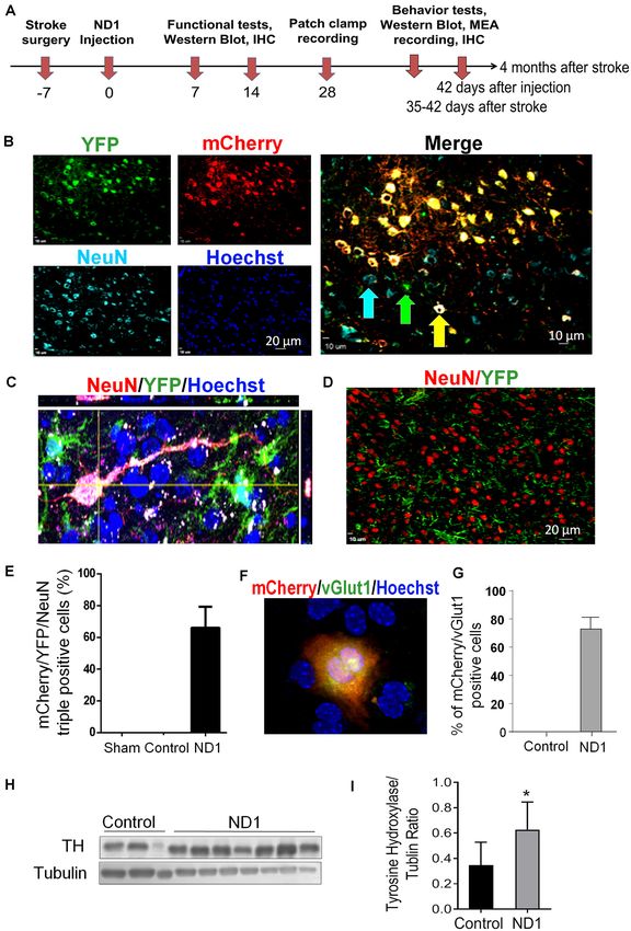
Jiang et al.                                                                                                Conversion of Astrocyte to Neurons After Stroke

INTRODUCTION                                                                        Based on the efficiency and efficacy of glial cell
                                                                                reprogramming, we and others experimented with several
Ischemic stroke is a devastating disease with limited therapies                 combinations of transcription factors and settled on the use of
available (Liu et al., 2014; Ginsberg, 2016; Catanese et al.,                   the single neural transcription factor NeuroD1 (Guo et al., 2014;
2017). Stem cell transplantation has emerged as a promising                     Gangal et al., 2017; Jiang et al., 2019b; Chen et al., 2020; Liu
regenerative therapy for stroke due to its potential for                        et al., 2020). Targeting astrocytes for neuronal reprogramming
repairing damaged brain structures and improving functional                     with different viral vectors has been tested in several animal
recovery (Liu et al., 2014; Wei et al., 2017). However, cell                    models of neurodegenerative diseases including ischemic stroke
transplantation therapies face multiple obstacles including                     with varying success (Guo et al., 2014; Rivetti di Val Cervo
the hosts’ immune systems, poor transplanted cell survival,                     et al., 2017; Yamashita et al., 2019; Chen et al., 2020; Yavarpour-
inappropriate migration/homing and differentiation, and the                     Bali et al., 2020). The exploration of this approach in animal
lack of specificity or integration into endogenous brain networks               disease models is at an early stage. The efficacy of neuronal
(Liu et al., 2014; Wei et al., 2017). Some clinical trials have also            conversion and its contribution to neuronal circuitry repair,
reported inconsistent results in the efficacy of cell transplantation           the mechanisms involved in the regenerative process, and the
therapies (Hatakeyama et al., 2020).                                            functional benefits of this therapy have not been well defined.
    In the adult brain, neurogenesis mainly exists in two                       Concurrently, the specificity, reliability, and clinical potential of
regenerative niches: the subventricular zone (SVZ) and                          this novel regenerative gene therapy have sparked much debate.
the sub-granular zone (SGZ; Ohab and Carmichael, 2008).                         Investigations spanning various routes of administration and
Unfortunately, endogenously generated cells are insufficient for                in animal models across the lifespan have yielded inconsistent
enacting meaningful repair of damaged brain structures (Gogel                   results (Gresita et al., 2019). Our investigation examined the
et al., 2011). On the other hand, glial cells such as astrocytes                viability of reprogramming of astrocytes in vitro and in vivo.
exhibit great proliferative capacity (Khakh and Sofroniew, 2015).               Reprogramming therapy was tested in a focal ischemic stroke
Astroglia are normally responsible for maintaining physiological                model of rats, induced by occlusion of distal branches of
homeostasis and supporting surrounding tissues by providing                     the middle cerebral artery (MCA) accompanied with partial
structural, trophic, and metabolic support to neurons while                     reperfusion (Wei et al., 1995; Jiang et al., 2017). This focal
also playing a role in modulating synaptic activity (Chen and                   ischemic stroke represents common clinical cases of relatively
Swanson, 2003; Gibbs et al., 2008). Resident astrocytes in the                  small infarction followed by spontaneous or thrombolysis-
brain remain mitotic throughout the lifespan and undergo                        induced partial recirculation (Hakim et al., 1987; Jorgensen
rapid gliosis in response to injury. This characteristic response               et al., 1994; Neumann-Haefelin et al., 2004). The well-defined
provides a rich source of cells adjacent to the site of injury                  structure-function relationship of the damaged sensorimotor
(Liu and Chopp, 2016). After an ischemic insult, astrogliosis                   cortex is suitable for specific functional assessments during
initially confers neuroprotective effects by forming a barrier to               the acute, subacute, and chronic stages of stroke. After a
limit toxic substances (Pekny and Nilsson, 2005). As the gliotic                stroke, we transduced ND1 using a lentivirus vector rather
tissue persists in subacute and chronic stages, the formation                   than other viral serotypes such as an adeno-associated virus
of a glial scar in the peri-infarcted region acts as physical and               (AAV) to preserve finer control over the scope of infection
chemical barriers that release inhibitory paracrine factors which               to study the mechanics of reprogramming on local circuitry
limit neuro-regeneration in and around the ischemic region                      and to limit the therapy to only the injured tissue. Neuronal
(Sofroniew and Vinters, 2010; Pekny and Pekna, 2016). Thus, the                 network repair and functional recovery were confirmed using
accumulation of reactive astrocytes and astrogliosis during later               comprehensive assessments and behavioral tests up to 4 months
phases after stroke is viewed as a major obstacle for regenerative              after stroke.
therapy involving either endogenous or transplanted cells.
    Several master transcription factors such as Neurogenic                     MATERIALS AND METHODS
differentiation 1 (NeuroD1 or ND1), Neurogenin2 (Ngn2),
Olig2, and Ascl1 have been identified for their role in controlling             Animals
cell fate (Heinrich et al., 2010; Li and Chen, 2016). These                     A total of 72 C57BL/6 adult mice (male, 26–28 g, n = 6–8 per
discoveries provide a breakthrough opportunity to utilize                       experimental group) were used in this investigation. For in vitro
endogenous and autologous cell substrates for regenerative                      direct reprogramming experiments, we dissected astrocytes from
therapy. More recently, the idea of direct reprogramming of                     P1 C57 mouse pups. The near-pure astrocyte (>98%) cultures
non-neuronal cells allows for the trans-differentiation of glial                featured little contamination by other cell types (Choi et al.,
cells (astrocytes, microglia, and oligodendrocytes) into induced                1987). Astrocyte to neuron reprogramming using a GFAP
neurons (iNeurons) without passing through a stem cell stage                    promoter creates a closed-loop feedback system whereby initial
(Vignoles et al., 2019; Chen et al., 2020). Theoretically, this is a            translation of ectopic ND1 is high in hypertrophied reactive
more efficient way to obtain desirable endogenous neurons from                  astrocytes but diminishes as the cell reprograms into neurons
a large cellular pool for ‘‘on-site’’ repair in the brain (Torper et al.,       (Choudhury and Ding, 2016). For in vivo experiments, we
2013; Vignoles et al., 2019). Being post-mitotic cells, iNeurons are            crossed GFAP-Cre with Rosa-YFP mice to generate animals
not tumorigenic, which offers another advantage over naïve and                  that exhibit constitutively active YFP expression in astrocytes
differentiating stem cells.                                                     regardless of cell phenotype changes; this property allowed

Frontiers in Aging Neuroscience | www.frontiersin.org                       2                                    March 2021 | Volume 13 | Article 612856
Jiang et al.                                                                                             Conversion of Astrocyte to Neurons After Stroke

tracking the cell fate of reprogrammed cells from start to finish.       Ngn2-BamHI-F                50 -GGG GGATCC ATGTTCGTCA AATCTGAGA-30
In these mice, Cre recombinase expression is controlled by a             Ngn2-KpnI-myc-EcoRI-R       50 -CCCGAATTCT CACAGATCCT CTTCAGAGAT
GFAP promoter to inverts a loxP-flanked YFP in astrocytes. The                                       GAGTTTCTGC TCGGTACCGA TACAGTCCCT
resulting YFP reporter is both astrocyte-specific and remains                                        GGCGAGGG-30
                                                                         ND1-BamHI-F                 50 -GGTGCCTTGCTATTCTAAGACGC-30
activated independently of cell fate (McLellan et al., 2017).            ND1-EcoRI-R                 50 -GCAAAGCGTCTGAACGAAGGAG-30
When astrocytes are reprogrammed into induced neurons,
                                                                         Note: F, Forward primer; R, Reverse primer.
they no longer express GFAP but continue to express the
YFP promoter while preexisting neurons do not express YFP.
The specificity of Cre recombinase under a human GFAP                    Generate ND1 Plasmid by Cloning
promoter in mice has been thoroughly characterized. Specificity          ND1 Under GFAP Promoter With mCherry
defined as the proportion of co-labeled S100β and reporter               Tag Into the FUGW Plasmid
positive cells over the total number of reporter positive                Lentivirus has been established as an effective method of
cells in the mouse has been reported as high as 96.02%                   transduction both in vitro and in vivo (Naldini et al., 1996;
(Park et al., 2018).                                                     Blömer et al., 1997; Taoufik et al., 2007). Under a GFAP
    The animal protocol (DAR 2003027) was approved by                    promoter, we packaged control GFP-FUGW and ND1-FUGW
the Institutional Animal Care and Use Committee (IACUC)                  into a lentiviral vector. HEK 293FT cells were used as the
of Emory University School of Medicine. Animal procedures                substrate for lentiviral production (Tiscornia et al., 2006;
followed institutional guidelines that meet NIH standards of             Ansorge et al., 2009). We achieved a viral titer of at
principles of laboratory animal care (NIH publication No. 86-            least 2.48 × 10⁷ which was suitable for use in vitro and
23). Animals were randomly assigned to different experimental            in vivo. The virus was harvested from culture media two
groups and data were analyzed under blinded conditions.                  times resulting in 144 ml total. To reach a high titer for
                                                                         in vivo use, the virus was concentrated using two rounds of
NeuroD1 Lentiviral Production,                                           ultracentrifugation to concentrate first from 144 to 1.2 ml
                                                                         and then from 1.2 to 30 µl. Phage titer was calculated by
Purification, and Titer Calculation                                      infecting triplicate HEK 293FT cell cultures with 1:10 and
To express genes together with mCherry using a single plasmid,           1:100 dilutions of concentrated virus and then fixed and stained
pEGIP was modified by replacing the GFP-IRES-Puro sequence               for mCherry.
with BamHI-EcoRI-IRES-mCherry-WPRE (EIMW). pEGIP was
a gift from Linzhao Cheng (Addgene plasmid #26777; Zou
et al., 2009). The mCherry tag was ligated to FUGW using                 Astrocyte Cultures and Lentivirus Infection
two-step overlap PCR. A purified ND1 fragment was ligated                Astrocytes were dissected and cultured as described (Li
into the FUGW plasmid. Verification of correct ligation and              et al., 2013). Briefly, cerebral cortex from postnatal day
plasmid generation was confirmed using PCR and DNA                       1 (P1), and cells were cultured in a medium consisting
sequence analysis. Plasmid production utilized Stbl3 bacteria            of Dulbecco’s Modification of Eagle’s Medium (DMEM,
and DNA was purified using Qiagen Miniprep and Maxiprep                  Corning, Manassas, VA, USA), 15% FBS (Sigma), MEM
kits. Transduction efficacy was assayed compared to control              non-essential amino acids (Life Technologies), 3.5 mM glucose
GFP-FUGW in HEK 293FT cell cultures that were fixed and                  (Sigma). One-week post-plating, cells were dissociated using
stained for both mCherry and ND1. To prevent interference                trypsin-EDTA (Life Technologies) and passaged onto poly-
from the mCherry reporter with ectopic ND1 translation,                  D-lysine (Sigma) and laminin (Sigma) coated coverslips
an internal ribosome entry site (IRES) preceding mCherry                 (80,000 cells per well for 24-well plates) in the same
ensures that it is translated separately from ND1 (Martinez-             medium. A 4-day-long shaking procedure helped to remove
Salas et al., 2001). The resulting ND1 and mCherry products              microglia and neurons. By the end of this regimen, astrocyte
are independent and able to be transported throughout the cell           activation and GFAP expression were induced uniformly
separately. While both proteins are translated in the cytoplasm,         across the culture by the addition of lipopolysaccharide
transcription factors such as ND1 contain nuclear localization           (LPS). Neurons contained in initial cultures died within
signals while mCherry remains throughout the cytoplasm                   10 days in culture. GFAP-ND1 expression is reliant on
(Petersen et al., 2002).                                                 GFAP-positive astrocytes of the reactive state. We confirmed
    Cloning experiments were performed in triplicates. A                 that 98.4% of the cells were positive for GFAP in our P10
minimum of eight samples were required during cloning                    astrocyte cultures.
steps and three bacterial strains were sequenced to isolate a               Lentivirus was added into astrocyte cultures immediately after
mutation-free strain. Of these, two contained point mutations,           passage. One day post-transduction, the medium was completely
and one was mutation-free. All subsequent experiments                    replaced to minimize exposure to viral adjuvants into a
used the mutation-free clone. In transduction experiments,               medium consisting of DMEM/F-12 (Life Technologies), 3.5 mM
three wells of HEK-293 cells were used to examine efficacy.              glucose, penicillin/streptomycin (Life Technologies), B27 (Life
DNA concentrations were assessed using Gen5 ultraviolet                  Technologies), and 20 ng/ml brain-derived neurotrophic factor
spectrophotometry by BioTek (Naldini et al., 1996). Plasmids             (BDNF, Sigma). After an initial media change, half-media
were isolated using the following primers:                               changes occurred every 3–4 days subsequently.

Frontiers in Aging Neuroscience | www.frontiersin.org                3                                        March 2021 | Volume 13 | Article 612856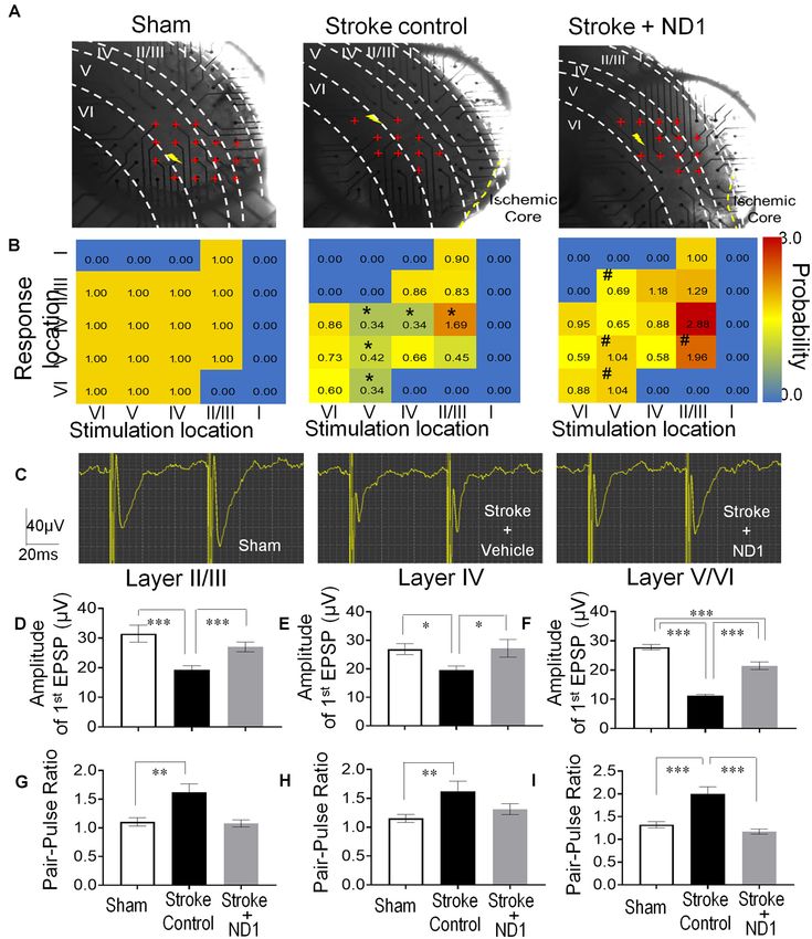
Jiang et al.                                                                                       Conversion of Astrocyte to Neurons After Stroke

Electrophysiological Examination of                                     incomplete (spontaneous and post-thrombolytic) recanalization
                                                                        after an ischemic attack occurs in 30–70% of clinical cases
ND1 Converted Cells In vitro                                            at different times after the onset of ischemia (Hakim et al.,
Astrocyte cultures from 14 to 42 days in vitro after
                                                                        1987; Jorgensen et al., 1994; Barber et al., 1998; Neumann-
ND1 transduction were examined using whole-cell patch-
                                                                        Haefelin et al., 2004). Few animal models of ischemic stroke
clamp recordings on converted cells identified by the
                                                                        featuring partial reperfusion have been available. In this regard,
morphological phenotype of axonal outgrowth with neurite
                                                                        the investigation on this stroke model possesses high face validity
extension. Recordings were performed using an EPC9 amplifier
                                                                        for translational research.
(HEKA; Elektronik) were performed at 21–23◦ C. The external
                                                                            Body temperature was monitored during surgery and
solution contained (in mM): 135 NaCl, 5 KCl, 1 MgCl2 , 2 CaCl2 ,
                                                                        recovery period using a rectal probe and maintained at 37◦ C
10 HEPES, and 10 Glucose at a pH of 7.4. Recording electrodes
                                                                        on a homoeothermic blanket in a ventilated incubator. Overall
pulled from borosilicate glass pipettes (Sutter Instrument) had a
                                                                        mortality resulting from ischemic stroke surgery was less than
tip resistance between 5 and 8 MO when filled with the internal
                                                                        2%. Before and after surgery the painkiller meloxicam was
solution (in mM): 140 KCl, 2 MgCl2 , 1 CaCl2 , 2 Na2 ATP,
                                                                        administered orally at a dosage of 5 mg/kg. Animals were housed
10 EGTA, and 10 HEPES at a pH of 7.2. Action potentials were
                                                                        with four to five mice per cage, with ad libitum access to food and
recorded under the current-clamp mode using the Pulse software
                                                                        water. At different time points after stroke, mice were sedated
(HEKA, Elektronik).
                                                                        with overdose isoflurane and sacrificed by decapitation.

MicroElectrode Array (MEA) Recording in                                 Stereotaxic Injection
Brain Slices                                                            Stereotaxic injection of control and ND1 lentivirus was
A high-resolution MEA2100-system (MultiChannel Systems,                 performed using a 10 µL Hamilton GASTIGHTTM syringe
Reutlingen, Germany) was used to record field excitatory                (Hamilton Company, NV). Injection locations included three
postsynaptic potentials (fEPSP) in brain slices. The MEA                areas around the infarcted region (Stereotaxic coordinates: AP
chamber (60pMEA200/30iR-Ti, MultiChannel Systems GmbH,                  −0.5, ML +3.2, DV +2.2) at a cortical depth of 0.5 mm and the
Reutlingen, Germany) is composed of a 6-mm high glass ring              sub-ventricular zone (SVZ; Stereotaxic coordinates: AP 0, ML
and an 8 × 8 Titanium nitride electrode grid (59 electrodes and         +1.5, DV +2.5). Infected cells could be observed up to a distance
1 internal reference electrode) with an electrode diameter of 30        of 3–4 mm from the injection site.
µm and spacing of 200 µm. The brain slice was transferred to the            At time points of 24 and 48 h after the onset of
MEA chamber that was perfused with oxygenated aCSF at a rate            MCAO, animals were sacrificed for assessment of brain
of 6–8 ml/min and stabilized at 34◦ C for at least 10 mins before       infarct formation. 2,3,5-triphenyltetrazolium chloride (TTC;
recording. For evoked fEPSPs, electric stimuli (±1.5 V, 10 ms)          Sigma–Aldrich) staining was used to reveal damaged/dead brain
were applied every 30 s and responses were simultaneously               tissue as previously described (Wang et al., 2014). Brains were
monitored in 58 locations. Pair-pulse facilitation was recorded         removed and placed in a brain matrix then sliced into 1-mm
by two electric stimuli (±1.0 V, 10 ms) with different intervals        coronal sections. Slices were incubated in 2% TTC solution
(20, 40, 60, 80, 100, 200 ms). The slopes of fEPSPs were                at 37◦ C for 5 min, then stored in 10% buffered formalin for
analyzed with Multi-Channel Analyzer V 2.6.0 (Multichannel              24 h. Digital images of the caudal aspect of each slice were
Systems) and GraphPad Prism 6 (GraphPad Software, San Diego,            obtained by a flatbed scanner. Infarct, ipsilateral hemisphere,
CA, USA).                                                               and contralateral hemisphere areas were measured using ImageJ
                                                                        software (NIH, Bethesda, MD, USA). The indirect method
Focal Ischemic Stroke in Mice                                           (subtraction of residual right hemisphere cortical volume from
A focal cerebral ischemic stroke targeting the right sensorimotor       cortical volume of the intact left hemisphere) was used for infarct
cortex was induced as previously described (Choi et al., 2012;          volume calculation. Infarct measurements were performed under
Li et al., 2013). Mice were anesthetized with 3% isoflurane             double-blind conditions.
and maintained using 1.5% isoflurane supplemented with
regular air during surgery. Cortical ischemia was achieved              Western Blotting Analysis
by permanent occlusion of the distal branches of the right              Western blot analysis was used to detect the expression of trophic
middle cerebral artery (MCA) supplying the sensorimotor cortex.         factors brain-derived neurotrophic factor (BDNF), FGF10, PSD-
The MCA occlusion was paired with 7-min ligation of both                95, tyrosine hydroxylase (TH), NFKB, and GFAP (n = 3–6).
common carotid arteries (CCAs) to cause sufficient reduction            Brain cortical tissue was lysed in a lysis buffer containing
of local cerebral blood flow (LCBF) in the sensorimotor                 0.02 M Na4 P2 O7 , 10 mM Tris-HCl (pH 7.4), 100 mM NaCl,
region and followed by partial reperfusion. This relatively             1 mM EDTA (pH 8.0), 1% Triton, 1 mM EGTA, 2 mM
small stroke targets a well-defined brain structure, i.e., the          Na3 VO4 , and a protease inhibitor cocktail (Sigma–Aldrich). The
sensorimotor cortex including the barrel cortex (Wei et al.,            supernatant was collected after centrifugation at 15,000 g for
1995; Jiang et al., 2017). According to the epidemiological             10 min at 4◦ C. Protein concentration was determined with a
data from American Heart Association (AHA), small strokes               bicinchoninic acid assay (Pierce Biotechnology, Rockford, IL,
are common and represent about 40% of all stroke cases                  USA). Equivalent amounts of total protein, 15–20 µl were
(Roger et al., 2011). Importantly, partial reperfusion due to           added per lane, were separated by molecular weight on an

Frontiers in Aging Neuroscience | www.frontiersin.org               4                                   March 2021 | Volume 13 | Article 612856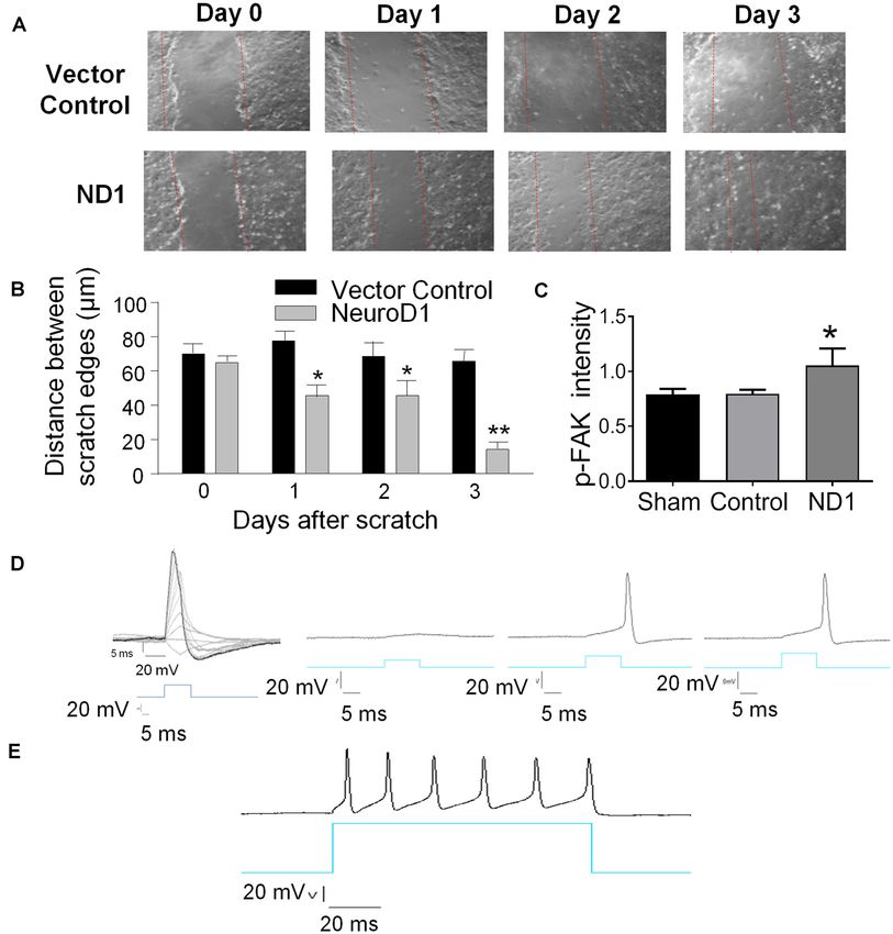
Jiang et al.                                                                                        Conversion of Astrocyte to Neurons After Stroke

SDS-polyacrylamide gradient gel, and then transferred to a                  For both ICC and IHC, on the next day, the samples were
polyvinyl difluoride (PVDF) membrane. The blot was incubated             washed three times with PBS for 5 min, then reacted with the
in 5% bovine serum albumin (BSA) for at least 1 h and                    secondary antibodies Alexa Fluorr 488 goat anti-mouse or
then reacted with primary antibodies at 4◦ C for overnight.              rabbit (1:300; Life Technologies, Grand Island, NY, USA)
The primary antibodies used in this investigation included:              and Cy3-conjugated donkey anti-rabbit (1:300; Jackson
anti-BDNF antibody (1:2,000; Cell Signaling, Danvers, MA,                ImmunoResearch Laboratories, West Grove, PA, USA) or
USA), anti-FGF-10 antibody (1:500, Abcam, Cambridge, MA,                 Cy5-conjugated donkey anti-mouse or rabbit (1:400; Jackson
USA), anti-PSD-95 (1:750, Abcam), anti-TH antibody (1:1,000;             ImmunoResearch Laboratories) for 80 min at room temperature.
Cell Signaling, Danvers, MA, USA), anti-FGF-10 antibody                  After three washes with PBS, nuclei were stained with Hoechst
(1:500, Abcam), anti-GFAP antibody (1:500, Abcam), rabbit                33342 (1:20,000; Molecular Probes, Eugene, OR, USA) for
anti-actin antibody (1:500, Abcam), rabbit anti-beta tubulin             5 min as a counterstain; and then the brain sections were
antibody (1:500, Abcam) and rabbit anti-cleaved caspase-3                mounted, coverslipped, imaged, and photographed under a
(1:500; Cell Signaling). After washing with Tris-buffered                fluorescent microscope (BX51, Olympus, Japan) or a laser
saline with Tween (TBST), membranes were incubated with                  scanning confocal microscope (Carl Zeiss Microimaging, Inc.,
AP-conjugated or HRP-conjugated secondary antibodies (GE                 Thornwood, NY, USA).
Healthcare, Piscataway, NJ, USA) for 1–2 h at room temperature.             For H&E, brains were sectioned with a vibratome and coronal
After final washing with TBST, the signals were detected with            sections (20 µm) of mouse brains were collected from the
bromochloroidolylphosphate/nitroblue tetrazolium (BCIP/NBP)              sensorimotor cortex (brains damaged during removal were not
solution (Sigma–Aldrich) or film. Signal intensity was measured          sectioned). Every 6th section and a total of 10 sections per brain
by ImageJ and normalized to the actin signal intensity.                  were selected for H&E staining from the anterior start to the
                                                                         posterior end of the injury site.
Immunocytochemistry and
Immunohistochemical Staining                                             Astroglia Scar Measurement
For immunocytochemistry (ICC), cell cultures were fixed using            Glial scar measurements were taken using ImageJ against GFAP
10% formalin buffer, washed with −20◦ C precooled ethanol:               immunofluorescence contiguous to the ischemic core. The
acetic acid (2:1) solution for 10 min, and finally permeabilized         thickness of the glial scar was measured using the average
with 0.2% Triton-X 100 solution for 5 min. All slides were               distance across hypertrophied astrocytes lining the ischemic core
washed three times with PBS (5 min each) after each step. Then,          based on a method previously described (Cai et al., 2017). GFAP
tissue sections were blocked with 1% fish gelatin (Sigma–Aldrich)        area and intensity were determined using the ImageJ analysis of
in PBS for 1 h at room temperature, and subsequently                     the area of GFAP-positive fluorescence in the glial scar region
incubated with the primary antibody: mouse anti-Tuj1 (1:500;             respectively. Median GFAP intensity was measured using a
Covance/Biolegend, CA, USA), rabbit synaptophysin (1:500;                100-pixel width rectangle across six different regions of the glial
Ab32127, Abcam, Cambridge, MA, USA), rabbit anti-synapsin I              scar to measure median GFAP intensity across the area.
(1:500; Millipore, Billerica, MA, USA), and rabbit anti-mCherry
(1:400; Abcam) overnight at 4◦ C.                                        Cell Death Assessment
    For immunohistochemistry (IHC), frozen brain tissues were            A terminal deoxynucleotidyl transferase dUTP nick end labeling
sliced into 10 µm-thick coronal sections using a cryostat                (TUNEL) assay kit was used to examine cell death by detecting
vibratome (Leica CM 1950; Leica Microsystems, Buffalo Grove,             fragmented DNA in 10-µm-thick coronal fresh frozen sections
IL, USA). The sections were dehydrated on a slide warmer                 as described previously (Lee et al., 2014). After fixation (10%
for 30 min, fixed with 10% formalin buffer, washed with                  buffered formalin for 10 min and then ethanol:acetic acid
−20◦ C precooled ethanol: acetic acid (2:1) solution for 10 min,         (2:1) solution for 5 min) and permeabilization (0.2% Triton
and finally permeabilized with 0.2% Triton-X 100 solution                X-100 solution), brain sections were incubated in equilibration
for 5 min. All slides were washed three times with PBS                   buffer for 10 min. Recombinant terminal deoxynucleotidyl
(5 min each) after each step. Then, tissue sections were                 transferase (rTdT) and nucleotide mixture were then added
blocked with 1% fish gelatin (Sigma–Aldrich) in PBS for                  on the slide at 37◦ C for 60 min in the dark. Reactions
1 h at room temperature, and subsequently incubated with                 were terminated by 2× SSC solution for 15 min. Nuclei
the primary antibody: mouse anti-NeuN (1:400; Millipore,                 were counterstained with Hoechst 33342 (1:20,000; Molecular
Billerica, MA, USA), rabbit anti-ND1 (1:400; Millipore, Billerica,       Probes) for 5 min. Cell counting was performed following
MA, USA), chicken anti-GFAP (1:400; Abcam), mouse anti-                  the principles of design-based stereology (Lee et al., 2014).
iba-1 (1:400; Sigma–Aldrich), and rabbit anti-Beclin (1:5,000;           Systematic random sampling was used to ensure accurate
Abcam, Cambridge, MA, USA), goat anti-collagen type IV                   and non-redundant cell counting. Eight brain sections per
(1:400; Millipore), rabbit anti-mCherry (1:400; Abcam), rabbit           animal were collected at 90 µm distance between sections for
anti-vGluT1 (1:500; Abcam), rabbit anti-GAD67 (1:500; Abcam),            non-overlapping multistage random sampling. For each animal,
rabbit anti-Iba1 (1:400; Abcam), rabbit anti-vGluT1 (1:500;              6 ‘‘areas of interest’’ regions per slide were selected. Each
Abcam), mouse anti-GFP (1:500; Sigma–Aldrich), Oct4 (1:250,              field was scanned at 200× magnifications for cell counting.
Cell Signaling, Danvers, MA, USA) and Klf4 (1:500, Abcam)                ImageJ (NIH) was used to analyze each picture. All experiments
overnight at 4◦ C.                                                       were performed in a blinded fashion, so the data collector

Frontiers in Aging Neuroscience | www.frontiersin.org                5                                   March 2021 | Volume 13 | Article 612856
Jiang et al.                                                                                           Conversion of Astrocyte to Neurons After Stroke

and data analysis were performed without knowledge of                      during the earlier mobility bouts also are not counted as mobility.
experimental groups.                                                       The same blinded observer was used to assess mobility in all
                                                                           experiments (Ge et al., 2012; Tsai et al., 2012).
Functional and Behavioral Tests
Rotarod Test                                                               Experimental Design and Statistical Analysis
Rats were placed on an accelerating rotarod cylinder (Economex,            Significant time and effort were initially spent to test several
Columbus In., Columbus, OH, USA) and the length of time the                master transcription factors including NeuroD1 (ND1),
animals remained on the rotarod was measured. The speed was                Neurogenin2 (Ngn2; Supplementary Figure 1, Gangal et al.,
slowly increased from 4 to 40 rpm in 5 min. The animals were               2017; Jiang et al., 2019a,b), and Ascl1 to identify effective
trained for 3 days before stroke surgery. The mean duration on             methods to efficiently carry out glia to neuron reprogramming.
the device was recorded with three measurements (Wittenberg                ND1 was eventually selected based on more than 2 years of
et al., 2007; Spurlin and Nelson, 2017).                                   in vitro and in vivo experiments and verification of combinations
Corner Test                                                                as well as individual applications of these factors. In cell culture
The corner test monitors unilateral whisker deficits. Mice were            experiments, at least three batches of cultures were tested per
allowed to roam freely in a star-shaped arena with 30◦ angles.             experiment, and the purity of astrocyte cultures was ensured
Upon entering a corner, the mouse would rear and turn toward               by both GFAP staining and specific culture media/protocols.
the wall, which was sensed by intact whisker sensations. In                Multiple examinations were performed to independently
our right sensorimotor cortex ischemic stroke model, mice lost             replicate the virus infection efficacy and conversion efficiency,
sensation in their left whiskers. The percentage of right turns was        dose-response relationship, and cell phenotypes. In stroke
measured before and after stroke (Calabresi et al., 2003; McCrary          animal experiments, the well-characterized focal ischemic stroke
et al., 2019).                                                             model with partial reperfusion represents common clinical cases
                                                                           (Wei et al., 2017; Zhong et al., 2020) and suitable for functional
Forced Swimming Test                                                       and behavioral tests. A specialized researcher with extensive
The forced swim test (FST) was carried out to measure depressive           rodent microsurgery training performed all ischemic surgery
behaviors. Mice were dropped individually into a plexiglass                to ensure highly consistent ischemic insult in the sensorimotor
cylinder (height: 30 cm, diameter: 22.5 cm) filled with water to           cortex. Another experienced researcher performed all virus
a depth of 15 cm and maintained at 23–25◦ C. In this test, after           injection procedures to ensure consistent virus infections in the
the initial vigorous activity of 2 min, mice acquired an immobile          same brain region. The neuronal phenotype of reprogrammed
posture which was characterized by motionless floating in the              cells was confirmed by at least two specific markers and the
water and made only those movements necessary to keep the                  co-localization of cellular markers was examined by 3-D
head above water. The duration of immobility was recorded in               microscopy. We identified mature neurons not only rely on
seconds during the last 4 min of the 6 min test. All mice received         morphology and cell markers but also functional activity using
a 15-min training session under similar conditions 24 h before             electrophysiological recordings. In cell counting analysis, we
the formal test (Kronenberg et al., 2014; Frechou et al., 2019).           followed stereology principles by separating analyzed by at least
Sucrose Preference Test                                                    100 µm to avoid double counting cells. In these experiments,
The mice were given access to both water and a sucrose solution,           sample sizes were six to eight per group and at least six sections
and their preference for the sucrose solution was quantified.              were examined for each brain. In whole-cell recordings,
Briefly, mice were deprived of food and water for 20 h. One bottle         over 20 cells per group were inspected; MEA recordings
of water and one containing 1% sucrose were simultaneously                 examined multiple brain slices from three to four brains per
placed in the cages and were freely accessible to the mice for 3 h.        group. Functional and behavioral tests were performed using
The position of the two bottles (left or right side of the cage) was       6–14 animals per group, depending on the outcomes of Power
varied randomly from trial to trial. The volume of each liquid             analysis and our previous investigations. Data analysis was
was measured before and after each trial, and sucrose preference           performed under blinded or double-blinded conditions.
was calculated according to the following equation: sucrose                    All data were analyzed for normality using the D’Agostino
preference = (sucrose consumption)/(sucrose consumption +                  and Pearson omnibus normality test (where n > 7) and
water consumption) × 100 (Heins et al., 2002; Grealish et al.,             Kolmogorov–Smirnov normality test (where n < 7). Unpaired
2016).                                                                     Student’s t-test or Fisher’s test were used for pairwise
                                                                           comparisons. Comparisons of multiple groups were analyzed
Tail Suspension Test (TST)                                                 using one- or two-way ANOVA followed by post hoc Tukey’s test
Mice were taped to a crossbar by their tails and their behavior            or other tests as indicated in figure legends. In these statistical
was recorded for 10 min. In this test, after the initial vigorous          reports, the variance between populations was tested using
activity of 2 min, mouse behavior was during an analysis period            Bartlett’s test, F-factor, and p-values are provided. All results are
of 6 min. The duration of immobility was recorded in seconds               expressed as Mean ± SEM Statistical comparisons were finished
during the time. In our laboratory, small movements that are               using Graph Pad Prism 6 (Graph Pad Software, Inc., San Diego,
confined to the front legs but without the involvement of the              CA, USA). In general, p < 0.05 was considered significant for all
hind legs are not counted as mobility. Additionally, oscillations          comparisons; the exact p-value is reported for some important
and pendulum-like swings that are due to the momentum gained               comparisons as a reference for readers.

Frontiers in Aging Neuroscience | www.frontiersin.org                  6                                    March 2021 | Volume 13 | Article 612856
Jiang et al. Conversion of Astrocyte to Neurons After Stroke
RESULTS Western blot analysis revealed that the phosphorylated focal
adhesion kinase (p-FAK), a key mediator in cell adhesion and
Among single transcription factor-mediated direct migration, was significantly increased (Figure 2C).
reprogramming candidate genes, both ND1 and In electrophysiological examinations, the whole-cell
Ngn2 demonstrated an ability to reprogram reactive astrocytes recording was performed in mCherry-positive converted
but ND1 was selected based on both efficiency and efficacy cells 28 days after ND1 transduction. In the current-clamp
(Supplementary Figure 1). A mCherry tagged ND1 lentivirus mode, gradually increased membrane depolarization triggered
was generated under a mouse GFAP promoter and used for both the firing of action potentials (Figure 2D). We patched a total
in vitro and in vivo transduction. A viral titer of GFAP-ND1 was of 10 mCherry positive cells with neuronal morphology; all cells
calculated to be approximately 4.8 × 10⁷ units/µl by using a exhibited sodium and potassium currents and action potentials
107 dilution of virus that yielded ∼92% infection in HEK293FT upon membrane depolarization. Prolonged depolarization
cells. Astrocyte cultures were infected at 7 days in vitro (DIV generated firing of repetitive action potentials, resembling
7) and followed for up to 8 weeks post-infection to examine functional activities of neuronal cells (Figure 2E).
cell fate and efficiency of transduction and conversion. After
lentiviral transduction, successfully infected cells could be Focal Ischemic Stroke in Mice and
monitored over time by the expression a mCherry reporter mCherry-ND1 Lentivirus Injection
carried by the ND1 virus (Figures 1A–D). Infected cells began to Focal ischemic stroke of the right sensorimotor cortex was
adopt neuronal features around 2 weeks following transduction, induced in adult GFAP-Cre × Rosa-YFP mice. Astrocytes in
including the extension of one or more long processes that this mouse express the YFP reporter regardless of cell phenotype
are uncharacteristic of astrocytes (Figure 1C). While neuronal changes. This property is favorable for tracking astrocytes as they
axonal processes are often elongated beyond the length of the cell switch between inter-lineage phenotypes. After ischemic stroke,
body (>100 µm), astrocytes are characterized by multiple shorter robust astrocyte accumulation took place in the peri-infarct
and spread-out processes. Staining using the neuronal lineage region, and the ischemic core gradually forms a tissue cavity
market Tuj1 verified this morphological difference between 2–3 weeks later (Kanekar et al., 2012; Pivonkova and Anderova,
mCherry-positive astrocytes and immature neuron-like cells 2017). Proliferating astrocytes around the damaged tissue
by morphological examination (Figures 1D,H). On the other provided an abundant cell supply for reprogramming. Lentivirus
hand, astrocytes infected with the empty vector did not exhibit containing mCherry-ND1 was injected into the peri-infarct
any of these alterations (Figure 1E). The observed cell lineage region 7 days after ischemia, this time point was selected to avoid
switch from ectoderm to endoderm is a time-dependent process. the acute phase when astrocytes are thought to be protective
By 4 weeks, 58.6% of astrocytes were positive for the immature yet the virus transduction still catches hypertrophied astrocytes
neuronal marker Tuj1 (Figure 1F). Consistent with the expected before the formation of a glial scar (Figure 3A).
reduction in cell proliferation and survival rate of converted
cells under culture conditions, the cell numbers in cultures that Reprogramming of Reactive Astrocytes
received ND1 transduction were drastically less than that in and Neuronal Conversion After Ischemic
control cultures infected by the empty vector where astrocytes Stroke
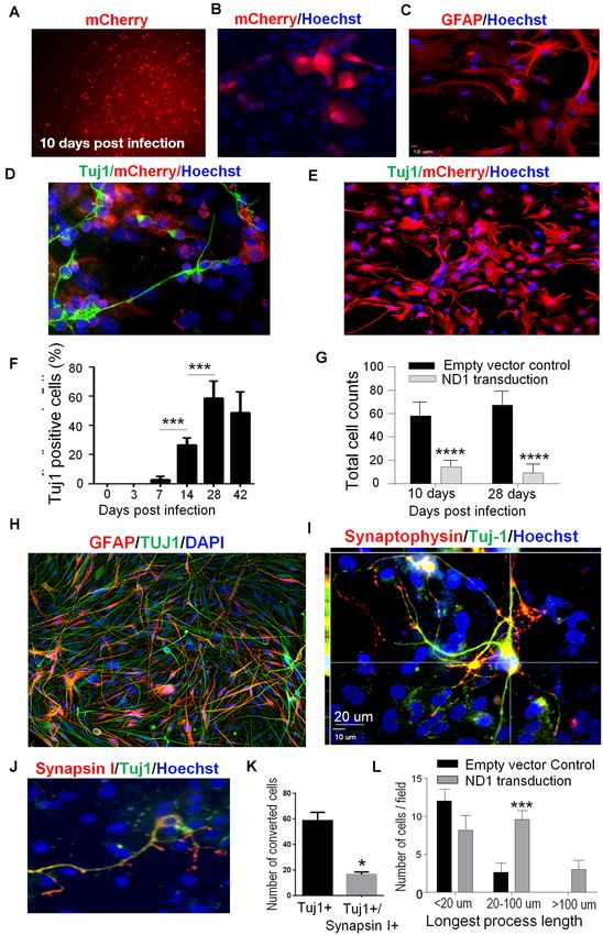
continued to proliferate (Figure 1G). At 28 days after ND1 lentivirus injection to the peri-infarct
region, approximately 10% of total astrocytes were YFP/mCherry
In vitro Conversion of Astrocytes to double-positive cells. We identified cells labeled by YFP
Neuronal Cells (identifying their astrocyte origin), mCherry (infected by
To confirm the maturity of astrocytes-converted iNeurons ND1 lentivirus), and NeuN (mature neuron marker; Figure 3B).
in vitro, we stained converted cells with the synaptic vesicle The combination of these three markers in single cells indicated
protein synaptophysin. As a marker for synapse formation, successful viral-mediated reprogramming of endogenous
synaptophysin expression was detected in converted cells astrocytes to neurons. Co-labeling of NeuN and transduction
(Figures 1H,I). In the next experiment, we co-infected marker YFP in a single converted cell was also confirmed using
ND1 with a synapsin-GFP reporter to monitor cells. confocal microscopy, showing a pyramidal neuronal phenotype
GFP expression emerged at 14 days post-transduction. In with a single axon and dendritic arbors (Figure 3C). In contrast,
co-labeling experiments of Tuj1 and synapsin-1, around 20% cells infected by the empty control vector showed separated
of Tuj1-positive cells expressed the synaptic protein synapsin-1 cell populations of neurons and infected astrocytes, illustrating
(Figures 1J,K). During neuronal maturation, converted high specificity of the viral promoter and no conversion in the
cells developed significantly longer processes (>20–100 µm) absence of ectopic ND1 (Figure 3D). In cell counts, around 66%
compared to cells infected by the control lentivirus, inspected at of infected cells became mCherry/YFP/NeuN triple-positive
6 weeks after transduction (Figure 1L). iNeurons (Figure 3E). Immunohistochemical examination
Interestingly, reprogrammed cells also featured increased revealed that most reprogrammed cells (∼80%) expressed
motility. In an in vitro scratch assay, reprogrammed cells moved vesicular glutamate transporters (vGLUTs), which are the
into the scratched area much faster than vehicle control cells characteristics of glutamatergic neurons (Figures 3F,G). Western
(Figure 2A). Three days later, the scratched area was significantly blotting of the peri-infarct tissue detected a significant increase
smaller compared to the control cultures (Figures 2A,B). in the expression of tyrosine hydroxylase (TH; Figures 3H,I),
Frontiers in Aging Neuroscience | www.frontiersin.org 7 March 2021 | Volume 13 | Article 612856Jiang et al. Conversion of Astrocyte to Neurons After Stroke
FIGURE 1 | In vitro direct reprogramming of astrocytes to induced neurons. Mouse astrocyte cultures were transduced with NeuroD1 (ND1) or empty vector control
and stained for different cell markers. (A) Ten days after lentiviral transduction, successfully infected cells were monitored over time by the expression of the
fluorescent mCherry reporter (red), implicating GFAP-ND1-IRES-Ubi-mCherry expression in these astrocytes. (B–D) Enlarged images of infected cells 14 days after
transduction. Infected cells expressed the reporter gene mCherry (B), GFAP stain revealing an astrocyte-like morphology of astrocytes before reprogramming (C). In
image (D), some cells develop the neuronal lineage marker Tuj1 (green) and adopted either a unipolar or bipolar morphology that is uncharacteristic of astrocytes. (E)
A representative image of control culture with the empty vehicle virus showing the lack of Tuj-1 expression in infected cells (mCherry, red). There were also no
alterations to the morphology of astrocytes. (F) Time course and reprogramming rate of the expression of the immature neuron marker Tuj1 in infected astrocytes up
to 42 days after transduction. One-way ANOVA (F (5,36) = 77.91) followed by Holm–Sidak’s multiple comparisons test. N = 3 independent cell culture batches,
(Continued)
Frontiers in Aging Neuroscience | www.frontiersin.org 8 March 2021 | Volume 13 | Article 612856Jiang et al. Conversion of Astrocyte to Neurons After Stroke
regenerative activities in the peri-infarct region. Some newly
FIGURE 1 | Continued
converted iNeurons (mCherry/YFP/NeuN triple-positive cells)
***p < 0.001. (G) Quantified cell counts of total cell numbers 10 and 28 days
after transduction with ND1 or vector control. The reduced cell number
were even observed inside of the core region where no neuronal
implied attenuated cell proliferation after the ND1 transduction and neuronal cells would normally be expected to survive at this delayed
conversion. Two-way ANOVA (interaction: F (1,10) = 2.377, p = 0.1542; time: post-stroke time point (Figure 5J).
F (1,10) = 0.174, p = 0.6854; treatment: F (1,10) = 358.9, ****p < 0.0001). (H) An
immunostaining image shows coexpression of Tuj1 and GFAP in cultured Astrocyte Reprogramming Improved
astrocytes 6 weeks after transduction. (I) Confocal microscopy of an enlarged
image from 6 weeks post-transduction, showing complex processes that are
Synaptic Transmission in the Peri-infarct
positive for Tuj-1 and synaptophysin colocalized with mCherry. Those cells Region
under the culture condition did not show positivity to NeuN; however, they The MultiElectrode array (MEA) system was used to record
expressed the synaptic protein synaptophysin. (J) At 6 weeks after
evoked extracellular post-synaptic potentials (EPSPs) in brain
transduction, astrocytes undergoing direct reprogramming extended
processes and developed nerve terminals positive to the pre-synaptic protein
slices of sham control, stroke control of empty vector, and stroke
synapsin I (red). (K) The bar graph shows quantified data that at 6 weeks with ND1 transduced mice (Figure 6A). In peri-infarct locations
post-transduction, 58.7% of reprogrammed cells (mCherry-positive) 6 weeks (42 days) after stroke, the responsive area covered by
expressed Tuj-1 and 17.4% of these cells also expressed synapsin 1. n = 3, 59 recording electrodes and the amplitude of EPSPs evoked
paired t-test, *p < 0.05 vs. Tuj1 only cells. (L) Measurements of the length of
by a stimulation electrode were noticeably absent or reduced
extended processes from converted cells. There were drastically increased in
the length of processes of cells received ND1 transduction. N = 3, two-way (Figure 6B). In the similar areas of slices from stroke mice
ANOVA (interaction: F (3,9) = 8.427, ***p < 0.001). that received ND1 transduction, normal or enhanced EPSPs
were detected in multiple locations (Figure 6B). To further
understand changes at the synaptic level, we examined the
which is consistent with the notion that activation of the
synaptic plasticity by paired-pulse facilitation (PPF). In PPF
Wnt/β-catenin pathway and ND1 upregulates TH expression
recordings, the second evoked EPSPs were large than the first
(Jiang et al., 2020). We did not observe the GABAergic neuronal
one in all three experimental groups (Figure 6C). In fact, in
marker glutamic acid decarboxylase 67 (GAD67; data are not
cortical layers II/III, layer IV, and layer V we observed even
shown). Measured at 14 days into the reprogramming process,
greater facilitation in stroke control slices, which was partly
none of the stemness markers Oct4 and Klf4 were detected in
due to a smaller size of the first EPSP in stroke control
transduced (mCherry-positive) cells (Supplementary Figure 2),
slices (Figures 6D–F). ND1 transduction prevented the smaller
supporting that ND1-mediated glia-to-neuron conversion did
initial EPSPs and the abnormally enhanced synaptic facilitation
not pass through an intermediate stem cell stage.
(Figures 6D–I). Similar increased synaptic facilitation has been
Neurotrophic/growth factors such as BDNF and FGF10 play
reported in human stroke patients (Kronenberg et al., 2014).
critical roles in important processes such as cell survival,
Increased synaptic modification such as ischemia-increased
proliferation, and maturation, angiogenesis, neurogenesis, and
long-term potentiation (LTP), has been called ‘‘pathological
wound healing, each of which plays an essential part in the
plasticity’’ (Frechou et al., 2019), which was ameliorated in
regenerative process (Matkar et al., 2017; Spurlin and Nelson,
ND1-treated slices (Figures 6G–I).
2017; Ghosh, 2019). Western blot assay in the peri-infarct region
detected significantly higher levels of BNDF and FGF10 in Astrocyte Reprogramming Improves
ND1 transfected mice compared to animals who received empty Functional Outcomes After Stroke
control vector (Figures 4A,B). Consistently, the ND1 treatment The focal ischemic stroke model results in well-defined motor
significantly attenuated the stroke-induced reduction of the and sensorimotor deficits detectable using the rotarod test and
synaptic protein PSD-95 expression in the post-stroke cortex corner test (Grealish et al., 2016). Three weeks after stroke, the
(Figure 4C). Meanwhile, the inflammatory marker protein time that mice balanced on a rotating beam was significantly
NFκB expression and the number of phagocytic Iba-1-positive shorter compared to sham control mice (Figure 7A). Stroke mice
microglia were significantly lower in the ND1 treated brains than that received the ND1 treatment, however, remained on the beam
in stroke mice that received a control vector (Figures 4D, 5A,B significantly longer than stroke mice received the empty control
and Supplementary Figure 3). vector (Figure 7A). In the corner test, rodents normally make
an equal left and right turn in their exploratory behavior. Stroke
Reprogramming of Reactive Astrocytes animals, however, show biased turns to one direction due to the
and Reduced Glial Scar After Ischemic unilateral injury to the right sensorimotor cortex (Figure 7B).
Stroke Significant improvement in the turning behavior was seen with
In stroke mice that received lentiviral ND1 administration, the stroke mice received the ND1 treatment (Figure 7B). We and
number of YFP-positive cells in the peri-infarct area decreased others have shown that stroke can induce chronically developed
compared to that in the stroke control cortex (Figures 5C,D). phenotypes of post-stroke depression-like behavior (Kronenberg
The relative size, thickness and mean fluorescent intensity of et al., 2014; Frechou et al., 2019; Zhong et al., 2020). To further
the GRAP-positive glial scar were also reduced (Figures 5D–H). characterize long-term behavioral modifications, the sucrose
6 weeks after stroke, Western blot data revealed decreased solution preference test, forced swim test, and tail suspension
GFAP levels in the peri-infarct region of ND1-treated stroke test was performed 4 months after stroke. Stroke control mice
mice (Figure 5I). The reduced astrogliosis appeared to facilitate exhibited obvious depressive-like behaviors in all these tests
Frontiers in Aging Neuroscience | www.frontiersin.org 9 March 2021 | Volume 13 | Article 612856Jiang et al. Conversion of Astrocyte to Neurons After Stroke
FIGURE 2 | Reprogramming induced cellular changes and functional activities in vitro. The ND1-mediated astrocytes reprogramming was monitored in cultures and
functionally characterized. (A,B) Two weeks after ND1 virus or empty control vector transduction, astrocyte cultures were challenged by the scratch test to measure
the motility of reprogrammed cells. From day 1 after the scratch insult, the damaged area recovered significantly faster by migrating cells in cultures infected by
ND1 compared to the vector control culture. N = 3 cell cultures. Two-way ANOVA (Interaction: F (3,24) = 11.15, ∗∗∗ p < 0.0001; Time: F (3,24) = 18.66, ∗∗∗ p < 0.0001;
ND1: F (1,8) = 53.87, ∗∗∗ p < 0.0001) followed by Holm–Sidak’s multiple comparisons test: *p < 0.05 and **p < 0.01 for ND1 vs. empty vector control. (C) Effect of the
scratch test on the expression of migration factor focal adhesion kinase (FAK) in reprogrammed cells. In line with the increased motility 3 days after scratch,
ND1-infected cells within 200 µm of the scratch expressed significantly higher immunofluorescence of phosphorylated FAK (p-FAK). N = 3 per group. One-way
ANOVA, F (2,6) = 7.327, **p = 0.0245 followed by Holm–Sidak’s multiple comparisons test: *p < 0.05 vs. control. (D) mCherry-positive converted cells were subjected
to whole-cell recordings 28 days after infection. In the current-clamp mode, membrane depolarization induced by current injections evoked depolarization generated
action potentials. A hyperpolarization was observed upon the decay phase of the spikes, which is typical for neurons and suggestive of functional potassium channels
in these cells. n = 10. (E) A longer membrane depolarization pulse evoked the firing of a chain of action potentials that are characteristics of functional neurons.
while no significant depressive-like changes were observed in demonstrate the efficiency and the efficacy of the reprogramming
mice that received ND1 treatment (Figures 7C–E). process by ND1 as well as the neuronal activity of converted cells.
Using an established sensorimotor ischemic stroke mouse model,
DISCUSSION we demonstrate that delivery of ND1 to reactive astrocytes in the
peri-infarct region can be achieved in a temporally and spatially
The present investigation presents compelling evidence for the specific manner using a lentiviral vector. The ectopic expression
feasibility and effectiveness of utilizing reactive astrocytes as an of ND1 redirects reactive astrocytes into mature iNeurons,
endogenous cellular source for the generation of neuronal cells while reduced glial scar facilitates regenerative repair. MEA
to repair damaged brain structures. Our in vitro experiments ex vivo recording revealed that ND1-induced reprogramming
Frontiers in Aging Neuroscience | www.frontiersin.org 10 March 2021 | Volume 13 | Article 612856Jiang et al. Conversion of Astrocyte to Neurons After Stroke
FIGURE 3 | Conversion of astrocytes to mature neurons in vivo. Adult GFAP-Cre x Rosa-YFP mice were subjected to a focal ischemic insult to the right
sensorimotor cortex and subjected to control and ND1 treatments. (A) Timeline of in vivo reprogramming experiments. Animals received sham, stroke control, or
ND1 lentivirus injection to the peri-infarct region 7 days after stroke. empty control vector or ND1 lentivirus. Functional and psychological assessments were
performed different days after stroke. Six weeks later, brain coronel sections were subjected to immunohistochemical staining with cell phenotype markers. (B)
Representative images show cells expressing YFP (green, astrocytes), mCherry (red, ND1 infected cells), NeuN (light blue, mature neurons), and Hoechst 33342
(dark blue, nuclei of all cells). The enlarged merged image illustrates overlapped markers (yellow/orange), indicating astrocyte-converted iNeurons. (C) A high
(Continued)
Frontiers in Aging Neuroscience | www.frontiersin.org 11 March 2021 | Volume 13 | Article 612856Jiang et al. Conversion of Astrocyte to Neurons After Stroke
et al., 2014). This physical and chemical barrier during the later
FIGURE 3 | Continued
stage of stroke becomes a major hurdle for neuronal regeneration
magnificent confocal 3-D image showing a converted iNeuron with overlaid
markers mCherry (red, transfection marker), YFP (green, astrocytes origin),
and tissue repair. In contrast to this theory, a recent study in
and NeuN (blue, mature neuronal marker). The converted cell adopted a spinal cord injury model suggested that reactive astrocytes
neuronal morphology of an extended axon. (D) Control experiment where the play an essential role in neuronal repair (Anderson et al., 2018).
brain region was infected with empty control virus. Due to the lack of cell We presume that the role and process of astrogliosis may differ
reprogramming and lineage change, the neuronal and astrocytes populations
in the spinal cord and the brain. Nevertheless, the goal of
(NeuN of red color and YFP of green color) were distinctively located without
co-localization. (E) Quantified data of the image analysis of conversion therapy is not to eliminate astrocytes in the local
mCherry/YFP/NeuN triple-positive cells. No such cell could be seen in sham region. The strategy of converting only a portion of astrocytes
and empty control vector cells. There were over 66.06% of aims to the remaining cellular homeostasis and supporting role
mCherry/YFP/NeuN triple-positive cells among ND1-infected cells. N = 6 for of astrocytes while facilitating neuronal regeneration in the local
sham or control, n = 8 for ND1 group. (F,G) At 3–4 weeks after
microenvironment.
ND1 transduction, the glutamatergic neuronal marker vGLUT (green) was
detected and colocalized with mCherry (red; overlay color: yellow/orange) in Recent evidence suggests that low-level neurogenesis can
the converted cells. The bar graph in (F) shows that more than 72.9% of occur outside the two canonical niches in SVZ and SGZ.
converted cells expressed vGLUT. There was no vGLUT expression in empty Neuroblasts have been observed in the striatum, cortex, and
vector control cells. N = 8 animals per group. (H,I) The dopaminergic neuronal amygdala of rodents, rabbits, guinea pigs, and primates including
marker tyrosine hydroxylase was detected via western blot in peri-infarct
tissue from control and ND1 treated brains. The bar graph in (H) indicates a
humans (Ernst et al., 2014; Luzzati et al., 2014; Feliciano et al.,
significant increase of the tyrosine hydroxylase (TH) level in the ND1 treated 2015). Local generation of new neurons has also been reported
tissue compared to injection of the empty vector control. N = 3 independent in the cerebral cortex, where layer I progenitors traced by
brain samples for control and n = 7 for ND1. Unpaired t-test, *p < 0.05, retrovirus-mediated labeling were shown to produce neurons
t = 1.937, DF = 8; analysis of variance: F (2,6) = 1.433, p = 0.9279.
(Ohira et al., 2010). These events imply that endogenous
neurogenic activities do take place in different brain regions
can restore disrupted cortical neuronal networks and correct for tissue homeostasis and neuroplasticity. It is also likely that
pathological synaptic activity in the peri-infarct region. Finally, these same processes are enhanced after brain injuries such
we observed improved short- and long-term functional and as stroke (Magnusson et al., 2014). This had led some to
behavioral recovery with ND1 treatment, suggesting the potential suggest that astrocytes already represent quiescent neuronal
to improve multiple outcomes as a result of this innovative progenitors that can become activated after injury. Based on
therapy. this view, the reprogramming strategy provides a method of
Glia to neuron reprogramming has recently been tested enhancing this endogenous mechanism to further enforce the
in basic and translational research (Grealish et al., 2016; Li neurogenic competence of reactive astrocytes proximal to the site
and Chen, 2016). Early in vitro work demonstrated successful of injury.
reprogramming of postnatal astrocytes and NG2 cells into Reactive astrocytes in the peri-infarct region may arise
functional neurons through forced expression of Pax6, Ngn2, or from remote areas including the SVZ (Niu et al., 2013).
Ascl1 (Heins et al., 2002; Berninger et al., 2007; Heinrich et al., The present investigation was not intended to differentiate
2010). In the last few years, reactive astrocytes have been explored between local astrocytes and those that had migrated to the
as a reprogramming pool in the adult mouse brain (Torper et al., site. A major challenge and necessary task are to distinguish
2013; Liu et al., 2020). Astrocytes are normally quiescent, and converted iNeurons from preexisting neurons and their
proliferation is not activated without injury (Ge et al., 2012; Tsai relative contribution to tissue repair. Proteins that are
et al., 2012). In response to brain injury or neurodegeneration, specific to astrocytes are not typically expressed in mature
astrocytes become reactive and exhibit changes in morphology, neurons. Thus, an astrocyte-specific reporter cannot be
gene expression, and engage in rapid proliferation (Sofroniew used to trace converted cells. To this end, we employed a
and Vinters, 2010; Robel et al., 2011). Reactivity of astrocytes Cre-lox system to permanently switch on the expression
to brain injury provides an advantageous step in the expression of a fluorescent protein so that astrocytes could be traced
of transcription factors using the GFAP promoter to drive the throughout the reprogramming process regardless of cell
expression of ectopic transcriptions factors and reprogramming phenotype transformation. Our Cre-lox-dependent YFP
of these cells. expression is ‘‘locked-in’’ before the injury and before the
Reactive astrocytes in the peri-infarct region encase the injury upregulation of GFAP expression in astrocytes, enabling us
site and play a biphasic role during acute and chronic phases to determine the astrocyte origin of converted neurons by
of stroke (Sofroniew and Vinters, 2010; Robel et al., 2011). At YFP expression.
the early phase after ischemic stroke, accumulating astrocytes Alterations in the epigenetically regulated enzymes,
limit the expansion of tissue damage. At later phases after variations in DNA methylation, histone modifications,
stroke, the formation of a physical and chemical barrier between and chromatin accessibility have been shown to occur
the ischemic core and surrounding regions is inhibitory for in glial cells upon spontaneous neurogenic activation or
regeneration. The glial scar comprised primarily of astrocytes reprogramming (Endo et al., 2011; Holtzman and Gersbach,
secretes inflammatory and growth-inhibitory factors, i.e., TNF- 2018). Current reprogramming strategies can generate
α, Interleukins, and proteoglycan proteins that inhibit neurite both excitatory glutamatergic and inhibitory GABAergic
outgrowth (Karimi-Abdolrezaee and Billakanti, 2012; Huang neurons in the adult brain and spinal cord (Torper et al.,
Frontiers in Aging Neuroscience | www.frontiersin.org 12 March 2021 | Volume 13 | Article 612856You can also read