Principal Postulates of Centrosomal Biology. Version 2020
←
→
Page content transcription
If your browser does not render page correctly, please read the page content below
cells
Review
Principal Postulates of Centrosomal Biology.
Version 2020
Rustem E. Uzbekov 1,2, *            and Tomer Avidor-Reiss 3,4
 1    Faculté de Médecine, Université de Tours, 10, Boulevard Tonnellé, 37032 Tours, France
 2    Faculty of Bioengineering and Bioinformatics, Moscow State University, Leninskye Gory 73,
      119992 Moscow, Russia
 3    Department of Biological Sciences, University of Toledo, 3050 W. Towerview Blvd., Toledo, OH 43606, USA;
      Tomer.AvidorReiss@utoledo.edu
 4    Department of Urology, College of Medicine and Life Sciences, University of Toledo, Toledo, OH 43607, USA
 *    Correspondence: rustem.uzbekov@univ-tours.fr; Tel.: +33-2-3437-9692

 Received: 24 August 2020; Accepted: 21 September 2020; Published: 24 September 2020

 Abstract: The centrosome, which consists of two centrioles surrounded by pericentriolar material, is a
 unique structure that has retained its main features in organisms of various taxonomic groups from
 unicellular algae to mammals over one billion years of evolution. In addition to the most noticeable
 function of organizing the microtubule system in mitosis and interphase, the centrosome performs
 many other cell functions. In particular, centrioles are the basis for the formation of sensitive primary
 cilia and motile cilia and flagella. Another principal function of centrosomes is the concentration in
 one place of regulatory proteins responsible for the cell’s progression along the cell cycle. Despite the
 existing exceptions, the functioning of the centrosome is subject to general principles, which are
 discussed in this review.

 Keywords: centrosome; centriole; cilia; flagella; microtubules

1. Introduction
     Nearly 150 years ago, almost simultaneously, three researchers described in dividing cells two
symmetrically located structures that looked like a “radiance” and were called the centrosphere [1–3].
At the centrosphere’s focus, granules were sometimes visible, which were originally called
“polar corpuscles” [3]. Van Beneden and Nate [4] and independently Boveri [5] found that the polar
corpuscles do not entirely disappear after mitosis, but remain in interphase, often located near the
geometric center of the cell.
     Later, these granules were named centrioles [6]. Shortly after, Henneguy and von Lenhossék
showed that centrioles and basal bodies are the same structure at distinct functional stages.
They proposed a “hypothesis about the homology of centrioles and basal bodies of flagella” [7,8].
This idea was overlooked for many years but ultimately is correct. After the ultrastructure of the
centrosome was studied using transmission electron microscopy, centrioles (and basal bodies) were
shown to have a conserved structure in many types of cells—hollow cylinders containing triplets of
microtubules (MTs) in their walls [9–11]. The unique centrally symmetric structure of centrioles has
always generated amazing, and sometimes fantastic, hypotheses about the centrosome’s origin and
functions. Fundamental advances in molecular biology, immunocytochemistry, and high-resolution
light microscopy have allowed us to advance the understanding of the fundamental functions of this
organelle that is small in size, but most important for many functions in the cells.
     Summing up the many years of centrosome research, we formulated 12 postulates of centrosomal
biology in 2007 [12]. A few years later, these postulates were somewhat supplemented and developed

Cells 2020, 9, 2156; doi:10.3390/cells9102156                                        www.mdpi.com/journal/cells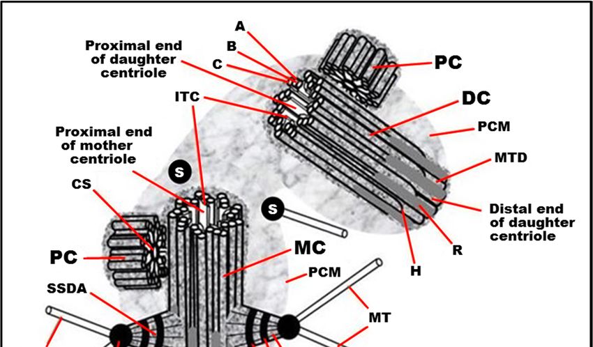
Cells 2020, 9, 2156                                                                                                              2 of 27

      Cells 2020, 9, x                                                                                                      2 of 26
in a book on the centrosome [13]. The discoveries of recent years make it necessary once again to return
      and developed in a book on the centrosome [13]. The discoveries of recent years make it necessary
to these postulates and update them.
      once again to return to these postulates and update them.
2. Postulates of Centrosomal Biology
      2. Postulates of Centrosomal Biology
2.1. The Centriole and the Basal Body Are Two Forms of the Same Organelle
      2.1. The Centriole and the Basal Body Are Two Forms of the Same Organelle
     The centrosome usually organizes and orients the mitotic spindle. The basal body nucleates and
          The centrosome usually organizes and orients the mitotic spindle. The basal body nucleates and
anchors  the cilium
     anchors          in interphase.
               the cilium               The centrosome
                               in interphase.               consistsconsists
                                                 The centrosome        of two centrioles      surrounded
                                                                                 of two centrioles             by pericentriolar
                                                                                                           surrounded      by
material  after mitosis
     pericentriolar        in the
                     material   afterG1 phaseinor
                                      mitosis     thethe
                                                      G1G0    phase
                                                          phase       of G0
                                                                  or the thephase
                                                                              cell cycle.     At the
                                                                                     of the cell      end
                                                                                                  cycle. At ofthethe
                                                                                                                   endinterphase
                                                                                                                       of the
before the next before
     interphase  mitosis  theinnext
                                the mitosis
                                     G2 phase    of the
                                             in the      cell cycle,
                                                     G2 phase   of thethe
                                                                       cellcell hasthe
                                                                            cycle,   twocellcentrosomes,       each having
                                                                                              has two centrosomes,       each two
centriolar
     havingcylinders    in its composition.
             two centriolar     cylinders in itsEach   centrioleEach
                                                  composition.     consists  of 9 MT
                                                                        centriole        triplets,
                                                                                   consists    of 9 and   it is associated
                                                                                                    MT triplets,    and it iswith
     associated
subdistal        with subdistal
          appendages,               appendages, distal
                           distal appendages,             appendages,
                                                    striated  roots, andstriated    roots,
                                                                           satellites (Figureand 1).
                                                                                                  satellites
                                                                                                     In some   (Figure
                                                                                                                 cases 1). In
                                                                                                                        or certain
     some
stages     cases
       of the     or certain stages
               development            of the development
                                  of organisms,     the walls ofof
                                                                 organisms,   the walls
                                                                   the centrioles     may ofcontain
                                                                                              the centrioles    may contain
                                                                                                       MT doublets        [14–16],
     MT doublets
MT singlets          [14–16],
              [17–19],   or noMT MTsinglets    [17–19],while
                                      at all [20,21],    or nomaintaining
                                                                 MT at all [20,21],     while symmetry.
                                                                                nine-beam        maintaining nine-beam
      symmetry.

      FigureFigure   1. Typical
               1. Typical         centrosome
                            centrosome        structure
                                          structure      in the
                                                     in the      S phase
                                                             S phase       of the
                                                                       of the   cellcell cycle
                                                                                      cycle  in in proliferatingmammalian
                                                                                                 proliferating    mammaliancells.
            cells. MC:
      MC: mother         mother centriole;
                      (mature)     (mature) centriole; DC: daughter
                                             DC: daughter              centriole;
                                                              centriole;            PC: procentriole;
                                                                          PC: procentriole;       PCM:PCM:      pericentriolar
                                                                                                          pericentriolar   material
            material   (pericentriolar  matrix); A: “A”  MT   of triplet; B: “B”  MT     of triplet; C: “C” MT
      (pericentriolar matrix); A: “A” MT of triplet; B: “B” MT of triplet; C: “C” MT of triplet; H: hook of “C”  of triplet; H: MT;
            hook   of “C”  MT;  MTD:   A-B  MT  duplex  (in the distal part  of centriolar    cylinder); ITC: internal
      MTD: A-B MT duplex (in the distal part of centriolar cylinder); ITC: internal triplets connections system        triplets
            connections system (scaffold structure), which include A-C linkers; CS: cartwheel structure (an axis
      (scaffold structure), which include A-C linkers; CS: cartwheel structure (an axis with spokes); SDA:
            with spokes); SDA: sub-distal appendage; HSDA: head of sub-distal appendage; SSDA: the stem of
      sub-distal appendage; HSDA: head of sub-distal appendage; SSDA: the stem of sub-distal appendage
            sub-distal appendage (connected to three triplets in this case); S: satellites; SS: the striated structure
      (connected to three triplets in this case); S: satellites; SS: the striated structure of sub-distal appendage
            of sub-distal appendage stem; MT: microtubule; DA: distal appendage; HAD: head of distal
      stem;appendage;
             MT: microtubule;        DA: distal appendage; HAD: head of distal appendage; R: rib. From [12] with
                           R: rib. From [12] with modifications. Insertion: The fine ultrastructure of the MT triplet,
      modifications.
            showing protofilamentsThe
                          Insertion:        fine(the
                                        of MTs    ultrastructure    of the
                                                     data from [22,23]       MTused
                                                                          were    triplet,   showing
                                                                                       to make           protofilaments of MTs
                                                                                                  this drawing).
      (the data from [22,23] were used to make this drawing).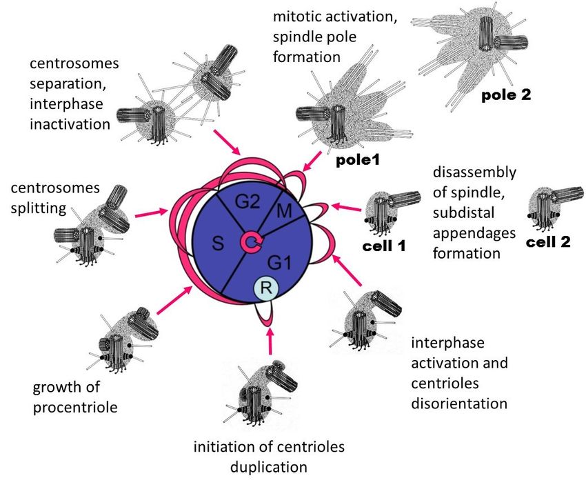
Cells 2020, 9, 2156                                                                                     3 of 27

      The structure of centrioles during the formation of cilia or flagella is conserved in the majority
organisms and cell types—9 MT triplets (aka “9×3”) with no central MTs (aka “+0”) that is summarized
by the formula “9×3 + 0”. In rare cases, cilia appear to form a basal body consisting of doublets
MTs [24–26]. The MT triplets are made three tubules named the MT “A”, the MT “B”, and the MT C”.
Only MT “A” has a closed rounded shape on a transverse section and consists of 13 protofilaments, as
most cellular MTs [27,28]. MT “B” has the shape of an arc adjacent to MT “A” and having three or four
common protofilaments with it. MT “C” also has the shape of an arc adjacent to MT “B” and has three
or four common protofilaments with it. The number of protofilaments in MT “B” and MT “C” can be
10 or 11 in centrioles from different organisms, and the total number of protofilaments in one triplet
can thus be from 33 to 35 [10,28–32]. There are suggestions that protofilaments number 11 of MT “B”
and MT “C” may have a special biochemical composition [28].
      The centriolar cylinders’ central nine-fold symmetry is established during the formation of the
“cartwheel” structure [10,33]. This symmetry is generated by lateral interaction of the N-terminal
domains of SAS-6 protein dimers [34,35]. The cartwheel structure disassembles in mature centrioles as
cells exit mitosis of vertebrates [33,36] but remains in mature centrioles of insects [37].

2.2. The Centriole Is a Polar Structure with Two Morphologically and Functionally Different Ends
      The centriole’s distal end (where the plus ends of the MT are) can serve as the site of cilia formation,
and it associates with distal appendages. A complex of proteins, including CP110 and CEP97, is found
at this end and supports cilium formation [38].
      Subdistal appendages are also more often formed closer to the distal end of the centriole.
The number of distal appendages (for centrioles of centrosome) and their homologous structures in
basal bodies—alar sheets—are always equal to nine [39,40]. The number of subdistal appendages
(the first original name of these structures was pericentriolar satellites [41]) is variable for different
types of cells and various conditions and ranges from 0 to 13 [42–44].
      The foot is the homologous structure of subdistal appendage in the basal body. One to two feet
are found per basal body, and they are located closer to their distal end [40,45,46]. The foot base is
connected to two or three triplets in a basal body [47]. In the basal bodies of functionally active cilia,
feet are located in the plane of the cilia beat [48].
      New centriolar cylinders (procentrioles) usually appear perpendicular to the mother centriole’s
surface closer to its proximal end [33], where the minus ends of the MT triplets are located.
      The pericentriolar material can also be located asymmetrically, surrounding only the centriole
proximal part [36,49]. The MT “A” centriole’s proximal end is covered with a conical structure with a
morphology similar to the γ-tubulin ring complex (γ-TuRC) [50].
      The polarity of the centriole is manifested in the orientation of some of its components.
When viewed from the proximal end of the centriole, the MT triplets are always twisted counterclockwise
(in the direction from the inner MT “A” to the outer MT “C”), and the distal appendages are always
twisted clockwise [12,51] (Figure 1).

2.3. The Outer Diameter of the Distal Part of the Centriolar Cylinder Is Smaller Than That of the Proximal End
     There are two reasons for the diameter differences between the centriole ends. (i) The MT triplets
transform into doublets of MT as MT “C” is usually shorter than MT “A” and MT “B”. (ii) The angle of
inclination of the MT triplets (the line passing through the centers MT “A” and MT “B”) to the radius
of the centrioles at the distal end of the centrioles is 80–90 degrees (the doublets lie almost in a circle).
At the proximal end of the centriole, the triplets’ inclination to the radius is 50–55 degrees (the triplets
are deployed like turbine blades). The inner radius of the lumen (the distance from the center of the
centriole to MT “A”) does not change. In contrast, the outer radius (the distance from the center of the
centriole to MT “C”) increases due to the unfolding of the triplets [36]. This twist’s angle appears to be
controlled by the A-C linkers in the proximal segment and an inner scaffold at the distal portion [52].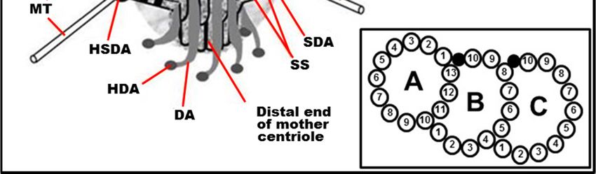
Cells 2020, 9, 2156                                                                                  4 of 27

2.4. The Centriolar Cylinder Length Is Highly Regulated
     The centriolar cylinder’s length is precisely set and usually ranges from 200 to 700 nm depending
on cell type and cell cycle phase [53,54]. However, in some cell types, extremely long centrioles can
be observed [10,55,56]. The growth of centrioles in length is regulated by a complex of proteins,
including SAS4/CPAP, POC1, and POC5 [57–60]. Overexpression of CPAP or its interaction partners,
CEP120 and SPICE1 (Spindle and Centriole Associated Protein 1), lead to the assembly of excessively
long centrioles. The protein CP110 is an antagonist to the CPAP; CP110 caps the distal end of the
growing centriole [61–64].

2.5. In the Centrosome of Proliferating Cells, Two Centrioles Differ Structurally and Functionally
     Only the older (mother) centriole has appendages at the distal end. Only the mother centriole
has subdistal appendages. MT nucleating centers are located predominantly on or near the
mother centriole. Gamma-tubulin [65,66] is the basis of two types of protein complexes that
nucleate MTs—the large Gamma-TuRC [67] and small Gamma-TuSC [68,69]. There are other protein
complexes on the centrosome that anchor the MTs. These complexes include CAP 350/FOP/EB1 [70],
PCM/BBS4/ninein/centrin/pericentrin [71], and ninein/ODF2/Cep170/centriolin/epsilon-tubulin [72].
MT nucleation and MT attachment activities are located in the subdistal appendages’ heads, on the
surface of the centrioles, and in the pericentriolar material. Centriolar satellites are dense rounded
structures containing protein complexes of the pericentriolar material located near centrioles and
undergo cell cycle-dependent assembly and disassembly [73–76]. They move towards the centrosome
along MTs in a dynein-dependent manner; participate in targeting of centrin, pericentrin, and ninein to
the centrosome; and are implicated in ciliogenesis [75,77,78].

2.6. The Proximal Ends of the Mother and Daughter Centrioles Are Connected via a Bundle of Thin Fibers
     The Proximal Ends of the Mother and Daughter Centrioles are Connected Via A Bundle of
Thin Fibers [79–81]. The composition of this ligament includes proteins rootletin, beta-catenin,
and C-NAP1 [82–84]. Severing this connection can lead to centriole separation. The regulation
of this separation is controlled by Nek2 kinase [84]. Separation of mother and daughter centrioles after
mitosis is a necessary prerequisite for the start of centrioles duplication [85].
     The separation of the mother and daughter centrioles in G1 phase of the cell cycle is a different
process from the separation and divergence of the two centrosomes before mitosis. The last process
differently depends on the intactness of MTs and actin microfilaments [86] and is controlled by
AuroraA [87–90], p34cdc2 [91], and Plk1 [92–94] kinases. In the process of centrosome separation
and movement during the formation of the two poles of the spindle, several types of motors are
involved: the cytoplasmic dynein–dynactin–NUMA complex [95–97], Xklp2 [98], kinesin-related
motors pEg5 [89,91,99], and XCTK2 [100].

2.7. The Centriole Lumen Helps in Stabilizing the Centriole
      Centriolar MTs have much greater stability than spindle or interphase cytosolic MTs. They are not
depolymerized either by anti-MT drugs or by exposure to cold [101–103]. However, MT triplets
can be disassembled by high (1–2 M NaCl or KCl) salt concentrations once centrioles are
isolated [104]. This stability of MT centrioles is associated with stabilizing proteins such as tektins
between microtubules [105,106], post-translational polyglutamylation of centriolar tubulin [107,108],
and presence of scaffold structures inside the centriole lumen.
      Inside the centriolar cylinder, an interconnected system of ligaments connects the MT
triplets [10,29,32,39,73]. The structure of these connections changes from the proximal end to the distal
one. MT “A” has a connective with MT “C” of the neighboring triplet. In addition, inward directions
from each MT “A” are strands of electron-dense material that are interconnected, with MT “A” and
A–C bundles. At the ultrastructural level, the centriolar cylinder’s lumen looks “empty” near the
Cells 2020, 9, 2156 5 of 27
Cells 2020, 9, x 5 of 26
proximal end and filled with electron-dense material at the distal lumen. The protein composition of
the A–C linker is unknown. The distal lumen scaffold is made of POC1B, POC5, FAM161A, CETN,
WDR90 [52,109]. Mutating some of these protein results in destabilization of the centriole cylinder
and WDR90 [52,109]. Mutating some of these protein results in destabilization of the centriole
[110].
cylinder [110].
2.8. Maintaining a Cylindrical Shape of Centrioles Can Be Independent of MT Triplets
2.8. Maintaining a Cylindrical Shape of Centrioles Can Be Independent of MT Triplets
Centrioles do not have MTs in haploid male larvae trophocytes and hypodermal cells of wasps
Centrioles do not have MTs in haploid male larvae trophocytes and hypodermal cells of wasps
Anisopteromalus calandrae [20]. However, their MT-free centrioles’ shape and size are similar to
Anisopteromalus calandrae [20]. However, their MT-free centrioles’ shape and size are similar to canonical
canonical centrioles with MT triplets of adult (imago) insects or late larvae of different wasps’ types
centrioles with MT triplets of adult (imago) insects or late larvae of different wasps’ types (Figure 2).
(Figure 2). The centriole’s symmetry is maintained, and they contain nine prongs of electron-dense
The centriole’s symmetry is maintained, and they contain nine prongs of electron-dense material
material in their walls forming the structure of the “cogwheel” [20]. One possibility is that at this
in their walls forming the structure of the “cogwheel” [20]. One possibility is that at this stage of
stage of development, the genes encoding the proteins responsible for the construction of MT triplets
development, the genes encoding the proteins responsible for the construction of MT triplets have not
have not yet turned on. Alternatively, the MTs disassembled after the centriole formed. Practically
yet turned on. Alternatively, the MTs disassembled after the centriole formed. Practically identical
identical cogwheel structure without MTs triplets was also found in the base of mature spermatozoa
cogwheel structure without MTs triplets was also found in the base of mature spermatozoa flagella of
flagella of wasps Cotesia congregata. It replaced the “normal” centriole during spermiogenesis, which
wasps Cotesia congregata. It replaced the “normal” centriole during spermiogenesis, which is present
is present in spermatids [21]. Prongs of cogwheel structure are visible between triplets or doublets of
in spermatids [21]. Prongs of cogwheel structure are visible between triplets or doublets of MT in
MT in centrioles of other insects, particularly in Drosophila. It was shown that the protein SAS4 [111]
centrioles of other insects, particularly in Drosophila. It was shown that the protein SAS4 [111] is
is concentrated in these regions.
concentrated in these regions.
After processing the isolated centrioles from bovines spleen by 2 M salts, the triplets’ MTs are
After processing the isolated centrioles from bovines spleen by 2 M salts, the triplets’ MTs are
completely disassembled [104]. Holes were found at the MT sites (Figure 3); still, the centrioles’
completely disassembled [104]. Holes were found at the MT sites (Figure 3); still, the centrioles’
cylindrical shape is preserved. The authors called the electron-dense structure surrounding triplets
cylindrical shape is preserved. The authors called the electron-dense structure surrounding triplets
“centriolar rim” [104]. Later, was reported that the PCM is not homogeneous and that some
“centriolar rim” [104]. Later, was reported that the PCM is not homogeneous and that some centrosomal
centrosomal proteins are closely associated with the centriole to form a distinct PCM compartment
proteins are closely associated with the centriole to form a distinct PCM compartment called the
called the “PCM tube”. In contrast, other proteins are more peripheral [112,113]. These MT
“PCM tube”. In contrast, other proteins are more peripheral [112,113]. These MT surrounding protein
surrounding protein complexes help maintain the cylindrical shape and provide mechanical strength
complexes help maintain the cylindrical shape and provide mechanical strength in MT’s absence.
in MT’s absence.
Figure 2. Centriole structure in larvae of two wasps: (a) larvae of Nasonia vitripennis, where prongs of
Figure 2. Centriole structure in larvae of two wasps: (a) larvae of Nasonia vitripennis, where prongs
cogwheel structure are visible between triplets of MT; and (b) early larvae of Anisopteromalus calandrae,
of cogwheel structure are visible between triplets of MT; and (b) early larvae of Anisopteromalus
where the centriole has a cogwheel without MT. The cartwheel structure is not clearly visible in
calandrae, where the centriole has a cogwheel without MT. The cartwheel structure is not clearly
the centriole lumen. View from the distal end of the centriole. Scale bar: 100 nm. From [20]
visible in the centriole lumen. View from the distal end of the centriole. Scale bar: 100 nm. From [20]
with modifications.
with modifications.Cells 2020, 9, 2156 6 of 27
Cells 2020, 9, x 6 of 26
Figure 3. Rotation images of centriole and centriolar rim. Nine photographs with a rotation angle of
Figure 3. Rotation images of centriole and centriolar rim. Nine photographs with a rotation angle of
40◦ were superimposed to obtain images: (a) centriole; and (b) centriolar rim after 1 M KC1 treatment.
40° were superimposed to obtain images: (a) centriole; and (b) centriolar rim after 1 M KC1 treatment.
Scale bar: 100 nm. From [104] with modifications.
Scale bar: 100 nm. From [104] with modifications.
2.9. The Structure and Activity of Centrosomes Differ in Interphase and Mitotic Cells
2.9. The Structure and Activity of Centrosomes Differ in Interphase and Mitotic Cells
The subdistal appendages and primary cilia disappear [36,114], and an amorphous mitotic halo
The
surrounds subdistal appendages
the centrosome and primary
in mitosis. cilia disappear
In addition, [36,114], and an amorphous
the centrosome-associated interphase mitotic
MTshalo
are
surrounds the centrosome in mitosis. In addition, the centrosome-associated
completely depolymerized before mitosis. Finally, mitotic MT asters form around each of the two interphase MTs are
completely
centrosomesdepolymerized
during prophase. before
These mitosis. Finally, with
MTs, together mitotic MTMTs
other asters form around
nucleating each
activities, of the two
organize and
centrosomes during prophase. These MTs, together with other MTs nucleating
form the mitotic spindle in metaphase [78]. All mitotic MTs have their minus ends near the centrosome activities, organize
and form three
but have the mitotic
options spindle in metaphase
for localizing [78]. end:
their plus All mitotic
(1) MTMTs plushaveend istheir minusinends
directed the near the
opposite
centrosome
direction from butthehave three options
chromosomes andfor localizingnear
is localized their
theplus
cell end: (1) MT plus end
membrane—astral MTs; is (2)
directed
MT plus in end
the
opposite direction from the chromosomes and is localized near the cell
is associated with the kinetochore of chromosomes—kinetochore MTs; and (3) MT plus end interacts membrane—astral MTs; (2)
MT
withplus endcoming
an MT is associated
from with the kinetochore
the opposite pole of theof chromosomes—kinetochore
spindle—interzonal MTs. The MTs; andalso
halo (3) MT plus
contains
end interacts with an MT coming from the opposite pole of the spindle—interzonal
many short MTs, which probably later transform into one of three types. Mitotic MTs have dynamic MTs. The halo
also contains many short MTs, which probably later transform into one of
and biochemical characteristics different from interphase MTs; in particular, they are less resistant tothree types. Mitotic MTs
have dynamic anddrugs
anti-microtubule biochemical
[115]. The characteristics different from
ability of centrosomes interphase
to nucleate MTMTs; in particular,
in mitosis increasesthey are
several
less resistant to anti-microtubule drugs [115]. The ability of centrosomes
times during preparation to cell division (G2 phase of the cell cycle) in a process named centrosome to nucleate MT in mitosis
increases
maturation several times during preparation to cell division (G2 phase of the cell cycle) in a process
[36,116,117].
named centrosome maturation [36,116,117].
2.10. New Centrioles Are Usually Formed in Association with Mother Centrioles but Can Be Formed without
Preexisting
2.10. CentrioleAre
New Centrioles De Novo
Usually Formed in Association with Mother Centrioles but Can Be Formed without
Preexisting Centriole De Novo
Duplication of centrioles occurs only one time per cell cycle; on each mature centriole, only one
procentriole appears
Duplication (Figure 4).occurs
of centrioles The accuracy
only one oftime
regulation
per cellofcycle;
these processes
on each matureis controlled by aonly
centriole, complex
one
of proteins that
procentriole were (Figure
appears first identified
4). The in Caenorhabditis
accuracy elegans:ofZYG-1
of regulation these [118,119],
processes SPD-2 [19,120,121],
is controlled by a
SAS-4 [122,123],
complex of proteinsSAS-5 that[124],
were and SAS-6
first [125,126].
identified In human cells,
in Caenorhabditis the ZYG-1
elegans: ZYG-1 homolog
[118,119],isSPD-2
PLK4
kinase [127]; in
[19,120,121], Drosophila
SAS-4 [122,123], cells, it is SAK/PLK4
SAS-5 [124], and SAS-6kinase[125,126].
[128]. More In than
human 30 proteins
cells, thehave ZYG-1already been
homolog
described
is that are
PLK4 kinase somehow
[127]; involved
in Drosophila in the
cells, duplication
it is SAK/PLK4ofkinase centrioles
[128].[129].
More Inthan
mammals and insects,
30 proteins have
centriolebeen
already duplication
describedstarts by CEP152/Asterless
that are somehow involvedthat recruits
in the PLK4 of
duplication [130–132].
centriolesCEP152
[129]. Inform a ring
mammals
together
and withcentriole
insects, CEP63 and CEP57 around
duplication starts bytheCEP152/Asterless
proximal part of the thatmother
recruitscentriole [133,134]. CEP152
PLK4 [130–132].
form a ring together with CEP63 and CEP57 around the proximal part of the mother centriole
[133,134].Cells 2020, 9, 2156 7 of 27
Cells 2020, 9, x 7 of 26
Figure 4. The relationship of the cell (nuclear) cycle and centriolar cycle. R, restriction point.
Figure 4. The relationship of the cell (nuclear) cycle and centriolar cycle. R, restriction point.
Centriole duplication is independent of DNA replication [135–137] and starts near the end of the
Centriole
G1 phase duplication
(Figure 4) of theiscell
independent of DNA
cycle [138,139]. replication
At this time, a [135–137]
complex of and starts near reactions
biochemical the end of in
the
theG1cellphase (Figuredependent
is launched 4) of the cell
oncycle [138,139].
the cell’s At this
size, the time, aofcomplex
presence externalof biochemical
growth factors,reactions
the growthin
the cell is launched
conditions dependent
of the cell, on the cell’swith
and its interaction size,surrounding
the presence cellsof external growth
[140–142]. factors,
It has been the growth
shown that
conditions
the Cyclinof the cell,
D/CDK 4/6and its interaction
complex with surrounding
phosphorylates cells [140–142].
the pRB protein, It hasits
which loses been shown
ability that the
to bind the
Cyclin D/CDKactivation
transcription 4/6 complexfactorphosphorylates
EF2. The released the pRB protein, factor
transcription which EF2loses its ability
activates the to bind the
synthesis of
transcription activation
Cyclin E and Cyclin factorstarts
A, which EF2. the
Theprocess
released transcription
of duplication of factor EF2 Thus,
centrioles. activates
DNA thereplication
synthesisandof
Cyclin
centriole E and Cyclin A,
duplication arewhich starts
regulated bythe process
a single of duplication
cytoplasmic of centrioles.
mechanism. However, Thus, DNA replication
centriole duplication
and
beginscentriole
earlier duplication are regulated
than DNA replication [138],by a single cytoplasmic
suggesting mechanism.
additional regulatory However, centriole
mechanisms.
duplication
In some begins
cases,earlier than DNA
new centrioles replication
form without[138], suggesting
preexisting additional
centrioles. In theregulatory mechanisms.
ciliary epithelium cells,
which In have
somehundreds
cases, newofcentrioles
cilia, theform without
centrioles preexisting
form centrioles.[46,143].
via deuterostomes In the ciliary epitheliumcan
Deuterosomes cells,
be
which
assembledhaveautonomously
hundreds of cilia, fromthe centrioles
parental form via
centrioles bydeuterostomes
de novo centriole [46,143]. Deuterosomes
amplification can be
in multiciliated
assembled autonomously
cells [144,145]. from embryonic
In murine early parental centrioles by de novo
development, centriole
centrioles amplification
appear de novo, butinthemulticiliated
mechanism
cells [144,145].
is unclear [146]. In murine
In cells withearly embryonic
centrioles, development,
centrioles elimination centrioles appear dedoes
by micro-irradiation novo,notbut the
prevent
mechanism
procentrioleisformation
unclear [146].
at S In cells In
phase. with centrioles,
contrast, centrioles
in this elimination
case, many by micro-irradiation
new centrioles does
are formed during
not
the prevent
S phase procentriole formation
[147]. In addition, at S phase.
centrioles In contrast,
can appear withoutin progenitors
this case, many duringnew centrioles are
parthenogenetic
formed
developmentduring[148].
the S phase [147]. In addition, centrioles can appear without progenitors during
parthenogenetic development
These observations suggest[148].
that centrioles can assemble independently of preexisting centrioles,
and These
the role observations
of centriolesuggest that centrioles
duplication can assemble
is to restrict the number independently
of assembling of preexisting
centrioles tocentrioles,
only one
and
newthe role of centriole duplication is to restrict the number of assembling centrioles to only one
centriole.
new centriole.Cells 2020, 9, 2156 8 of 27
2.11. The 9,
Cells 2020, Centrosomes
x Have Four Types of MT Nucleating Activity 8 of 26
Many of the centrosome’s critical function is mediated by MTs that are nucleated by it at different
2.11. The Centrosomes Have Four Types of MT Nucleating Activity
cell types and cell cycle phases. This includes four main MTs nucleating activities:
Many of the centrosome’s critical function is mediated by MTs that are nucleated by it at
(1) Formation
different of two
cell types andtypes of interphase
cell cycle MTs:
phases. This (i) a radial
includes fourMTs
mainsystem around theactivities:
MTs nucleating centriole; and (ii)
non-centrosomal (free) cytoplasmic MTs that were polymerized on the centrosome
(1) Formation of two types of interphase MTs: (i) a radial MTs system around the centriole; and later
and
released to the cytoplasm.
(ii) non-centrosomal (free) cytoplasmic MTs that were polymerized on the centrosome and later
(2) Formation
released of three types of mitotic spindle microtubules: (i) astral MTs; (ii) kinetochore MTs;
to the cytoplasm.
and (iii) interzonal
(2) Formation of three MTs.types of mitotic spindle microtubules: (i) astral MTs; (ii) kinetochore MTs;
(3) Formation
and (iii) interzonal of MT
MTs.of procentrioles.
(3) Formation
(4) Formation MTsofofMT
ciliaoforprocentrioles.
flagellum, or a related structure, known as the centriolar adjunct, that is
(4) Formation
found MTs of spermatids
in mammalian cilia or flagellum, or a related structure, known as the centriolar adjunct,
[149,150].
that is found in mammalian spermatids [149,150].
The more mature mother centrioles usually form the primary cilia with the formula 9×2 + 0
The more mature mother centrioles usually form the primary cilia with the formula 9 × 2 + 0
(Figure 5). The centrioles that arose in the current cell cycle and therefore are more immature form
(Figure 5). The centrioles that arose in the current cell cycle and therefore are more immature form
the motile cilia. Motile cilia have the formula 9×2 + 2 with the nine doublets MTs of the wall and
the motile cilia. Motile cilia have the formula 9 × 2 + 2 with the nine doublets MTs of the wall and
two central MTs (Figure 4). The mature (proximal) centriole forms a centriolar adjunct in mammalian
two central MTs (Figure 4). The mature (proximal) centriole forms a centriolar adjunct in mammalian
spermatids [150,151]. The centriolar adjunct has a formula similar to the primary cilium (9×2.5 + 0)
spermatids [150,151]. The centriolar adjunct has a formula similar to the primary cilium (9 × 2.5 + 0)
(Figure 5). Simultaneously, the centriolar adjunct forms in the spermatid, the daughter (distal) centriole
(Figure 5). Simultaneously, the centriolar adjunct forms in the spermatid, the daughter (distal)
form a motile flagellum with the formula 9×2 + 2 (Figure 5).
centriole form a motile flagellum with the formula 9 × 2 + 2 (Figure 5).
Figure5.5.Four
Figure Fourtypes
typesofof MT-contained
MT-contained structures:
structures: (a) proximal
(a) proximal centriole
centriole in the in thepig
early early pig spermatid—
spermatid—formula
“9×3 + 0”; (b) centriolar adjunct in the early pig spermatid—formula “9×2.5 + 0”; (c) flagellum
formula “9 × 3 + 0”; (b) centriolar adjunct in the early pig spermatid—formula “9 × 2.5 + 0”; pig
in early (c)
flagellum in early pig
spermatid—formula “9×2 + 2”; and (d) the cilia-like
spermatid—formula “9 × 2structure
+ 2”; and (d) the cilia-like
structurally similar tostructure structurally
the primary cilium of
similar tointhe
mammals theprimary cilium
spermatid of theofwasp
mammals in the spermatid
Anisopteromalus of the wasp
calandrae—formula + 0”. Scale bar:
Anisopteromalus
“9×2 calandrae—
(a–d) 100
formula
nm. (a–c)“9 × 2 [150];
from + 0”. Scale
and (d)bar: (a–d)
from 100 nm. (a–c) from [150]; and (d) from [20].
[20].
2.12.
2.12. The
The Complete
Complete Process
Process of
of Centrioles
Centrioles Maturation
Maturation from from Procentriole
ProcentrioletotoMother
MotherCentriole
CentrioleTakes
TakesMore
MoreThan
One and a Half Cell Cycles in Duration
Than One and a Half Cell Cycles in Duration
The complete process of centrioles maturation from procentriole to mother centriole takes
The complete process of centrioles maturation from procentriole to mother centriole takes more
more than one and a half cell cycles in duration [36]. The exact timing of procentriole initiation
than one and a half cell cycles in duration [36]. The exact timing of procentriole initiation is debatable
is debatable [152]. The percentage of cells with procentrioles significantly exceeded the S-phase
[152]. The percentage of cells with procentrioles significantly exceeded the S-phase percentage in the
percentage in the cell cycle in synchronized HeLa cells. This difference may indicate that the process of
cell cycle in synchronized HeLa cells. This difference may indicate that the process of procentriole
procentriole formation began before DNA replication start [153]. It was shown later that the initiation
formation began before DNA replication start [153]. It was shown later that the initiation of centriole
of centriole duplication could occur even in the absence of a nucleus in cytoplasts and enucleated
duplication could occur even in the absence of a nucleus in cytoplasts and enucleated sea urchin
sea urchin zygotes [137,154]. Procentrioles were found near mother centrioles two hours before the
zygotes [137,154]. Procentrioles were found near mother centrioles two hours before the start of DNA
start of DNA replication in pig kidney cell line, suggesting the procentrioles start to form before DNA
replication in pig kidney cell line, suggesting the procentrioles start to form before DNA replication
replication [138,139]. However, whether this observation is universal in other cell types is unknown.
[138,139]. However, whether this observation is universal in other cell types is unknown.
After its initial appearance, the centriole grows gradually to the mother’s size during the S
After its initial appearance, the centriole grows gradually to the mother’s size during the S phase
phase and G2 phase of the cell cycle (Figure 4). It becomes a daughter centriole after mitosis in the
and G2 phase of the cell cycle (Figure 4). It becomes a daughter centriole after mitosis in the newly
newly formed cell. The centriole becomes a mature mother centriole, acquiring a complete set of its
formed cell. The centriole becomes a mature mother centriole, acquiring a complete set of its cell
cell activities
activities after
after thethe second
second mitosis
mitosis in in
itsitslife
life[36,152].
[36,152].The
Thecell
cell(nuclear)
(nuclear)cycle
cycle and
and the
the centriolar
centriolar
(centrosomal) cycle are mutually coordinated (Figure 4) at least at two critical points
(centrosomal) cycle are mutually coordinated (Figure 4) at least at two critical points in the cell in the cell cycle:
cycle:
the end of the G1 and the end of the G2 phase [13,155]. In addition, some critical events take place in
mitosis during a process termed “centriole to centrosome conversion” [156]. Moreover, many
proteins involved in the regulation of the cell cycle are concentrated in the centrosome. InterferenceCells 2020, 9, 2156 9 of 27
the end of the G1 and the end of the G2 phase [13,155]. In addition, some critical events take place in
mitosis during a process termed “centriole to centrosome conversion” [156]. Moreover, many proteins
involved in the regulation of the cell cycle are concentrated in the centrosome. Interference with this
regulation leads to perturbation of cell cycle progression, ultimately leading to overproliferation or
degeneration [157].
2.13. The Centrosome Is the Center of the Organization of Actin Microfilaments in the Cell
This novel function, “actin organization center”, is in addition to the classic centriole function
in MT organizing [158]. The centrosome occupies a central position in the cytoplasm of many types
of cells. This position is associated with other interconnected cytoskeleton elements, such as actin
microfilaments. This position is regulated by the balance of the tension forces associated with the
cytoplasmic dynein [159] and the repulsive forces caused by the growth of MT [160,161]. In addition,
the centrosome position depends on the interaction of centrosomal MTs with the actomyosin complex,
and on the activity of the actomyosin complex itself [162–164]. Besides this, the intermediate filaments’
architecture is indirectly dependent on the centrosome since the intermediate filaments system collapsed
during the depolymerization of MT by colchicine or nocodazole [165,166].
2.14. Centrioles Can Gain an Atypical Structure and Composition and Become Undetected Using Standard
Expectations and Techniques
Centrioles are remodeled and earn distinct novel structures in a species-specific manner that
makes them difficult to detect [167,168]. In some insects, the sperm atypical centrioles are lacking
MT [25,169,170]. In contrast, in human and bovine sperm, the MTs are present, but they are
splayed around two novel rod structures [170]. Like canonical centrioles, these atypical centrioles
recruit PCM, form centrosomes and asters, and participate in spindle formation in the zygote.
In addition, canonical centrioles become undetected, including during myogenesis, oogenesis, and mice
spermatogenesis [171–173]. In amoeboflagellate Naegleria, centriolar cylinders are formed de novo
during the transition from centriole-less amoebae to flagellates with basal bodies from precursor
complexes that act as radiometry centrioles [174,175]. Therefore, centrioles can gain novel structure
and composition while performing many of their classical functions.
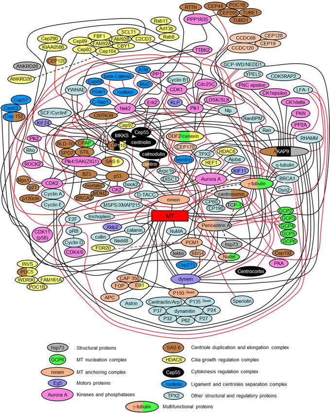
2.15. The Centrosome Is a Polyfunctional, Multi-Protein, and Cell Regulation Complex; Some of Its Proteins
Simultaneously Regulate Several Intracellular Processes
The presented interactome scheme in Figure 6 is only a partial depiction of all centrosomal proteins
and their interactions. It is intended to be a simplified diagram that helps to assess the complexity of
the centrosome’s biochemical organization. Indeed, each year, more and more centrosomal proteins
are characterized. These proteins include proteins that mediate or regulate processes such as signaling
pathways, nucleation and anchoring of microtubules, centriole duplication, separation of the daughter
centriole from the mother centriole, conversation of centrioles to basal bodies, and nucleating of cilia
and flagella. It is often impossible to separate the functions performed by the same protein in different
aspects of centrosome functioning.
A single universal classification for centrosomal proteins is unsubtle since they can be organized
according to several parameters. Many of them have multiple characteristics for any parameters such
as localization, timing, and activity. In the case of localization, some proteins, such as tubulins, are both
in the centrioles and pericentriolar material, or the cilium axoneme. In terms of timing, some proteins
always present in the centrosome. In contrast, other proteins appear in the centrosome only at specific
periods of the cell cycle or at a particular cell differentiation stage. In terms of activity, the centrosomal
proteins can be classified according to their biochemical activity—kinases, phosphatases, motors,
and structural proteins.
Centrosomal proteins do not exist on their own, but form complex, often interconnected,
functional complexes. Figure 6 shows how some of these complexes are related to each other.Cells 2020, 9, 2156 10 of 27
Cells 2020, 9, x 10 of 26
Figure 6.6.Scheme
Figure Schemeof protein interactions
of protein and functional
interactions protein complexes
and functional in centrosome
protein complexes in (interactome)
centrosome
([13] with modifications).
(interactome) Red and blackRed
([13] with modifications). linesand
mark centrosomal
black lines markprotein interactions.
centrosomal protein interactions.
The
The following
followingpublications werewere
publications used used
to prepare the presented
to prepare interactomeinteractome
the presented version: [13,19,38,49,
version:
52,57,63,68–71,75,83,84,89,92,93,98,126,127,130,133,134,141,176–355].
[13,19,38,49,52,57,63,68–71,75,83,84,89,92,93,98,126,127,130,133,134,141,176–355].
3.
3. Questions
Questions and
and Perspectives
Perspectives
The
The centrosome
centrosomecontinues
continuestotoremain
remain “the
“thecentral enigma
central enigmaof cell biology”
of cell [30],[30],
biology” although manymany
although data
have been obtained in recent years on many of its various aspects. The exact time of the
data have been obtained in recent years on many of its various aspects. The exact time of the onsetonset of centriole
duplication
of centrioleinduplication
the cell cycle
inand
the the
celltemporal
cycle andrelationship of this
the temporal process with
relationship of DNA replication
this process withremains
DNA
replication remains unclear. It is also not entirely clear to what extent the principles of centriole
biology are maintained in various cell types. For example, there is no certainty about the mechanismCells 2020, 9, 2156 11 of 27
unclear. It is also not entirely clear to what extent the principles of centriole biology are maintained in
various cell types. For example, there is no certainty about the mechanism of formation of centrioles
in the early development of mammals: How is the only proximal centriole of the spermatozoon
transformed into four centrioles at two poles of mitotic division at later stages of development? What is
the nature of the differences between this process in mice and other mammals [356]? What are the
evolutionary and molecular mechanisms underlying these centriole specific properties [357]? Many of
these aspects will be clarified in the near future, and the role of the centrosome, as the main regulatory
center of the cell—a kind of “cell processor” [358]—will become more evident.
Author Contributions: R.E.U. wrote the original draft of the manuscript; and R.E.U. and T.A.-R. wrote and edited
the paper. Both authors have read and agreed to the published version of the manuscript.
Funding: This work was supported by grant R03 HD087429 from the Eunice Kennedy Shriver National Institute
of Child Health & Human Development (NICHD).
Acknowledgments: The authors are grateful to Elena Nadezhdina for helpful discussion and for providing
Figure 3. Data were obtained with the assistance of the IBiSA Electron Microscopy Facility of Tours University and
the University Hospital of Tours. Many thanks are given to Andreas Merdes, Greenfield Sluder, Pierre Gönczy,
and Jadranka Loncarek for comments on the manuscript, which helped us to complement and better structure the
review content.
Conflicts of Interest: The authors declare no conflict of interest.
References
1. Flemming, W. Studien in der entwicklungsgeschichte der najaden. Sitz. Akad. Wissensch Wien. 1875, 71,
81–147.
2. Hertwig, O. Beitrage zur kenntniss der bildung, befruchtung und theilung des thierischen eies. Morphol. Jb.
1875, 1, 347–434. [CrossRef]
3. Van Beneden, E. Recherches sur les dicyémides, survivants actuels d’un embranchement des mésozoaires.
Bull. Acad. Roy. Méd. Belg. 1876, 41, 1160–1205.
4. Van Beneden, E.; Neyt, A. Nouvelles recherches sur la fecondation et la division mitoshes l’ascaride
megalocephale. Bull. Acad. R. Méd. Belg. 1887, 14, 1–81. Available online: https://orbi.uliege.be/handle/2268/
160175 (accessed on 23 September 2020).
5. Boveri, T. Die Bildung der Richtungskörper bei Ascaris megalocephala und Ascaris lumbricoides. Zellen-Studien
Heft 1; Verlag von Gustav Fischer: Jena, Germany, 1887; Available online: https://www.biozentrum.uni-
wuerzburg.de/zeb/research/topics/theodor-boveri/cell-studies/ (accessed on 23 September 2020).
6. Boveri, T. Ueber das Verhalten der Centrosomen bei der Befruchtung des Seeigel-Eies, nebst allgemeinen
Bemerkungen über Centrosomen und Verwandtes. Verhandl. Phys.-Med. Ges. Würzburg. 1895, 29, 1–75.
Available online: https://www.biozentrum.uni-wuerzburg.de/fileadmin/07020100/2018/Downloads_PDF/
Boveri_1895a.pdf (accessed on 23 September 2020).
7. Henneguy, L.F. Sur les rapports des cils vibratiles avec les centrosomes. Arch. Microsc. Morph. Exp. 1898, 1,
481–496.
8. Von Lenhossék, M. Uber flimmerzellen. Verh. Anat. Ges. Kiel. 1898, 12, 106–128.
9. Afzelius, B. Electron microscopy of the sperm tail; results obtained with a new fixative. J. Biophys.
Biochem. Cytol. 1959, 5, 269–278. [CrossRef]
10. Gibbons, I.R.; Grimstone, A.V. On flagellar structure in certain flagellates. J. Biophys. Biochem. Cytol. 1960, 7,
697–716. [CrossRef]
11. Brinkley, B.R.; Stubblefield, E. Ultrastructure and interaction of the kinetochore and centriole in mitosis
and meiosis. In Advances in Cell Biology; Prescott, D.M., Mc Conkey, E., Eds.; Appleton-Century Crofts:
New York, NY, USA, 1970; Volume 1, pp. 119–184.
12. Uzbekov, R.; Prigent, C. Clockwise or anticlockwise? Turning the centriole triplets in the right direction!
FEBS Lett. 2007, 581, 1251–1254. [CrossRef]
13. Uzbekov, R.E.; Alieva, I.B. Centrosome: History of Study and New Discoveries. From Cytoplasmic Granule to the
Center of Intracellular Regulation; Moscow University Press: Moscow, Russia, 2013.Cells 2020, 9, 2156 12 of 27
14. Sulston, J.E.; Albertson, D.G.; Thomson, J.N. The caenorhabditis elegans male: Postembryonic development
of nongonadal structures. Dev. Biol. 1980, 78, 542–576. [CrossRef]
15. Riparbelli, M.G.; Dallai, R.; Callaini, G. The insect centriole: A land of discovery. Tissue Cell 2010, 42, 69–80.
[CrossRef] [PubMed]
16. Gottardo, M.; Callaini, G.; Riparbelli, M.G. The drosophila centriole–conversion of doublets into triplets
within the stem cell niche. J. Cell Sci. 2015, 128, 2437–2442. [CrossRef] [PubMed]
17. Favard, P. Evolution des ultrastructures cellulaires au cours de la spermatogenese de l’ascaris
(ascaris megalocephala, schrank = parascaris equorum, goerze). Ann. Des Sci. Nat. Zool. Et Biol. Anim. 1961,
12, 52–152.
18. Wolf, N.; Hirsh, D.; McIntosh, J.R. Spermatogenesis in males of the free-living nematode,
caenorhabditis elegans. J. Ultrastruct. Res. 1978, 63, 155–169. [CrossRef]
19. Pelletier, L.; O’Toole, E.; Schwager, A.; Hyman, A.A.; Muller-Reichert, T. Centriole assembly in caenorhabditis
elegans. Nature 2006, 444, 619–623. [CrossRef]
20. Uzbekov, R.; Garanina, A.; Bressac, C. Centrioles without microtubules: A new morphological type of
centriole. Biol. Open 2018, 7, bio036012. [CrossRef]
21. Uzbekov, R.; Garanina, A.S.; Burlaud-Gaillard, J.; Bressac, C. The flagellum of the shortest spermatozoon
in the animal kingdom. Elongation and shortening of the axoneme in the process of spermiogenesis of
the parasitic wasp cotesia congregata. In Flagella and Cilia: Types, Structure and Functions; Uzbekov, R., Ed.;
Nova Science Publishers, Inc.: New York, NY, USA, 2018; pp. 83–108.
22. McNitt, R. Centriole ultrastructure and its possible role in microtubule formation in an aquatic fungus.
Protoplasma 1974, 80, 91–108. [CrossRef]
23. Greenan, G.A.; Keszthelyi, B.; Vale, R.D.; Agard, D.A. Insights into centriole geometry revealed by
cryotomography of doublet and triplet centrioles. ELife 2018, 7, e36851. [CrossRef]
24. Jana, S.C.; Girotra, M.; Ray, K. Heterotrimeric kinesin-ii is necessary and sufficient to promote different
stepwise assembly of morphologically distinct bipartite cilia in drosophila antenna. Mol. Biol. Cell 2011, 22,
769–781. [CrossRef]
25. Gottardo, M.; Pollarolo, G.; Llamazares, S.; Reina, J.; Riparbelli, M.G.; Callaini, G.; Gonzalez, C. Loss of
centrobin enables daughter centrioles to form sensory cilia in drosophila. Curr. Biol. 2015, 25, 2319–2324.
[CrossRef] [PubMed]
26. Nechipurenko, I.V.; Berciu, C.; Sengupta, P.; Nicastro, D. Centriolar remodeling underlies basal body
maturation during ciliogenesis in caenorhabditis elegans. ELife 2017, 6, e25686. [CrossRef] [PubMed]
27. Ringo, D.L. The arrangement of subunits in flagellar fibers. J. Ultrastruct. Res. 1967, 17, 266–277. [CrossRef]
28. Tilney, L.G.; Bryan, J.; Bush, D.J.; Fujiwara, K.; Mooseker, M.S.; Murphy, D.B.; Snyder, D.H. Microtubules:
Evidence for 13 protofilaments. J. Cell Biol. 1973, 59, 267–275. [CrossRef]
29. André, J. Le centriole et la région centrosomienne. J. Microsc. 1964, 3, 1–23.
30. Wheatley, D.N. The Centriole: A Central Enigma of Cell Biology; Elsevier Biomedical Press: Amsterdam,
The Netherlands; New York, NY, USA, 1982.
31. Ou, Y.; Rattner, J.B. The centrosome in higher organisms: Structure, composition, and duplication.
Int. Rev. Cytol. 2004, 238, 119–182.
32. Guichard, P.; Hachet, V.; Majubu, N.; Neves, A.; Demurtas, D.; Olieric, N.; Fluckiger, I.; Yamada, A.; Kihara, K.;
Nishida, Y.; et al. Native architecture of the centriole proximal region reveals features underlying its 9-fold
radial symmetry. Curr. Biol. 2013, 23, 1620–1628. [CrossRef]
33. Gall, J.G. Centriole replication. A study of spermatogenesis in the snail viviparus. J. Biophys. Biochem. Cytol.
1961, 10, 163–193. [CrossRef]
34. Kitagawa, D.; Vakonakis, I.; Olieric, N.; Hilbert, M.; Keller, D.; Olieric, V.; Bortfeld, M.; Erat, M.C.; Fluckiger, I.;
Gonczy, P.; et al. Structural basis of the 9-fold symmetry of centrioles. Cell 2011, 144, 364–375. [CrossRef]
35. Van Breugel, M.; Hirono, M.; Andreeva, A.; Yanagisawa, H.A.; Yamaguchi, S.; Nakazawa, Y.; Morgner, N.;
Petrovich, M.; Ebong, I.O.; Robinson, C.V.; et al. Structures of sas-6 suggest its organization in centrioles.
Science 2011, 331, 1196–1199. [CrossRef]
36. Vorobjev, I.A.; Chentsov, Y.S. Centrioles in the cell cycle. I. Epithelial cells. J. Cell Biol. 1982, 93, 938–949.
[CrossRef]
37. Friedlander, M.; Wahrman, J. Giant centrioles in neuropteran meiosis. J. Cell Sci. 1966, 1, 129–144. [PubMed]Cells 2020, 9, 2156 13 of 27
38. Spektor, A.; Tsang, W.Y.; Khoo, D.; Dynlacht, B.D. Cep97 and cp110 suppress a cilia assembly program. Cell
2007, 130, 678–690. [CrossRef] [PubMed]
39. Szollosi, D. The structure and function of centrioles and their satellites in the jellyfish phialidium gregarium.
J. Cell Biol. 1964, 21, 465–479. [CrossRef] [PubMed]
40. Anderson, R.G. The three-dimensional structure of the basal body from the rhesus monkey oviduct. J. Cell Biol.
1972, 54, 246–265. [CrossRef] [PubMed]
41. Bernhard, W.; de Harven, E. L’ultrastructure du centriole et d’autres éléments de l’appareil achromatique.
In Vierter Internationaler Kongress fur Elektronenmikroskopie, 1958; Bargmann, W., Möllenstedt, G., Niehrs, H.,
Peters, D., Ruska, E., Wolpers, C., Eds.; Springer: Berlin, Germany, 1960; Volume 2, pp. 217–227.
42. Stubblefield, E.B.; Brinkley, R. Architecture and function of the mammalian centriole. In Formation and Fate of
Cell Organelles; Warren, K.B., Ed.; Academic Press Inc.: New York, NY, USA, 1967; pp. 175–218.
43. Vorobjev, I.A.; Chentsov, Y.S. The ultrastructure of the centrosome in the cells of hematopoietic tissues of
axolotl. Tsitologiia 1977, 19, 598–603.
44. Uzbekov, R.; Alieva, I. Who are you, subdistal appendages of centriole? Open Biol. 2018, 8, 180062. [CrossRef]
45. Doolin, P.F.; Birge, W.J. Ultrastructural organization of cilia and basal bodies of the epithelium of the choroid
plexus in the chick embryo. J. Cell Biol. 1966, 29, 333–345. [CrossRef]
46. Sorokin, S.P. Reconstructions of centriole formation and ciliogenesis in mammalian lungs. J. Cell Sci. 1968, 3,
207–230.
47. Wolfe, J. Basal body fine structure and chemistry. Adv. Cell Mol. Biol. 1972, 2, 151–192.
48. Gibbons, I.R. The relationship between the fine structure and direction of beat in gill cilia of a lamellibranch
mollusc. J. Biophys. Biochem. Cytol. 1961, 11, 179–205. [CrossRef] [PubMed]
49. Ou, Y.; Rattner, J.B. A subset of centrosomal proteins are arranged in a tubular conformation that is reproduced
during centrosome duplication. Cell Motil. Cytoskelet. 2000, 47, 13–24. [CrossRef]
50. Guichard, P.; Chretien, D.; Marco, S.; Tassin, A.M. Procentriole assembly revealed by cryo-electron tomography.
EMBO J. 2010, 29, 1565–1572. [CrossRef] [PubMed]
51. O’Hara, P.T. Spiral tilt of triplet fibers in human leukocyte centrioles. J. Ultrastruct. Res. 1970, 31, 195–198.
[CrossRef]
52. Le Guennec, M.; Klena, N.; Gambarotto, D.; Laporte, M.H.; Tassin, A.M.; van den Hoek, H.; Erdmann, P.S.;
Schaffer, M.; Kovacik, L.; Borgers, S.; et al. A helical inner scaffold provides a structural basis for centriole
cohesion. Sci. Adv. 2020, 6, eaaz4137. [CrossRef] [PubMed]
53. Avidor-Reiss, T.; Gopalakrishnan, J. Cell cycle regulation of the centrosome and cilium. Drug Discov. Today.
Dis. Mech. 2013, 10, e119–e124. [CrossRef] [PubMed]
54. Basiri, M.L.; Blachon, S.; Chim, Y.C.; Avidor-Reiss, T. Imaging centrosomes in fly testes. J. Vis. Exp.
2013, e50938. [CrossRef]
55. Schreiner, A.; Schreiner, K.E. Űber die entwicklung der männlichen geschlechtszellen von myxine glutinosa (l.).
Arch. Biol. 1905, 21, 183–357.
56. Guichard, P.; Desfosses, A.; Maheshwari, A.; Hachet, V.; Dietrich, C.; Brune, A.; Ishikawa, T.; Sachse, C.;
Gonczy, P. Cartwheel architecture of trichonympha basal body. Science 2012, 337, 553. [CrossRef]
57. Azimzadeh, J.; Hergert, P.; Delouvee, A.; Euteneuer, U.; Formstecher, E.; Khodjakov, A.; Bornens, M. Hpoc5 is
a centrin-binding protein required for assembly of full-length centrioles. J. Cell Biol. 2009, 185, 101–114.
[CrossRef]
58. Blachon, S.; Cai, X.; Roberts, K.A.; Yang, K.; Polyanovsky, A.; Church, A.; Avidor-Reiss, T. A proximal
centriole-like structure is present in drosophila spermatids and can serve as a model to study centriole
duplication. Genetics 2009, 182, 133–144. [CrossRef] [PubMed]
59. Kohlmaier, G.; Loncarek, J.; Meng, X.; McEwen, B.F.; Mogensen, M.M.; Spektor, A.; Dynlacht, B.D.;
Khodjakov, A.; Gonczy, P. Overly long centrioles and defective cell division upon excess of the sas-4-related
protein cpap. Curr. Biol. 2009, 19, 1012–1018. [CrossRef] [PubMed]
60. Tang, C.J.; Fu, R.H.; Wu, K.S.; Hsu, W.B.; Tang, T.K. Cpap is a cell-cycle regulated protein that controls
centriole length. Nat. Cell Biol. 2009, 11, 825–831. [CrossRef]
61. Schmidt, T.I.; Kleylein-Sohn, J.; Westendorf, J.; Le Clech, M.; Lavoie, S.B.; Stierhof, Y.D.; Nigg, E.A. Control of
centriole length by cpap and cp110. Curr. Biol. 2009, 19, 1005–1011. [CrossRef] [PubMed]You can also read