Repetitive RNAs as Regulators of Chromatin-Associated Subcompartment Formation by Phase Separation
←
→
Page content transcription
If your browser does not render page correctly, please read the page content below
Review

                        Repetitive RNAs as Regulators of
                        Chromatin-Associated Subcompartment
                        Formation by Phase Separation

Lukas Frank and Karsten Rippe
Division of Chromatin Networks, German Cancer Research Center (DKFZ) and Bioquant, 69120 Heidelberg, Germany

Correspondence to Karsten Rippe: Karsten.Rippe@dkfz.de.
https://doi.org/10.1016/j.jmb.2020.04.015
Edited by Brian Luke

Abstract
Repetitive RNA (repRNA) sequences emerge as important regulators of the dynamic organization of genomic
loci into membrane-less subcompartments with distinct nuclear functions. These domains include sites of
active transcription like the nucleolus as well as (peri)centromeric and telomeric satellite repeats. Recent
studies point to an important role of repRNAs in complex with proteins to promote a phase separation-driven
formation of chromatin domains. We review how key features of the phase separation process can be
revealed by different experimental approaches and discuss the associated structure–function relationships for
chromatin subcompartments that involve repRNA.
        © 2020 The Authors. Published by Elsevier Ltd. This is an open access article under the CC BY license (http://
                                                                             creativecommons.org/licenses/by/4.0/).

Genome Organization by                                         and the surrounding nucleoplasm, similar to the
                                                               demixing of oil drops in water. It is driven by
Phase Separation                                               multivalent interactions and frequently involves
                                                               RNA interactions with low complexity intrinsically
   The eukaryotic nucleus has a typical diameter of            disordered regions (IDRs) of a protein that change
10–20 μm without internal membranes, and proteins              the association properties of the complex [21–26].
can transverse through it by diffusion within sec-             IDRs are also commonly found in many chromatin-
onds. Nevertheless, the nucleus is structured into             binding proteins [27] and are positively correlated
subcompartments (also referred to asmembrane-                  with the propensity of a given protein to undergo an
less organelles or nuclear bodies) that can dynam-             LLPS [28]. Here, we focus on the role of repetitive
ically partition the genome in a self-organizing               RNA (repRNA) sequences for regulating the forma-
manner [1–5]. RNA plays a crucial role as an                   tion of chromatin-associated subcompartments via
architectural factor at ribosomal genes in the                 different types of PS. These RNAs comprise tandem
nucleolus, (peri)centromeres, telomeres or the inac-           repeats that are present at satellite DNA elements
tive X-chromosome as described in a number of                  like (peri)centromeres and telomeres. In addition,
reviews [6–10]. Based on initial studies of the                they can originate from interspersed repeat se-
nucleolus, the model of a liquid droplet-like structure        quences of transposable element (TE) sequences
established by a liquid–liquid phase separation                that constitute up to ~ 40% of mammalian genomes
(LLPS) has been developed that rationalizes a                  [29]. Some TEs are transcribed into abundant RNA
highly dynamic but yet confined chromatin subcom-              species with specific functions in nuclear compart-
partment organization [11–13]. LLPS as well as                 mentalization as discussed in further detail below,
other phase separation (PS) mechanisms are                     while others have evolved to function as cis-
described in detail in a number of excellent reviews           regulatory elements for protein coding gene net-
[14–17], and distinctive features in the context of            works [30]. Given the high copy number, clustered
chromatin subcompartment formation have been                   distribution and partial self-complementarity,
discussed recently [18–20]. LLPS creates two liquid-           repRNAs bear the potential to function as platforms
like phases, a droplet-like cellular subcompartment            for locally concentrating protein factors and to
0022-2836/© 2020 The Authors. Published by Elsevier Ltd. This is an open access article under the CC BY license (http://
creativecommons.org/licenses/by/4.0/).                                  Journal of Molecular Biology (2020) 432, 4270-4286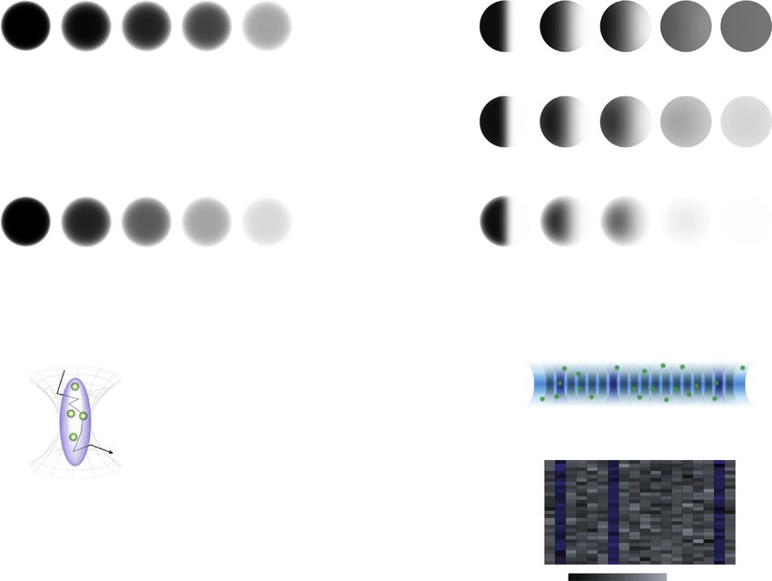
4271

nucleate PS processes: (i) The repRNA sequences             mechanisms that include liquid–liquid, liquid–gel
may provide a scaffold with locally enriched binding        and liquid–solid phase transitions, and it is not
sites for interacting proteins, especially for those that   straightforward to distinguish between them in the
contain tandem repeats. (ii) They may interact with         native cellular environment [14,17,37]. With these
themselves via self-complementary sequence motifs           caveats in mind, we here discuss three different
to provide additional interactions in RNA–protein           prototypic PS scenarios, LLPS, liquid–gel phase
assemblies. (iii) By interaction with IDRs, repRNAs         separation (LGPS) and polymer–polymer phase
might regulate their association into liquid droplets       separation (PPPS) that are informative for the
as mentioned above. (iv) They could be involved in          mechanism of an RNA-driven chromatin subcom-
cross-linking of regions of the nucleosome chain to         partment formation and are conceptually different
induce the compaction of chromatin domains. In this         from “simple” binding to a cluster of binding sites
review, we discuss the role of repRNA in PS-driven          (Figure 1). The three different PS mechanisms
formation of chromatin subcompartments by differ-           share a common origin with respect to their targeting
ent mechanisms.                                             to certain genomic regions via specific binding sites.
                                                            This chromatin binding step could involve RNAvia
                                                            transcription factors that bind certain RNA second-
The Different Flavors of Chromatin PS                       ary motifs [10], RNA–DNA hybrids (R-loops) or
Processes                                                   RNA–DNA triplexes [38]. However, the relevance of
                                                            these interactions in the context of PS, e.g., as
   Chromatin subcompartments are frequently                 nucleation sites for an LLPS/LGPS or as linkers
assigned based on the local enrichment of a given           between chromatin segments in PPPS processes,
marker protein, e.g., nucleolin (NCL) or nucleophos-        remains to be established.
min (NPM) for the nucleolus [31], promyelocytic                For LLPS, the presence of liquid-like properties of
leukemia (PML) protein for PML nuclear body                 the subcompartment is a defining feature. It empha-
complexes at telomeres [32] or heterochromatin              sizes that macromolecules constantly rearrange
protein 1 (HP1) for pericentric heterochromatin             their position with respect to each other in a random
compartments [33]. In the context of cellular PS,           manner like molecules in a liquid, in a volume
the term “biomolecular condensates” or simply               confined by the PS boundary. This behavior is to be
“condensates” has been coined and implies assem-            distinguished from the fast exchange of a protein
bly of macromolecules into a membrane-less                  between a chromatin bound and unbound state,
supramolecular complex by PS [15,34]. A local               which would simply reflect transient binding with
enrichment of a factor with more or less sharp              short residence times. Liquid droplets formed by
concentration boundaries to the surrounding nucle-          association of oppositely charged molecules like
oplasm on fluorescence microscopy images into               negatively charged RNA interacting with positively
what traditionally has been termed nuclear “foci,”          charged proteins/peptides are referred to as “coac-
“puncta,” “bodies” or “speckles” is frequently taken        ervates” [39]. The PS process can also involve a
as an initial finding that could point toward a PS          liquid–gel transition [14,17,22,40]. For gels that
process being involved. However, it is noted that a         comprise a hydrophilic cross-linked polymer net-
more formal definition would require it to demon-           work, the term “hydrogel” is used to emphasize it has
strate that indeed a given physical property, e.g., the     a high water content. An example for this state would
protein concentration, is uniform throughout the            be an agarose gel as it is used for gel electropho-
subcompartment. For example, PML complexes                  resis of nucleic acids. Hydrogels can form in an
might appear as homogeneous liquid-like droplets            RNA-dependent manner and might regulate tran-
at diffraction-limited image resolution but rather          scription and chromatin organization as discussed
organize into spherical shell-like structure [35].          in further detail below [40–43]. For LGPS, the mixing
Likewise, the PS boundary should display a steep            of gel constituting components within the PS
concentration increase that can be distinguished            compartment is largely reduced as compared to
from the enrichment due to “simple” binding to a            LLPS. Finally, a third type of PS can occur with
cluster of sites on the nucleosome chain. Finally, the      respect to the conformation of the nucleosome chain
examination of a cellular subcompartment in steady          and is referred to here as PPPS. It is characterized
state is not sufficient to conclude that it is indeed a     by a sharp transition of the chain from an open
PS product rather than being formed by coales-              random coil conformation into an ordered and
cence of smaller structures, active transport pro-          “collapsed” chromatin globule as it is induced by
cesses or local macromolecule synthesis [36].               embedding the polymer in a bad solvent [44]. If this
Accordingly, using the terms “phase separation”             process involves only parts of a polymer, it is called
and “biomolecular condensates” needs to be justi-           a block copolymer microphase separation [45]. It is
fied beyond simply reporting a local enrichment of a        driven by an attractive interaction between seg-
specific factor in a chromatin associated domains.          ments. For the nucleosome chain, the latter can
Finally, PS covers a broad range of different               involve localized bridging interactions mediated by
4272
Nucleosome
chain
Chromatin
binding site
RNA
Protein
Binding site cluster Electrostatic cat
Phase separation (PS)
Liquid-liquid phase Liquid-gel phase Polymer-polymer phase
separation (LLPS) separation (LGPS) separation (PPPS)
Oil drops in water Jelly Sticky tape
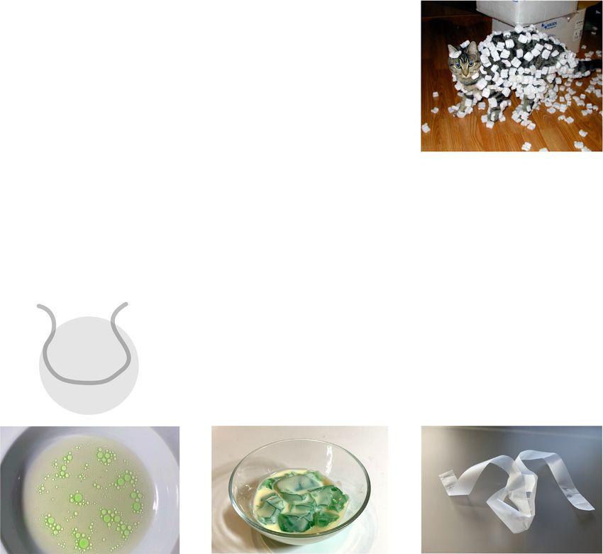
Figure 1. Different types of RNA-driven PS of chromatin domains. The clustered binding of proteins and RNA to
genomic loci alone can compartmentalize activities by locally confining enrichment of certain factors on the nucleosome
chain as shown at the top of the figure. This is illustrated by the “electrostatic cat” example. Due to the triboelectric effect,
the electrically charged cat's fur acts as a “binding site cluster” for the styrofoam chips that are distributed in its environment
(original image by Sean McGrath, NB, Canada). In the context of PS processes, clustered chromatin binding sites are
essential to target a PS process to a certain chromatin locus. They can serve as nucleation sites for the further
accumulation of protein and RNA by the LLPS or LGPS mechanisms depicted at the bottom. In LLPS, factors constantly
re-arrange like solvent in a liquid, but clearly separate from the surroundings like in the “oil drops in water” model. An LGPS
comprises organization into gel-like assemblies, which remain percolated by the solvent (“jelly” model). In a PPPS
process, proteins and/or RNA form a linker that bridges two segments of the nucleosome chain. If these attractive
interactions become sufficiently strong, they can induce a local collapse of the nucleosome chain into a “chromatin
globule.” This process is represented by the “sticky tape” model where parts of the adhesive tape stick together. Note that
the three different mechanisms could also occur in combination. For further details and functional differences, see text and
Table 1.
chromosomal proteins [46–49]. It is noted that pensity to polymerize into labile amyloid-like fibrils,
LLPS,LGPS and PPPS are not mutually exclusive e.g., via beta-sheet stacking [52]. This seemingly
and that in reality frequently contributions from controversial observation indicates that self-
different mechanisms are present. For example, assembly of intrinsically disordered proteins can
many low-complexity protein domains can form occur through different PS mechanisms, potentially
liquid-like droplets [50,51] but also have the pro- also with phase transitions that are mixed and/or4273
Table 1. Features of chromatin subcompartments formed by different PS mechanisms
Binding site cluster LLPS LGPS PPPS
a
Preferential internal mixing No Yes No No
Chemical environment same as in
Yes No Mostly yes Mostly yes
nucleoplasmb
Coalescencec No Yes Yes Yes
Chromatin compactiond No Possible Possible Yes
Dependence of size on concentration None (except for None (for persistent
Expansion Expansion
increasee unspecific binding) bridging interactions)
Concentration fluctuation buffer
None High Low Low
capacityf
Exclusion dependence of other Dominated by solubility Dominated by Dominated by
Fully accessible
factorsg in the phase particle size particle size
Exchange rate with other factorsh Fast Slow Fast Fast
The table refers to the four different cases depicted in Figure 1 in their pure form. In reality, different combinations and intermediate states
occur. For example, clusters of specific chromatin binding sites occur as part of the LLPS, LGPS and PPPS mechanisms.
a
Macromolecules in LLPS subcompartments have per definition liquid-like properties so that their mixing within the compartment
should occur fast. For gel- or chromatin globule-like states the constituting factors organize into more rigid structures. This does not
exclude a simultaneously occurring exchange with molecules from the outside.
b
In an LLPS, the accumulation of RNA and protein into a homogenous liquid-like droplet creates a chemical microenvironment that is
different from that of the nucleoplasm, whereas in an LGPS and PPPS scenario, the compartment is still percolated by nucleoplasm.
c
All three types of PS can exhibit coalescence.
d
Chromatin compaction in a globule state is a defining feature of PPPS. LLPS or LGPS can occur without any change of the chromatin
compaction state. However, they might induce a local environment that favors or initiates PPPS mediated compaction.
e
LLPS and LGPS are not size buffered. Increasing concentration of the PS driving factor either expands liquid-like droplet volume in
order to reach its steady-state concentration or extends the gel structure as additional binding partners associate to the existing assembly.
The PPPS domain is size-buffered over the concentration regime that does not significantly affect bridging interactions.
f
LLPS has the unique property that the liquid droplet state creates stable concentrations inside and outside the compartment.
g
Passive access to the subcompartment from the surrounding nucleoplasm can be regulated by chemical properties (e.g, charge,
LLPS) or size with compartment barriers acting as “molecular sieves” (LGPS, PPPS).
h
LGPS and PPPS subcompartments are percolated with soluble factors from the surrounding nucleoplasm (unless restricted by their
size) so that they can exchange fast.
change over time. Such a behavior was shown for defines the accessible space [55]. Accordingly, factors
both native and light-inducedFUS protein droplets, within a LGPS and PPPS subcompartment can
which can “mature” from a liquid-like state to exchange fast with the surrounding space. The size
exhibiting gel-like behavior (LGPS), up to even of LGPS domains will also fluctuate with concentration
undergoing amyloid fibril-like aggregation, also as for LLPS. For the PPPS mechanism, however,
termed liquid–solid PS [53,54]. domain size should be constant over the concentration
range that leaves the driving attractive interactions
between chromatin segments mostly unaffected.
Experimental Approaches to Study PS Since these and other key hallmarks of PS subcom-
Processes partments are functionally relevant and need to be
experimentally addressed, we summarize strategies to
The mechanisms depicted in Figure 1 all segregate measure them in the following.
chromatin into distinct and mostly spherical structures,
but the properties of the resulting assemblies are very Concentration dependence and coalescence/
different (Table 1). Molecules that form by LLPS tend
dispersion
to stay within the resulting liquid droplets with fast
internal mixing, create a specific chemical microenvi-
In vitroPS analysis
ronment and insulate chromatin regions from each
other. An LLPS compartment is concentration-buffered An essential part of any characterization of cellular
as it maintains a constant concentration of molecules subcompartments in the context of PS processes is
in its interior against fluctuations from the outside. identifying conditions for which a well-mixed solution
However, the droplet size follows concentration undergoes a PS [56]. This issue can be directly
changes so that the chromatin content increases at addressed in vitro by varying relevant parameters
higher concentrations. This feature could thus present (macromolecule composition, concentration, tem-
a mechanism for spreading a given chromatin state. perature, pH, ionic strength) to generate a phase
LGPS and PPPS subcompartments on the other hand diagram that defines the regime for which a PS is
are percolated with soluble factors from the surround- observed. These experiments typically comprise an
ing nucleoplasm, but the size of macromolecules analysis of liquid droplet formation or other types of4274
macromolecular associates by microscopy or other additional binding partners associate to the existing
methods [56,57]. Examples for studies of RNA- scaffold. A collapsed chromatin globule formed by
dependent droplet formation are given in Refs. [25, PPPS, however, should behave differently and remain
39, 58]. In order to quantitate the concentration constant in size unless protein/RNA concentration
dependence of the PS process, the turbidity of the affect the attractive properties of chromatin segments
solution can be determined by absorbance spectros- within the PS domain.
copy as described previously [57,59,60]. It involves
measurements at wavelength where protein and
Coalescence and dispersion
nucleic acids do not absorb, e.g., 340 nm and
above. The resulting signal reflects scattering of light The fusion/fission of liquid droplets is a crucial LLPS
in dependence of particle size and can be exploited to feature inherent to its fluid-like properties. It is not
trace the formation of droplets or other particle types. In observed for domains that originate from (cooperative)
the context of the RNA-dependentchromatin- binding to chromatin (Figure 1). The coalescence/
associatedPS discussed here, one challenge is it to dispersion process can be directly observed by
adequately represent the chromatin part in vitro. Mono- microscopy, both in vitro and in living cells as done
and oligonucleosome particles can be reconstituted in for the nucleolus [11,13,76] or for ectopically formed
vitro and have been shown to undergo an LLPS RNA–protein assemblies [25]. Interestingly, also col-
themselves [61–63]. However, it is difficult to envision lapsed polymer regions in PPPS that are separated by
how such a transition could occur in the context of a freely fluctuating chain parts can undergo coalescence
human chromosome that is connected by a continuous after collision [77]. This process, however, requires that
nucleosome chain that varies between 5 and 24 nm in the attractive interactions between polymer segments
diameter [64] and stably occupies a distinct territory in are interchangeable between different globule regions
the nucleus [65]. In this environment, the nucleosome and can dynamically rearrange on the time scale
translocations on the second to minute time scale are observed. For an LGPS, dynamic reorganization might
confined to local movements of chain segments within be very slow (especially for the case of chemically
a radius of about 70–80 nm [66–69]. Accordingly, cross-linked hydrogels), leading to a kinetically trapped
chromatin appears in the cell as a mostly immobile state that would be incompatible with coalescence.
scaffold as also reflected by the hour-long persistence
of fluorescence bleach patterns of histones and lack of Structure, dynamics, and physicochemical
local positional changes [70–73]. How these pertinent properties
cellular chromatin features can be adequately repre-
sented in in vitro experiments for studying the assembly Subcompartment structure
of a chromatin subcompartment is currently an open
question. A critical test whether the formation of Fluorescent microscopy-based approaches provide
chromatin droplets observed with mono- and oligonu- a wealth of information on RNA-dependentPS within
cleosomes occurs also with longer nucleosome chains the endogenous cellular environment. Candidate
would be to study the dependence of LLPS propensity RNAs can be transcribed in vitro (and also fluorescently
on chain length. Technically, this can be accomplished labeled if desired) and delivered into cells by micro-
by preparing native chromatin fragments up to 60–70 injection or transfection to examine their coalescence/
nucleosomes in size from human cell lines (e. g., [74]) dispersion behavior or association with specific pro-
or chromatin reconstituted in vitro with Drosophila teins over time [78]. The sequence specific tagging and
extracts on DNAs that can exceed 40 kb (or 220 fluorescent labeling of RNA with autofluorescent
nucleosomes) in length [75]. domains can be accomplished by engineering a high-
affinity protein binding sequence in one or more copies
into the RNA of interest ([79,80] and references
therein). Examining the subcompartment's internal
Response to concentration changes
structure by fluorescence microscopy, preferably at
The response to concentration changes of protein super-resolution, provides crucial mechanistic informa-
and nucleic acids is a crucial parameter for PS tion. A homogeneous distribution of components is
diagrams that can be examined directly in vitro as indicative of LLPS, while the observation of distinct
described above. In the cell, over-expression or micro-domains would argue against a liquid-like state,
knockdown of phase-separating protein and RNAs in at least at the level of the subcompartment.
conjunction with quantitative microscopy readouts of
local concentrations and subcompartment size is highly
Dynamics and accessibility of phase-separated
informative. For LLPS, the total cellular droplet volume
factors in the nucleus
scales with the cellular concentration of the phase-
separating factors, while their concentration within the As discussed above, the kinetics of internal mixing
droplet is buffered [15]. For LGPS, the gel structure and exchange with the nuclear environment distinguish
should also expand at increasing concentration as between different PS mechanisms. This process can4275
(a) Full-bleach FRAP Half-bleach FRAP
Bleach of subcompartment Impermeable (only internal exchange)
1
Intensity
non-bleached
bleached 0
Intermediate
1
Intensity
1
Intensity
0 0
Time
Bleach of reference region Permeable (unrestricted exchange)
1
Intensity
0
Time t/τD 0 1 2
0.01 0.05 0.1 0.5 1 Time t/τD
(b) Point FCS Line FCS
Auto-correlation
Subcompartment
GAC(τ)
GAC(τ)
(2) (7) (17)
Detection elements
Log (Lag time τ)
Nucleoplasm
GAC(τ)
Cross-correlation
Time t
(2) (7) (2) (17)
Cytoplasm
GXC(τ)
Intensity
GAC(τ)
Time Log (Lag time τ) Intensity Log (Lag time τ)
Figure 2. Experimental approaches to study protein mobility features informative about PS mechanisms by fluorescence
fluctuation microscopy methods. (a) FRAP experiments that bleach the complete subcompartment (“full-bleach FRAP,” left
panel) in comparison to a reference region in the nucleoplasm reveal differences in protein–protein and protein-chromatin
interaction. Right: Half-bleach FRAP. By bleaching only half of the subcompartment internal mixing and permeability of the
boundary can be evaluated. The transport of molecules between the two parts reflects internal mixing, which can be then
compared to the exchange with molecules from the surrounding nucleoplasm. Simulated temporal intensity traces for low,
intermediate and high permeability are depicted, for a time axis normalized for differences in the diffusion coefficient by division to
the diffusion time τD. Further details on this approach are given by Erdel et al. [90] from which the figure panel was adapted. (b) In
normal FCS fluorescent proteins enter and leave the excitation volume of a confocal microscopy by diffusion (“point FCS”). The
resulting local fluctuations in the fluorescence signal are detected and used to compute auto-correlation (AC) functions that reveal
spatially resolved protein mobility by subsequent measurements at different cellular loci, e.g., the subcompartment of interest, the
nucleoplasm and the cytoplasm. Right: FCS with line-illumination (“Line FCS”) allows it to compare protein mobility throughout the
compartment and surrounding regions, either by repetitive fast scanning or, as depicted by parallelized multifocal fluorescence
signal detection [55,81,82]. By applying correlation analysis to the fluorescence signal recorded at a given detector pixel, AC
curves can be calculated at every pixel position. Correlation of signals from spatially separated detection volumes are evaluated
by computing cross-correlation (XC) curves (e.g., signals of detection volumes 2 and 7 or detection volumes 2 and 17). In this
manner, the transport in and out a compartment can be directly measured. The figure panel was adapted from Baum et al. [55].
be measured in living cells by single particle tracking partments. By locally bleaching fluorescently tagged
and a variety of fluorescence bleaching and correlation marker proteins of interest, its exchange can be
spectroscopy approaches to compare the mobility determined and compared to other regions as, for
inside/outside of the compartment of interest and to example, done in our previous work for proteins that
measure transport in and out of the compartment mark mouse pericentric heterochromatin [85,86]. Inter-
[4,81–83]. For structures in the 1-μm size range and actions can be identified via a reaction–diffusion
above, fluorescence recovery after photobleaching analysis that are specific for a nuclear subcompartment
(FRAP) can be applied. While it has been recently of interest and absent in other regions of the nucleus
argued that FRAP would not be suited to test liquid-like [87]. In another type of FRAP experiment, only part of
properties [84], we do not share this view. Rather we the subcompartment is bleached to directly measure
consider FRAP measurements according to the the contribution of fluorescence recovery due to internal
different experiments depicted in Figure 2(a) as highly mixing of proteins [54,88–90] (Figure 2(a)). A recent
informative on protein mobility properties associated application of this approach demonstrates how the
with different mechanistic features of nuclear subcom- preferential internal mixing (or low permeability at the4276
boundary) of a subcompartment can be quantitated accomplished by fusion or tethering of a protein of
and compared between different types of chromatin interest to a domain that can be induced to promote
subcompartments [90]. A method providing comple- association into liquid droplets. Subsequently, the
mentary information to FRAP is fluorescence correla- stability and properties of these structure can be
tion spectroscopy (FCS) that can be conducted at measured over time to quantify the capacity of
different points in the cell (inside versus outside of the candidate factors to phase separate (Figure 3(a)).
subcompartment of interest), along a line or at a light- An attractive system for performing such experiments
sheet plane [81,82] (Figure 2(b)). The multi-pointFCS are droplets formed by light-induced oligomerization
methods are particularly informative to directly measure of the CRY2-derivedPHR domain from plants [100].
protein transport boundaries for a given marker protein Such “opto-droplet” systems have proven useful to
via a spatial cross-correlation analysis by simultaneous induce the assembly of liquid-like RNA/protein body
measurements at different cellular locations [55], and to assess the PS propensity of proteins
although this approach has been hardly exploited in [53,90,101]. Another system to study the
the context of PS processes. Finally, assessment of the concentration-dependent RNA–protein PS in the cell
local particle exclusion behavior with tracers, e.g., GFP has been induced recently and employs the formation
monomers and oligomers, can reveal whether com- of an ectopic multivalent scaffold of a protein of
partment accessibility is dominated by particle size interest [25]. With this system, endogenous RNAs
versus chemical properties like charge or hydrophobic were found to locally enrich and to nucleate an LLPS.
interactions [55,91,92]. For nucleoli, a prototypic To dissect the role of RNA for assembling of a
example for a liquid-like subcompartment, it was chromatin-associated subcompartment, different
demonstrated that nucleolar exclusion of wild- strategies are depicted in Figure 3(b). Local enrich-
typeGFP could be completely reverted by addition of ment of an RNA of interest on chromatin can be
small arginine-rich peptides conferring a strong positive achieved by ectopic transcription [102] or tethering
charge [93]. RNA to repetitive sequences [103] to nucleate the
formation of “RNA bodies". This approach can also
be applied to test for RNA-induced histone modifi-
Chemical properties of phase-separated cation and compaction/decondensation of chromatin
subcompartments domains that would be associated with a PPPS
transition [79]. Alternatively, RNAs can be tethered
For an LLPS subcompartment that contains a fluid- to endogenous loci by using the CRISPR-Display
like mixture of proteins and RNA, one would expect method, which involves dead-Cas9 (dCas9) target-
distinct changes of the chemical microenvironment ing by fusion of target RNAs to a sgRNA [104].
that manifest themselves by differences in viscosity, These approaches can also be combined with opto-
pH, dielectric permittivity, hydrophobicity, macromo- droplet systems (Figure 3(a)) and targeting of the
lecular crowding etc. These parameters are highly droplet inducing factor to chromatin via dCas9 [101].
relevant for the potential acceleration of the biochem- The resulting RNA-induced chromatin subcompart-
ical reactions due to special microenvironment created ments can be examined by using the above-
by a given subcompartment [94], and a number of mentioned assays to gather information about a PS
molecular sensors that exhibit a change in their mechanism in the endogenous cellular environment.
emission intensity or spectrum in response to the
different features are available [95–97]. To probe the
intracellular viscosity, the rotational diffusion coefficient RepRNAs Involved in Establishing
can be measured by different methods that include (i) Active/Silenced Chromatin Domains
time-resolved anisotropy using fluorescence micros-
copy [98], (ii) extrapolation to short diffusion times The inherently multivalent repRNAs represent
coefficient of GFP tracers with measurements by multi- genome organizing factors that can interact simulta-
scale fluorescence cross-correlation spectroscopy neously with multiple IDRs of chromosomal proteins
[55,99] or (iii) polarization-sensitiveFCS to measure and thus drive PS processes. Indeed, an increasing
the local viscosity experienced by a given protein from number of studies supports such a role for repRNAs
its rotation diffusion time [90]. and a number of specific cases are discussed in the
following.
Ectopic induction of liquid droplets and RNA
bodies in living cells RNA-mediated nucleolus organization
In order to investigate the link between RNA and The nucleolus represents a prototypic model for an
nuclear bodies formed by PS within the cell, a number LLPS induced droplet-like structure that becomes
of approaches can be used. For a mechanistic separated from the nucleoplasm and has NCL,NPM
analysis, it is also important to precisely control the and fibrillarin as key marker proteins [105–107]. It
PS process and follow it over time. This can be contains hundreds of nucleolar proteins and large4277
(a) Light-induced liquid droplet formation
Light-inducible Candidate
Droplet abundance
interaction protein
domain Stabilized
droplet
Unstable
ON OFF droplet
RNA "Opto-droplet"
Time after light pulse
(b) RNA at lacO/tetO arrays dCas9 RNA targeting Ectopic RNA body
dCas9
RNA binder
DNA binder
sgRNA chimera
sgRNA Target
Figure 3. Strategies to induce ectopic RNA bodies to characterize their PS properties. (a) Light-induced formation of
opto-droplets to assess the propensity of candidate proteins interacting with RNAs to form RNA–protein bodies [53]. The
protein of interest is fused to the PHR domain, which promotes an LLPS upon illumination with blue light. The resulting
RNA–protein bodies can be studied with respect to their stability in the absence of the light trigger. In a similar fashion,
candidate RNAs with engineered high-affinity protein binding sites (e.g., MS2 loops) can be assembled into light-
dependent droplets by fusion of PHR to the corresponding RNA binder (e.g., MS2 coat protein). (b) Formation of ectopic
RNA bodies induced by tethering RNA to chromatin at lacO or tetO arrays with LacI/TetR fusion constructs at endogenous
loci via the CRISPR-Display method [104]. The chromatin-tethered RNAs can nucleate a PS process to form a distinct
chromatin subcompartment that can be studied with respect to its biophysical properties like, for example, internal protein
mobility.
amounts of rRNA that are produced from long rDNA multivalent protein interactions that are modulated
repeats within the nucleolar organizer regions by interactions with its RNA components, while the
[31,108] with typical volumes of 50 μm 3 (sum of all relatively low concentration of rDNA appears to be
nucleoli) and 600 μm 3 (nucleus) measured for relevant for nucleating this process [114]. At the
human cell lines [109]. Many of the rDNA genes onset of mitosis, the nucleolus disassembles ([115]
are transcriptionally silenced via repressive chroma- and references therein). Its reassembly requires
tin modifications, but the active and silenced states RNA polymerase (Pol) I activity and the restart of
are interspersed [110]. The average protein concen- rRNA production. In the absence of Pol I transcrip-
tration is about 2-fold higher in the nucleolus as tion, segregated mininucleoli with NCL, fibrillarin,
compared to the nucleoplasm with values of 0.20 g/ and pre-rRNAs are formed at the nucleolar organizer
ml and 0.11 g/ml, respectively [111]. The nucleolar regions of the genome [116]. For the nucleation of
RNA concentration is ~ 100-fold higher than the such droplets, rRNA plays a crucial role [107,117].
surrounding part of the nucleus [112], which would Inhibition of Pol I activity during interphase induces a
correspond to approximate concentrations of 10 8 decrease in the size of nucleoli and rDNA condenses
and 10 6 nucleotides/μm 3 (or 50 and 0.5 mg/ml, into nucleolar caps [118]. Interestingly, a full disper-
respectively) for a value of 3 pg RNA in the nucleus sion of nucleoli throughout the nucleoplasm was only
[113]. In contrast, the estimated DNA content of the observed when Pol II instead of Pol I was inhibited
nucleolus is about 20 times lower than the nuclear [78], which also impaired post-mitotic assembly
average. The human rDNA repeats are 43–45 kb [115]. This process was linked to Alu element-
long and present in 300 copies [108]. This amounts containing RNAs (aluRNAs) originating from introns
to about 26 Mb of rDNA in a diploid human cell of Pol II transcripts that were enriched in nucleoli. It
nucleus, which yields DNA concentrations of was suggested that aluRNAs can shift the equilibri-
0.5 Mb/μm 3 (or 0.6 mg/ml) versus an average of um between the coalescent and dispersed state of
12 Mb/μm 3 in other regions of the nucleus. Thus, the nucleoli via an LLPS transition by interacting with
dominating contributions for the LLPS-driven struc- IDRs of NCL and NPM [78,115]. The relatively small
ture of the nucleolus are likely to arise from prenucleolar bodies containing NCL,NPM and rRNA4278
appear to require the association with Pol IIaluRNA nucleosome free unspecific DNA binding sites and
transcripts as the “glue” to assemble them into not by LLPS [134].
complete nucleoli. In addition to having a role in
nucleolus formation, multi-phase separation was RNA from TEs
suggested as a model that functionally compartmen-
talizes rDNA transcription, rRNA processing and Repetitive DNA sequences have been implicated
rRNA-ribosomal protein assembly within the fully in PS processes as reviewed recently [135]. With
assembled nucleolus [13]. respect to the corresponding repRNA counterparts,
L1 and Alu sequences are among the most
RNA polymerase II subcompartments abundantly transcribed TE species. Interestingly,
proteins encoded by TEs preferentially associate in
Transcriptionally active subcompartments of Pol II cis with TE-derived RNAs (e.g., Gag, ORF1 proteins)
have been termed “transcription factories” based on and show a high degree of structural disorder [136].
the initial observation that multiple Pol II complexes The L1 and Alu RNA sequences arising from TEs
organize into distinct nuclear foci [119,120]. Tran- accumulate in distinct nuclear foci that mostly do not
scription factories comprise Pol II, transcription colocalize with chromatin [137–139]. However, a
factors, promoter/enhancer DNA and RNA, and euchromatin-associatedRNA fraction in human cells
numerous studies have characterized their features was characterized that comprised predominantly L1
as well as their role as self-assembling organizers of sequences [140]. The loss of these sequences from
the genome [2,5,121–123]. Recently, IDR containing euchromatin induces an aberrant chromatin distri-
components of transcription factories were reported to bution and PPPS-like behavior. This finding is in line
undergo an LLPS transition in vitro to form “transcrip- with the observation that also a class of RNA from
tional condensates” in different mixtures and nucleat- coding transcripts is needed to maintain an “open”
ed by DNA in some instances. This process was and decondensed chromatin state [141]. The L1 and
described for the carboxy-terminal domain (CTD) of Alu RNA-dependent global euchromatin organiza-
Pol II in dependence of its phosphorylation [124–126]. tion as reported [140] was dependent on the nuclear
An LGPS transition of the CTD to form a matrix scaffold attachment factor-A (SAF-A, also
phosphorylation-dependent hydrogel nucleated by called HNRPU), which has been shown to regulate
RNA was observed for the CTD interacting with chromosome structure through interaction with
TAF15 and FUS IDRs [40–42]. In addition to the CTD, nuclear RNA [142]. Notably, overexpression of a
also transcriptional activators like MED1, BRD4, dominant-negativeSAF-A mutant protein induced
OCT4, GCN4, β-catenin, STAT3 and SMAD3 dis- the dissociation of RNA and chromatin compaction
played an LLPS in vitro [127–130]. LLPS features [140]. Thus, SAF-A and repRNAs may counteract a
were also observed in the cell upon binding of PPPS leading to a collapsed chromatin state. SAF-A
transcription factors TAF15 and SP1 to ectopic arrays has a RNA-specific binding domain, an IDR and,
of lacO repeats in dependence of an over-expression based on additional functional properties, an LGPS
of these proteins [131]. Interestingly, also endoge- of SAF-A and interacting RNAs to form a hydrogel
nously tagged MED1 and BRD4 [127] as well MED19 has been proposed to rationalize its function as an
and the RBP1 Pol II core unit RBP1 [132] colocalized euchromatin organizer [43].
into nuclear foci that were associated with enhancers
and transcriptionally active chromatin. Thus, a num- Satellite repeat transcription at pericentric
ber of studies point to the formation of Pol II heterochromatin
transcription factories by PS. Interestingly, an analysis
of the RNA content of Pol II factories found a Pericentric repeat sequences assemble into com-
substantial enrichment of Alu and LINE1 (L1) RNA pacted heterochromatin domains in mouse and
[133]. In the light of the finding that RNA can nucleate Drosophila cells and are termed chromocenters
CTD interactions [42], this raises the possibility that due to their intense fluorescence observed after
these repRNA sequences are involved in establish- DAPI staining. In recent work, it was concluded that
ing the Pol II subcompartment structure. Thus, it will HP1 drives an LLPS via multivalent interactions at
be important to further characterize interactions chromocenters [59,143]. However, alternative mech-
between RNA, the CTD and transcription factor anisms exist [18,86] and recent experimental evi-
IDRs in Pol II transcription factories and evaluate dence argues against an HP1-dependentLLPS for
them with respect to functionally relevant PS mouse pericentric heterochromatin [90]. Rather, the
features within the endogenous cellular context. In results point to a PPPS of the nucleosome chain
addition, alternative chromatin binding mechanisms from a decondensed state into a collapsed chroma-
need to be considered. For example, it was shown tin globule that is independent of HP1. Another
that enrichment of Pol II and transcription factors in component of chromocenters, the lysine methyl-
replication compartments of the herpes simplex transferase KMT5C (SUV4–20H2) that sets the
virus appeared to be mostly driven by the creation of H4K20me3 modification, is much more tightly4279
bound than HP1 in these subcompartments with an ing of repA induces H3K27me3 [79]. Xist shows a
immobile fraction N 90% on the minute time scale spatial association with PRC2 in the nucleus [150]
[86]. Interestingly, SUV4–20H2 was reported to and also drives the formation of an Xi-specific
display a preferential internal recovery at pericentric chromatin subcompartment structure via recruitment
heterochromatin loci in half-bleachFRAP experi- of PRC1 [151]. In addition, L1 RNA has been shown
ments indicative of a mechanism that confines to conspire with Xist in the assembly and spreading
translocations of SUV4–20 to this region [89]. of the Xi nuclear compartment [152]. It involves a
RNAs derived from the major satellite repeats dual role of L1 loci as nucleation sites for X
(mSat) that constitute mouse pericentric heterochro- chromosome inactivation and of nascent L1 tran-
matin are involved in stabilizing these domains scripts expressed from Xi to promote silencing of
[144–146]. The mSat RNAs contribute to the certain genes.
retention of SUV39H1 and SUV39H2 methyl- Recently, it was hypothesized that Xist induces an
transferases that sets the histone H3 trimethylation LLPS process that leads to X-chromosome inacti-
at lysine 9 by RNA-nucleosome association and vation [153]. In this context, a number of points are
RNA–DNA hybrid formation. Thus, mSat transcrip- noted: (i) Xist assembles into distinct nuclear foci
tion at chromocenters could provide an intrinsic [148,153]. (ii) The complex interactome of Xist and
mechanism to re-establish silencing if spurious its repA repeat point to its capability to promote
transcription of this large constitutive heterochroma- multivalent interactions [149]. (iii) IDR-containing
tin region should occur. Such an activity could be proteins are enriched in the Xist interactome [153].
achieved by simply binding SUV39H enzyme in an (iv) The PRC1 protein chromobox 2 (CBX2), a
RNA-dependent manner to chromatin in the ab- member of the CBX protein family, has been
sence of a PS transition (Figure 1, top). Another role shown to form liquid-like droplets in vitro and nuclear
for mSat RNAs at chromocenters involves the SAFB foci in the cell that overlap with H3K27me3 [154,155]
nuclear matrix protein. According to a recent study, and could be linked to Polycomb bodies [156]. (v)
SAFB modulates chromatin condensation and sta- Xist foci are frequently located adjacent or partially
bilizes heterochromatin foci in mouse cells via the overlapping to SAF-A foci [148], which has been
interactions of its R/G-rich region with mSat RNAs as proposed to undergo an LGPS [43] and interacts
well as other heterochromatin-associated repeat also directly with Xist [149,157]. Thus, a number of
transcripts [147]. SAFB-GFP formed droplets in links to potential LLPS/LGPS features of the
vitro that associated into larger clusters upon XistRNA subcompartment exist, which await further
addition of mSat RNA. Based on this and other experimental examination. It is also noted that the Xi
findings, the authors proposed that some kind of chromosome displays a condensed chromatin struc-
SAFB PS would occur at chromocenters and support ture that segregates it from the surrounding autoso-
the accumulation of more loosely bound HP1. mal chromatin [148]. This compaction is indicative of
However, given the controversy with regard to a PPPS in which the Xi chromatin chain over the
intracellular LLPS of HP1 [59,90,143], the mecha- length of the complete chromosome displays attrac-
nism by which mSat RNA-driven interactions with tive intra-chromosomal interactions that could be
SAFB contribute to chromocenter stability and/or mediated by Xist and L1 RNA to induce a collapsed
structure in the cell remain to be dissected in further globule state.
detail.
Telomeres and TERRA
Xist and L1 RNA-driven X chromosome inactiva-
tion Telomere repeats are maintained in some cancers
by an alternative lengthening of telomeres (ALT)
The inactive X chromosome (Xi) in female pathway in the absence of telomerase. One ALT
mammalian cells represents a large transcriptionally hallmark is the transcription of telomere repeats into a
repressed chromatin subcompartment. It displays a long non-codingRNA termed TERRA [158,159].
distinct morphology upon DNA staining on micros- Another well-established cytological marker of ALT
copy images as a so-called Barr body that localizes is the association of the PML protein with some
to the nuclear or nucleolar periphery ([148] and telomeres to form distinct telomeric subcompartments
references therein). The establishment of this referred to as an ALT-associatedPML nuclear bodies
silenced heterochromatic state is mediated by the (APB) [32] that have TERRA enriched [160]. APBs
Xist long non-codingRNA (17 kb in length in human) have been shown to mediate telomere clustering in
that contains the conserved repeat A (repA) region ALT [161–163] and the coalescence/dispersion of
with up to nine repeated elements. Xist and its repA telomere-telomere [162,164], PML-telomere [68,164]
region can associate with components of the and APB-APB [165] subcompartments has been
Polycomb repressive complex 1 and 2 (PRC1/2), demonstrated in living ALT cells. In line with these
which are involved in Xi heterochromatinization in a findings, a recent study reported LLPS-like features of
complex manner [149]. Furthermore, ectopic tether- artificially engineered APB-like subcompartments that4280 promoted telomere clustering as well as other ALT- to recruit the SUV39H methylase to maintain like phenotypes [166]. Thus, the TERRA enriched silencing of these loci [144,145]. In addition, the APB and telomere subcompartments in ALT cells HP1-H3K9me3-SUV39H epigenetic circuit is also display PS characteristics. Independent of APBs and involved in the silencing of centromeric repeat the ALT pathway, TERRA itself has the capacity to transcripts that were linked to telomerase activity segregate into distinct micrometer-sized nuclear foci regulation in embryonic stem cells [180]. Accord- as visualized by fluorescence in situ hybridization and ingly, telomeric, centromeric and pericentromeric live-cell imaging [167,168]. Given the repetitive nature repRNAs might share common interactions with of TERRA as well as its ability to form intra- and proteins involved in H3K9me3-mediated hetero- intermolecular G-quadruplex structures [169], it is not chromatin formation that lead to the establishment surprising that TERRA can act as a scaffold for of repressive chromatin subcompartments at these interactions with a variety of factors. The diffusive loci. properties of TERRA in living cells are confined in telomere-neighboring regions of the nucleus, which Regulation of RNA-dependent PS may represent functional compartmentalization of telomere factors [170]. In fact, TERRA was shown to The nuclear subcompartments described above be involved in nucleation of telomerase molecules into dynamically respond to different cellular conditions. clusters prior to their recruitment at a short telomere in Chromocenters and nucleoli, for instance, disas- yeast [171]. TERRA is expressed and accumulates semble during cell divisions and are re-established into a nuclear focus following critical telomere during interphase. Thus, the question arises how the shortening. The TERRA body then nucleates the assembly process is regulated. One mechanism to recruitment of telomerase components, which are achieve this are cell cycle dependent posttransla- subsequently loaded onto the shortened telomere in tional protein modifications that affect protein–RNA late S-phase. Similar functional principles apply to interactions. Protein phosphorylation, for example, other factors requiring dynamic exchange at telo- was shown to control the ability of protein–RNA meres in response to the cellular state. The protein mixtures studied in vitro to undergo an LLPS hnRNPA1, for instance, participates in the exchange [39,124,125] or LGPS [40–42]. Likewise, sumoyla- of the single-strandDNA-binding protein RPA for tion was suggested to modulate telomere clustering POT1 at telomeres in late S-phase [172]. Localization by LLPS-driven segregation of chromosomal ends and activity of hnRNPA1 at telomeres are counter- into PML-body like subcompartments in vivo [166]. acted by binding to TERRA during early S-phase. In Modification of proteins with the nucleic acid-like line with the above mechanism, a recent study molecule poly(ADP-ribose) (parylation) was implicat- provides evidence for sequestering hnRNPA1 into ed in regulating the liquid PS potential of the stress telomere-proximalTERRA foci [170]. Intriguingly, granule protein hnRNP A1 [181]. In addition, liquid-like hnRNPA1 was also identified as a low complexity- properties of some DNA damage foci components domainRNA-binding protein that can undergo LLPSin were recently described [182] and linked to parylation vitro and in vivo [173]. Accordingly, it is tempting to as a regulatory modification [183]. Multivalent interac- speculate that TERRA acts in manner conceptionally tions between RNA and proteins could be influenced similar to the NEAT1 non-codingRNA, which can also by RNA modifications. The presence of multiple induce the formation of phase-separated para- N6-methyladenosines was shown to enhance the PS speckles via interactions with NONO and SFPQ potential of cytosolic N6-methyladenosines-binding proteins [174]. In fact, TERRA can associate with proteins, which then partition into specific subcom- telomeric chromatin via stable RNA–DNA hybrids, partments [184]. Finally, it has been demonstrated which in turn are suppressed by NONO and SFPQ that highly structured RNAs can bind a large amount proteins [175]. of proteins, which affects the distribution of their Finally, it is noted that TERRA is linked to the interactors into different phases [185]. In this scenario, trimethylation of histone H3 at lysine 9 (H3K9me3) the expression levels of such a protein sequestering as a characteristic heterochromatic mark in a RNA would be the factor that modulates the PS complex manner [176–179]. In human cells, the process. H3K9me3 density is inversely related with telo- meric TERRA levels [177,178]. At the same time, it has been reported that TERRA interacts with the Conclusion histone methyltransferase SUV39H1 that sets this mark [179] as well as with the heterochromatin repRNAs can interact with proteins and other RNA proteins HP1α and HP1β that preferentially bind to molecules (including self-interactions), associate nucleosomes that have the H3K9me3 modification with chromatin through multiple mechanisms and [176]. These features of TERRA are reminiscent to act as scaffolding sequences. These features make the above-mentionedmajor-satellite repeat tran- them prime candidates for playing a role in a PS- scripts at chromocenters that have been reported driven formation of chromatin subcompartments via
4281

different mechanisms as discussed above. In line          the Deutsche Forschungsgemeinschaft (DFG) with-
with this view, an increasing number of studies           in the Priority Program 2191 “Molecular Mechanisms
invoke different PS mechanisms to rationalize the         of Functional Phase Separation” via grant RI1283/
assembly of protein and RNA components at certain         16-1 to KR.
chromatin loci. One aspect in this context that so far
has received little attention is the link between                                     Received 15 November 2019;
repRNAs and viral RNAs as many cellular repeats                              Received in revised form 14 April 2020;
are of viral origin. RNA viruses have evolved to                                             Accepted 14 April 2020
exploit their genome's secondary structure for                                        Available online 19 April 2020
efficient control of packaging and replication by
highly optimized interactions of RNAs and binding                                                            Keywords:
proteins. Recent studies in measles [186], influenza                                               genome organization;
A [187] and vesicular stomatitis virus [188] suggest                                                nuclear architecture;
that PS processes could be a general mechanism                                           liquid–liquid phase separation;
exploited by viruses to survive and replicate within                            intrinsically disordered protein region;
their hosts.                                                                                               RNA repeats
   While in vitro experiments provide ample evidence
that mixtures of protein, RNA and DNA can indeed                                                 Abbreviations used:
undergo an LLPS or LGPS, it is next to impossible to      LLPS, liquid–liquid phase separation; PS, phase separa-
reproduce the complex macromolecule mixtures and           tion; IDR, intrinsically disordered region; repRNA, repeti-
solution conditions that are present in the cell. Thus,         tive RNA; TE, transposable element; NCL, nucleolin;
it is crucial to complement in vitro experiments with           NPM, nucleophosmin; PML, promyelocytic leukemia;
direct measurements in living cells to test for the        HP1, heterochromatin protein 1; LGPS, liquid–gel phase
presence or absence of functionally relevant PS              separation; PPPS, polymer–polymer phase separation;
features along the lines discussed above. The             FRAP, fluorescence recovery after photobleaching; FCS,
specifics of the PS mechanism that is proposed or             fluorescence correlation spectroscopy; CTD, carboxy-
tested in a given study need to be clearly defined and    terminal domain; ALT, alternative lengthening of telomere;
compared to (cooperative) binding of proteins and                          APB, ALT-associated PML nuclear bodies.
RNA to a chromatin scaffold as the “null hypothesis.”
Thus, distinctive PS features need to be experimen-
tally demonstrated that go beyond a local enrich-
ment of a given factor into nuclear foci. Toward this     References
goal, identifying and characterizing the functional
                                                             [1] T. Misteli, The concept of self-organization in cellular
relevant “material properties” of chromatin subcom-              architecture, J. Cell Biol. 155 (2001) 181–185.
partment are highly informative and provide valuable         [2] P.R. Cook, Predicting three-dimensional genome struc-
information on their own. Protein and RNA compo-                 ture from transcriptional activity, Nat. Genet. 32 (2002)
sition, concentration dependence, subcompartment                 347–352.
access, viscosity, pH, hydrophobicity, charge densi-         [3] T. Misteli, Beyond the sequence: cellular organization of
ty, macromolecular crowding, etc. are essential                  genome function, Cell. 128 (2007) 787–800.
parameters to rationalize how the efficiency of              [4] M. Wachsmuth, M. Caudron-Herger, K. Rippe, Genome
certain chemical reactions can be locally increased              organization: balancing stability and plasticity, Biochim.
[94]. Perturbing key features identified in this manner          Biophys. Acta 2008 (1783) 2061–2079.
                                                             [5] P.R. Cook, D. Marenduzzo, Transcription-driven genome
and evaluating the corresponding reaction changes                organization: a model for chromosome structure and the
could then serve to demonstrate that a given                     regulation of gene expression tested through simulations,
subcompartment indeed creates a specific environ-                Nucleic Acids Res. 46 (2018) 9895–9906.
ment that modulates a specific genome-associated             [6] Y.S. Mao, B. Zhang, D.L. Spector, Biogenesis and function
activity. By comparing the properties of PS process-             of nuclear bodies, Trends Genet. 27 (2011) 295–306.
es, outlining experimental strategies to assess them         [7] M. Caudron-Herger, K. Rippe, Nuclear architecture by RNA,
and discussing these aspects in the context of                   Curr. Opin. Genet. Dev. 22 (2012) 179–187.
specific examples, we hope to stimulate further work         [8] M. Dundr, Nuclear bodies: multifunctional companions of
in this exciting area of research.                               the genome, Curr. Opin. Cell Biol. 24 (2012) 415–422.
                                                             [9] R.S. Nozawa, N. Gilbert, RNA: nuclear glue for folding the
                                                                 genome, Trends Cell Biol. 29 (2019) 201–211.
                                                           [10] X. Li, X.D. Fu, Chromatin-associated RNAs as facilitators of
                                                                 functional genomic interactions, Nat Rev Genet. 20 (2019)
                                                                 503–519.
Acknowledgments                                            [11] C.P. Brangwynne, T.J. Mitchison, A.A. Hyman, Active
                                                                 liquid-like behavior of nucleoli determines their size and
  We thank Maïwen Caudron-Herger and Gernot                      shape in Xenopus laevis oocytes, P Natl Acad Sci USA. 108
Längst for discussion. This work was supported by                (2011) 4334–4339.
You can also read