Molecular Characterization and Survive Abilities of Salmonella Heidelberg Strains of Poultry Origin in Brazil
←
→
Page content transcription
If your browser does not render page correctly, please read the page content below
ORIGINAL RESEARCH
                                                                                                                                                    published: 18 June 2021
                                                                                                                                           doi: 10.3389/fmicb.2021.674147

                                             Molecular Characterization and
                                             Survive Abilities of Salmonella
                                             Heidelberg Strains of Poultry Origin
                                             in Brazil
                                             Roberta T. Melo 1* † , Newton N. Galvão 2† , Micaela Guidotti-Takeuchi 1 ,
                                             Phelipe A. B. M. Peres 1 , Belchiolina B. Fonseca 1 , Rodrigo Profeta 3 ,
                                             Vasco A. C. Azevedo 3 , Guilherme P. Monteiro 1 , Bertram Brenig 4 and Daise A. Rossi 1
                                             1
                                              Faculty of Veterinary Medicine, Federal University of Uberlândia, Uberlândia, Brazil, 2 Ministry of Agriculture, Livestock
                                             and Supply, Rio de Janeiro, Brazil, 3 Department of Genetics, Ecology and Evolution (GEE), Federal University of Minas
                                             Gerais, Belo Horizonte, Brazil, 4 Institute of Veterinary Medicine, University of Göttingen, Göttingen, Germany

                          Edited by:
                Virgínia Farias Alves,       The aim of the study was to evaluate the genotypic and phenotypic characteristics
Universidade Federal de Goiás, Brazil
                                             of 20 strains of S. Heidelberg (SH) isolated from broilers produced in southern Brazil.
                        Reviewed by:
        Monique Ribeiro Tiba-Casas,
                                             The similarity and presence of genetic determinants linked to virulence, antimicrobial
           Adolfo Lutz Institute, Brazil     resistance, biofilm formation, and in silico-predicted metabolic interactions revealed this
               Carlos Manuel Franco,         serovar as a threat to public health. The presence of the ompC, invA, sodC, avrA, lpfA,
                University of Santiago
               de Compostela, Spain          and agfA genes was detected in 100% of the strains and the luxS gene in 70% of
                      Fabio Campioni,        them. None of the strains carries the blaSHV , mcr-1, qnrA, qnrB, and qnrS genes. All
        University of São Paulo, Brazil
                                             strains showed a multidrug-resistant profile to at least three non-β-lactam drugs, which
                  *Correspondence:
                     Roberta T. Melo
                                             include colistin, sulfamethoxazole, and tetracycline. Resistance to penicillin, ceftriaxone
           roberta-melo@hotmail.com          (90%), meropenem (25%), and cefoxitin (25%) were associated with the presence
     † These   authors have contributed      of blaCTX−M and blaCMY−2 genes. Biofilm formation reached a mature stage at 25
                    equally to this work
                                             and 37◦ C, especially with chicken juice (CJ) addition. The sodium hypochlorite 1%
                   Specialty section:
                                             was the least efficient in controlling the sessile cells. Genomic analysis of two strains
         This article was submitted to       identified more than 100 virulence genes and the presence of resistance to 24 classes
           Antimicrobials, Resistance
                                             of antibiotics correlated to phenotypic tests. Protein-protein interaction (PPI) prediction
                   and Chemotherapy,
               a section of the journal      shows two metabolic pathways correlation with biofilm formation. Virulence, resistance,
             Frontiers in Microbiology       and biofilm determinants must be constant monitoring in SH, due to the possibility of
           Received: 02 March 2021           occurring infections extremely difficult to cure and due risk of the maintenance of the
            Accepted: 12 May 2021
           Published: 18 June 2021
                                             bacterium in production environments.
                             Citation:       Keywords: biofilms, antimicrobial resistance, whole genome sequencing, virulence, salmonellosis
                Melo RT, Galvão NN,
  Guidotti-Takeuchi M, Peres PABM,
             Fonseca BB, Profeta R,         INTRODUCTION
Azevedo VAC, Monteiro GP, Brenig B
     and Rossi DA (2021) Molecular
                                            Salmonellosis is one of the main enteric bacterial diseases in humans. Its public health importance
Characterization and Survive Abilities
    of Salmonella Heidelberg Strains
                                            is mainly associated with food-borne infection, usually caused by the consumption of chicken meet
            of Poultry Origin in Brazil.    that are often colonized by zoonotic serovars (Elhariri et al., 2017). In the United States, Salmonella
        Front. Microbiol. 12:674147.        spp. affects approximately 1.2 million people each year, resulting in 23,000 hospitalizations and 450
   doi: 10.3389/fmicb.2021.674147           deaths (CDC, 2018). In the European Union, it is the second leading cause of food-borne diseases,

Frontiers in Microbiology | www.frontiersin.org                                       1                                              June 2021 | Volume 12 | Article 674147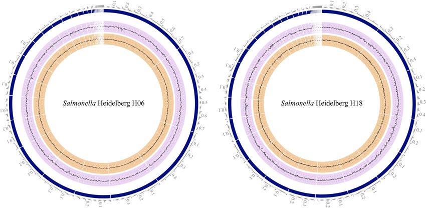
Melo et al.                                                                                               Characterization of Salmonella Heidelberg

with Campylobacter spp. being the first (EFSA, 2019). Whereas             different factors that influence the increase in its prevalence,
in Brazil, Salmonella spp. infections accounted for more than             resistance, and virulence must be studied for the effective
30% of all food-borne outbreaks between 2000 and 2017 (Brasil.            execution of control measures.
Ministério da Saúde, 2018).                                                  Biofilm formation is an essential factor that has an impact on
    S. Heidelberg (SH) is among the most prevalent serovars               the contamination of food during its processing. Sessile cells are
involved in human salmonellosis in North America, Europe, and             more resistant than planktonic cells to antimicrobials and typical
Brazil (Antunes et al., 2016; Deblais et al., 2018; Duarte, 2019).        sanitation processes. Few studies were conducted to characterize
The incidence of human infections by this serovar increased 25%           biofilms of this serovar (Borges et al., 2018; Silva et al., 2019).
between 1996 and 2015, even with the decrease of 9% in the total          Considering the importance and the emergence of SH, this study
number of serovar-unspecified salmonellosis cases (Foley and              aimed to evaluate the virulence, resistance, and phylogenetic
Lynne, 2008; Alzwghaibi et al., 2018; Duarte, 2019). In addition,         profile, as well as biofilm formation of SH strains of poultry
emerging microbial threats such as multi-drug resistant (Parisi           origin, discussing their possible danger to public health.
et al., 2018; Gupta et al., 2019) and virulent (Giuriatti et al.,
2017; Nakao et al., 2018; Yuki et al., 2019) strains have increased
the global concern about this serovar. Studies have shown the             MATERIALS AND METHODS
presence of multi-drug resistance to β-lactam antibiotics such
as third-generation cephalosporins (Parisi et al., 2018), along           Sample Collection and Identification
with very invasive strains, causing severe salmonellosis that has         Bacterial isolations were carried out between the years 2017 and
progressed to sepsis and endocarditis in identified outbreaks in          2018 in broiler batches, aged 11–46 days, in eight industrial units
the United States (Nakao et al., 2018; Yuki et al., 2019).                (A, B, C, D, E, F, G, and H) that received the animals from
    The molecular mechanisms involved in SH virulence are                 five different producers (1, 2, 3, 4, and 5). They were isolated
complex and different genes are involved in adhesion (lpfA;               from a Brazilian company, with a complete production cycle and
agfA), invasion (ompC; invA), and colonization (avrA). They               integration system, good standardization and regulatory practices
enable survival and multiplication of the microorganism in                verified by federal inspection, and qualified for national and
host cells, which initiate a series of subsequent events that             international trade.
trigger the disease (Suzuki, 1994). Furthermore, the ability to              Twenty SH strains were analyzed, stored on nutrient agar
form biofilms can be regulated by these and other genes, such             (NA, Difco), isolated from 17 poultry shed trawl swabs, one
as those that interfere with the quorum sensing mechanism                 fecal sample, one cecum sample and one sample from breast
(luxS) and allow communication between bacterial cells (Borges            (Table 1). Biochemical identification (ISO, 2002) and serotyping
et al., 2018). Together, factors that favor virulence and biofilm         were performed by the Enterobacteria Laboratory of the Oswaldo
formation can create an even more favorable environment for the           Cruz Foundation in the state of Rio de Janeiro (IOC/FIOCRUZ,
exchange of plasmid genes (Deblais et al., 2018; Oladeinde et al.,        Rio de Janeiro, Brazil).
2019), such as those encoding resistance to quinolone (qnrA,
qnrB, and qnrS), polymyxins (mcr-1), and β-lactam (blaTEM ,               Antimicrobial Susceptibility
blaSHV , blaCTX−M , and blaCMY−2 ) that can express resistance            The minimum inhibitory concentrations (MICs) were
to different classes of antibiotics that intensify the difficulty         determined for antimicrobials commonly used in human and
in treatment in case of infections (Monte et al., 2019). The              veterinary medicine. The tests were carried out in triplicates. The
growing concern about the impact of antimicrobial resistance              tested antimicrobials were ampicillin (AMP), amoxicillin-
for a wide range of drugs on animal bacteria of public health             clavulanate (AMO), colistin (COL), ceftriaxone (CFT),
importance also includes serovar SH. The increased resistance to          ciprofloxacin (CIP), meropenem (MER), sulfamethoxazole
the β-lactams such as penicillin and cephamycin (Monte et al.,            (SUL), and tetracyclin (TET). The test for cefoxitin (30 µg) (FOX)
2019), carbapenems (Mthembu et al., 2021) and broad-spectrum              was done by the disk diffusion method, just to complement the
cephalosporins (das Neves et al., 2020), as well as to other              discussion. The bacterial inoculum was prepared in NaCl
groups of non-β-lactam antimicrobials such as tetracyclines (das          solution 0.9% and the turbidity was adjusted to 0.5 on the
Neves et al., 2020; Souza et al., 2020), quinolones (Monte et al.,        McFarland scale (5 × 10⁸ CFU/mL). The suspension of the
2019), sulfonamides (Rodrigues et al., 2020), and polymyxins              bacterial inoculum was sown on a Müller Hinton (MH, Difco)
(Werlang et al., 2018) amplify the severity of clinical cases             agar plate up to 15 min after preparation. The plates were
that require hospitalization, as they are important drugs for             incubated at 35◦ C for a period of 16–24 h aerobically. The
therapeutic use.                                                          tested concentrations were: 0.5, 1, 2, 4, 8, 16, 32, and 64 mg
    Brazil is the largest exporter of chicken-based products              L−1 for all antimicrobials, except for sulfamethoxazole (16, 32,
Associação Brasileira de Proteína Animal (ABPA, 2020), with               64, 128, 256, 512, 1,024, and 2,048). The cutoff points were:
automated production and strict sanitary control for Salmonella           AMP > 32, AMO > 8/2, COL > 2, CIP > 0.6, CFT > 2,
spp. (Brasil, 2016; Mouttotou et al., 2017). Despite this, it is          MER > 4, SUL > 512, TET > 16 mg L−1 according to CLSI
important to highlight the increasing number of SH isolates               guidelines and recommendations for enterobacteria [Clinical
in poultry products, a consequence of the increase in the                 and Laboratory Standards Institute [CLSI], 2019]. Media with
genetic variability of this serotype (Webber et al., 2019). Thus,         the absence of bacteria and S. Typhimurium ATCC 14028 were
considering its wide distribution and variety of reservoirs, the          used as sterility control and positive controls, respectively.

Frontiers in Microbiology | www.frontiersin.org                       2                                     June 2021 | Volume 12 | Article 674147
Melo et al. Characterization of Salmonella Heidelberg
TABLE 1 | Data from SH strains isolated from Brazilian poultry samples, during the years 2017 and 2018, resistance phenotypic, and genotypic.
Id Date Company Producer Age of Material Resistance profile luxS and resistance genes (R -
collection animals bla)
(days)
H10 02/15/2018 A 1 26 Bed trawl COL, SMX, TET, AMP, CFT (P3) luxS
H11 02/15/2018 A 1 26 Bed trawl COL, SMX, TET, AMO, CFT, AMP, CIP (P6) luxS/CMY-2 (R1)
H12 02/15/2018 A 1 26 Bed trawl COL, SMX, TET, AMO, CFT, AMP (P5) luxS/CMY-2 (R1)
H15 02/16/2018 A 2 27 Bed trawl COL, SMX, TET, AMO, CFT, AMP, MER (P7) CMY-2 (R1)
H16 02/16/2018 A 3 26 Bed trawl COL, SMX, TET, AMP, CFT (P3) luxS/CMY-2 (R1)
H17 02/16/2018 A 4 26 Bed trawl COL, SMX, TET, AMO, CFT, MER (P4) luxS/CMY-2 (R1)
H18 02/16/2018 A 5 27 Bed trawl COL, SMX, TET, AMP, CFT (P3) CMY-2 (R1)
H19 02/16/2018 A 5 27 Bed trawl COL, SMX, TET, AMP, CFT (P3) CMY-2 (R1)
H03 02/09/2018 B 1 24 Bed trawl COL, SMX, TET, AMO, CFT, AMP, MER (P7) luxS/CMY-2 (R1)
H04 02/09/2018 B 2 21 Bed trawl COL, SMX, TET, AMO, CFT, AMP (P5) luxS/CMY-2 (R1)
H01 02/07/2018 C 1 11 Bed trawl COL, SMX, TET, AMP, MER (P2) luxS/TEM, CMY-2 (R4)
H02 02/08/2018 D 1 25 Bed trawl COL, SMX, TET, AMO, CFT, AMP (P5) luxS/CMY-2 (R1)
H05 02/09/2018 D 2 21 Bed trawl COL, SMX, TET, AMO, CFT, AMP (P5) luxS/CMY-2 (R1)
H06 11/29/2017 E 1 26 Bed trawl COL, SMX, TET, AMO, CFT, AMP, CIP (P6) luxS/CTX-M (R2)
H07 12/01/2017 F 1 40 Fecal sample COL, SMX, TET, AMO, CFT, AMP (P5) luxS/CMY-2, CTX-M (R3)
H20 02/16/2018 F 2 44 Bed trawl COL, SMX, TET, AMO, CFT, AMP, MER (P7) CMY-2 (R1)
H08 12/23/2017 G 1 46 Cecum COL, SMX, TET, AMP (P1) –
H09 02/15/2018 H 1 46 Chest COL, SMX, TET, AMP, CFT (P3) luxS/CMY-2 (R1)
H13 02/16/2018 H 2 24 Bed trawl COL, SMX, TET, AMP, CFT (P3) –
H14 02/16/2018 H 3 34 Bed trawl COL, SMX, TET, AMO, CFT, AMP (P5) luxS/CMY-2 (R1)
Total N (%) P1: 1 (5%); P2: 1 (5%); P3: 6 (30%); P4: 1 (5%); P5: R1: 14 (70%); R2: 1 (5%); R3: 1
6 (30%); P6: 2 (10%); P7: 3 (15%) (5%); R4: 1 (5%)
N (%), number and percentage of resistant strains; AMO, amoxicillin with clavulanic acid; COL, colistin; CFT, ceftriaxone; CIP, ciprofloxacin; AMP, ampicillin; MER,
meropenem; TET, tetracyclin; SMX, sulfamethoxazole; P, profile; R, bla: resistomes described by the bla genes.
PCR Detection of Virulence and Uberlândia was used as a positive control, for the presence of
Resistance Genes antimicrobial resistance genes.
Amplification was performed in a thermocycler (Eppendorf ), R
Extraction and purification of genomic DNA (gDNA) was
with an initial denaturation cycle at 94◦ C for 5 min and 35
performed using the Wizard Genomic DNA Purification
amplification cycles: denaturation at 94◦ C for 45 s, annealing
Kit (Promega). The quantification of DNA was performed
according to Table 2, extension at 72◦ C for 90 s, with final
using NanoDropTM 2000/2000c Spectrophotometer (Thermo
extension at 72◦ C for 10 min. The amplification conditions for
ScientificTM ). PCR reactions were performed using 10 ng
the antimicrobial resistance genes to β-lactams differed in terms
of purified DNA. The presence of ompC (biosynthesis
of the number of cycles (30) and annealing conditions (Table 2).
of outer membrane protein C; invasion), avrA (effector
For the qnrA, qnrB, and qnrS genes the initial denaturation was
protein-colonization), sodC (elimination of free radicals), invA
95◦ C for 10 min, followed by 35 denaturation cycles at 95◦ C
(invasion), sefA (fimbriae-adhesion), agfA (fimbriae-biofilm),
for 1 min, annealing (Table 2), extension at 72◦ C for 90 s, and
lpfA (fimbriae-adhesion) and luxS (quorum-sensing mechanism)
finally, final extension at 72◦ C for 10 min. The amplified products
genes was determined by PCR. The presence of the genes
were submitted to electrophoresis in 1.5% agarose gel, using the
blaTEM , blaSHV, blaCTX−M , blaCMY−2 (linked to beta-lactamases
TBE 0.5x running buffer (Invitrogen) and as a molecular weight
production), qnrA, qnrB, and qnrS (linked to quinolone
standard, the 100 bp marker (Invitrogen).
resistance), and mcr-1 (linked to colistin resistance) were also
tested in all strains.
PCR reactions were conducted with GoTaq Green Master R
Mix kit (Promega), contemplating a final volume of 25 µL. For Genotyping by Pulsed-Field Gel
each reaction, a volume of 12.5 µL of Green Mix, 10.5 µL of Electrophoresis
MiliQ water, 1 µL of R/F primers (concentrations described in To verify the genetic relatedness of isolates, pulsed-field gel
Table 2), and 1 µL of DNA at 10 ng/µL were used. S. Enteritidis electrophoresis (PFGE) analysis was executed using the PulseNet
ATCC 13076 and ultrapure water were used as positive and protocol, as previously recommended (CDC, 2017). Bacteria
negative controls, respectively, for the analysis of virulence genes. grown at 37◦ C overnight on Tryptic Soy Agar (TSA) (OXOID ) R
A Klebsiella pneumoniae strain, previously tested, provided by the were suspended in tubes containing 2 mL of phosphate buffered
Molecular Microbiology Laboratory of the Federal University of saline (PBS: 0.01 M phosphate buffer; pH 7.2; 0.85% NaCl). After
Frontiers in Microbiology | www.frontiersin.org 3 June 2021 | Volume 12 | Article 674147Melo et al. Characterization of Salmonella Heidelberg
TABLE 2 | Primers used to identify specific genes in SH strains.
Gene Concentration Amplicon Primer 50 –30 T. annealing Reference
(bp)
ompC 10 pmol 204 ATCGCTGACTTATGCAATCG 58◦ C/30 s Jawad and Al-Charrakh, 2016
CGGGTTGCGTTATAGGTCTG
avrA 20 pmol 385 GTTATGGACGGAACGACATCGG 62◦ C/30 s Prager et al., 2003
ATTCTGCTTCCCGCCGCC
sodC 20 pmol 500 ATGAAGCGATTAAGTTTAGCGATGG 62◦ C/30 s Sanjay et al., 2010
TTTAATGACTCCGCAGGCGTAACGC
invA 10 pmol 284 GTGAAATTATCGCCACGTTCGGGCAA 58◦ C/30 s Oliveira et al., 2002
TCATCGCACCGTCAAAGGAACC
sefA 10 pmol 488 GATACTGCTGAACGTAGAAGG 50◦ C/30 s Oliveira et al., 2002
GCGTAAATCAGGATCTGCAGTAGC
agfA 10 pmol 350 TCCACAATGGGGCGGCGGCG 66◦ C/30 s Collinson et al., 1993
CCTGACGCACCATTACGCTG
lpfA 10 pmol 250 CTTTCGCTGCTGAATCTGGT 50◦ C/30 s Webber et al., 2019
CAGTGTTAACAGAAACCAGT
luxS 20 pmol 1080 GATAATCCTGAACTAAGCTTCTCCGC 62◦ C/30 s Choi et al., 2007
GGTTATGAGAAAAGCATGCACCGATCA
blaTEM 10 pmol 643 CAGCGGTAAGATCCTTGAGA 50◦ C/45 s Chen et al., 2004
ACTCCCCGTCGTGTAGATAA
blaSHV 10 pmol 714 GGCCGCGTAGGCATGATAGA 56◦ C/45 s Chen et al., 2004
CCCGGCGATTTGCTGATTTC
blaCMY −2 10 pmol 870 TGGCCGTTGCCGTTATCTAC 59◦ C/53 s Chen et al., 2004
CCCGTTTTATGCACCCATGA
blaCTX−M 10 pmol 593 TGGGTRAARTARGTSACCAGAAYCAGCGG 58◦ C/60 s Monstein et al., 2007
CCCCCGCTTATAGAGCAACAACAA
Mcr-1 10 pmol 309 CGGTCAGTCCGTTTGTTC 58◦ C/40 s Liu et al., 2016
CTTGGTCGGTCTGTAGGG
qnrA 10 pmol 580 AGAGGATTTCTCACGCCAGG 54◦ C/60 s Cattoir et al., 2007
TGCCAGGCACAGATCTTGAC
qnrS 10 pmol 428 GCAAGTTCATTGAACAGGGT 54◦ C/60 s Cattoir et al., 2007
TCTAAACCGTCGAGTTCGGCG
qnrB* 10 pmol 264 GGMATHGAAATTCGCCACT 54◦ C/60 s Cattoir et al., 2007
TTTGCYGYYCGCCAGTCGA
T, temperature.
*Able to amplify the qnrB1 to qnrB6 variants.
agarose blocking, gDNA digestion was performed with 30 U of and H18 – Table 1 and Figure 1). To evaluate the cell adherence
Xba1 enzyme (Invitrogen ) for 2 h at 37◦ C.
R
of the selected strains, bacterial inoculum (104 CFU/mL–
The DNA fragments were separated on 1% agarose gel OD600 = 0.22–0.28) was centrifuged (5,000 rpm, 10 min, 4◦ C)
(SeaKem Gold ) in 0.5X TBE buffer in CHEF DRIII (Bio-Rad ,
R R
and washed twice (NaCl 0.9%). Then, 20 mL of Tryptic Soy Broth
California, United States) for 18 h with the following parameters: (TSB) (Merck ) was added, and in parallel, 20 mL of TSB was
R
200 V, 120◦ angle, 6 V/cm gradient and 14◦ C buffer temperature. supplemented with 5% chicken juice (CJ) to simulate industry
The gels were stained with ethidium bromide, photographed conditions and compare both situations (Brown et al., 2014).
under UV light in a transilluminator (Loccus Biotechnology ) R
Qualitative analysis of the biofilms was performed
and evaluated using the BioNumerics program. according to recommendations (Kudirkienë et al., 2012),
with modifications, including eight replicates in each of the
three repetitions. Briefly, 200 µL of the bacterial suspension in
Biofilm Production TSB medium and TSB enriched with CJ was added in 96-well
Qualitative and quantitative phenotypic analyzes of the sessile plates. For biomass formation, the plates were incubated for
structure at temperatures of 4, 25, and 37◦ C were performed 24 h at different temperatures (4, 25, and 37◦ C) under agitation
using two phylogenetically different strains, from different (100 rpm). After incubation, the washed and dried total biomass
origins and with differing virulence and resistance profiles (H06 was measured by fixing with 0.1% Violet Crystal (LaborClin),
Frontiers in Microbiology | www.frontiersin.org 4 June 2021 | Volume 12 | Article 674147Melo et al. Characterization of Salmonella Heidelberg
FIGURE 1 | Comparative dendrogram of 20 SH strains, constructed from PFGE results considering the isolation site, date of collection, and the presence or
absence of specific genes and antimicrobial resistance, using the Dice similarity coefficient with 1.5% tolerance and UPGMA method with 0.80% optimization. I-VI,
pulsotypes; Id, Identification. Genes: only those that showed differences between the strains. Profile: antimicrobial resistance profile (P1-P7; Table 1).
followed by elution with alcohol-acetone solution (80:20 v/v formed in glass spheres with a diameter of 5 mm, in TSB
ethanol-acetone) (Dinamica ). R
respecting the growth conditions described above. After biomass
The determination of the biofilm formation index (BFI) was formation, the samples were fixed with 2.5% glutaraldehyde and
carried out according to Stepanović et al. (2000) from the results 2.5% paraformaldehyde in 0.1 M PBS buffer (pH 7.4) overnight at
of reading the OD600 . As cutoff (ODc), the average value of 4◦ C. Samples were washed three times with PBS buffer. The beads
OD600 equivalent of 0.044, obtained by adding the average were post-fixed with 1% osmium tetroxide for 1 h and washed
optical density of negative controls (which is 0.007) the offset three times with PBS buffer. The spheres were dehydrated in a
value of the standard multiplied by three (0.0124 × 3), was series of ethanol solutions (30, 40, 50, 60, 70, 80, and 90% and
used. The classification of biofilms was determined as follows: then three times at 100%) for 15 min in each step.
(a) non-existent: OD600 ≤ 0.044 (less than ODc); (b) weak: The samples were dried on CPD (Critical Point Drying)
0.088 ≤ OD600 > 0.044 (up to two times higher than ODc); (c) (CPD 030, Baltec, Germany) using liquid carbon dioxide as the
moderate: 0.176 ≤ OD600 > 0.088 (between two to four times transition fluid, then coated with a 20 nm thick gold layer (SCD
the ODc value); and (d) strong: OD600 > 0.176 (greater than four 050, Baltec, Germany) and visualized in MEV VP Zeiss Supra 55
times the ODc value). FEG SEM operating at 20 kV.
Quantitative adhesion (2 h of incubation) and biofilm tests
(24 h of incubation) were determined by the number of CFUs Biofilm Inhibition Assay
of sessile cells. Biofilms were formed under the same conditions To examine the interaction between H06 and H18 biofilms
described above for the qualitative test with three replicates per with disinfectant components (0.8% peracetic acid, 1% sodium
repetition. After the formation of the adhered mass and biofilm, hypochlorite and 1% chlorhexidine), a modified protocol
the wells were washed twice with 0.9% NaCl solution and the followed by Nguyen and Yuk (2013) was used.
biomass was removed by scraping the wells for 90 s. The cell An aliquot of 100 µL (107 cells) was inoculated on the surface
suspension obtained was subjected to serial dilutions and seeded of a sterile cellulose membrane with porosity of 0.45 µm and
on TSA agar plates to obtain the CFU number. 47 mm in diameter, on a TSA plate (Merck ). The plates were
R
incubated at 37◦ C and had the membrane transferred to a new
Scanning Electron Microscopy plate every 24 h for 3 days. Subsequently, the membrane was
The ultrastructure of the sessile form of H06 and H18 was placed in a flask containing 20 mL of TSB broth with the
evaluated by Scanning Electron Microscope (SEM), using a concentrations of the disinfectant components. After incubation
modified method followed by Brown et al. (2014). Biofilms were at 37◦ C for 15 min, the membrane was washed three times with
Frontiers in Microbiology | www.frontiersin.org 5 June 2021 | Volume 12 | Article 674147Melo et al. Characterization of Salmonella Heidelberg
phosphate buffer (PBS), followed by treatment in 25 mL of 0.1% Pathways significantly enriched in KEGG PATHWAY database2
trypsin for 15 min at room temperature. Then, the resulting were then checked.
solution underwent serial dilutions for subsequent counting on
TSA plates, at 37◦ C for 24 h. Data Analyses
In addition to descriptive statistics, the quantitative assays with
Genomic Sequencing biofilms tests were compared by one-way analysis of variance
The strains H06 and H18 had their genomes sequenced. First, (One-way ANOVA). Two variables’ comparisons were performed
gDNA was obtained through Wizard Genomic DNA Purification using student’s t-test and Fisher exact test. Confidence level of
Kit (Promega). A 450 bp read library was constructed using 95% was considered, using the GraphPad Prism software, version
NEBNext Fast DNA Fragmentation and Library Preparation Kit 8.0 (GraphPad Software, United States).
(New England Biolabs, Ipswich, NE, United States) and quality Analysis for dendrogram construction was conducted using
checked by the Agilent 2100 Bioanalyzer. BioNumerics software. The comparison of the band patterns
The gDNA library was then sequenced by 2 × 150 bp paired- was performed by the UPGMA analysis method, using the Dice
end sequencing on an Illumina HiSeq 2500 sequencing platform similarity coefficient with a tolerance of 1.5% in the comparison
(Illumina, San Diego, CA, United States). Read quality was of the position of the bands.
assessed using FastQC (Andrews, 2010). De novo assembly was
performed using Unicycler (v3.1) (Wick et al., 2017). Assembly
quality statistics were generated using QUAST (Gurevich et al., RESULTS
2013). Initial scaffolding was performed using SSPACE (Boetzer
et al., 2011) and the gaps closed using GapFiller (Nadalin et al., Virulence Factors and Resistance
2012). S. Heidelberg SH14-009 genome sequence obtained from
Profiles
GenBank (accession CP016581.1) was used as the reference
to generate final scaffold in CONTIGuator (Galardini et al., All strains (100%) of the study carried the ompC, invA,
2008). Genome completeness was assessed using Benchmarking sodC, avrA, lpfA, and agfA genes, but only 70% (14/20) of
Universal Single-Copy Orthologs (BUSCO) v4.0.6 (Simao et al., them had the luxS gene. No strains showed the presence of
2015). The presence of plasmid-predicted contigs was accessed the sefA gene. Table 3 shows the percentage of strains with
using gplas (Arredondo-Alonso et al., 2020). Its results, along phenotypic resistance to the tested drugs, demonstrating that
with completed circular contigs predicted by Unicycler, were the highest susceptibility (p < 0.05) was identified for CIP
merged to obtain all predicted plasmid sequences (circular (90%), MER (75%), and AMO (40%). All strains were resistant
and uncircular contigs). Then, BLASTn against the non- to COL, TET, and SMX, being characterized as multidrug
redundant database of NCBI was used to confirm the plasmid resistant (MDR) strains.
prediction. Identified plasmids had their sequences extracted The identified resistance profiles (Table 1) show resistance
and the remaining contig sequences were considered part of the to at least four classes of antimicrobials. 60% (12/20) of the
bacterial chromosome. Annotation was performed using Prokka strains showed resistance to at least six drugs and include profiles
(Seemann, 2014). The sequenced SH genomes were deposited P4, P5, P6, and P7. The resistome analysis did not identify the
at the DDBJ/ENA/GenBank under the accession numbers presence of the blaSHV , qnrA, qnrB, and qnrS genes in any of
JAGFJR000000000 (H06) and JAGEOP000000000 (H18). the strains. However, all of them showed phenotypic resistance
to the classes of penicillin (AMP and AMO) and/or cefens (CFT,
In silico Genomic Characterization FOX). This result was consistent with the genotype (R1 to R4)
ABRicate (v 0.9.9) (Seemann, 2020) was used to screen virulence related to the presence of one or more of the bla genes tested in
and resistance genes. This tool allows the use of multiple 17 (85%) from them.
databases. Search for virulence genes was completed using the The presence of bla genes co-produced in this group
VFDB database (Chen et al., 2016). Resistance genes were was identified in 2/17 (11.8%), corresponding to the R3 and
screened using the CARD database (Jia et al., 2017). The VFDB R4 resistomes, determinants of the phenotypic resistance to
database includes 2,597 curated genes related to virulence factors penicillins (R4), and to the cefens in R3. For R2 and R3 resistomes
whereas the CARD database includes 2,617 genes related to showed the presence of the blaCTX−M gene found in 2/20 (10%),
antimicrobial resistance. Hits were only considered for gene expressing phenotypic resistance to penicillin and CFT. Only one
lengths ≥75% and identities of ≥75%. strain (1/20–5.0%) (R4) presented the blaTEM gene and expressed
Presence of chromosomal mutations was predicted using phenotypic resistance to ampicillin in the MIC test.
ResFinder v. 4.1 with default settings (90% selected percentage The blaCMY−2 gene was the most prevalent (16/20 – 80%)
identity threshold and 60% selected minimum coverage length), and was identified in the R1 and R3 resistomes. In addition
available at the Centre for Genomic Epidemiology1 . to determining phenotypic penicillin resistance, unanimous
Virulence genes related to biofilm formation selected by resistance to CFT was observed. In parallel, disc diffusion
manual curation was used as input to generate an association assay showed that the four strains resistant to cefoxitin (FOX)
protein-protein network in STRING (Szklarczyk et al., 2019). presented this gene in their resistome (data not shown).
1 2
https://cge.cbs.dtu.dk/services/ResFinder/ https://www.genome.jp/kegg/pathway.html
Frontiers in Microbiology | www.frontiersin.org 6 June 2021 | Volume 12 | Article 674147Melo et al. Characterization of Salmonella Heidelberg
Clustering of Strains adhesion process (p < 0.05) at temperatures of 25 and 37◦ C, but
The dendrogram constructed from the PFGE results was the presence of CJ did not favor this process at the quantitative
compared considering the isolation site, date of collection, and level (p = 0.124) (Table 4).
genotypic and phenotypic characteristics evaluated. Similarity The 2-h incubation period was sufficient for the initial
analysis of the 20 strains of SH presented six pulsotypes (I – establishment of the biofilm structure, except for the temperature
VI; Figure 1) that showed genotypic similarities above 80%. In of 4◦ C, which allows us to infer that lower temperatures may
opposition, six profiles showed little genetic proximity ( 0.05) in
Characterization of SH Biofilms all assays, it was observed that for the strain H06 there was a
Strains H06 and H18 demonstrated the same ability to adhere significant increase in TSB + CJ counts when compared to TSB
when inoculated in TSB and TSB + 5% CJ (p = 0.153). counts at 25◦ C, but the reverse was observed at 4 and 37◦ C. For
Considering the initial inoculum of approximately 103 CFU/well, both strains, 4◦ C was the temperature that least favored bacterial
it was found that there was an increase in counts after the multiplication. More efficient replication capacity in the sessile
TABLE 3 | Distribution of MIC and resistance index of SH.
Antibiotic concentration (mg/L) AMP AMO TET COL CFT MER CIP SMX* Antibiotic concentration (mg/L)*
2,048
Total of resistant R (%) 19a (95.0) 12ac (60.0) 20a (100.0) 20a (100.0) 18a (90.0) 5bc (25.0) 2b (10.0) 20a (100.0) Total of resistant R (%)
AMO, amoxicillin with clavulanic acid; COL, colistin; CFT, ceftriaxone; CIP, ciprofloxacin; AMP, ampicillin; MER, meropenem; TET, tetracyclin; SMX, sulfamethoxazole; ___
(line), cutoff point according to Clinical and Laboratory Standards Institute [CLSI], 2019; dark gray, resistant strains; light gray, strains with intermediate resistance; R (%),
resistance index.
Different letters on the line indicate a significant difference (Fisher’s Test p < 0.05).
*Specific concentrations and results for the sulfamethoxazole test (SMX).
TABLE 4 | Mean counts (Log CFU/mL) and BFI (Biofilm Formation Index) obtained in the tests for the two strains of SH under different temperature conditions.
Test Strain Media Initial inoculum 4◦ C 25◦ C 37◦ C
Adhesion Both mean TSB 3.22 ± 0.29 a 3.99 ± 0.21 a 4.05 ± 0.12 b 4.81 ± 0.33 b
TSB + CJ 3.25 ± 0.32 a 3.74 ± 0.12 a 4.64 ± 0.25 b 4.86 ± 0.43 b
Biofilm Both mean TSB 3.22 ± 0.29 a 4.895 ± 0.35 b 5.825 ± 0.51 c 6.471 ± 0.49 c
TSB + CJ 3.25 ± 0.32 a 4.456 ± 0.5 b 6.294 ± 0,0.3 c 6.529 ± 0.41 c
Biofilm H06 TSB – 5.130 ± 0.14* a I 6.373 ± 0.19* b I 7.256 ± 0.02* b
TSB + CJ – 4.485 ± 0.38* a 7.274 ± 0.03* b 7.206 ± 0.01* b
Biofilm H18 TSB – 4.731 ± 0.36 a I 5.459 ± 0.22 b I 6.320 ± 0.10 b
TSB + CJ – 4.436 ± 0.35 a 5.263 ± 0.06 b 6.452 ± 0.01 b
BFI (Class.) Both mean TSB – 0.0353 (N) 0.1611 (M) 0.1622 (M)
TSB + CJ – 0.0329 (N) 0.3491 (S) 0.3970 (S)
Class., classification by BFI; N, non-existent; M, moderate; S, strong.
Different letters indicate significant difference compared to the initial inoculum, the test one-way ANOVA (adhesion test). Different letters indicate significant difference,
one-way ANOVA test (biofilm test).
*Indicate significant when comparing the culture media TSB and TSB + CJ, student’s t test (biofilm test in individual strain). Iindicate significant difference when strains
are compared in relation to temperature, student’s t test (biofilm test in compare strains).
Frontiers in Microbiology | www.frontiersin.org 7 June 2021 | Volume 12 | Article 674147Melo et al. Characterization of Salmonella Heidelberg FIGURE 2 | SEM images of two strains of SH at temperatures of 4 (A,B), 25 (C,D), and 37◦ C (E,F) in TSB. form, considering the conditions supplemented or not with CJ, formation were observed with more intense extracellular matrix was observed at 25 and 37◦ C for strain H06 (Table 4). production at 25◦ C, while in Figures 2E,F the presence of a The joint evaluation of the strains demonstrated that the CJ three-dimensional structure of the matrix becomes more evident, did not influence the number of bacteria present in the biofilm at indicating the development of a mature biofilm at 37◦ C. the three temperatures tested, but favored the greater production Greater biomass production was observed at 25 and 37◦ C in of extracellular matrix at 25 and 37◦ C. Similarly, the increase in both strains. The characteristic of the matrix in these biofilms temperature from 4 to 25◦ C or 37◦ C increased (p < 0.05) the indicates a dense and compact structure. However, the inhibition number of bacteria adhered in biofilms (Table 4). of biofilm formation by chemical agents demonstrated the same SEM showed changes in the biomass formed in both behavior in both strains for all treatments (p > 0.2). None of strains cultivated in TSB at different temperatures (Figure 2). the treatments promoted total elimination of viable cells, but Figures 2A,B show a minimum synthesis of extracellular matrix all agents significantly reduced the counts (Figure 3) of SH in fibers in isolated bacteria, indicating the incapacity to form relation to the untreated biofilm (7.23 log CFU/mL). biofilm at 4◦ C. This result is consistent with the qualitative The treatment with sodium hypochlorite 1% reduced assay found in the biofilms. In Figures 2C,D, the initial biofilm the number of sessile bacteria to 4.83 log CFU/mL, while Frontiers in Microbiology | www.frontiersin.org 8 June 2021 | Volume 12 | Article 674147
Melo et al. Characterization of Salmonella Heidelberg
FIGURE 3 | Graph of biofilm counts for both strains of SH (log of CFU mL− 1 ) in control and maintained for 15 min in 0.8% peracetic acid (AP), 1% sodium
hypochlorite (HS) and 1% chlorhexidine (CX). *p < 0.0001; **p < 0.001 using one-way ANOVA for counts between treatments.
FIGURE 4 | Circular graphical display of the distribution of the contigs of Salmonella Heidelberg H06 and H18 genomes assembly. From outer to inner rings, we
present contigs (in Mbp), GC content, and GC skew, produced by Circos software.
peracetic acid 0.8% reduced about 3.51 log cycles compared 52.17%, while strain H18 had 43 contigs, with the total length of
to untreated biofilm. The use of chlorhexidine 1% had the 4,838,250 bp and an average G + C content of 52.11% (Figure 4).
greatest effect on the control of the sessile structure with a Findings of BUSCO analysis in the genomes of strains H06 and
viable biomass of 2.69 log CFU/mL, representing a reduction of H18 showed 99.5 and 99.5% completeness, respectively, using the
4.53 log cycles. Enterobacterales dataset based on conserved orthologous genes
among species within the order.
Gene prediction and annotation of the Salmonella strains
Genome Overview and in silico resulted in 4,431 and 4,544 predicted protein coding sequences
Prediction (CDS), for strains H06 and H18, respectively, 76 transfer RNA
The assembled chromosome of strain H06 had 35 contigs, with (tRNA) genes for both strains, and four ribosomal RNA (rRNA)
the total length of 4,731,348 bp and an average G + C content of genes in strain H06, while five in H18.
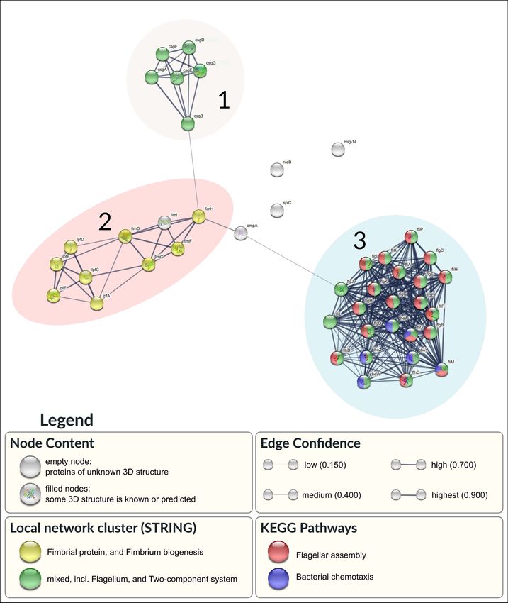
Frontiers in Microbiology | www.frontiersin.org 9 June 2021 | Volume 12 | Article 674147Melo et al. Characterization of Salmonella Heidelberg FIGURE 5 | Association network in STRING of the 44 selected proteins. Network nodes represent proteins. Proteins with enriched pathways are colored by red (Flagellar assembly) and blue (Bacterial chemotaxis). Additional node clustering is highlighted in yellow (Fimbria biogenesis) and green (Flagellum and Two-component system). Edges represent predicted protein-protein association with specific confidence score, according to its thickness. Plasmid prediction of the assembled contigs of SH genomes system. Manual screening for virulence genes coding for resulted in three plasmids on strain H06 and six on strain H18. proteins related to biofilm formation resulted in 44 proteins The results of the genomic virulence typing for both isolates (Supplementary Table 2). Interactions between them are shown are shown in Supplementary Table 1. Among the two isolates, in Figure 5. 135 different virulence genes were identified. The majority A total of 49 different resistance genes were identified, of the genes (127/135) were present in both genomes. Only belonging to 24 classes of antibiotics. Five of these genes eight genes were exclusively found in the genome of strain [ANT (300 )-IIa, AAC (3)-Via, sul1, blaCTX−M−2 , and blaCMY−59 ] H18. All of them (sptP, prgH, prgI, prgJ, prgK, orgA, orgB, are strain specific (the first four in H06 and the last one in and orgC) code for proteins related to a type III secretion H18) (Supplementary Table 3). The presence of plasmid genes Frontiers in Microbiology | www.frontiersin.org 10 June 2021 | Volume 12 | Article 674147
Melo et al.                                                                                                  Characterization of Salmonella Heidelberg

associated with resistance to aminoglycosides (aac3, aac6, and              as blaCTX−M , is highlighted by their ability to produce
ant3), sulfonamides (sul1 and sul2), β-lactams (blaCTX−M−2 and              extended-spectrum β-lactamases (ESBLs). Considering the
blaCMY−2 ), fosfomycin (fosA7), and tetracycline (tetA), featuring          importance of this antibiotic class as the first-choice treatment of
an alarming multi-resistance profile.                                       salmonellosis, the presence of resistant strains is alarming. It is
   The gene blaCTX−M−2 was found only in the genome of the                  estimated that approximately 26,000 infections and 1,700 deaths
strain 6H, while blaCMY−2 only appeared in the strain 18H. None             occur annually in the United States caused by ESBL-producing
of the strains carries the qnrB gene. Point mutations in DNA                strains, generating enormous hospital expenses to the country.
gyrAse subunit A (gyrA; Ser83Phe) and DNA topoisomerase IV                  Recently, the number of cases reporting ESBL-producing
subunit C (parC; Thr57Ser) conferring resistance to nalidixic acid          Salmonella has increased worldwide, becoming one of the major
and ciprofloxacin were observed in both strains. No mutation                public health concerns. blaCTX and blaCMY−2 are the most
was detected in gyrAse subunit B (gyrB) and topoisomerase                   identified genes in isolates of food-producing animals (Biffi et al.,
IV subunit E (parE). Mutation in gyrA occurred at the                       2014; Qiao et al., 2017; Jeon et al., 2019).
eighty-third codon, where the amino acid serine was substituted                The four identified resistomes (Table 1) revealed a high
by phenylalanine (gyrA; Ser83Phe). Mutation in parC occurred                level of resistance to amoxicillin, ampicillin and/or ceftriaxone.
at the fifty-seventh codon, where the amino acid threonine was              Although the blaCTX−M gene is reported with a higher incidence
substituted by serine (parC; Thr57Ser).                                     of Salmonella in poultry products (Moura et al., 2018; Perin et al.,
                                                                            2020), our study shows that its frequency was low (2/20–10%)
                                                                            and corroborates with recent studies carried out with samples
DISCUSSION                                                                  Brazilian samples (van den Berg et al., 2019; Souza et al., 2020).
                                                                            Gram-negative ESBL-producing and hyper-producing AmpC
Antimicrobial Resistance Profile                                            bacteria have also stood out for their resistance to carbapenems,
Our findings demonstrated higher phenotypic antimicrobial                   such as meropenem, identified in 20% (4/20) of our strains, when
resistance against to AMO, AMP, COL, CFT, SMX, and TET                      associated with other mechanisms, such as hyper expressed efflux
commonly used in veterinary and human medicine routine when                 systems (Andrade and Darini, 2017).
compared to results obtained in Brazil and Argentina (Borsoi                   The phenotypic co-resistance to TET and SUL identified in
et al., 2009; Aravena et al., 2019).                                        all strains in the present study may be associated with the
    There are no reports of studies in Brazil on resistance                 expression of tetA as well as the sul1 and sul2 genes, detected
to colistin in SH in birds, but the previous permission to                  in strains H06 and H18 by WSG. tetA gene, like the tet gene
use this drug in poultry production generated a selection                   complex, has wide distribution in several serovars of Salmonella
pressure of resistant strains that remained, even after the                 (Michael et al., 2006; Delgado-Suárez et al., 2019), including
ban of its use in 2016 (BRASIL, 2016). Other serovars with                  SH (Keefer et al., 2019). Tetracyclines have a great capacity for
this phenotype have been identified and associated with                     intracellular diffusion, reversibly preventing protein synthesis
the presence of the plasmid-mediated mcr gene (Borowiak                     (Brasil. Ministério da Saúde, 2007; Luiz et al., 2008). The sul1 and
et al., 2019; Trujillo-Soto et al., 2019) that codes for                    sul2 genes encode forms of enzymes (dihydropteroate synthase)
phosphoethanolamine transferase (pEtN transferase) and                      that are not inhibited by sulfonamides (Lynne et al., 2008). In
changes the LPS, target of this antibiotic, inhibiting the action           parallel, we also identified the plasmid pSH-04-1 in H06 and H18,
of colistin (Baron et al., 2016). However, this gene was not                which was associated with resistance to sulfonamides, β-lactams,
detected in our strains, the ugd gene was identified in the H06             aminoglycosides, and tetracycline in SH (Hoffmann et al., 2014;
and H18 strains by WSG, and may also be the cause of the                    Keefer et al., 2019).
observed phenotype since it encodes polymyxin resistance                       Although we did not identify qnr in any of the strains, nor
(Mouslim and Groisman, 2003).                                               via WSG, we can associate the resistance of 2/20 (10%) to this
    Similar to colistin resistance, 90% of the studied strains              drug with the presence of point mutations in gyrA at codons
were resistant to third-generation cephalosporins, in particular            Ser83Phe and parC; Thr57Ser, in H06, that conferring phenotypic
for ceftriaxone, an important drug used to treat children                   resistance to ciprofloxacin. The same mutation regions that
with salmonellosis (das Neves et al., 2020). The presence of                confer resistance to quinolones have also been identified by
the blaCMY−2 gene is consistent with this phenotype, due                    Monte et al. (2019) in SH.
to the overproduction of AmpC induced by the presence                          In addition to the genes already mentioned, four of them
of cephalosporin and cephamycin that exhibit resistance to                  (aac3, aac6, ant3, and fosA7) were also previously reported in SH
extended-spectrum cephalosporins, such as ceftriaxone, and to               strains isolated from clinical, environmental and animal samples
cefoxitin (identified in four of the 20 strains – data not shown)           (Hoffmann et al., 2014; Keefer et al., 2019) with high potential
(De Angelis et al., 2020). The high prevalence of SH AmpC                   for plasmid gene transfer, via conjugation, in species of the order
strains highlights the endemic occurrence of β-lactam resistance            Enterobacterales in the intestinal microbiota (Lim et al., 2013).
in North and South America (Edirmanasinghe et al., 2017; Monte                 The aac3, aac6, and ant3 genes are directly involved in
et al., 2019; van den Berg et al., 2019; Souza et al., 2020).               resistance to aminoglycosides and acts to alter the function of
    β-lactam antibiotics act on the bacterial cell wall, interfering        the bacterial ribosome by binding to its 30S fraction, inhibiting
with the synthesis of the peptidoglycan layer. Bacterial resistance         protein synthesis or producing defective proteins. The identified
mediated by plasmids belonging to the bla subgroups, such                   resistance plasmids encode proteins that act in enzymatic

Frontiers in Microbiology | www.frontiersin.org                        11                                      June 2021 | Volume 12 | Article 674147
Melo et al. Characterization of Salmonella Heidelberg
modifications of antibiotics, mainly gentamicin (Brasil. all, 58.2% are environmental and chicken-isolated samples, while
Ministério da Saúde, 2007; Luiz et al., 2008; Noh et al., 2019). only 6.2% are described as clinical isolates (Rehman et al., 2019).
The presence of fosA7 (recently reported – 2017) is alarming
because its sequence was found in only 36 of the approximately Specific Virulence Genes
40,000 genomes in the NCBI database. 75% of them belong Previous studies with SH isolated from various sources between
to serovar SH. In addition, studies show that this gene easily 1985 and 2011, from the United States and Brazil, shows that
integrates with Salmonella chromosomal DNA (Keefer et al., virulence factors are highly conserved for this serovar (Hoffmann
2019; Rehman et al., 2019). Fosfomycin is used to treat systemic et al., 2014). Our results are consistent with these findings, as 6/8
diseases caused by members of the Enterobacteriaceae family genes were identified in all strains.
with advanced resistance to antimicrobial drugs, including ESBL The ompC and invA genes are used to characterize the genus
producers (Hou et al., 2012). The presence of the fosA7 gene and the ability to invade host tissues, respectively, and the
was identified via WGS, co-existing with blaCTX−M (strain presence in all strains was expected. Together with the agfA
H06) and blaCMY−2 (strain H18). There are other reports that and lpfA genes, they were previously identified in all Salmonella
identified fosfA7/blaCMY−2 in SH (Rehman et al., 2019) and isolates in Brazil and Chile (Rowlands et al., 2014; Aravena et al.,
fosA3/blaCMY−2 in other enterobacteria (Zhao et al., 2015). This 2019). Both encode fimbriae-related proteins that carry essential
shows that these genes can be co-selected and transported in adhesins that are associated with cell attachment to abiotic
plasmids IncFIIa and F33: A-: B- for blaCTX−M (Hou et al., 2012; surfaces and biofilm formation for later intestinal colonization
Cunha et al., 2017) and HD0149 for blaCMY−2 blaCMY-2 (Zhao and expression of virulence, which indicates potential risk after
et al., 2015), intensifying the horizontal transfer of genes and infection (Yoo et al., 2013).
MDR profile in SH (Rehman et al., 2017) and other enterobacteria The sefA gene also encodes a fimbria-related protein (SefA)
(Zhao et al., 2015). that is associated with the adhesion process. It is restricted to
The MDR profile identified in all strains intensifies the public group D serovars, Enteritidis, Dublin, Moscow, and Blegdon
health alert, since there was an outbreak involving multidrug- serotypes (Amini et al., 2010). Thus, the absence of this gene
resistant SH in the United States (Nakao et al., 2018). Between in SH was expected, but its presence was investigated due to
January 2015 to November 2017, 56 people were infected in 15 the possibility of genetic recombination, which can occur when
United States’ states by multidrug-resistant SH strains (β-lactams, the sefABCD genes present G + C around 35.2%, suggesting
fluoroquinolones, tetracyclines, aminoglycosides, sulfonamides, horizontal transfer (Bäumler et al., 1997; Mendonça et al., 2020).
and carbapenems), from veterinary environments (CDC, 2018). The avrA gene encodes effector proteins essential for infection
In Brazil, similar results were found by other researchers, and bacterial proliferation, escaping the host’s immune system.
with resistance in SH to different antibiotic classes, such as This mechanism is possible through the induction of cellular
penicillins, cephalosporins, quinolones, streptomycin, nalidixic apoptosis that limits the inflammatory response to infection
acid, cefotaxime, cefoxitin, ceftiofur, and tetracyclines (Monte (Labriola et al., 2018). It is a highly conserved gene in
et al., 2019; Souza et al., 2020). Salmonella spp. of public health importance, including SH
The identification of five strains (25%) resistant to seven (Borsoi et al., 2009).
tested drugs (P6 and P7) indicates the indiscriminate use of Despite the high potential for adhesion and the biofilm
antimicrobials and neglect of methods of preventing biofilm initialization process, it is possible that part of the strains
formation, which facilitates recombination and the exchange of (30%–6/20) produce poorly stable structures or with low
genes linked to the resistance and expression of efflux pumps bacterial load due to the absence of the luxS gene, as observed
(Mouttotou et al., 2017). The current emergence of SH and its comparatively for the H18 strain (-luxS) and H06 (+luxS)
dissemination in Brazil has demonstrated critical data related to (Table 4). The LuxS protein is linked to the production of
its microevolution and the implementation of control measures autoinducers that accumulate in the extracellular environment
(Voss-Rech et al., 2019). In general, the presence of (i) resistance and signal the quorum sensing mechanism that allows bacterial
genes acquired via recombination, (ii) efflux pumps (with 27 reorientation aiming at ensuring the organization, the bacterial
genes identified in H06 and H18) and permeability barrier (pmrF, population at the quantitative level, the stability and maturity of
present in H06 e H18), and (iii) exposure to sublethal doses the sessile structure (Parveen and Cornell, 2011). Although we
as in metaphylaxis procedures are commonly associated to the observed similar biomass for the two strains, the bacterial count
appearance of multi-drug resistance (MDR) in the serovar SH was significantly higher (p < 0.05) for the H06 sessile strain at
(Deblais et al., 2018). temperatures of 25 and 37◦ C due to a probable expression of the
To assess the overall prevalence of clinical and environmental luxS gene.
isolates of S. enterica harboring the identified plasmids, data from
the NCBI – Pathogen detection database3 were analyzed. Among Genetic Similarity Between Strains
the 205,032 genomes of Salmonella enterica selected, only 0.4– Each pulsotype gathered two or three strains, demonstrating
21.1% showed at least one of these genes. However, the serovar that they belong to the same origin (or producer) whose
SH showed the highest prevalence of these genes (64.4%). Among genotype was disseminated to different industrial units.
The exception was pulsotype IV, whose strains had all the
common epidemiological and molecular characteristics.
3
https://www.ncbi.nlm.nih.gov/pathogens/ In this case, the local maintenance of the microorganism
Frontiers in Microbiology | www.frontiersin.org 12 June 2021 | Volume 12 | Article 674147Melo et al. Characterization of Salmonella Heidelberg
in different samples can be linked to cross contamination stage of biofilm formation. The closed ultra-structure composed
(Melo et al., 2020). of channels indicates a homogeneous biofilm, typical of the
The profile of antimicrobial resistance was variable within all mature sessile structure with the presence of interconnections
pulsotypes, since it is a phenotypic characteristic that depends that assist in the nutrient distribution inside the cellular
directly on the different ways in which the bacteria modulate the aggregates, as well as in the drainage of metabolic waste
protein expression (Etter et al., 2019). Bacterial gene expression (Donlan and Costerton, 2002).
is regulated in order to prevent unnecessary protein production In the biofilm inhibition test, the three tested chemical agents,
and its phenotype can undergo rapid changes according to the widely used in the industrial routine, reduced the number of
conditions to which they are exposed (Bhardwaj et al., 2021). sessile cells. Still, none of them was capable of eliminating all
We emphasize the distinct pattern presented by the H08 the microorganism (Figure 3). Tolerance to different sanitizers
strain, isolated from a 46-days old animal (older), characterized suggests the development of intrinsic or extrinsic adaptive
as the most phylogenetically distinct strain that was not grouped mechanisms allowing the survival of SH, arising from the
with any resistome and presented the greatest phenotypic inappropriate use in the industrial routine due sublethal exposure
susceptibility (P1 - Table 1). The greater susceptibility can be to these biocides that can lead to bacterial adaptation. Also, it
explained by the older age of the sampled chicken, due to a can positively influence biofilm production, promoting bacterial
possible decrease in bacteria resistance to antimicrobials during survival even in harsh environments, since the bacterial adaptive
the rearing period, when the use of antimicrobials is not done response mechanisms to stress are activated in these conditions
or is reduced (Moreno et al., 2019), different from the profile (Pereira, 2014; Capita et al., 2017).
observed in younger chickens such as the H01 strain. The efficiency of a chemical sanitizing agent is proven by a
3.0 log reduction in the count of bacterial cells. Thus, sodium
Phenotype of Biofilms by SH hypochlorite 1% was the only product that did not reach this
The sessile structures of SH showed an effective increase in score since it promoted an average of 2.4 log reduction. The
their intensity, according to the BFI, at higher temperatures low effectiveness detected for this agent can also be associated
(25 and 37◦ C). According to de Melo et al. (2021), the use of with molecular factors such as the presence of RpoS and Dps
higher temperatures also favors the production of biofilms in S. genes, which were identified in both strains (H06 and H18) by
Minnesota. The influence of temperature on the establishment WGS in our study. These oxidative stress-related genes are highly
of biofilms is described as a serovar- and strain-dependent expressed in sodium hypochlorite-resistant S. enterica strains at
characteristic. Although thermal stress at low temperatures is a concentration of 200 ppm. In parallel, the properties of this
an important trigger for the production of biofilms (Stepanović sanitizer can be altered according to the pH variation and with
et al., 2003; Ivana et al., 2015; Piras et al., 2015), the test at the presence of organic matter that alternates the availability of
4◦ C may have made bacterial multiplication infeasible due to hypochlorous acid, reducing its efficiency (Allan Pfuntner, 2011;
its proximity to the minimum growth temperature (Matches Ritter et al., 2012).
and Liston, 1968). The inclusion of CJ promoted a significant Although the use of chemical compounds provides benefits to
reinforcement in bacterial biomass, increasing on average 0.2114 disinfection, these agents have the limitation of not destroying
the BFI when compared to the value of the non-supplemented the residual structures of the biofilm matrix, which can facilitate
samples, except at 4◦ C (Table 4). This increase can represent bacterial resurgence or even the maintenance of these structures
the major problem of food contamination. The use of biofilms on the surfaces. Thus, special efforts are required for the complete
analysis model with CJ approaches the condition only found in removal of SH biofilms adapted to biocides. Probably, hygiene
the environment processing of carcasses and represents the major plans that combine cleaning measures focused on eliminating the
source of bacterial contamination in the processing surfaces. extra polymeric matrix combined with the use of different agents,
CJ contains a complex mixture of carbohydrates (0.06%) and and the periodic rotation of sanitizers, respecting the periods
protein (2.79%), giving an ideal medium for the proliferation between sanitizations, will be efficient in the growth control of
and survival of bacteria and allows the formation of micro- these microorganisms (Ohsumi et al., 2015; Cardoso, 2019).
layers on the surfaces that aid in bacterial fixation. The influence
of the increase promoted by the CJ was also identified in the Prediction in SH
Campylobacter jejuni, S. Typhimurium, S. Enteritidis, and S. Antimicrobial resistance and biofilm formation have been linked
Minnesota (Cappitelli et al., 2014; Li et al., 2017; Melo et al., to the virulence and persistence of bacterial isolates (Alcántar-
2017; de Melo et al., 2021). Despite the increase of extracellular Curiel et al., 2018). The protein-protein interaction (PPI) results
matrix, CJ did not increase the number of viable cells, which is a show that 41 of the selected 44 proteins form an association
strain-dependent characteristic. network (Figure 5). Three main clusters can be visualized in the
Scanning electron microscope showed that the two strains network (Figure 5 – 1, 2, and 3).
of SH produce similar biofilm structure at the different Cluster 1 included the complete set of genes involved in
temperatures tested. At 25 and 37◦ C, the structure of the the production of Curli proteins. The Curli protein is a type
biofilm matrix was very similar, with a more compact and stable of extracellular fiber produced by some bacteria that serves
architecture, in addition to the presence of a regular coverage to promote cell adhesion and invasion, biofilm formation,
along the surface, consistent with the maintenance of several and environmental persistence. Curli assembly involves the six
microcolonies. In contrast, at 4◦ C, only the production of a few proteins encoded by the curli specific genes (csg) A, B, D, E, F,
extracellular matrix fibers was observed, indicating the initial and G (Green et al., 2016).
Frontiers in Microbiology | www.frontiersin.org 13 June 2021 | Volume 12 | Article 674147You can also read