Nested oscillatory dynamics in cortical organoids model early human brain network development - bioRxiv
←
→
Page content transcription
If your browser does not render page correctly, please read the page content below
bioRxiv preprint first posted online Jun. 29, 2018; doi: http://dx.doi.org/10.1101/358622. The copyright holder for this preprint
(which was not peer-reviewed) is the author/funder, who has granted bioRxiv a license to display the preprint in perpetuity.
It is made available under a CC-BY-NC-ND 4.0 International license.
Nested oscillatory dynamics in cortical organoids model
early human brain network development
Cleber A. Trujillo1,2,*, Richard Gao3,*, Priscilla D. Negraes1,2,*,
Isaac A. Chaim2, Alain Domissy2, Matthieu Vandenberghe4, Anna Devor4,5, Gene W. Yeo2,6,7,
Bradley Voytek3,8, ¶ & Alysson R. Muotri1,2,8,9, 
1
University of California San Diego, School of Medicine, Department of Pediatrics/Rady
Children's Hospital San Diego, La Jolla, California 92093, USA.
2
University of California San Diego, School of Medicine, Department of Cellular & Molecular
Medicine, La Jolla, California 92093, USA.
3
University of California San Diego, Department of Cognitive Science, Neurosciences Graduate
Program, Institute for Neural Computation, La Jolla, California 92093, USA.
4
University of California San Diego, Department of Radiology, Department of Neurosciences, La
Jolla, California 92093, USA;
5
Harvard Medical School, Massachusetts General Hospital, Martinos Center for Biomedical
Imaging, Charlestown, MA 02129, USA.
6
Molecular Engineering Laboratory, Agency for Science, Technology and Research, Singapore,
Singapore.
7
Department of Physiology, Yong Loo Lin School of Medicine, National University of Singapore,
Singapore, Singapore.
8
University of California San Diego, Kavli Institute for Brain and Mind, La Jolla, California 92093,
USA.
9
Center for Academic Research and Training in Anthropogeny (CARTA), La Jolla, California
92093, USA.
* These authors contributed equally to this work.
Correspondence to:
Dr. Muotri, 2880 Torrey Pines Scenic Drive, La Jolla, CA 92093. MC0695, Email:
muotri@ucsd.edu, or
Dr. Voytek, 9500 Gilman Drive, La Jolla, CA 92093. MC0515, Email: bvoytek@ucsd.edu.bioRxiv preprint first posted online Jun. 29, 2018; doi: http://dx.doi.org/10.1101/358622. The copyright holder for this preprint
(which was not peer-reviewed) is the author/funder, who has granted bioRxiv a license to display the preprint in perpetuity.
It is made available under a CC-BY-NC-ND 4.0 International license.
Trujillo, Gao, Negraes et al.
SUMMARY
Structural and transcriptional changes during early brain maturation follow fixed
developmental programs defined by genetics. However, whether this is true for functional network
activity remains unknown, primarily due to experimental inaccessibility of the initial stages of the
living human brain. Here, we developed cortical organoids that spontaneously display periodic
and regular oscillatory network events that are dependent on glutamatergic and GABAergic
signaling. These nested oscillations exhibit cross-frequency coupling, proposed to coordinate
neuronal computation and communication. As evidence of potential network maturation,
oscillatory activity subsequently transitioned to more spatiotemporally irregular patterns, capturing
features observed in preterm human electroencephalography (EEG). These results show that the
development of structured network activity in the human neocortex may follow stable genetic
programming, even in the absence of external or subcortical inputs. Our approach provides novel
opportunities for investigating and manipulating the role of network activity in the developing
human cortex.
KEYWORDS: brain organoids, network oscillations, stem cells, phase-amplitude coupling,
preterm electroencephalography (EEG), Methyl-CpG-binding protein 2 (MECP2).
2bioRxiv preprint first posted online Jun. 29, 2018; doi: http://dx.doi.org/10.1101/358622. The copyright holder for this preprint
(which was not peer-reviewed) is the author/funder, who has granted bioRxiv a license to display the preprint in perpetuity.
It is made available under a CC-BY-NC-ND 4.0 International license.
Trujillo, Gao, Negraes et al.
HIGHLIGHTS
● Early development of human functional neural networks and oscillatory activity can
be modeled in vitro.
● Cortical organoids exhibit phase-amplitude coupling between delta oscillation (2
Hz) and high-frequency activity (100-400 Hz) during network-synchronous events.
● Differential role of glutamate and GABA in initiating and maintaining oscillatory
network activity.
● Developmental impairment of MECP2-KO cortical organoids impacts the
emergence of oscillatory activity.
● Cortical organoid network electrophysiological signatures correlate with human
preterm neonatal EEG features.
eTOC
Brain oscillations are a candidate mechanism for how neural populations are temporally organized
to instantiate cognition and behavior. Cortical organoids initially exhibit periodic and highly regular
nested oscillatory network events that eventually transition to more spatiotemporally complex
activity, capturing features of late-stage preterm infant electroencephalography. Functional neural
circuitry in cortical organoids exhibits emergence and development of oscillatory network
dynamics similar to those found in the developing human brain.
3bioRxiv preprint first posted online Jun. 29, 2018; doi: http://dx.doi.org/10.1101/358622. The copyright holder for this preprint
(which was not peer-reviewed) is the author/funder, who has granted bioRxiv a license to display the preprint in perpetuity.
It is made available under a CC-BY-NC-ND 4.0 International license.
Trujillo, Gao, Negraes et al.
INTRODUCTION
Diverse and hierarchical cellular networks develop into circuits with patterns of functional
spatiotemporal activity to form the human brain. Neural oscillations, a prominent, rhythmic brain
signal found across species, robustly track cognitive, behavioral, and disease states (Buzsáki and
Draguhn, 2004; Fries, 2005; de Hemptinne et al., 2015; Henriques and Davidson, 1991; Khan et
al., 2013; Uhlhaas and Singer, 2010), and have long been leveraged in cognitive and systems
neuroscience due to their ubiquity and accessibility. These complex network dynamics emerge
early in development, and is unclear if shaped exclusively by biological programming prenatally
(Blankenship and Feller, 2010; Johnson, 2001; Power et al., 2010). In vitro and in vivo rodent
studies have shown that a conserved repertoire of organized network activity, such as traveling
waves, giant depolarizing potentials, and early network oscillations, develop according to a
consistent timeline prior to and immediately after birth (Allene et al., 2008; Khazipov and
Luhmann, 2006; Uhlhaas et al., 2010). However, due to an inability to interrogate the
electrophysiology of intact embryonic brains, it remains unknown whether the same happens in
humans. As a result, our knowledge about human brain functional development rests upon
observations from nonhuman model systems (Power et al., 2010).
Organoids generated from induced pluripotent stem cells (iPSC) have emerged as a
scaled-down and three-dimensional model of the human brain, mimicking various developmental
features at the cellular and molecular levels (Camp et al., 2015; Lancaster and Knoblich, 2014;
Lancaster et al., 2013; van de Leemput et al., 2014; Luo et al., 2016; Mariani et al., 2012; Paşca
et al., 2015; Qian et al., 2016; Renner et al., 2017). Despite recent advances in the understanding
of their vast cellular diversity, there is no evidence that these organoids develop complex and
functional neural network activity that resembles early human brain formation (Birey et al., 2017;
Quadrato et al., 2017). Therefore, researchers have not yet clearly determined whether organoids
are a suitable model for neural network dynamics (Kelava and Lancaster, 2016; Pașca, 2018).
Here, we use human iPSCs to generate cortical organoids that exhibit evolving and nested
4bioRxiv preprint first posted online Jun. 29, 2018; doi: http://dx.doi.org/10.1101/358622. The copyright holder for this preprint
(which was not peer-reviewed) is the author/funder, who has granted bioRxiv a license to display the preprint in perpetuity.
It is made available under a CC-BY-NC-ND 4.0 International license.
Trujillo, Gao, Negraes et al.
oscillatory network dynamics over the span of several months. We subsequently investigated the
molecular basis of human brain oscillatory activity formation, maintenance, and temporal control
by gene targeting. Finally, we applied unsupervised machine learning to evaluate the similarity
between electrophysiological activity patterns of the in vitro model and human preterm neonatal
electroencephalogram (EEG). Our findings suggest that organoid models are suitable for the
investigation of the physiological basis of network formation at early and late stages of the human
brain development. This prolonged evaluation of cortical organoid activity expands our
understanding of the emergence of network-level neurodynamics in humans.
RESULTS
Generation of functional cortical organoids
Despite the structural and transcriptional similarities between brain organoids and the
developing nervous system, the emergence of higher-level complex network activity comparable
to the living human brain remains largely untested (Figure 1A). To investigate the formation of a
functional network, we promoted cortical specification by modifying previously described
protocols (Paşca et al., 2015; Thomas et al., 2016) (Figure 1B, see Methods for details). At the
beginning of differentiation, an abundance of proliferative neural progenitor cells (NPCs) (Ki67+,
SOX2+ and Nestin+) that self-organized into a polarized neuroepithelium-like structure was
observed. Similar to human cortical development in vivo, the proliferative zone around a lumen
delimited by β-catenin+ cells was surrounded by progenitor cells. Progressively, the organoids
increased in size and in the proportion of mature neurons (NeuN+ and MAP2+) to ultimately
develop into concentric multi-layer structures composed of NPCs, intermediate progenitors
(TBR2+, also known as EOMES), and lower (CTIP2+, also known as BCL11B) and upper
(SATB2+) cortical layer neurons (Figure 1B-E and S1A-C). Although the initial fraction of glial
cells was less than 5%, this population increased to about 30-40% after 6 months of differentiation
(Figure 1D, 1E and S1D-H). The neurons exhibit dendritic protrusions and synaptic structures
5bioRxiv preprint first posted online Jun. 29, 2018; doi: http://dx.doi.org/10.1101/358622. The copyright holder for this preprint
(which was not peer-reviewed) is the author/funder, who has granted bioRxiv a license to display the preprint in perpetuity.
It is made available under a CC-BY-NC-ND 4.0 International license.
Trujillo, Gao, Negraes et al.
(Figure 1F and 1G).
To characterize the cellular diversity of a cortical organoid, we performed single-cell gene
expression profiling in 6-month-old organoids and used unbiased clustering to classify the main
existing cell types. From two independent differentiation replicates (Figure S2A-D), seven distinct
clusters were characterized based on their differential gene expression patterns including:
progenitors, glia, and cortical neurons, which could be further subdivided into lower and upper
layer based on the expression of the layer-specific markers CTIP2 and SATB2, respectively
(Figure 1H-K, S2E, S2F and Table S1).
Emergence of nested oscillatory network activity
In addition to the observed cellular diversity and expression of synaptic markers, we
interrogated the presence of functional network activity. We performed weekly extracellular
recordings of spontaneous electrical activity using multi-electrode arrays (MEA). Six-week-old
cortical organoids were plated per well in 12-well MEA plates contains 64 platinum
microelectrodes with 30 µm of diameter spaced by 200 µm, yielding a total of 512 channels. We
separately analyzed single-channel and population firing characteristics derived from channel-
wise spike times, and the local field potential (LFP); a measure of aggregate synaptic currents
and other slow ionic exchanges (Buzsáki et al., 2012) (Figure 2A). The spikes from each channel
do not represent putative single-unit action potentials. Since the spatial resolution of MEA
electrodes was sparse, the total population spiking of a well was submitted for further analysis,
rather than individual spike trains. Over the course of 10 months, organoids exhibited consistent
increases in electrical activity, as parametrized by channel-wise firing rate, burst frequency, and
spike synchrony (Figure 2B-D and S3A-E), which indicates a continually-maturing neural network
(Chen et al., 2009; Lisman, 1997). Additionally, the variability between replicates over 40 weeks
of differentiation was significantly lower compared to iPSC-derived neurons in monolayer cultures
(Figure 2C inset and S3E).
6bioRxiv preprint first posted online Jun. 29, 2018; doi: http://dx.doi.org/10.1101/358622. The copyright holder for this preprint
(which was not peer-reviewed) is the author/funder, who has granted bioRxiv a license to display the preprint in perpetuity.
It is made available under a CC-BY-NC-ND 4.0 International license.
Trujillo, Gao, Negraes et al.
During individual recordings, cultures displayed a robust pattern of activity, switching
between long periods of quiescence and short bursts of spontaneous network-synchronized
spiking (hereafter referred to as “network events”). These network events are periodic (~0.05 Hz)
but infrequent early in development (~2 months), occurring roughly every 20 seconds and
decayed monotonically after the initial onset, similar to previously reported network “oscillations”
in primary cultures and organoids (Figure 2E). From 4-months onwards, a secondary peak
emerged 300-500 ms after the initial network activation, leading to the presence of a nested fast
oscillatory (2-3 Hz) pattern up to 6-months in culture (Figure 2F and Figure S4A-G). Notably, this
robust fast timescale nested oscillation was not observed in 3D neurospheres, suggesting that
the spherical arrangement of neurons is insufficient for the emergence of nested oscillations
(Figure S4H-J). The regular oscillatory activity during network events transitioned to stronger, yet
more variable, oscillations over time. To quantify this network complexity, we tracked the
regularity (coefficient of variation of inter-event intervals, CV) and the spatial and temporal
correlation between spontaneous network events. The inter-event interval CV consistently
increased over 10 months of differentiation (Figure 2G), from extremely regular latencies (CV ≅
0) at 2 months to irregular, Poisson-like (CV ≅ 1) at 10 months. This indicates increased variability
between consecutive network events initiation. Additionally, spatial and temporal irregularity on a
shorter time-scale (within-event) also increased with development, suggesting a breakdown of
deterministic population dynamics from the onset of network events (Figure S4G).
Periodic oscillatory activity is often defined as a “bump” over the characteristic 1/f
background noise in the power spectral density (PSD) of extracellular signals above-and-beyond
the aperiodic 1/f signal (Buzsáki et al., 2013; Gao et al., 2017). In organoid LFPs, we observed
both prominent oscillatory peaks in the low-frequency range (1-4 Hz) and in the aperiodic signal
characteristic of neural recordings (Ben-Ari, 2001; Voytek et al., 2015). The development of
oscillatory activity in cortical organoids over time was quantified by computing the PSD for each
LFP recording (Figure 2H, inset). Oscillatory power in the delta range (1-4 Hz) increased for up
7bioRxiv preprint first posted online Jun. 29, 2018; doi: http://dx.doi.org/10.1101/358622. The copyright holder for this preprint
(which was not peer-reviewed) is the author/funder, who has granted bioRxiv a license to display the preprint in perpetuity.
It is made available under a CC-BY-NC-ND 4.0 International license.
Trujillo, Gao, Negraes et al.
to 24 weeks in culture, tapering off slightly in subsequent recordings and plateauing during the
last 10 weeks. This inverted-U trajectory reflects the network’s initial acquisition of oscillatory
modes at steady frequencies and the dispersion of this regularity at later time points. The LFP
results reveal the development of the cortical organoid cultures across different network states:
from sparse activity with extreme rigidity and regularity, to one that acquires repetitive, perhaps
overly-regular oscillatory patterns (Voytek and Knight, 2015), until it finally reaches a stage of
higher spatiotemporal complexity and variability that is reminiscent of self-organized networks
(Tetzlaff et al., 2010) (Figure S4C-G).
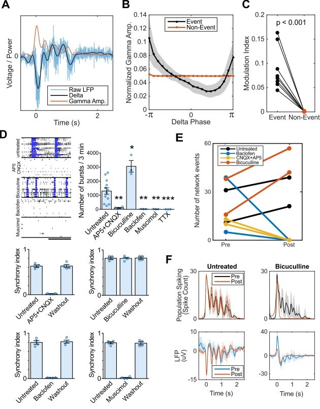
Oscillatory coordination of neural ensembles and its synaptic mechanisms
Oscillatory dynamics in the functioning brain have been postulated to coordinate spiking
across neural ensembles. In the LFP and other mesoscopic brain signals, this manifests as a
phenomenon known as cross-frequency phase-amplitude coupling (PAC) (Voytek and Knight,
2015), wherein the high-frequency content of the LFP is entrained to the phase of slow oscillations
(Manning et al., 2009; Miller et al., 2007; Mukamel et al., 2005). PAC in the neocortex and
hippocampus has been shown to be functionally relevant in a range of behaviors and neurological
disorders (de Hemptinne et al., 2015; Voytek and Knight, 2015; Voytek et al., 2015). In the
organoids, we observed greater PAC between oscillatory delta (1-4 Hz) and broadband gamma
activity (100-400 Hz, see Methods) during network events compared to quiescent periods (Figure
3A-C).
We further evaluated the role of glutamatergic and GABAergic synaptic transmission in
forming oscillations by pharmacological intervention. Organoid neural networks were susceptible
to both glutamate receptor antagonists (AP5 and CNQX; NMDA and AMPA/kainate, respectively)
and GABA receptor agonists (muscimol, GABAA; baclofen, GABAB) by significantly reducing the
number of spikes and bursts, with a subsequent extinction of synchronous activity. The electrical
activity was abolished in the presence of tetrodotoxin (TTX) (Figure 3D and 3E). Notably,
8bioRxiv preprint first posted online Jun. 29, 2018; doi: http://dx.doi.org/10.1101/358622. The copyright holder for this preprint
(which was not peer-reviewed) is the author/funder, who has granted bioRxiv a license to display the preprint in perpetuity.
It is made available under a CC-BY-NC-ND 4.0 International license.
Trujillo, Gao, Negraes et al.
blockade of GABAergic transmission by bicuculline increased the number of network-
synchronized events and did not affect peak population firing rates, but abolished nested 2 Hz
oscillatory activity by erasing subsequent reverberant peaks (Figure 3F). The findings suggest
that GABA transmission is crucial for the maintenance, but not the initiation of faster oscillatory
activity. This is consistent with accounts of inhibition rhythmically coordinating pyramidal
populations activity during early development (Opitz et al., 2002).
MECP2 is essential for the timely emergence of network oscillations
In addition to modeling the typically-developing brain, cortical organoids can also shed
light on the mechanism behind functional deficits in neurodevelopmental disorders (Birey et al.,
2017; Lancaster et al., 2013; Thomas et al., 2016). Normal nested oscillatory network dynamics
in the brain are often shown to break down in psychiatric and neurological conditions (Uhlhaas
and Singer, 2010). However, the mechanisms by which that happens and its impact on the circuit
are difficult to elucidate. Thus, we next investigated whether cortical organoids could be used to
model oscillatory network defects. Previous work evidenced that patients with autism spectrum
disorder exhibit reduced alpha oscillation power (8-12 Hz) and evoked low-gamma (40-60 Hz)
response, as well as reduced PAC (Khan et al., 2013; Mohammad-Rezazadeh et al., 2016).
Mutations in the Methyl-CpG-binding protein 2 (MECP2) gene lead to a severe disruption in
cortical development that account for many symptoms of Rett syndrome, autism, schizophrenia
and other neurological disorders (Amir et al., 1999; Cohen et al., 2002; Du et al., 2016; Liu et al.,
2016; Wen et al., 2017). MECP2 is involved on the epigenetic regulation of target genes by
binding to methylated CpG dinucleotides promoter regions, acting as a transcriptional modulator
(Figure 4A).
To model MECP2 deficiency during neurodevelopment, we generated a pluripotent stem
cell model from two different cell lines, each carrying a distinct MECP2 mutation that results in a
nonfunctional protein (Zhang et al., 2016). Human MECP2-mutant neurons in vitro exhibit fewer
9bioRxiv preprint first posted online Jun. 29, 2018; doi: http://dx.doi.org/10.1101/358622. The copyright holder for this preprint
(which was not peer-reviewed) is the author/funder, who has granted bioRxiv a license to display the preprint in perpetuity.
It is made available under a CC-BY-NC-ND 4.0 International license.
Trujillo, Gao, Negraes et al.
synapses, smaller soma size, altered calcium signaling and electrophysiological defects
compared to controls (Marchetto et al., 2010). Based on the observed reduction in the number of
layer V neurons in Mecp2-mutant mice (Stuss et al., 2012) and documented clinical data of
microcephaly in Rett syndrome patients (Amir et al., 1999), we sought to examine transcriptomics,
cellular and structural differences using MECP2-KO cortical organoids. We observed a significant
decrease in the diameter of MECP2-KO organoids, neuronal protrusions or spine-like density and
synaptic puncta at later stages of differentiation (Figure 4B-D). Additionally, and similar to the
Mecp2-mutant mice (Stuss et al., 2012), a significant reduction in the proportion of CTIP2+ and
SATB2+ neurons was observed by targeted single-cell analysis (Figure 4E-G) and corroborated
by immunostaining (Figure 4H). MECP2-KO cortical organoids also showed reduced neural
activity leading to an absence of network oscillations when compared to isogenic control
organoids at the same age (Figure 4I and 4J). The inability to entrain into a functionally connected
network at early stages of development might underlie the core deficits found in MECP2-deficient
related disorders. More importantly, these results highlight the contribution of specific genes in
the timely emergence of oscillatory activity.
Organoid network development recapitulates preterm EEG
Despite emergence of complex oscillatory network activity in organoids, it is unclear
whether the spontaneous developmental trajectory observed is representative of programmed
early neurodevelopment. While network activity from organoids does not exhibit the full temporal
complexity seen in adults, the pattern of alternating periods of quiescence and network-
synchronized events resembles electrophysiological signatures present in preterm human infant
EEG. During trace discontinu (Tolonen et al., 2007), quiescent periods are punctuated by high-
amplitude oscillations (spontaneous activity transients, SATs) lasting a few seconds. Intervals of
complete quiescence disappear as infants become of term, and the EEG is dominated by
continuous and low-amplitude desynchronized activity in adult brains (Figure 5A and S5A).
10bioRxiv preprint first posted online Jun. 29, 2018; doi: http://dx.doi.org/10.1101/358622. The copyright holder for this preprint
(which was not peer-reviewed) is the author/funder, who has granted bioRxiv a license to display the preprint in perpetuity.
It is made available under a CC-BY-NC-ND 4.0 International license.
Trujillo, Gao, Negraes et al.
Because of the inability to interrogate the electrophysiology of intact human embryonic
brains, we attempt to quantitatively compare network activity in cortical organoids to preterm
human EEG, by applying an unsupervised regularized regression model (with cross-validation)
(L1 & L2 regularized, ElasticNet) on a dataset of 567 preterm neonatal EEGs, ranging from 24 to
38 post-conception weeks (PCW) (Stevenson et al., 2017). Specifically, the training dataset
consists of a subset of the preterm EEG data after we discarded features not sensibly computed
from organoid LFPs, such as interhemispheric synchrony and frequency-dependent filtering
properties of the skull (see Table S2 for a full list of included and rejected features). The remaining
features correspond to aspects of spontaneous activity transient (SAT) timing, such as SATs per
hour and SAT duration, which were similarly computed on organoid LFPs after network event
detection.
Initially, the regression model was only optimized to anticipate preterm infant age based
on their own brain features, and has not seen any organoid data whatsoever up to this point.
Then, after unbiased training, we submitted analogous features computed from cortical organoid
LFPs to the model for neurodevelopmental correlation (Figure 5B). Notably, brain organoids past
28 weeks in culture exhibit similar developmental trajectories of electrophysiological features as
preterm neonates (Figure 5C). Next, we examined the similarities between brain organoids and
preterm humans by looking at each specific feature (Figure 5C and 5D). Of all features, “SATs
per hour” (“events per hour” in organoids) showed high similar values and growth, while “root-
mean-square SAT duration” showed a similar decline (but not in absolute value) over 25 to 38
weeks in both datasets (Figure 5E, S5B and 5C). Therefore, while the developmental trajectory
of cortical organoids is not identical to that of the fetal brain, a machine learning model trained
only on preterm neonatal EEG features was able to demonstrate that the observed network
electrophysiological features may share similarities representative of genetically programmed
developmental timelines.
11bioRxiv preprint first posted online Jun. 29, 2018; doi: http://dx.doi.org/10.1101/358622. The copyright holder for this preprint
(which was not peer-reviewed) is the author/funder, who has granted bioRxiv a license to display the preprint in perpetuity.
It is made available under a CC-BY-NC-ND 4.0 International license.
Trujillo, Gao, Negraes et al.
DISCUSSION
Development of functional human brain networks is an activity-dependent process guided
by genetic and molecular programs, shaped by the emerging cellular diversity. Neonatal neural
networks share many features with adult brains, despite the fundamental structural differences
(Power et al., 2010). Even though the chronological stages of the human cortical network
formation are not well understood, it is suggested that emerging cognitive functions during infancy
are a result of different brain regions and environmental cues (Johnson, 2001). However, in utero
development is vital for the establishment of neuronal circuitry and healthy functioning of the brain.
The second and third trimester of human gestation are when the corticothalamic network is
formed via transient connections of the subplate GABAergic neurons and the emergence of
synchronized network activity (Kostović and Judaš, 2010). Thus, early cortical functional
maturation follows an independent sensory-input pathway, guided by spontaneous activity and
associated with synaptic regulating mechanisms (Uhlhaas et al., 2010).
Here we report the formation of small-scale functional electrophysiological networks in
human cortical organoids, with some similar features to those observed in the developing brain.
While we do not claim functional equivalence between the organoids and a full neonatal cortex,
the current results represent the first step towards an in vitro model that captures some of the
complex spatiotemporal oscillatory dynamics of the human brain. Robust extracellular electrical
activity was established at earlier stages and progressively developed into an organized
oscillatory network. As such, we show that some features of early functional network dynamics
(e.g., spontaneous activity transients) can be recapitulated by an in vitro model of the developing
cortex, with no additional constraints other than structural and genetic similarities. This offers
initial evidence for a convergent experience-independent neurodevelopmental program of the
neocortex prior to birth. Given the potential roles of synchronized and oscillatory network
dynamics in coordinating information flow between developed cortical brain regions during human
cognition (Uhlhaas et al., 2010), these results highlight the potential for cortical organoids to
12bioRxiv preprint first posted online Jun. 29, 2018; doi: http://dx.doi.org/10.1101/358622. The copyright holder for this preprint
(which was not peer-reviewed) is the author/funder, who has granted bioRxiv a license to display the preprint in perpetuity.
It is made available under a CC-BY-NC-ND 4.0 International license.
Trujillo, Gao, Negraes et al.
advance our understanding of functional electrophysiology, brain development, and neuro-
genetic disorders. The MECP2 deficiency, leading to cellular and network defects, is an example
of how this platform can be used to identify important genes that are essential for the timely
emergence of oscillations in brain organoids. Finally, our findings may ultimately reframe the
ethical discussions on human brain organoid research and offer an innovative link between
microscale organoid physiology and cognitive neuroscience.
13bioRxiv preprint first posted online Jun. 29, 2018; doi: http://dx.doi.org/10.1101/358622. The copyright holder for this preprint
(which was not peer-reviewed) is the author/funder, who has granted bioRxiv a license to display the preprint in perpetuity.
It is made available under a CC-BY-NC-ND 4.0 International license.
Trujillo, Gao, Negraes et al.
REFERENCES
Allene, C., Cattani, A., Ackman, J.B., Bonifazi, P., Aniksztejn, L., Ben-Ari, Y., and Cossart, R.
(2008). Sequential Generation of Two Distinct Synapse-Driven Network Patterns in
Developing Neocortex. J. Neurosci. 28, 12851–12863.
Amir, R.E., Van den Veyver, I.B., Wan, M., Tran, C.Q., Francke, U., and Zoghbi, H.Y. (1999).
Rett syndrome is caused by mutations in X-linked MECP2, encoding methyl-CpG-binding
protein 2. Nat. Genet. 23, 185–188.
Ben-Ari, Y. (2001). Developing networks play a similar melody. Trends Neurosci. 24, 353–360.
Birey, F., Andersen, J., Makinson, C.D., Islam, S., Wei, W., Huber, N., Fan, H.C., Metzler,
K.R.C., Panagiotakos, G., Thom, N., et al. (2017). Assembly of functionally integrated
human forebrain spheroids. Nature 545, 54–59.
Blankenship, A.G., and Feller, M.B. (2010). Mechanisms underlying spontaneous patterned
activity in developing neural circuits. Nat. Rev. Neurosci. 11, 18–29.
Buzsáki, G., and Draguhn, A. (2004). Neuronal oscillations in cortical networks. Science 304,
1926–1929.
Buzsáki, G., Anastassiou, C.A., and Koch, C. (2012). The origin of extracellular fields and
currents — EEG, ECoG, LFP and spikes. Nat. Rev. Neurosci. 13, 407–420.
Buzsáki, G., Logothetis, N., and Singer, W. (2013). Scaling Brain Size, Keeping Timing:
Evolutionary Preservation of Brain Rhythms. Neuron 80, 751–764.
Camp, J.G., Badsha, F., Florio, M., Kanton, S., Gerber, T., Wilsch-Bräuninger, M., Lewitus, E.,
Sykes, A., Hevers, W., Lancaster, M., et al. (2015). Human cerebral organoids
recapitulate gene expression programs of fetal neocortex development. Proc. Natl. Acad.
Sci. 112, 201520760.
Chen, L., Deng, Y., Luo, W., Wang, Z., and Zeng, S. (2009). Detection of bursts in neuronal
spike trains by the mean inter-spike interval method. Prog. Nat. Sci. 19, 229–235.
Cohen, D., Lazar, G., Couvert, P., Desportes, V., Lippe, D., Mazet, P., and Héron, D. (2002).
MECP2 mutation in a boy with language disorder and schizophrenia. Am. J. Psychiatry
159, 148–149.
Du, F., Nguyen, M.V.C., Karten, A., Felice, C.A., Mandel, G., and Ballas, N. (2016). Acute and
crucial requirement for MeCP2 function upon transition from early to late adult stages of
brain maturation. Hum. Mol. Genet. 25, 1690–1702.
Fries, P. (2005). A mechanism for cognitive dynamics: neuronal communication through
neuronal coherence. Trends Cogn. Sci. 9, 474–480.
Gao, R., Peterson, E.J., and Voytek, B. (2017). Inferring synaptic excitation/inhibition balance
from field potentials. Neuroimage 158, 70–78.
Gore, A., Li, Z., Fung, H.-L., Young, J.E., Agarwal, S., Antosiewicz-Bourget, J., Canto, I.,
Giorgetti, A., Israel, M.A., Kiskinis, E., et al. (2011). Somatic coding mutations in human
induced pluripotent stem cells. Nature 471, 63–67.
de Hemptinne, C., Swann, N.C., Ostrem, J.L., Ryapolova-Webb, E.S., San Luciano, M.,
Galifianakis, N.B., and Starr, P.A. (2015). Therapeutic deep brain stimulation reduces
cortical phase-amplitude coupling in Parkinson’s disease. Nat. Neurosci. 18, 779–786.
Henriques, J.B., and Davidson, R.J. (1991). Left frontal hypoactivation in depression. J.
Abnorm. Psychol. 100, 535–545.
Johnson, M.H. (2001). Functional brain development in humans. Nat. Rev. Neurosci. 2, 475–
483.
Kelava, I., and Lancaster, M.A.A. (2016). Stem Cell Models of Human Brain Development. Cell
Stem Cell 18, 736–748.
Khan, S., Gramfort, A., Shetty, N.R., Kitzbichler, M.G., Ganesan, S., Moran, J.M., Lee, S.M.,
Gabrieli, J.D.E., Tager-Flusberg, H.B., Joseph, R.M., et al. (2013). Local and long-range
functional connectivity is reduced in concert in autism spectrum disorders. Proc. Natl.
14bioRxiv preprint first posted online Jun. 29, 2018; doi: http://dx.doi.org/10.1101/358622. The copyright holder for this preprint
(which was not peer-reviewed) is the author/funder, who has granted bioRxiv a license to display the preprint in perpetuity.
It is made available under a CC-BY-NC-ND 4.0 International license.
Trujillo, Gao, Negraes et al.
Acad. Sci. U. S. A. 110, 3107–3112.
Khazipov, R., and Luhmann, H.J. (2006). Early patterns of electrical activity in the developing
cerebral cortex of humans and rodents. Trends Neurosci. 29, 414–418.
Kostović, I., and Judaš, M. (2010). The development of the subplate and thalamocortical
connections in the human foetal brain. Acta Paediatr. 99, 1119–1127.
Lancaster, M.A., and Knoblich, J.A. (2014). Generation of cerebral organoids from human
pluripotent stem cells. Nat. Protoc. 9, 2329–2340.
Lancaster, M. a, Renner, M., Martin, C.-A., Wenzel, D., Bicknell, L.S., Hurles, M.E., Homfray, T.,
Penninger, J.M., Jackson, A.P., and Knoblich, J. a (2013). Cerebral organoids model
human brain development and microcephaly. Nature 501, 373–379.
van de Leemput, J., Boles, N.C., Kiehl, T.R., Corneo, B., Lederman, P., Menon, V., Lee, C.,
Martinez, R.A., Levi, B.P., Thompson, C.L., et al. (2014). CORTECON: a temporal
transcriptome analysis of in vitro human cerebral cortex development from human
embryonic stem cells. Neuron 83, 51–68.
Lisman, J. (1997). Bursts as a unit of neural information: making unreliable synapses reliable.
Trends Neurosci. 20, 38–43.
Liu, Z., Li, X., Zhang, J.-T., Cai, Y.-J., Cheng, T.-L., Cheng, C., Wang, Y., Zhang, C.-C., Nie, Y.-
H., Chen, Z.-F., et al. (2016). Autism-like behaviours and germline transmission in
transgenic monkeys overexpressing MeCP2. Nature 530, 98–102.
Luo, C., Lancaster, M.A., Castanon, R., Nery, J.R., Knoblich, J.A., and Ecker, J.R. (2016).
Cerebral Organoids Recapitulate Epigenomic Signatures of the Human Fetal Brain. Cell
Rep. 17, 3369–3384.
Manning, J.R., Jacobs, J., Fried, I., and Kahana, M.J. (2009). Broadband Shifts in Local Field
Potential Power Spectra Are Correlated with Single-Neuron Spiking in Humans. J.
Neurosci. 29, 13613–13620.
Marchetto, M.C.N.N., Carromeu, C., Acab, A., Yu, D., Yeo, G.W., Mu, Y., Chen, G., Gage, F.H.,
and Muotri, A.R. (2010). A model for neural development and treatment of Rett syndrome
using human induced pluripotent stem cells. Cell 143, 527–539.
Mariani, J., Simonini, M. V., Palejev, D., Tomasini, L., Coppola, G., Szekely, A.M., Horvath, T.L.,
and Vaccarino, F.M. (2012). Modeling human cortical development in vitro using induced
pluripotent stem cells. Proc. Natl. Acad. Sci. 109, 12770–12775.
Miller, K.J., Leuthardt, E.C., Schalk, G., Rao, R.P.N., Anderson, N.R., Moran, D.W., Miller, J.W.,
and Ojemann, J.G. (2007). Spectral Changes in Cortical Surface Potentials during Motor
Movement. J. Neurosci. 27, 2424–2432.
Mohammad-Rezazadeh, I., Frohlich, J., Loo, S.K., and Jeste, S.S. (2016). Brain connectivity in
autism spectrum disorder. Curr. Opin. Neurol. 29, 137–147.
Mukamel, R., Gelbard, H., Arieli, A., Hasson, U., Fried, I., and Malach, R. (2005). Coupling
Between Neuronal Firing, Field Potentials, and fMRI in Human Auditory Cortex. Science
309, 951–954.
Nageshappa, S., Carromeu, C., Trujillo, C.A., Mesci, P., Espuny-Camacho, I., Pasciuto, E.,
Vanderhaeghen, P., Verfaillie, C.M., Raitano, S., Kumar, A., et al. (2016). Altered neuronal
network and rescue in a human MECP2 duplication model. Mol. Psychiatry 21, 178–188.
Opitz, T., De Lima, A.D., and Voigt, T. (2002). Spontaneous Development of Synchronous
Oscillatory Activity During Maturation of Cortical Networks In Vitro. J. Neurophysiol. 88,
2196–2206.
Paşca, A.M., Sloan, S.A., Clarke, L.E., Tian, Y., Makinson, C.D., Huber, N., Kim, C.H., Park, J.-
Y., O’Rourke, N.A., Nguyen, K.D., et al. (2015). Functional cortical neurons and astrocytes
from human pluripotent stem cells in 3D culture. Nat. Methods 12, 671–678.
Pașca, S.P. (2018). The rise of three-dimensional human brain cultures. Nature 553, 437–445.
Pedregosa, F., Varoquaux, G., Gramfort, A., Michel, V., Thirion, B., Grisel, O., Blondel, M.,
Prettenhofer, P., Weiss, R., Dubourg, V., et al. (2011). Scikit-learn: Machine Learning in
15bioRxiv preprint first posted online Jun. 29, 2018; doi: http://dx.doi.org/10.1101/358622. The copyright holder for this preprint
(which was not peer-reviewed) is the author/funder, who has granted bioRxiv a license to display the preprint in perpetuity.
It is made available under a CC-BY-NC-ND 4.0 International license.
Trujillo, Gao, Negraes et al.
Python. J. Mach. Learn. Res. 12, 2825–2830.
Power, J.D., Fair, D.A., Schlaggar, B.L., and Petersen, S.E. (2010). The Development of Human
Functional Brain Networks. Neuron 67, 735–748.
Qian, X., Nguyen, H.N., Song, M.M., Hadiono, C., Ogden, S.C., Hammack, C., Yao, B.,
Hamersky, G.R., Jacob, F., Zhong, C., et al. (2016). Brain-Region-Specific Organoids
Using Mini-bioreactors for Modeling ZIKV Exposure. Cell 165, 1238–1254.
Quadrato, G., Nguyen, T., Macosko, E.Z., Sherwood, J.L., Min Yang, S., Berger, D.R., Maria,
N., Scholvin, J., Goldman, M., Kinney, J.P., et al. (2017). Cell diversity and network
dynamics in photosensitive human brain organoids. Nature 545, 48–53.
Quiroga, R.Q., Nadasdy, Z., and Ben-Shaul, Y. (2004). Unsupervised spike detection and
sorting with wavelets and superparamagnetic clustering. Neural Comput. 16, 1661–1687.
Renner, M., Lancaster, M.A., Bian, S., Choi, H., Ku, T., Peer, A., Chung, K., and Knoblich, J.A.
(2017). Self- organized developmental patterning and differentiation in cerebral organoids.
EMBO J. 36, 1316–1329.
Stevenson, N.J., Oberdorfer, L., Koolen, N., O’Toole, J.M., Werther, T., Klebermass-Schrehof,
K., and Vanhatalo, S. (2017). Functional maturation in preterm infants measured by serial
recording of cortical activity. Sci. Rep. 7, 12969.
Stuss, D.P., Boyd, J.D., Levin, D.B., and Delaney, K.R. (2012). MeCP2 Mutation Results in
Compartment-Specific Reductions in Dendritic Branching and Spine Density in Layer 5
Motor Cortical Neurons of YFP-H Mice. PLoS One 7, e31896.
Tetzlaff, C., Okujeni, S., Egert, U., Wörgötter, F., and Butz, M. (2010). Self-organized criticality
in developing neuronal networks. PLoS Comput. Biol. 6, e1001013.
Chailangkarn, T., Trujillo, C.A., Freitas, B.C., Hrvoj-Mihic, B., Herai, R.H., Yu, D.X., Brown, T.T.,
Marchetto, M.C., Bardy, C., McHenry, L., et al. (2016). A human neurodevelopmental
model for Williams syndrome. Nature 536, 338–343.
Thomas, C.A., Tejwani, L., Trujillo, C.A., Negraes, P.D., Herai, R.H., Mesci, P., Macia, A., Crow,
Y.J., and Muotri, A.R. (2017). Modeling of TREX1-Dependent Autoimmune Disease using
Human Stem Cells Highlights L1 Accumulation as a Source of Neuroinflammation. Cell
Stem Cell 21, 319–331.e8.
Tolonen, M., Palva, J.M., Andersson, S., and Vanhatalo, S. (2007). Development of the
spontaneous activity transients and ongoing cortical activity in human preterm babies.
Neuroscience 145, 997–1006.
Tort, A.B.L., Komorowski, R., Eichenbaum, H., and Kopell, N. (2010). Measuring Phase-
Amplitude Coupling Between Neuronal Oscillations of Different Frequencies. J.
Neurophysiol. 104, 1195–1210.
Uhlhaas, P.J., and Singer, W. (2010). Abnormal neural oscillations and synchrony in
schizophrenia. Nat. Rev. Neurosci. 11, 100–113.
Uhlhaas, P.J., Roux, F., Rodriguez, E., Rotarska-Jagiela, A., and Singer, W. (2010). Neural
synchrony and the development of cortical networks. Trends Cogn. Sci. 14, 72–80.
Voytek, B., and Knight, R.T. (2015). Dynamic network communication as a unifying neural basis
for cognition, development, aging, and disease. Biol. Psychiatry 77, 1089–1097.
Voytek, B., Kayser, A.S., Badre, D., Fegen, D., Chang, E.F., Crone, N.E., Parvizi, J., Knight,
R.T., and D’Esposito, M. (2015). Oscillatory dynamics coordinating human frontal
networks in support of goal maintenance. Nat. Neurosci. 18, 1318–1324.
Wen, Z., Cheng, T.-L., Li, G.-Z., Sun, S.-B., Yu, S.-Y., Zhang, Y., Du, Y.-S., and Qiu, Z. (2017).
Identification of autism-relatedMECP2mutations by whole-exome sequencing and
functional validation. Mol. Autism 8, 43.
Zhang, Z.-N.Z.-N., Freitas, B.C.B.C., Qian, H., Lux, J., Acab, A., Trujillo, C.A.C.A., Herai,
R.H.R.H., Huu, V.A.N., Wen, J.H.J.H., Joshi-Barr, S., et al. (2016). Layered hydrogels
accelerate iPSC-derived neuronal maturation and reveal migration defects caused by
MeCP2 dysfunction. 113, 3185–3190.
16bioRxiv preprint first posted online Jun. 29, 2018; doi: http://dx.doi.org/10.1101/358622. The copyright holder for this preprint
(which was not peer-reviewed) is the author/funder, who has granted bioRxiv a license to display the preprint in perpetuity.
It is made available under a CC-BY-NC-ND 4.0 International license.
Trujillo, Gao, Negraes et al.
SUPPLEMENTAL INFORMATION
Supplemental information includes Supplemental Experimental Procedures, 5 Supplemental
Figures, 2 Supplemental Tables can be found with this article online.
ACKNOWLEDGMENTS
This work was supported by grants from the California Institute for Regenerative Medicine (CIRM)
DISC1-08825 and DISC2-09649, the National Institutes of Health through the R01MH108528,
R01MH094753, R01MH109885, R01MH100175, R56MH109587, a SFARI grant #345469, and a
NARSAD Independent Investigator Grant to A.R.M. B.V is supported by a Sloan Research
Fellowship, the Whitehall Foundation (2017-12-73), and the National Science Foundation
(1736028). I.A.C. is a San Diego IRACDA Fellow supported by National Institutes of Health
(NIH)/NIGMS K12 GM06852 Award. G.W.Y. and A.R.M. are supported by U19MH1073671, part
of the National Cooperative Reprogrammed Cell Research Groups (NCRCRG) to Study Mental
Illness. R.G is supported by the Natural Sciences and Engineering Research Council of Canada
(NSERC PGS-D), UCSD Kavli Innovative Research Grant (IRG), Frontiers for Innovation
Scholars Program, and Katzin Prize. We thank Patrick S. Cooper for his help on the Australian
EEG Database. We thank Dr. V.Taupin for her expert assistance with electron microscopy.
AUTHOR CONTRIBUTIONS
C.A.T., R.G. and P.D.N. should be considered co-first authors as they each designed the
experiments and conducted the analyses with input from A.R.M., B.V.; P.D.N. and C.A.T.
generated and characterized the cortical organoids and performed the MEA recordings. C.A.T.
performed C1 single-cell analyses and synaptic quantification. I.A.C. performed and analyzed
10X Genomics single-cell experiments. A.Do. processed 10X Genomics single-cell data. G.W.Y.
led and funded the single-cell RNA-seq analyses. M.V. and A.De. performed the functional
experiment. P.D.N. analyzed the MEA data using the Axion Biosystems Neural Metrics Tool. R.G.
17bioRxiv preprint first posted online Jun. 29, 2018; doi: http://dx.doi.org/10.1101/358622. The copyright holder for this preprint
(which was not peer-reviewed) is the author/funder, who has granted bioRxiv a license to display the preprint in perpetuity.
It is made available under a CC-BY-NC-ND 4.0 International license.
Trujillo, Gao, Negraes et al.
performed the custom MEA and EEG analyses. A.R.M. and B.V. should be considered co-senior
authors as they contributed equally to directing the overall study design, with A.R.M. leading the
cortical organoid development and analyses, and B.V. leading the electrophysiological design
and analyses. C.A.T., R.G., P.D.N., B.V. and A.R.M wrote the manuscript. All authors reviewed
the manuscript for publication.
CONFLICT OF INTERESTS
Dr. Muotri is a co-founder and has equity interest in TISMOO, a company dedicated to genetic
analysis focusing on therapeutic applications customized for autism spectrum disorder and other
neurological disorders with genetic origins. The terms of this arrangement have been reviewed
and approved by the University of California San Diego in accordance with its conflict of interest
policies.
AUTHOR INFORMATION
Correspondence and requests for materials should be addressed to muotri@ucsd.edu or
bvoytek@ucsd.edu.
18bioRxiv preprint first posted online Jun. 29, 2018; doi: http://dx.doi.org/10.1101/358622. The copyright holder for this preprint
(which was not peer-reviewed) is the author/funder, who has granted bioRxiv a license to display the preprint in perpetuity.
It is made available under a CC-BY-NC-ND 4.0 International license.
Trujillo, Gao, Negraes et al.
FIGURES & FIGURE LEGENDS
Figure 1. Cellular and molecular development of human cortical organoids. (A) Overview of
human neural network formation and dynamics evaluation using organoids. (B) Schematic of the
protocol used to generate cortical organoids. Scale bar, 200 µm. (C) Organoid growth during
different developmental stages. (D) Representative immunostainings showing proliferating NPCs
19bioRxiv preprint first posted online Jun. 29, 2018; doi: http://dx.doi.org/10.1101/358622. The copyright holder for this preprint
(which was not peer-reviewed) is the author/funder, who has granted bioRxiv a license to display the preprint in perpetuity.
It is made available under a CC-BY-NC-ND 4.0 International license.
Trujillo, Gao, Negraes et al.
(Ki67+ and Nestin+), lower (TBR1+ and CTIP2+) and upper (SATB2+) cortical layer neurons and
glial cells (GFAP+) overtime. Scale bar, 50 µm. (E) Population analysis of specific markers
indicating stages of maturation and multiple neuronal subtypes. The data are shown as mean ±
s.e.m. (n = 8). (F) Representative image of a pyramidal neuron (left panel); dendritic structures
are observed in cells transduced with the SYN:EGFP reporter (middle panel; scale bar, 5 µm).
Immunohistochemical detection of the synaptic protein Syn1 (right panel; scale bar, 50 µm). (G)
Electron microscopy of synaptic structures in 4-month-old cortical organoids (blue). (H) t-
distributed stochastic neighbor embedding (tSNE) plot of 3,491 cells from 6-month-old organoids.
Colors denote seven main cell clusters. (I) tSNE plots depicting cell-type specific marker
expression levels (red denotes higher expression). (J) Heatmap of average expression for
representative gene markers by cluster and cell-type (see also Figure S4). (K) Violin plots showing
transcript levels for representative markers of each cluster (see Figure S3 for additional markers).
20bioRxiv preprint first posted online Jun. 29, 2018; doi: http://dx.doi.org/10.1101/358622. The copyright holder for this preprint
(which was not peer-reviewed) is the author/funder, who has granted bioRxiv a license to display the preprint in perpetuity.
It is made available under a CC-BY-NC-ND 4.0 International license.
Trujillo, Gao, Negraes et al.
Figure 2. Oscillatory network dynamics in long-term cortical organoids. (A) Schematic of
the organoid signal processing pipeline. Raw MEA data is analyzed as population spiking and
LFP separately. Synchronous network events are highlighted in yellow. (B) Raster plot of network
spiking activity after 1.5 and 6 months of maturation. A 3-s interval of activity over 5 channels is
shown in the upper right corners. (C) Cortical organoids show elevated and continuously
increasing mean firing rate compared to 2D monolayer neurons (n = 8 organoid cultures, and n =
12 for 2D neurons). Inset, correlation of the firing rate vector over 12 weeks of differentiation (from
8 to 20) between pairs of cultures showing reduced variability among organoid replicates. (D)
Temporal evolution of cortical organoid network activity. Detailed definitions and further
parameters are presented in Figure 5B and 5C. (E) Time series of population spiking and LFP
21bioRxiv preprint first posted online Jun. 29, 2018; doi: http://dx.doi.org/10.1101/358622. The copyright holder for this preprint
(which was not peer-reviewed) is the author/funder, who has granted bioRxiv a license to display the preprint in perpetuity.
It is made available under a CC-BY-NC-ND 4.0 International license.
Trujillo, Gao, Negraes et al.
during network events in cortical organoid development. Each trace represents a single event
during the same recording session. (F) Oscillatory dynamics within network events develop
nonlinearly, following an inverted-U trajectory. (G) Increase of network variability dynamics
throughout development. (H) Oscillatory power increases up to the 25th week in culture and
plateaus at 30 weeks. Inset, Oscillatory power is calculated by fitting a straight line (dashed) over
the aperiodic portion of the PSD and taken as the height of narrow peaks rising above the linear
fit. The data shown in C, D, F, G and H are presented as mean ± s.e.m. *P < 0.05, **P < 0.01,
***P < 0.001, unpaired Student’s t-test (C), quadratic (F) and linear (G) regression.
22bioRxiv preprint first posted online Jun. 29, 2018; doi: http://dx.doi.org/10.1101/358622. The copyright holder for this preprint
(which was not peer-reviewed) is the author/funder, who has granted bioRxiv a license to display the preprint in perpetuity.
It is made available under a CC-BY-NC-ND 4.0 International license.
Trujillo, Gao, Negraes et al.
Figure 3. Cortical organoid serves as a model of functional oscillations and their synaptic
mechanisms. (A-C) Phase-amplitude coupling is observed in organoid LFP during network
events, a phenomenon proposed to mediate neural communication in vivo. (A) Example of raw
LFP during a network event decomposed into its low-frequency component (1-4 Hz delta) and the
amplitude envelope of the high-frequency, broadband gamma component (200-400 Hz). Analysis
23bioRxiv preprint first posted online Jun. 29, 2018; doi: http://dx.doi.org/10.1101/358622. The copyright holder for this preprint
(which was not peer-reviewed) is the author/funder, who has granted bioRxiv a license to display the preprint in perpetuity.
It is made available under a CC-BY-NC-ND 4.0 International license.
Trujillo, Gao, Negraes et al.
was repeated for 100-200 Hz with near identical effect size and significance. (B) Normalized
gamma amplitude binned by delta phase during network events (black) shows greater modulation
depth by low frequency delta than during non-event periods (red). (C) Phase-amplitude coupling
during network events is significantly greater than non-event periods in all batches. (D) Effect of
selective drug treatments on neuronal electrical activity in 6-month-old organoids. Representative
raster plots and burst measurements of untreated and treated organoids. Scale bar, 20 s.
Exposure to AP5 + CNQX, baclofen and muscimol reversibly extinguish the network bursts
(synchrony), while no changes were promoted by bicuculline. (E-F) Pharmacological perturbation
of oscillatory activity during network events in 6-month-old organoids. Application of bicuculline
increases the number of network events, while CNQX + AP5 and baclofen completely abolish
synchronized network events. Bicuculline blocks oscillatory network activity but not the network
event itself. Data are shown as mean ± s.e.m.; unpaired Student’s t-test.
24bioRxiv preprint first posted online Jun. 29, 2018; doi: http://dx.doi.org/10.1101/358622. The copyright holder for this preprint
(which was not peer-reviewed) is the author/funder, who has granted bioRxiv a license to display the preprint in perpetuity.
It is made available under a CC-BY-NC-ND 4.0 International license.
Trujillo, Gao, Negraes et al.
Figure 4. MECP2 contribution to the emergence of network oscillations. (A) MECP2-
knockout neurons (MECP2-KO) show reduced spine-like density and soma size compared to
controls. (B) Organoid diameter quantification (CTR, n = 210 organoids; KO, n = 333 organoids).
(C) Spine-like density and (D) synaptic puncta are reduced in MECP2-KO neurons. Scale bar, 50
µm. (E-H) Targeted single-cell analysis of neural markers and cortical layer-related genes over
defined control Ct value. In 3-month-old cortical organoids, a significant decrease in the number
of CTIP2+ and SATB2+ neurons was observed. (I) MECP2-KO cortical organoids show
decreased mean firing rate after 5 months of maturation (n = 6 organoid cultures). (J) Lack of
oscillatory network events in 5-month-old MECP2-KO organoids. Each trace represents a single
event during the same recording session. For B, C, D, G, H, I and J, data are shown as mean ±
s.e.m.; *P < 0.05, **P < 0.01, ***P < 0.001, unpaired Student’s t-test.
25bioRxiv preprint first posted online Jun. 29, 2018; doi: http://dx.doi.org/10.1101/358622. The copyright holder for this preprint
(which was not peer-reviewed) is the author/funder, who has granted bioRxiv a license to display the preprint in perpetuity.
It is made available under a CC-BY-NC-ND 4.0 International license.
Trujillo, Gao, Negraes et al.
Figure 5. Organoid network dynamics mimic premature neonates after 28 weeks of
maturation. (A) Representative LFP trace from cortical organoid, highlighting instances of
network events (yellow). Comparable events between periods of quiescence (discontinuous
network dynamics) are shown in human preterm neonate EEG at 35 weeks gestational age, while
a different pattern of continuous activity is observed in adult EEG. SAT: spontaneous activity
transient. (B) Schematic of unsupervised machine learning pipeline: 9 EEG features from 39
premature babies (n = 567 recordings) between 25 and 38 PCW were used to train and cross-
26bioRxiv preprint first posted online Jun. 29, 2018; doi: http://dx.doi.org/10.1101/358622. The copyright holder for this preprint
(which was not peer-reviewed) is the author/funder, who has granted bioRxiv a license to display the preprint in perpetuity.
It is made available under a CC-BY-NC-ND 4.0 International license.
Trujillo, Gao, Negraes et al.
validate a regularized regression model (ElasticNet) to first determine neonate brain age, which
was then applied directly to organoid LFP features. (C) Machine-determined organoid “brain age”
plotted against actual organoid age. Black stars denote time points where mean determined age
is not significantly different from actual age under 1-sample t-test (P < 0.05, n = 8). (D) Resampled
Pearson’s correlation coefficient between age and electrophysiological features for both organoid
and premature neonates show different degrees of developmental similarity for individual
features. (E) EEG/LFP features over time for organoids and premature neonates show various
levels of similarity.
27You can also read