Vitamin D deficiency in chronic liver disease
←
→
Page content transcription
If your browser does not render page correctly, please read the page content below
Submit a Manuscript: http://www.wjgnet.com/esps/                                   World J Hepatol 2014 December 27; 6(12): 901-915
   Help Desk: http://www.wjgnet.com/esps/helpdesk.aspx                                                        ISSN 1948-5182 (online)
   DOI: 10.4254/wjh.v6.i12.901                                            © 2014 Baishideng Publishing Group Inc. All rights reserved.

                                                                                                                            REVIEW

Vitamin D deficiency in chronic liver disease

Paula Iruzubieta, Álvaro Terán, Javier Crespo, Emilio Fábrega

Paula Iruzubieta, Álvaro Terán, Javier Crespo, Emilio Fá-
brega, Gastroenterology and Hepatology Unit, Marqués de Val-             Core tip: (Vitamin D and liver disease) vitamin D defi-
decilla University Hospital, Instituto de Investigación Marqués          ciency has been frequently reported in many causes of
de Valdecilla, 39008 Santander, Cantabria, Spain                         chronic liver disease and has been associated with the
Author contributions: Iruzubieta P, Terán Á, Crespo J and Fá-            development and evolution of non-alcoholic fatty liver
brega E contributed to this paper.                                       disease (NAFLD) and chronic hepatitis C (CHC) virus
Correspondence to: Emilio Fábrega, MD, Gastroenterology                  infection. The role of vitamin D in the pathogenesis of
and Hepatology Unit, Marqués de Valdecilla University Hospital,          NAFLD and CHC is not completely known, but it seems
Instituto de Investigación Marqués de Valdecilla, Avenida Valde-
                                                                         that the involvement of vitamin D in the activation and
cilla s/n, 39008 Santander, Cantabria, Spain. digfge@humv.es
Telephone: +34-07-3442202544 Fax: +34-07-3442202544                      regulation of both innate and adaptive immune systems
Received: August 29, 2014          Revised: October 14, 2014             and its antiproliferative effect may explain its impor-
Accepted: November 7, 2014                                               tance in these liver diseases.
Published online: December 27, 2014

                                                                         Iruzubieta P, Terán Á, Crespo J, Fábrega E. Vitamin D deficiency
                                                                         in chronic liver disease. World J Hepatol 2014; 6(12): 901-915
Abstract                                                                 Available from: URL: http://www.wjgnet.com/1948-5182/full/
                                                                         v6/i12/901.htm DOI: http://dx.doi.org/10.4254/wjh.v6.i12.901
Vitamin D is an important secosteroid hormone with
known effect on calcium homeostasis, but recently there
is increasing recognition that vitamin D also is involved
in cell proliferation and differentiation, has immunomod-
ulatory and anti-inflammatory properties. Vitamin D de-                  INTRODUCTION
ficiency has been frequently reported in many causes of                  Vitamin D insufficiency and deficiency are prevalent in
chronic liver disease and has been associated with the                   almost half the healthy population of developed coun-
development and evolution of non-alcoholic fatty liver                   tries[1]. Most experts define vitamin D insufficiency as
disease (NAFLD) and chronic hepatitis C (CHC) virus                      a 25(OH)D level below 75 nmol/L (30 ng/mL) and
infection. The role of vitamin D in the pathogenesis of                  deficiency as levels below 50 nmol/L (20 ng/mL). It is
NAFLD and CHC is not completely known, but it seems                      estimated that one billion people suffer from deficiency
that the involvement of vitamin D in the activation and                  or insufficiency of vitamin D[2]. In the United States, be-
regulation of both innate and adaptive immune systems                    tween 25% and 50% of the adult population has vitamin
and its antiproliferative effect may explain its impor-
                                                                         D deficiency[3]. In patients with chronic liver diseases, the
tance in these liver diseases. Published studies provide
                                                                         prevalence of vitamin D deficits is much higher and prac-
evidence for routine screening for hypovitaminosis D in
                                                                         tically universal[4]. Up to 93% of patients with chronic liv-
patients with liver disease. Further prospectives studies
demonstrating the impact of vitamin D replacement in
                                                                         er disease have insufficient vitamin D levels, and almost
NAFLD and CHC are required.                                              one-third of these show severe deficiency[5].
                                                                             The outcome of vitamin D deficiency in terms of
© 2014 Baishideng Publishing Group Inc. All rights reserved.             osteoporosis, osteomalacia and increased fracture risk
                                                                         is well known[6,7]. Furthermore, the association between
Key words: Cholecalciferol; Vitamin D; Hepatitis C; Liver                vitamin D deficiency and the development of infections,
fibrosis; Liver disease; Interferon; Sustained virological               cardiovascular, autoimmune and degenerative diseases
response; Nonalcoholic fatty liver disease; Nonalcoholic                 and several types of cancer (colon, prostate and breast
steatohepatitis                                                          cancer) has also been reported[8]. Vitamin D is important

            WJH|www.wjgnet.com                                     901                        December 27, 2014|Volume 6|Issue 12|
Iruzubieta P et al . Vitamin D and liver disease
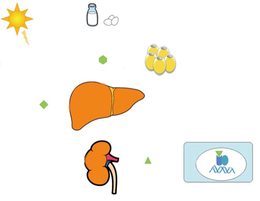
Diet ary
intake
UVB
7-dehydrocholesterol Storage in
adipocytes
Vitamin D3/D2
Vitamin D3
Epidermis
CYP2R1
CYP27A1
25(OH)D
25-hidroxylation
Bound to
DBP
CYP27B1
VDR RXR
1α,25(OH)2D
Cell
1α hidroxylation
Figure 1 Vitamin D synthesis. VDR: Vitamin D receptor; DBP: Vitamin D-binding protein; UVB: Ultraviolet radiation; RXR: Retinoid X receptor.
in calcium homeostasis and has also been implicated in tive form of vitamin D. It has a half-life of 2-3 wk and is
the mechanisms of cellular proliferation, differentiation a useful measure of vitamin D levels because it reflects
and immunomodulation[9]. These effects are noted in the the total amount of vitamin D from dietary sources, sun
pathogenesis and treatment of many chronic liver diseas- exposure and conversion from fatty deposits of the liver,
es. In this review, we will focus on vitamin D functions and its concentration in plasma is the most reliable indi-
involved in the development of chronic liver disease and cator of vitamin D status[11]. This vitamin D metabolite,
on the relationship between vitamin D deficiency and the like others, is a low-solubility lipophilic molecule that
two main causes of chronic liver disease: chronic hepati- moves through the bloodstream attached to plasmatic
tis C (CHC) virus infection and non-alcoholic fatty liver proteins, the most prevalent of which is vitamin D-bind-
disease (NAFLD). ing protein (DBP), also known as Gc. Up to 88% of
An evidence-based approach was used for this review. serum 25(OH)D is attached to a DBP, a protein synthe-
MEDLINE search was performed to September 2014 sized mainly in the liver that has anti-inflammatory and
using the following MeSH terms: liver diseases, vitamin D, immunomodulating functions independent of its role as
cholecalciferol, hepatitis C, Chronic, nonalcoholic fatty a vitamin D transporter[12,13]. 25(OH)D is hydroxylated
liver disease. Searches were limited to English language in the proximal tubules of the kidney by 1α-hydroxylase
articles. References of suitable articles were searched for (CYP27B1) that form 1α,25(OH)2D or calcitriol, the
other appropriate articles. most biologically active and powerful metabolite of vi-
tamin D[1]. CYP27B1 activity has been observed in the
kidney and other tissues, including the liver, fat tissue
VITAMIN D SYNTHESIS and the cells of the innate immune system[14]. Finally,
Under normal conditions, biogenesis from epidermal 24-hydroxylase, which is most abundant in the intestine
cells is the main source of vitamin D. In the skin, ultravi- and the kidney, catabolizes the calcitriol into an inactive
olet radiation from sun exposure transforms 7-dehydro- metabolite that is excreted in bile[15] (Figure 1).
cholesterol, a metabolite of cholesterol, into pre-vitamin 1α,25(OH)2D has a half-life of 4 h. It is transported
D3, which is transformed into vitamin D3 (cholecalcif- via attachment to plasmatic proteins such as DBP and, as
erol). A small portion of vitamin D comes from dietary mentioned previously, conducts most of the biological
sources, such as milk and eggs, in the form of vitamin D2 effects of vitamin D by directly and indirectly controlling
(ergocalciferol) and D3 that is absorbed in the intestine the expression of over 200 genes linked to angiogenesis,
by biliary acids[1,10]. Vitamin D synthesized from skin and apoptosis, proliferation, differentiation and immuno-
from dietary sources may be stored in the adipocytes, modulation[1,16,17]. The biological effects of vitamin D are
or it may undergo hepatic 25-hydroxylation. This latter mediated by binding to the vitamin D receptor (VDR),
process is mediated by isoforms of the P450 cytochrome belongs to the superfamily of nuclear steroid hormone
(CYP2R1, CYP27A1), the 25-hydroxylases, which pro- receptors, which is expressed in more than 30 tissues,
duce 25-hydroxyvitamin D [25(OH)D] or calcidiol. The including the liver, the pancreatic islet cells, the epithelial
metabolite 25(OH)D, most abundant in blood, is an inac- cells of the gastrointestinal tract and the immune system
WJH|www.wjgnet.com 902 December 27, 2014|Volume 6|Issue 12|Iruzubieta P et al . Vitamin D and liver disease
cells[18]. Hence, vitamin D deficiency may be involved PTH[34,35]; (5) the DBP protein may buffer the levels of
in several processes, such as cancer, diabetes mellitus free vitamin D which is correlated with the levels of ac-
(DM) and cardiovascular and autoimmune diseases[19-26]. tive vitamin D, this prevents intoxication[36]. Additionally,
Furthermore, the immune system cells, including macro- DBP prevents catabolism and excretion of the hormone.
phages, dendritic cells, and T and B lymphocytes, express The DBP levels decrease in liver disease, nephrotic syn-
CYP27A1 or CYP27B1 enzymes and thus can metabo- drome and malnutrition; despite this modification, the
lize 25(OH)D to calcitriol. Calcitriol will then have an concentration of 1α,25(OH)2D remains constant; and
autocrine or paracrine function[19,20]. Vitamin D favors the (6) 1α,25(OH)2D controls its own synthesis not only
innate response of the immune system and has a “self- through the increase of CYP24A1 expression, as men-
regulatory” effect by limiting the adaptive response. On tioned above, but also by directly or indirectly inhibiting
one hand, it stimulates the synthesis of antimicrobial CYP27B1 expression and providing a negative feedback
peptides (cathelicidin and beta-defensin) and the chemo- pathway.
taxis and phagocytosis of the macrophages. On the other Therefore, we can conclude that multiple factors
hand, it decreases the expression of class Ⅱ complex regulate vitamin D metabolism. The intake of vitamin D
molecules, co-stimulating molecules and the synthesis through diet or sun exposure is only one of many vari-
of Th1, Th2 and Th17 cytokines[19,20]. Finally, in addition ables that determine its activity, another of these variables
to acting as a transcription factor, VDR seems to induce are DBP levels, the local synthesis of 1α,25(OH)2D (the
fast non-genomic responses by activating cellular signal- autocrine or paracrine effect) and VDR expression.
ing pathways. In this sense, has been shown presence of
VDR in plasma membranes of intestinal, lung, kidney,
muscle cells and osteoblasts, where it efficiently binds VITAMIN D AND CHRONIC LIVER
1α,25(OH)2D[16,27,28]. DISEASE
As discussed previously, vitamin D plays an important
REGULATORY MECHANISMS OF role in reducing the risk of chronic diseases, including
DM type 2, several types of cancer, and cardiovascular,
VITAMIN D SYNTHESIS autoimmune and infectious diseases. This role most likely
The synthesis process of vitamin D includes regula- results from the local production of 1α,25(OH)2D and
tory mechanisms in each step, as follows: (1) in the its autocrine and paracrine actions in cellular proliferation
skin, excess of vitamin D3 is destroyed by sunlight, thus and differentiation, apoptosis, insulin and renin secretion
preventing vitamin D3 intoxication from excessive sun and interleukin (IL) and bactericidal protein produc-
exposure[29]; (2) the 25-hydroxylation of vitamin D is tion[1,16,17,19-23]. These effects may also be relevant in the
under-regulated. The levels of 25(OH)D increase ac- pathogenesis of chronic liver diseases.
cording to the intake of vitamin D; thus, plasmatic levels Vitamin D deficiency is extremely common in chronic
of 25(OH)D are used to regulate vitamin D status; (3) liver disease patients. Up to 93% of these patients have
in contrast, 1α-hydroxylase is highly regulated. Differ- some degree of vitamin insufficiency[4,5]. Even patients
ent factors are involved in its activity and expression, with mild liver disease are affected, although liver cirrhosis
including serum calcium and phosphate, parathyroid hor- patients more commonly suffer from severe deficiency.
mone (PTH) and fibroblast growth factor 23 (FGF23). Several studies in general populations have shown
An elevated calcium serum concentration suppresses that low levels of 25(OH)D significantly increase the risk
1α-hydroxylase directly and indirectly by decreasing the of mortality from all causes, including cardiovascular dis-
PTH levels[30]; elevated plasmatic phosphate also decreas- eases[37,38]. Regarding patients with chronic liver disease of
es the expression and activity of 1α-hydroxylase through varying etiologies, vitamin D deficiency has been associ-
a mechanism that is not yet understood. This increase in ated with increased mortality[39,40], bacterial infections[41],
serum phosphate seems to trigger an increase of FGF23 portal hypertension complications[42] and fibrosis sever-
that inhibits 1α,25(OH)2D synthesis[31]. Furthermore, ity[43,44]. However, because the liver plays an important
the synthesis and degradation of 1α,25(OH)2D is also role in the metabolism and pleiotropic functions of vita-
controlled by local factors such as cytokines and growth min D, the question is whether vitamin D deficiency is a
factors, although this local production has no effect on consequence of liver disease or a contributor to the liver
the blood levels[32,33]. In the case of the macrophages, the dysfunction.
expression of CYP27B1 and synthesis of 1α,25(OH)2D Severe liver disease decreases vitamin D hydroxylation
are induced by inflammatory cytokines, such as interferon and albumin and DBP production, all of which are linked
(IFN)γ, and by toll-like receptor (TLRs) ligands, such as to low levels of 25(OH)D. Nevertheless, the vitamin D
the lipopolysaccharide (LPS); (4) the 25-hydroxyvitamin deficiency in chronic liver disease is only partly the re-
D-24-hydroxylase (CYP24A1) catabolizes 1α,25(OH)2D sult of a synthesis dysfunction of the liver, as evidenced
to calcitroic acid, a biologically inactive bile-excreted by the fact that vitamin D deficiency is highly prevalent
metabolite[15]. The activity and expression of this en- in non-cirrhotic patients [4]. The levels of 25(OH)D
zyme, which is most abundant in intestine and kidney, is in cirrhotic patients normalize after vitamin D treat-
controlled by the levels of 1α,25(OH)2D, phosphate and ment, which indicates that the 25-hydroxylation is pre-
WJH|www.wjgnet.com 903 December 27, 2014|Volume 6|Issue 12|Iruzubieta P et al . Vitamin D and liver disease
served[45,46]; and although DBP is moderately decreased The available data suggest that vitamin D supple-
in cirrhosis[47], vitamin D metabolites require only 5% of ments could be beneficial in terms of morbimortal-
the DBP binding sites[48], indicating that liver dysfunction ity[70,71]. Most experts consider of at least 75 nmol/L (30
must be severe to decrease the DBP levels and contribute ng/mL) as the most advantageous 25(OH)D level for
to vitamin D deficiency. Therefore, vitamin D deficiency reducing the risk of fractures, prevention of cancer and
in chronic liver disease requires several causes in addi- the risk of hypertension, and between 90-120 nmol/L
tion to those mentioned above, including inadequate sun (36-48 ng/mL) as the most optimal level[71]. In fact, a
exposure, insufficient food intake, steroid use, jaundice- recent meta-analysis that included 73 cohort studies
related deterioration of vitamin synthesis on the skin (849412 participants) and 22 controlled and randomized
and decreased vitamin D absorption caused by intestinal studies with over 30716 participants showed that vitamin
edema secondary to portal hypertension or to cholestasis- D3 supplements significantly reduced mortality from any
induced bile salt disruption. cause among older adults[72]. Few published prospective
The observed association between vitamin D and studies have examined the effects of supplements in
liver disease is insufficient to establish a causal effect be- chronic liver disease, and the results to date are contra-
tween vitamin D deficiency and the severity of chronic dictory, most likely because of issues with study designs,
liver disease. Recent systematic and umbrella reviews the quantity of vitamin D administered, the pre- or
has cast doubt on any causal link between vitamin D post-treatment measurements used and the presence of
deficiency and non-skeletal health outcomes, suggesting genetic polymorphisms that influence the biological ac-
that vitamin D deficiency is a marker of ill-health, rather tivity of vitamin D. Nonetheless, vitamin D supplements
than an important factor implicated in the pathogenesis are currently recommended to decrease the skeletal ef-
of disease[49]. However, there is growing evidence that fects of vitamin D deficiency. In fact, the latest recom-
vitamin D is involved in the decrease of inflammation mendation suggest that a 25(OH)D level over 20 ng/mL
and fibrosis[43,50,51]. Proinflammatory signals in monocytes is sufficient to meet the vitamin D requirement[73]. How-
and macrophages may regulate the local metabolism of ever, the Endocrine Society Clinical Practice Guideline
vitamin D, auto-inducing the expression of CYP27B1 (ESCPG) suggested that vitamin D requirements may be
and the local production of 1α,25(OH)2D, and thus con- greater for sick patients than for healthy individuals and
trolling the excessive inflammatory response[33,52]. Almost blood level above 30 ng/mL may have additional health
90% of the tissue macrophages are in the liver[53], which benefits in reducing the risk of various disease condi-
suggests that the liver production of active vitamin D is tions[74]. In addition, the ESCPG suggest that 25(OH)D
affected during inflammatory diseases of the liver. Fur- should be measured in chronic liver disease patients to
thermore, VDR is expressed in both macrophages and identify those with levels under 20 ng/mL who would
other non-parenchymal cells and biliary epithelial cells[54]. benefit from vitamin D supplements to reduce the risk
After activation, these cells increases the expression of of bone fracture[74]. Similarly, the guidelines of the Eu-
cathelicidin, an antimicrobial peptide with anti-endotoxin ropean Association for the Study of the Liver recom-
activity[55], and inhibits the synthesis of biliary acids, thus mend calcium (1000-1200 mg/d) and vitamin D (400-800
protecting the hepatocytes from these acids[56,57]. There- UI/d) supplements for cholestatic liver disease patients,
fore, the relationship between vitamin D and hepatic although supplement use is supported by limited clinical
physiopathology may result from signaling disruptions in data[75]. In fact, despite the frequency of vitamin D de-
non-parenchymal liver cells or extrahepatic cells[58]. ficiency in liver disease patients, their calcium and PTH
It is important to mention that, together with diet serum concentration levels are normal, which contradicts
intake and sun exposure, genetic factors substantially the possibility that regulatory mechanism of calcium
contribute to variations in 25(OH)D levels[59,60]. Several metabolism is affected[76,77]. Our group has confirmed
simple nucleotide polymorphisms of genes involved in these results in cirrhotic patients of different etiologies;
the metabolism of VDR and vitamin D, such as DHCR7 these patients showed vitamin D deficiencies[78] but had
(encode the 7-dehydrocholesterol reductase enzyme), free vitamin D levels similar to those of healthy subjects
CYP2R1, CYP24A1 and GC (encode DBP), have been (unpublished data). Consequently, the unaffected free
strongly linked with the serum levels of 25(OH)D and vitamin D may be involved in the lack of correlation
its efficacy [59-62]. A recent study community-dwelling between the levels of 25(OH)D and calcium and PTH
black Americans, as compared with whites, had low and may maintain calcium homeostasis without caus-
levels of total 25(OH)D and DBP, resulting in similar ing secondary hyperparathyroidism[79]. For this reason,
concentrations of estimated bioavailable 25(OH)D. Ra- several authors indicate that the levels of total and free
cial differences in the prevalence of common genetic 25(OH)D should be measured to identify the vitamin D
polymorphisms provide a likely explanation for this ob- status in chronic liver disease patients[76]. Nonetheless,
servation[63]. Therefore, such genetic variations may be these patients have a high prevalence of bone mass loss
associated with the severity of chronic liver disease, and that can be explained by the previous data of vitamin D
several polymorphisms of the VDR gene associated with deficiency and by other interfering factors, such as the
primary biliary cirrhosis, autoimmune hepatitis, CHC and increase in pro-inflammatory cytokines[80-82], hypogonad-
hepatocellular carcinoma have been identified[64-69]. ism[83], elevated bilirubin levels[84] and steroid treatment[85].
WJH|www.wjgnet.com 904 December 27, 2014|Volume 6|Issue 12|Iruzubieta P et al . Vitamin D and liver disease
VITAMIN D FUNCTIONS AND THEIR cytokines IL-10 and transforming growth factor beta
(TGF-β)[107,109,110]. This ability to modulate the adaptive
IMPLICATIONS IN LIVER DISEASES immune system may explain the association between vi-
Vitamin D maintains the normal skeletal architecture and tamin D deficiency and the risk of autoimmune diseases
plays roles in the cardiovascular[86,87] and nervous sys- and liver damage.
tems[88,89] and cellular proliferation and differentiation[90,91]. Moreover, in vitro and in vivo studies of mouse models
Furthermore, vitamin D may be relevant in the physiopa- with liver fibrosis have reported that vitamin D has an
thology of chronic liver diseases because of its effect on anti-fibrotic effect due to ability to affect the pathological
the immune system and its anti-fibrotic effect[51,92,93]. process of liver fibrosis at several stages, such as: inhibi-
Several research lines suggest that vitamin D has ben- tion of injury trigger, suppression of hepatic stellate cells
eficial effects in liver diseases by activating and regulating activation and proliferation, reduction in accumulation of
innate and adaptive immunity.Vitamin D increases innate extracellular matrix and even degradation of collagen me-
immunity[23], stimulating the mechanisms associated with talloproteinases activation and tissue inhibitor matrix me-
the elimination of pathogen agents through the secre- talloproteinases (TIMPs) inhibition[92,93]. Moreover, Ding
tion of antibacterial proteins, such as cathelicidin and et al[111] revealed an intersecting VDR/SMAD genomic
beta-defensin, and favoring chemotaxis and macrophage circuit that regulates hepatic fibrogenesis and define a
phagocytosis[19,20,94,95]. An excessive immune response can role for VDR as an endocrine checkpoint to modulate
cause tissue damage; in this sense, vitamin D promotes the wound-healing response in liver and VDR ligands
an adequate innate immune response by regulating the as potential therapy for liver fibrosis[111]. In this regard, a
expression of several TLRs and by decreasing the pro- recent study in mice showed that the active metabolite of
duction of proinflammatory cytokines [52]. An inverse vitamin D-1α,25(OH)2D may prevent liver fibrosis in the
relationship between vitamin D levels and the expres- in-vivo model. However, it cannot ameloriate establish cir-
sion of TLR2, TLR4 and TLR9 in monocytes has been rhosis in an animal model[112].
observed, as has a decrease in the expression of these
innate immunity receptors after the administration of VITAMIN D AND CHRONIC HEPATITIS C
1α,25(OH)2D[52,96,97]. These three TLRs are primarily re-
lated to the inflammation and fibrosis of the liver. A high- VIRUS INFECTION
fat diet, alcohol consumption and structural changes in Epidemiological studies show that vitamin D deficiency
the intestinal mucosa resulting from chronic liver diseases may increase the risk of acquiring viral infections such as
(e.g., the loss of epithelial attachment, vascular congestion, influenza, human immunodeficiency virus and respiratory
defects of the mucosal immune system) alter the perme- infections[113]. Chronic hepatitis C virus (HCV) infection
ability of the mucosa, promoting an increase in intestinal is one of the main causes of chronic liver disease; it is
bacteria translocation[98-100] and bacterial products, such as estimated to affect 130 to 150 million people worldwide,
LPS, through the bloodstream; there, these bacteria bond a significant number of whom also develop cirrhosis and
to the TLRs, mainly TLR4, that are present such immune hepatic cancer[114]. A high percentage of these patients
cells as hepatocytes, biliary epithelial cells, dendritic cells (46% to 92%) have low vitamin D levels[50,115-117], and more
and hepatic stellate cells, triggering the synthesis of proin- than 25% suffer from severe deficiency[50,115,117]. It has
flammatory cytokines and fibrogenesis that ultimately re- been hypothesized that the high incidence of vitamin D
sult in liver damage[98,101]. However, vitamin D is involved deficiency in these patients may be caused by HCV’s effect
not only in the regulation of TLR expression but also in on direct or indirect 25-hydroxylation through cytokine
intestinal permeability; it plays a role in intestine epithelial induction or oxidative stress[118,119] and that the virus may
cell differentiation and in improving cell bonding[102,103], suppress 25(OH)D levels due to a disruption in lipid me-
thus decreasing the bacterial products in the liver. tabolism; as shown a recent study where HCV decreases
Regarding adaptive immunity, vitamin D seems to the production of 7-dehydrocholesterol, the endogenous
control an excessive immune response by decreasing precursor of vitamin D[120].
the expression of class Ⅱ HLA complex molecules and As discussed previously, vitamin D inhibits fibrosis
co-stimulator molecules and by modulating the T cell and modulates the innate and adaptive immune response,
response[19,20,104]. The activation of naïve T cells has been increases the production of antimicrobial peptides and
shown to be vitamin D-dependent[105]; furthermore, it inhibits proinflammatory cytokines. The anti-inflamma-
inhibits the development of Th1 (IL-2 and interferon- tory action of vitamin D[19,20,50,94,95,104,106-110] can explain the
gamma proinflammatory cytokine producers) and Th9 improved therapeutic results of IFN and ribavirin (RBV)
and increases the number of Th2 cells (IL-4, 5 and 10 after the administration of vitamin D supplements[121-123],
anti-inflammatory cytokine producers), thus affecting as some data indicate that proinflammatory cytokines and
the polarization of T helper cells [106-108]. Additionally, chemokines promote the persistence of HCV[124]. In this
1α,25(OH)2D prevents the development of Th17 cells by respect, a low Th1/Th2 ratio is an independent sustained
inhibiting IL-6 and IL-23 production from the dendritic viral response (SVR) factor in the treatment of the HCV
cells, and it induces the differentiation and expansion genotype 1[125], and 1α,25(OH)2D favors Th2 in this bal-
of regulatory T cells that secrete the anti-inflammatory ance, as mentioned previously[108]. Furthermore, several
WJH|www.wjgnet.com 905 December 27, 2014|Volume 6|Issue 12|Iruzubieta P et al . Vitamin D and liver disease
Table 1 Studies regarding vitamin D and hepatitis C virus
Ref. Year Design n HCV genotype Vitamin D deficiency Outcome P
Petta et al[50] 2010 Cohorts 197 1 73% Vitamin D levels (ng/mL): 0.05
SVR: 26.6
No SVR: 23.7
Bitetto et al[121] 2011 Cohorts 42 1 and no 1 Not stated SVR according to the vitamin D levels < 0.05
(ng/mL):
≤ 10 ng/mL: 10%
> 10 and ≤ 20 ng/mL: 30%
> 20 ng/mL: 50%
Bitetto et al[136] 2011 Cohorts 211 1-5 46.4% SVR according to the vitamin D levels 0.038
(ng/mL):
≤ 10 ng/mL: 50%
> 10 and ≤ 20 ng/mL: 60.9%
> 20 ng/mL: 69%
Lange et al[115] 2011 Cohorts 468 1-3 66% SVR (genotype 2/3): < 0.0001
Vitamin D deficit (< 10 ng/mL): 50%
Without deficiency: 81%
SVR (genotype 1) 0.45
Vitamin D deficit: 60%
Without deficiency: 54%
Nseir et al[133] 2011 Cohorts 80 1 Not stated Vitamin D levels (ng/mL): < 0.001
SVR: 42.1
No SVR: 27.3
Jazwinski et al[134] 2011 Cohorts 82 1 Not stated Vitamin D levels (ng/mL): 0.82
SVR: 23.3
No SVR: 19.3
Abu-Mouch et al[123] 2011 Randomized 72 1 59% (with vitamin D SVR: < 0.001
prospective supplementation) With vitamin D: 86%
60% (control group) Control group: 42%
Nimer et al[122] 2012 Randomized 50 2-3 60% (with vitamin D) SVR: < 0.001
prospective 50% (control group) With vitamin D: 95%
Control group: 77%
Lange et al[116] 2012 Cohorts 269 1-4 74% No significant association between 0.13
SVR and 25(OH)D serum levels
Kitson et al[137] 2013 Cohorts 274 1 48% Vitamin D levels (ng/mL): 0.03
SVR: 76.6
No SVR: 84.7
Esmat et al[140] 2014 Randomized 101 4 95% SVR: 0.22
prospective With vitamin D: 44%
Control group: 68.6%
Yokoyama et al[142] 2014 Randomized 84 1b Not stated SVR: 0.19
prospective With vitamin D: 64.3%
Control group: 50%
Grammatikos et al[138] 2014 Cohorts 398 1 Not stated Vitamin D levels (ng/mL) 0.09
SVR: 15.8
No SVR: 17.6
HCV: Hepatitis C virus; SVR: Sustained viral response.
in-vitro studies have considered vitamin D a direct HCV deficiency with a greater degree of necrosis and fibro-
antiviral agent[126-128]. Gal-Tanamy et al[127] showed that sis[40,50,68,131,132] and with a lower likelihood of a SVR to
vitamin D increases VDR expression and inhibits HCV IFN-based therapies[50,115,121,123,133-135]. In fact, all of the
replication in human hepatocytes by inducing the expres- patients who showed severe vitamin D deficiency had
sion of IFN beta and the IFN-stimulated gene (MxA) hardly any SVR, while 50% of those with normal levels
with different antiviral properties, thus producing a syn- or almost normal levels had SVR[50,121,123,136]. However
ergic effect with antiviral treatment[127]. In the same study, other studies failed to find ant relationship between base-
vitamin D or calcitriol added to the antiviral treatment line vitamin D level and SVR and fibrosis[116,137-140] (Table
had a synergic effect in the inhibition of HCV. In addi- 1). In addition, conflicting conclusions have been reached
tion, in recent clinical studies have described an associa- in two recent meta-analysis[130,141]. This may be due to lim-
tion between VDR polymorphisms on the response to itations of the studies included: (1) the small number of
IFN/RBV therapy in CHC[129,130]. patient; (2) majority had a cross-sectional studies that are
The relevance of vitamin D in CHC has been re- subject to bias due to the possibility of reverse causation;
ported in numerous studies that associated vitamin D (3) lack of vitamin D level assessment during therapy;
WJH|www.wjgnet.com 906 December 27, 2014|Volume 6|Issue 12|Iruzubieta P et al . Vitamin D and liver disease
and (4) characteristic of vitamin D assessment (seasonal- port of this hypothesis, some studies show that vitamin
ity, cut off values, methodology of vitamin D determina- D administration improves insulin secretion[145,157-160] and
tion, ethnicity). In contrast, vitamin D has been shown to that its use decreases IR in patients with end-stage renal
increase the probability of SVR when it is added to the disease[161]. Moreover, VDR polymorphisms have been
antiviral treatment[121-123,142] (Table 1). Thus, futher clinical associated with IR and have an effect on insulin secretion
investigation on the effect of vitamin D supplementation and on the fasting glucose concentration[162]. Addition-
in treating CHC are needed to confirm this item. ally, previous studies have shown that VDR knock-out
Furthermore, Bitteto et al [136] provided additional mice developed hepatic steatosis[163]. Finally, studies have
information in their study of the rs12979860 C/T poly- shown that vitamin D administration in mice activates
morphism of IL28B. In their study, vitamin D levels were the fibroblastic intestinal growth factor 15 (FGF15)
complementary to the rs12979860 C/T polymorphism (human ortholog FGF19). This intestinal hormone pre-
of the IL28B for predicting SVR in CHC patients in- vents IR and high-fat diet-induced obesity by inhibiting
fected with difficult-to-treat genotypes (1, 4, 5). Another CYP7A1, an essential enzyme in the physiopathology of
polymorphism, the CYP27B1-1260 polymorphism is also liver dyslipidemia[164]. This evidence suggests that vitamin
known to decrease the intracellular concentration of cal- D is linked to the development of NAFLD via its role
citriol in mononuclear cells and T lymphocytes[134] and is in glucose metabolism by accelerating the conversion
a known cofactor in immune response disruption in these of proinsulin to insulin, while vitamin D deficiency has
cells. In fact, the study by Lange and colleagues confirms been associated with pancreatic β cell dysfunction and a
the lack of SVR in patients infected with the HCV 1, 2 greater prevalence of type 2 DM[153,164-167].
and 3 genotypes who have this polymorphism[115]. This As in the case of CHC, vitamin D levels are lower
study also hypothesized that genotype 3 patients had low in patients with NAFLD compared with healthy con-
25(OH)D levels, in contrast with previously published trols[43,167-174]. In addition, vitamin D deficiency in obese
data[50,136]. We should, however, note that the definition of patients has been attributed to the accumulation of the
vitamin D deficiency differed among the three studies, a vitamin D in adipose tissue[175-177]. Furthemore, vitamin
factor that should be considered when interpreting these D levels are inversely correlated with the severity of ste-
results. atosis, necroinflammation and fibrosis independent of
Vitamin D also favors the HCV response by improv- age gender, BMI, Homeostatic Model Assesment of IR
ing the sensitivity to insulin [143-145]. Insulin resistance score and presence of metabolic syndrome[43,168,178]. In a
(IR) is considered one of the most important factors in recent clinical study of adults with NAFLD, Targher et
predicting HCV patients’ response to IFN and RBV[146], al[43] showed that the vitamin D levels had an effect on
and vitamin D is known to prevent DM type 2[144]. As the development of hepatic steatosis and in the severity
β-pancreatic cells contain VDR, vitamin D deficiency of the histological lesion. In fact, their hypothesis stated
may alter the balance between the intra- and extracellular that patients with greater inflammation and fibrosis had
calcium and interfere with insulin release[147]. lower vitamin D levels independent of other compo-
Therefore, in theory, vitamin D deficiency may be nents of the metabolic syndrome. This observation was
linked to a lack of response to anti-viral treatment, while later confirmed in pediatric patients[179,180] (Table 2). Still,
vitamin D supplementation may potentiate SVR. an association between vitamin D and NAFLD has been
demonstrated that is independent of BMI or IR and met-
abolic syndrome[43,157,162]. Although causal conclusions are
VITAMIN D AND NAFLD difficult to obtain from these studies, their results suggest
NAFLD is a pathological clinical entity that includes a that vitamin D deficiency plays a role in the development
broad spectrum of liver conditions from steatosis to and progression of fatty liver, especially in terms of its
nonalcoholic steatohepatitis (NASH) and cirrhosis[148] and anti-inflammatory potential. In fact, vitamin D reduces
NAFLD is one of the main causes of chronic liver dis- the risk for NAFLD in healthy men[181] and attenuates
ease in developed countries, affecting 20% to 30% of the high fat diet-induced hepatic steatosis in rats by modulat-
population[149,150]. Some NAFLD patients develop NASH ing lipid metabolism[182].
and cirrhosis, while most others do not experience dis- Vitamin D deficiency has been linked to a systemic
ease progression; however, the reason for these differ- increase in inflammation markers[183,184], and systemic
ences in progression are not known. NAFLD is generally inflammation may play a central role in the pathogenesis
related to at least one metabolic syndrome characteristic; and progression of NAFLD[185,186]. Increases in visceral
in fact, liver conditions are considered part of the syn- adiposity promote the release of fatty acids and proin-
drome, and although their pathogenesis is not yet known, flammatory cytokines and activate inflammation path-
IR is a key factor in its development[151,152]. Several studies ways in the liver, prompting proinflammatory cytokine
show a negative correlation between vitamin D levels and secretion that leads to liver damage[187]. Moreover, the
obesity, glucose intolerance, IR, metabolic syndrome and obesity promotes the onset of NAFLD due to increased
body mass index (BMI)[24-26,153-155]. Furthermore, vitamin hepatic lipid synthesis secondary to excess free fatty
D deficiency stimulates PTH, which has been linked to acids; subsequent association with oxidative stress on
IR and an increase in the acute-phase reactant[156]. In sup- mitochondrial and with the increase of proinflammatory
WJH|www.wjgnet.com 907 December 27, 2014|Volume 6|Issue 12|Iruzubieta P et al . Vitamin D and liver disease
Table 2 Studies regarding vitamin D and non-alcoholic fatty liver disease
Ref. Year Design n NAFLD diagnosis Vitamin D levels (ng/mL) P
Targher et al[43] 2007 Cohorts prospective 120 Liver biopsy Controls (60): 29.8 ± 6 0.001
Steatosis (10): 23.72 ± 8
NASH (50): 14.8 ± 9.2
Manco et al[179] 2010 Cohorts prospective 64 Liver US Without necroinflamation: 26.1 ± 10 0.16
With necroinflamation: 19.9 ± 9.8 < 0.001
Without fibrosis: 27.7 ± 10.3
With fibrosis: 17.1 ± 7.4
Barchetra et al[168] 2011 Cohorts prospective 262 Liver US Without NAFLD (100): 20.5 ± 9.7 < 0.001
NAFLD (162): 14.8 ± 9.2
Jablonski et al[169] 2013 Cohorts retrospective 1214 Liver US Controls (607): 34 ± 8 < 0.001
NAFLD (607): 30 ± 7
Kasapoglu et al[171] 2013 Cohorts prospective 613 Liver US Controls (275): 26,4 ± 9.8 < 0.05
NAFLD stage 1 (133): 20 ± 9.2
NAFLD stage 2 (106): 13.3 ± 6.7
NAFLD stage 3 (99): 8.8 ± 7.4
Black et al[170] 2014 Cohorts prospective 994 Liver US Without NAFLD (838): 30.8 ± 9.6 < 0.001
NAFLD (156): 26.8 ± 8.8
Yildiz et al[174] 2014 Cohorts prospective 101 Liver US Without NAFLD (43): 16.4 (IQR 12.4-24.8) 0.005
NAFLD grade 1 (41): 14.2 (IQR 9.5-21.2)
NAFLD grade 2 (17): 11.5 (IQR 7.5-16.7)
Dasarathy et al[178] 2014 Cohorts prospective 148 Liver biopsy Controls (39): 35.7 ± 6 < 0.01
Steatosis (67): 25 ± 11.3
NASH (81): 18.1 ± 8.4
Nobili et al[180] 2014 Cohorts prospective 73 Liver biopsy NASH (49) was associated with lower VD levels, < 0.001
i.e., -9.0 pg/mL when compared with that in
children without NASH (24)
Küçükazman et al[173] 2014 Cohorts prospective 211 Liver US Without NAFLD (57): 20 ± 13.6 < 0.001
NAFLD (154): 12.3 ± 8.9
US: Ultrasonography; IQR: Interquartile range; NAFLD: Non-alcoholic fatty liver disease; NASH: Nonalcoholic steatohepatitis.
cytokines can definitely trigger a progression of steato- creased hepatic stellate cell activity and TGF-β synthesis
sis to NASH and cirrhosis[188]. Studies in vivo and in vitro and stimulated the production of apolipoprotein E and
have clearly documented that steatosis reduces oxidative adiponectin. Together, these findings translate into a ben-
activity controlled by cytochrome P450[189]. These inflam- eficial effect on NAFLD, and a decrease in IR, steatosis,
matory processes may be blocked by increasing the levels apoptosis, inflammation and intrahepatic fibrosis was
of 25(OH)D, and the development and progression of hypothesized[196]. Thus, given the above-mentioned find-
NAFLD may stop. In fact, vitamin D supplements have ings, we can conclude that extrahepatic signaling affects
been shown to decrease inflammation markers[190-193] and fibrosis and inflammation[187] and that the vitamin D-VDR
increase anti-inflammatory cytokines[190]. It is known that axis may play a role in the initiation and progression of
vitamin D’s effects in the liver are not only exerted on the NAFLD.
hepatocytes, given that these cells express very little VDR Therefore, although the mechanisms of vitamin D’s
mRNA. In contrast, sinusoidal cells, Kupffer cells, he- control over hepatic lipid homeostasis and its link with in-
patic stellate cells and immune system cells express VDR flammation are not fully known, recent research lines pro-
mRNA that is functionally active. Therefore, vitamin D vide a more comprehensive understanding of its immune
deficiency may affect the activity/expression of macro- modulation capacity and of new therapeutic interventions
phages, dendritic cells and T and B lymphocytes by favor- for NAFLD.
ing oxidative stress and the production of proinflamma-
tory cytokines that lead to subclinical inflammation[18,19].
Furthermore, fibrosis is induced by TGF-β secretion that CONCLUSION
results from the increased secretion of the matrix metal- The pleiotropic effects of vitamin D indicate a rela-
loproteinase 9 inhibitor (TIMP-1)[194]. In fact, cell cultures tionship between its deficiency and numerous chronic
show that vitamin D has an anti-inflammatory and an diseases, such as DM, cardiovascular, autoimmune and
antifibrinolytic effect on hepatic stellate cells. Finally, ani- infectious diseases, several types of cancer and chronic
mal models show that more severe histological lesions of liver diseases. In the case of chronic liver diseases, vita-
NAFLD are associated with higher levels of mRNA of min D seems to modulate the innate and adaptive im-
TLR2, 4 and 9, proinflammatory cytokines and oxidative mune system, which explains the association. Specifically,
stress markers in rats with a high-fat diet and deficient in vitamin D deficiency has been associated with a greater
vitamin D[195]. A recent study of experimentally NAFLD- risk of portal hypertension complications, mortality and
induced rats showed that ultraviolet light exposure de- increased histological severity in NAFLD and CHC, and
WJH|www.wjgnet.com 908 December 27, 2014|Volume 6|Issue 12|Iruzubieta P et al . Vitamin D and liver disease
a lower probability of viral response to HCV treatment map of vitamin D receptor binding: associations with dis-
with IFN based therapies. In fact, clinical studies sug- ease and evolution. Genome Res 2010; 20: 1352-1360 [PMID:
20736230 DOI: 10.1101/gr.107920.110]
gest that these parameters may improve with vitamin D 18 Bookout AL, Jeong Y, Downes M, Yu RT, Evans RM, Man-
supplementation; however, prospective, randomized and gelsdorf DJ. Anatomical profiling of nuclear receptor expres-
placebo-controlled studies are required to establish firm sion reveals a hierarchical transcriptional network. Cell 2006;
conclusions. 126: 789-799 [PMID: 16923397]
19 Van Belle TL, Gysemans C, Mathieu C. Vitamin D in au-
toimmune, infectious and allergic diseases: a vital player?
REFERENCES Best Pract Res Clin Endocrinol Metab 2011; 25: 617-632 [PMID:
21872803]
1 Holick MF. Vitamin D deficiency. N Engl J Med 2007; 357: 20 Mora JR, Iwata M, von Andrian UH. Vitamin effects on the
266-281 [PMID: 17634462] immune system: vitamins A and D take centre stage. Nat Rev
2 Holick MF. Vitamin D: evolutionary, physiological and Immunol 2008; 8: 685-698 [PMID: 19172691]
health perspectives. Curr Drug Targets 2011; 12: 4-18 [PMID: 21 Garland CF, Garland FC, Gorham ED, Lipkin M, Newmark
20795941] H, Mohr SB, Holick MF. The role of vitamin D in cancer
3 Looker AC, Dawson-Hughes B, Calvo MS, Gunter EW, Sa- prevention. Am J Public Health 2006; 96: 252-261 [PMID:
hyoun NR. Serum 25-hydroxyvitamin D status of adolescents 16380576]
and adults in two seasonal subpopulations from NHANES 22 Vacek JL, Vanga SR, Good M, Lai SM, Lakkireddy D, How-
III. Bone 2002; 30: 771-777 [PMID: 11996918] ard PA. Vitamin D deficiency and supplementation and rela-
4 Fisher L, Fisher A. Vitamin D and parathyroid hormone in tion to cardiovascular health. Am J Cardiol 2012; 109: 359-363
outpatients with noncholestatic chronic liver disease. Clin [PMID: 22071212 DOI: 10.1016/j.amjcard.2011.09.020]
Gastroenterol Hepatol 2007; 5: 513-520 [PMID: 17222588] 23 Mathieu C, Badenhoop K. Vitamin D and type 1 diabetes
5 Arteh J, Narra S, Nair S. Prevalence of vitamin D deficiency mellitus: state of the art. Trends Endocrinol Metab 2005; 16:
in chronic liver disease. Dig Dis Sci 2010; 55: 2624-2628 [PMID: 261-266 [PMID: 15996876]
19960254 DOI: 10.1007/s10620-009-1069-9] 24 Song Y, Wang L, Pittas AG, Del Gobbo LC, Zhang C, Man-
6 Pérez-López FR. Vitamin D and its implications for muscu- son JE, Hu FB. Blood 25-hydroxy vitamin D levels and
loskeletal health in women: an update. Maturitas 2007; 58: incident type 2 diabetes: a meta-analysis of prospective stud-
117-137 [PMID: 17604580] ies. Diabetes Care 2013; 36: 1422-1428 [PMID: 23613602 DOI:
7 Looker AC, Mussolino ME. Serum 25-hydroxyvitamin D 10.2337/dc12-0962]
and hip fracture risk in older U.S. white adults. J Bone Miner 25 Bellan M, Guzzaloni G, Rinaldi M, Merlotti E, Ferrari C,
Res 2008; 23: 143-150 [PMID: 17907920] Tagliaferri A, Pirisi M, Aimaretti G, Scacchi M, Marzullo P.
8 Peterlik M, Cross HS. Vitamin D and calcium deficits pre- Altered glucose metabolism rather than naive type 2 diabe-
dispose for multiple chronic diseases. Eur J Clin Invest 2005; tes mellitus (T2DM) is related to vitamin D status in severe
35: 290-304 [PMID: 15860041] obesity. Cardiovasc Diabetol 2014; 13: 57 [PMID: 24618074
9 Nagpal S, Na S, Rathnachalam R. Noncalcemic actions of vi- DOI: 10.1186/1475-2840-13-57]
tamin D receptor ligands. Endocr Rev 2005; 26: 662-687 [PMID: 26 Afzal S, Brøndum-Jacobsen P, Bojesen SE, Nordestgaard
15798098] BG. Vitamin D concentration, obesity, and risk of diabetes:
10 DeLuca HF. Overview of general physiologic features and a mendelian randomisation study. Lancet Diabetes Endocrinol
functions of vitamin D. Am J Clin Nutr 2004; 80: 1689S-1696S 2014; 2: 298-306 [PMID: 24703048 DOI: 10.1016/S2213-8587(1
[PMID: 15585789] 3)70200-6]
11 Heaney RP. The Vitamin D requirement in health and 27 Boland RL. VDR activation of intracellular signaling path-
disease. J Steroid Biochem Mol Biol 2005; 97: 13-19 [PMID: ways in skeletal muscle. Mol Cell Endocrinol 2011; 347: 11-16
16026981] [PMID: 21664245 DOI: 10.1016/j.mce.2011.05.021]
12 Yamamoto N, Homma S. Vitamin D3 binding protein 28 Huhtakangas JA, Olivera CJ, Bishop JE, Zanello LP, Norman
(group-specific component) is a precursor for the macro- AW. The vitamin D receptor is present in caveolae-enriched
phage-activating signal factor from lysophosphatidylcho- plasma membranes and binds 1 alpha,25(OH)2-vitamin D3
line-treated lymphocytes. Proc Natl Acad Sci USA 1991; 88: in vivo and in vitro. Mol Endocrinol 2004; 18: 2660-2671 [PMID:
8539-8543 [PMID: 1924312] 15272054]
13 Metcalf JP, Thompson AB, Gossman GL, Nelson KJ, Koya- 29 Holick MF, Garabedian M. Vitamin D: photobiology, me-
ma S, Rennard SI, Robbins RA. Gcglobulin functions as a co- tabolism, mechanism of action, and clinical applications. In:
chemotaxin in the lower respiratory tract. A potential mech- Favus MJ, editor. Primer on the metabolic bone diseases and
anism for lung neutrophil recruitment in cigarette smokers. disorders of mineral metabolism. Washington, DC: Ameri-
Am Rev Respir Dis 1991; 143: 844-849 [PMID: 2008995] can Society for Bone and Mineral Research, 2006: 129-137
14 Townsend K, Evans KN, Campbell MJ, Colston KW, Ad- 30 Bland R, Walker EA, Hughes SV, Stewart PM, Hewison M.
ams JS, Hewison M. Biological actions of extra-renal 25-hy- Constitutive expression of 25-hydroxyvitamin D3-1alpha-
droxyvitamin D-1alpha-hydroxylase and implications for hydroxylase in a transformed human proximal tubule cell
chemoprevention and treatment. J Steroid Biochem Mol Biol line: evidence for direct regulation of vitamin D metabo-
2005; 97: 103-109 [PMID: 16081283] lism by calcium. Endocrinology 1999; 140: 2027-2034 [PMID:
15 Akeno N, Saikatsu S, Kawane T, Horiuchi N. Mouse vitamin 10218951]
D-24-hydroxylase: molecular cloning, tissue distribution, and 31 Bai XY, Miao D, Goltzman D, Karaplis AC. The autosomal
transcriptional regulation by 1alpha,25-dihydroxyvitamin dominant hypophosphatemic rickets R176Q mutation in
D3. Endocrinology 1997; 138: 2233-2240 [PMID: 9165006] fibroblast growth factor 23 resists proteolytic cleavage and
16 Messa P, Alfieri C, Rastaldi MP. Recent insights into vitamin enhances in vivo biological potency. J Biol Chem 2003; 278:
D and its receptor. J Nephrol 2011; 24 Suppl 18: S30-S37 [PMID: 9843-9849 [PMID: 12519781]
21623580] 32 Stoffels K, Overbergh L, Giulietti A, Verlinden L, Bouillon
17 Ramagopalan SV, Heger A, Berlanga AJ, Maugeri NJ, Lin- R, Mathieu C. Immune regulation of 25-hydroxyvitamin-D3-
coln MR, Burrell A, Handunnetthi L, Handel AE, Disanto G, 1alpha-hydroxylase in human monocytes. J Bone Miner Res
Orton SM, Watson CT, Morahan JM, Giovannoni G, Ponting 2006; 21: 37-47 [PMID: 16355272]
CP, Ebers GC, Knight JC. A ChIP-seq defined genome-wide 33 Liu PT, Stenger S, Li H, Wenzel L, Tan BH, Krutzik SR,
WJH|www.wjgnet.com 909 December 27, 2014|Volume 6|Issue 12|Iruzubieta P et al . Vitamin D and liver disease
Ochoa MT, Schauber J, Wu K, Meinken C, Kamen DL, Wag- bayashi T. Concentrations of vitamin D-binding protein and
ner M, Bals R, Steinmeyer A, Zügel U, Gallo RL, Eisenberg vitamin D metabolites in plasma of patients with liver cir-
D, Hewison M, Hollis BW, Adams JS, Bloom BR, Modlin RL. rhosis. J Nutr Sci Vitaminol (Tokyo) 1989; 35: 225-234 [PMID:
Toll-like receptor triggering of a vitamin D-mediated human 2585144]
antimicrobial response. Science 2006; 311: 1770-1773 [PMID: 48 White P, Cooke N. The multifunctional properties and char-
16497887] acteristics of vitamin D-binding protein. Trends Endocrinol
34 Chen KS, DeLuca HF. Cloning of the human 1 alpha,25- Metab 2000; 11: 320-327 [PMID: 10996527]
dihydroxyvitamin D-3 24-hydroxylase gene promoter and 49 Autier P, Boniol M, Pizot C, Mullie P. Vitamin D status and
identification of two vitamin D-responsive elements. Biochim ill health: a systematic review. Lancet Diabetes Endocrinol
Biophys Acta 1995; 1263: 1-9 [PMID: 7632726] 2014; 2: 76-89 [PMID: 24622671 DOI: 10.1016/S2213-8587(13)
35 Wu S, Finch J, Zhong M, Slatopolsky E, Grieff M, Brown AJ. 70165-7]
Expression of the renal 25-hydroxyvitamin D-24-hydroxylase 50 Petta S, Cammà C, Scazzone C, Tripodo C, Di Marco V,
gene: regulation by dietary phosphate. Am J Physiol 1996; Bono A, Cabibi D, Licata G, Porcasi R, Marchesini G, Craxí A.
271: F203-F208 [PMID: 8760262] Low vitamin D serum level is related to severe fibrosis and
36 Bouillon R, Van Assche FA, Van Baelen H, Heyns W, De low responsiveness to interferon-based therapy in genotype
Moor P. Influence of the vitamin D-binding protein on the 1 chronic hepatitis C. Hepatology 2010; 51: 1158-1167 [PMID:
serum concentration of 1,25-dihydroxyvitamin D3. Signifi- 20162613 DOI: 10.1002/hep.23489]
cance of the free 1,25-dihydroxyvitamin D3 concentration. J 51 Adams JS, Hewison M. Unexpected actions of vitamin D:
Clin Invest 1981; 67: 589-596 [PMID: 6894152] new perspectives on the regulation of innate and adaptive
37 Zittermann A, Iodice S, Pilz S, Grant WB, Bagnardi V, Gan- immunity. Nat Clin Pract Endocrinol Metab 2008; 4: 80-90
dini S. Vitamin D deficiency and mortality risk in the general [PMID: 18212810 DOI: 10.1038/ncpendmet0716]
population: a meta-analysis of prospective cohort stud- 52 Sadeghi K, Wessner B, Laggner U, Ploder M, Tamandl D,
ies. Am J Clin Nutr 2012; 95: 91-100 [PMID: 22170374 DOI: Friedl J, Zügel U, Steinmeyer A, Pollak A, Roth E, Boltz-
10.3945/ajcn.111.014779] Nitulescu G, Spittler A. Vitamin D3 down-regulates mono-
38 Pilz S, Tomaschitz A, März W, Drechsler C, Ritz E, Zit- cyte TLR expression and triggers hyporesponsiveness to
termann A, Cavalier E, Pieber TR, Lappe JM, Grant WB, pathogen-associated molecular patterns. Eur J Immunol 2006;
Holick MF, Dekker JM. Vitamin D, cardiovascular disease 36: 361-370 [PMID: 16402404]
and mortality. Clin Endocrinol (Oxf) 2011; 75: 575-584 [PMID: 53 Bilzer M, Roggel F, Gerbes AL. Role of Kupffer cells in host
21682758 DOI: 10.1111/j.1365-2265.2011] defense and liver disease. Liver Int 2006; 26: 1175-1186 [PMID:
39 Trépo E, Ouziel R, Pradat P, Momozawa Y, Quertinmont 17105582]
E, Gervy C, Gustot T, Degré D, Vercruysse V, Deltenre P, 54 Gascon-Barré M, Demers C, Mirshahi A, Néron S, Zalzal S,
Verset L, Gulbis B, Franchimont D, Devière J, Lemmers A, Nanci A. The normal liver harbors the vitamin D nuclear re-
Moreno C. Marked 25-hydroxyvitamin D deficiency is as- ceptor in nonparenchymal and biliary epithelial cells. Hepa-
sociated with poor prognosis in patients with alcoholic liver tology 2003; 37: 1034-1042 [PMID: 12717384]
disease. J Hepatol 2013; 59: 344-350 [PMID: 23557869 DOI: 55 D’Aldebert E, Biyeyeme Bi Mve MJ, Mergey M, Wendum D,
10.1016/j.jhep.2013.03.024] Firrincieli D, Coilly A, Fouassier L, Corpechot C, Poupon R,
40 Putz-Bankuti C, Pilz S, Stojakovic T, Scharnagl H, Pieber TR, Housset C, Chignard N. Bile salts control the antimicrobial
Trauner M, Obermayer-Pietsch B, Stauber RE. Association peptide cathelicidin through nuclear receptors in the hu-
of 25-hydroxyvitamin D levels with liver dysfunction and man biliary epithelium. Gastroenterology 2009; 136: 1435-1443
mortality in chronic liver disease. Liver Int 2012; 32: 845-851 [PMID: 19245866 DOI: 10.1053/j.gastro.2008.12.040]
[PMID: 22222013 DOI: 10.1111/j.1478-3231.2011.02735.x] 56 Han S, Li T, Ellis E, Strom S, Chiang JY. A novel bile acid-ac-
41 Malham M, Jørgensen SP, Ott P, Agnholt J, Vilstrup H, Borre tivated vitamin D receptor signaling in human hepatocytes.
M, Dahlerup JF. Vitamin D deficiency in cirrhosis relates to Mol Endocrinol 2010; 24: 1151-1164 [PMID: 20371703 DOI:
liver dysfunction rather than aetiology. World J Gastroenterol 10.1210/me.2009-0482]
2011; 17: 922-925 [PMID: 21412501 DOI: 10.3748/wjg.v17. 57 Schmidt DR, Holmstrom SR, Fon Tacer K, Bookout AL,
i7.922] Kliewer SA, Mangelsdorf DJ. Regulation of bile acid synthe-
42 Anty R, Tonohouan M, Ferrari-Panaia P, Piche T, Pariente A, sis by fat-soluble vitamins A and D. J Biol Chem 2010; 285:
Anstee QM, Gual P, Tran A. Low Levels of 25-Hydroxy Vita- 14486-14494 [PMID: 20233723 DOI: 10.1074/jbc.M110.116004]
min D are Independently Associated with the Risk of Bacte- 58 Khan AA, Chow EC, van Loenen-Weemaes AM, Porte RJ,
rial Infection in Cirrhotic Patients. Clin Transl Gastroenterol Pang KS, Groothuis GM. Comparison of effects of VDR
2014; 5: e56 [PMID: 24871371 DOI: 10.1038/ctg.2014.6] versus PXR, FXR and GR ligands on the regulation of
43 Targher G, Bertolini L, Scala L, Cigolini M, Zenari L, Falezza CYP3A isozymes in rat and human intestine and liver. Eur J
G, Arcaro G. Associations between serum 25-hydroxyvita- Pharm Sci 2009; 37: 115-125 [PMID: 19429418 DOI: 10.1016/
min D3 concentrations and liver histology in patients with j.ejps.2009.01.006]
non-alcoholic fatty liver disease. Nutr Metab Cardiovasc Dis 59 Shea MK, Benjamin EJ, Dupuis J, Massaro JM, Jacques PF, D’
2007; 17: 517-524 [PMID: 16928437] Agostino RB, Ordovas JM, O’Donnell CJ, Dawson-Hughes
44 Barchetta I, Carotti S, Labbadia G, Gentilucci UV, Muda B, Vasan RS, Booth SL. Genetic and non-genetic correlates of
AO, Angelico F, Silecchia G, Leonetti F, Fraioli A, Picardi A, vitamins K and D. Eur J Clin Nutr 2009; 63: 458-464 [PMID:
Morini S, Cavallo MG. Liver vitamin D receptor, CYP2R1, 18030310]
and CYP27A1 expression: relationship with liver histology 60 Hunter D, De Lange M, Snieder H, MacGregor AJ, Swami-
and vitamin D3 levels in patients with nonalcoholic steato- nathan R, Thakker RV, Spector TD. Genetic contribution
hepatitis or hepatitis C virus. Hepatology 2012; 56: 2180-2187 to bone metabolism, calcium excretion, and vitamin D and
[PMID: 22753133 DOI: 10.1002/hep.25930] parathyroid hormone regulation. J Bone Miner Res 2001; 16:
45 Skinner RK, Sherlock S, Long RG, Wilis MR. 25-Hydroxyl- 371-378 [PMID: 11204437]
ation of vitamin D in primary biliary cirrhosis. Lancet 1977; 1: 61 Wang TJ, Zhang F, Richards JB, Kestenbaum B, van Meurs
720-721 [PMID: 66518] JB, Berry D, Kiel DP, Streeten EA, Ohlsson C, Koller DL,
46 Compston JE. Hepatic osteodystrophy: vitamin D metabo- Peltonen L, Cooper JD, O’Reilly PF, Houston DK, Glazer NL,
lism in patients with liver disease. Gut 1986; 27: 1073-1090 Vandenput L, Peacock M, Shi J, Rivadeneira F, McCarthy
[PMID: 3530896] MI, Anneli P, de Boer IH, Mangino M, Kato B, Smyth DJ,
47 Masuda S, Okano T, Osawa K, Shinjo M, Suematsu T, Ko- Booth SL, Jacques PF, Burke GL, Goodarzi M, Cheung CL,
WJH|www.wjgnet.com 910 December 27, 2014|Volume 6|Issue 12|Iruzubieta P et al . Vitamin D and liver disease
Wolf M, Rice K, Goltzman D, Hidiroglou N, Ladouceur M, 2014; 348: g1903 [PMID: 24690623 DOI: 10.1136/bmj.g1903]
Wareham NJ, Hocking LJ, Hart D, Arden NK, Cooper C, Ma- 73 The National Academies Collection: Reports funded by
lik S, Fraser WD, Hartikainen AL, Zhai G, Macdonald HM, National Institutes of Health; Ross AC, Taylor CL, Yaktine
Forouhi NG, Loos RJ, Reid DM, Hakim A, Dennison E, Liu AL, Del Valle HB, editors. Institute of Medicine (US) Com-
Y, Power C, Stevens HE, Jaana L, Vasan RS, Soranzo N, Bo- mittee to Review Dietary Reference Intakes for Vitamin D
junga J, Psaty BM, Lorentzon M, Foroud T, Harris TB, Hof- and Calcium. The National Academies Collection: Reports
man A, Jansson JO, Cauley JA, Uitterlinden AG, Gibson Q, funded by National Institutes of Health. Dietary Reference
Järvelin MR, Karasik D, Siscovick DS, Econs MJ, Kritchevsky Intakes for Calcium and Vitamin D. Washington (DC): Na-
SB, Florez JC, Todd JA, Dupuis J, Hyppönen E, Spector TD. tional Academies Press (US), 2011 [PMID: 21796828]
Common genetic determinants of vitamin D insufficiency: 74 Holick MF, Binkley NC, Bischoff-Ferrari HA, Gordon CM,
a genome-wide association study. Lancet 2010; 376: 180-188 Hanley DA, Heaney RP, Murad MH, Weaver CM. Evalua-
[PMID: 20541252 DOI: 10.1016/S0140-6736(10)60588-0] tion, treatment, and prevention of vitamin D deficiency: an
62 Ahn J, Yu K, Stolzenberg-Solomon R, Simon KC, Mc- Endocrine Society clinical practice guideline. J Clin Endocri-
Cullough ML, Gallicchio L, Jacobs EJ, Ascherio A, Helzl- nol Metab 2011; 96: 1911-1930 [PMID: 21646368]
souer K, Jacobs KB, Li Q, Weinstein SJ, Purdue M, Virtamo J, 75 European Association for the Study of the Liver. EASL
Horst R, Wheeler W, Chanock S, Hunter DJ, Hayes RB, Kraft Clinical Practice Guidelines: management of cholestatic liver
P, Albanes D. Genome-wide association study of circulating diseases. J Hepatol 2009; 51: 237-267 [PMID: 19501929 DOI:
vitamin D levels. Hum Mol Genet 2010; 19: 2739-2745 [PMID: 10.1016/j.jhep.2009.04.009]
20418485 DOI: 10.1093/hmg/ddq155] 76 Corey RL, Whitaker MD, Crowell MD, Keddis MT, Aqel
63 Powe CE, Evans MK, Wenger J, Zonderman AB, Berg AH, B, Balan V, Byrne T, Carey E, Douglas DD, Harrison ME,
Nalls M, Tamez H, Zhang D, Bhan I, Karumanchi SA, Powe Vargas HE, Rakela J. Vitamin D deficiency, parathyroid
NR, Thadhani R. Vitamin D-binding protein and vitamin hormone levels, and bone disease among patients with end-
D status of black Americans and white Americans. N Engl stage liver disease and normal serum creatinine awaiting
J Med 2013; 369: 1991-2000 [PMID: 24256378 DOI: 10.1056/ liver transplantation. Clin Transplant 2014; 28: 579-584 [PMID:
NEJMoa1306357] 24628047 DOI: 10.1111/ctr.12351]
64 Tanaka A, Nezu S, Uegaki S, Kikuchi K, Shibuya A, Mi- 77 Chen CC, Wang SS, Jeng FS, Lee SD. Metabolic bone dis-
yakawa H, Takahashi S, Bianchi I, Zermiani P, Podda M, ease of liver cirrhosis: is it parallel to the clinical severity of
Ohira H, Invernizzi P, Takikawa H. Vitamin D receptor cirrhosis? J Gastroenterol Hepatol 1996; 11: 417-421 [PMID:
polymorphisms are associated with increased susceptibility 8743912]
to primary biliary cirrhosis in Japanese and Italian popula- 78 Terán A, Fábrega E, Moraleja I, Iruzubieta P, García-Inzueta
tions. J Hepatol 2009; 50: 1202-1209 [PMID: 19376604 DOI: MT, Crespo J, Amado JA, Pons-Romero F. Eje calcio-vitam-
10.1016/j.jhep.2009.01.015] ina D-PTH en la cirrosis hepática. Existe un hipoparatiroid-
65 Fan L, Tu X, Zhu Y, Zhou L, Pfeiffer T, Feltens R, Stoecker W, ismo relativo en el paciente cirrótico? Gatroenterol Hepatol
Zhong R. Genetic association of vitamin D receptor polymor- 2013; 36 (Congres 1): 83-84
phisms with autoimmune hepatitis and primary biliary cir- 79 Bikle DD, Halloran BP, Gee E, Ryzen E, Haddad JG. Free
rhosis in the Chinese. J Gastroenterol Hepatol 2005; 20: 249-255 25-hydroxyvitamin D levels are normal in subjects with liver
[PMID: 15683428] disease and reduced total 25-hydroxyvitamin D levels. J Clin
66 Vogel A, Strassburg CP, Manns MP. Genetic association of Invest 1986; 78: 748-752 [PMID: 3745436]
vitamin D receptor polymorphisms with primary biliary 80 Nakchbandi IA, van der Merwe SW. Current understand-
cirrhosis and autoimmune hepatitis. Hepatology 2002; 35: ing of osteoporosis associated with liver disease. Nat Rev
126-131 [PMID: 11786968] Gastroenterol Hepatol 2009; 6: 660-670 [PMID: 19881518 DOI:
67 Halmos B, Szalay F, Cserniczky T, Nemesanszky E, Lakatos 10.1038/nrgastro.2009.166]
P, Barlage S, Schmitz G, Romics L, Csaszar A. Association of 81 Axmann R, Böhm C, Krönke G, Zwerina J, Smolen J, Schett
primary biliary cirrhosis with vitamin D receptor BsmI geno- G. Inhibition of interleukin-6 receptor directly blocks osteo-
type polymorphism in a Hungarian population. Dig Dis Sci clast formation in vitro and in vivo. Arthritis Rheum 2009; 60:
2000; 45: 1091-1095 [PMID: 10877221] 2747-2756 [PMID: 19714627 DOI: 10.1002/art.24781]
68 Baur K, Mertens JC, Schmitt J, Iwata R, Stieger B, Eloranta JJ, 82 Fábrega E, Orive A, García-Suarez C, García-Unzueta M,
Frei P, Stickel F, Dill MT, Seifert B, Ferrari HA, von Eckard- Antonio Amado J, Pons-Romero F. Osteoprotegerin and
stein A, Bochud PY, Müllhaupt B, Geier A. Combined effect RANKL in alcoholic liver cirrhosis. Liver Int 2005; 25: 305-310
of 25-OH vitamin D plasma levels and genetic vitamin D re- [PMID: 15780054]
ceptor (NR 1I1) variants on fibrosis progression rate in HCV 83 Karan MA, Erten N, Tascioglu C, Karan A, Sindel D, Dilsen
patients. Liver Int 2012; 32: 635-643 [PMID: 22151003 DOI: G. Osteodystrophy in posthepatitic cirrhosis. Yonsei Med J
10.1111/j.1478-3231.2011.02674] 2001; 42: 547-552 [PMID: 11675684]
69 Falleti E, Bitetto D, Fabris C, Cussigh A, Fontanini E, Forna- 84 Janes CH, Dickson ER, Okazaki R, Bonde S, McDonagh AF,
siere E, Fumolo E, Bignulin S, Cmet S, Minisini R, Pirisi M, Riggs BL. Role of hyperbilirubinemia in the impairment of
Toniutto P. Vitamin D receptor gene polymorphisms and osteoblast proliferation associated with cholestatic jaundice.
hepatocellular carcinoma in alcoholic cirrhosis. World J Gas- J Clin Invest 1995; 95: 2581-2586 [PMID: 7769100]
troenterol 2010; 16: 3016-3024 [PMID: 20572305] 85 Mitra R. Adverse effects of corticosteroids on bone me-
70 Autier P, Gandini S. Vitamin D supplementation and total tabolism: a review. PM R 2011; 3: 466-471; quiz 471 [PMID:
mortality: a meta-analysis of randomized controlled trials. 21570035]
Arch Intern Med 2007; 167: 1730-1737 [PMID: 17846391] 86 Liu L, Chen M, Hankins SR, Nùñez AE, Watson RA, Wein-
71 Bischoff-Ferrari HA. Optimal serum 25-hydroxyvitamin D stock PJ, Newschaffer CJ, Eisen HJ. Serum 25-hydroxyvita-
levels for multiple health outcomes. Adv Exp Med Biol 2014; min D concentration and mortality from heart failure and
810: 500-525 [PMID: 25207384] cardiovascular disease, and premature mortality from all-
72 Chowdhury R, Kunutsor S, Vitezova A, Oliver-Williams cause in United States adults. Am J Cardiol 2012; 110: 834-839
C, Chowdhury S, Kiefte-de-Jong JC, Khan H, Baena CP, [PMID: 22658246 DOI: 10.1016/j.amjcard.2012.05.013]
Prabhakaran D, Hoshen MB, Feldman BS, Pan A, Johnson 87 Li YC, Qiao G, Uskokovic M, Xiang W, Zheng W, Kong
L, Crowe F, Hu FB, Franco OH. Vitamin D and risk of cause J. Vitamin D: a negative endocrine regulator of the renin-
specific death: systematic review and meta-analysis of obser- angiotensin system and blood pressure. J Steroid Biochem Mol
vational cohort and randomised intervention studies. BMJ Biol 2004; 89-90: 387-392 [PMID: 15225806]
WJH|www.wjgnet.com 911 December 27, 2014|Volume 6|Issue 12|You can also read