Endolithic Algae Affect Modern Coral Carbonate Morphology and Chemistry - Frontiers
←
→
Page content transcription
If your browser does not render page correctly, please read the page content below
ORIGINAL RESEARCH
                                                                                                                                      published: 03 December 2019
                                                                                                                                      doi: 10.3389/feart.2019.00304

                                             Endolithic Algae Affect Modern
                                             Coral Carbonate Morphology
                                             and Chemistry
                                             Stefan Krause 1* , Volker Liebetrau 1 , Gernot Nehrke 2 , Timo Damm 3 , Sebastian Büsse 4 ,
                                             Thomas Leipe 5 , Angela Vogts 5 , Stanislav N. Gorb 4 and Anton Eisenhauer 1
                                             1
                                              GEOMAR Helmholtz Centre for Ocean Research Kiel, Kiel, Germany, 2 Alfred Wegener Institute Helmholtz Centre for Polar
                                             and Marine Research, Bremerhaven, Germany, 3 Section Biomedical Imaging, Department of Diagnostic Radiology
                                             and Neuroradiology, University Hospital Schleswig-Holstein, Kiel, Germany, 4 Functional Morphology and Biomechanics,
                                             Zoological Institute, Kiel University, Kiel, Germany, 5 Leibniz Institute for Baltic Sea Research, Warnemünde, Germany

                                             While burial diagenetic processes of tropical corals are well investigated, current
                                             knowledge about factors initiating early diagenesis remains fragmentary. In the present
                                             study, we focus on recent Porites microatolls, growing in the intertidal zone. This growth
                           Edited by:
               Alberto Perez-Huerta,         form represents a model organism for elevated sea surface temperatures (SSTs) and
           The University of Alabama,        provides important but rare archives for changes close to the seawater/atmosphere
                        United States
                                             interface with exceptional precision on sea level reconstruction. As other coral growth
                        Reviewed by:
                                             forms, microatolls are prone to the colonization by endolithic green algae. In this
                 Kristine Lee DeLong,
            Louisiana State University,      case, the algae can facilitate earliest diagenetic alteration of the coral skeleton. Algae
                         United States       metabolic activity not only results in secondary coral porosity due to boring activities,
                         Nicola Allison,
             University of St Andrews,       but may also initiate reprecipitation of secondary aragonite within coral pore space, a
                      United Kingdom         process not exclusively restricted to microatoll settings. In the samples of this initial
                  *Correspondence:           study, we quantified a mass transfer from primary to secondary aragonite of around
                        Stefan Krause
                                             4% within endolithic green algae bands. Using δ18 O, δ13 C, Sr/Ca, U/Ca, Mg/Ca, and
                  skrause@geomar.de
                                             Li/Mg systematics suggests that the secondary aragonite precipitation followed abiotic
                   Specialty section:        precipitation principles. According to their individual distribution coefficients, the different
         This article was submitted to
                       Biogeoscience,
                                             isotope and element ratios showed variable sensitivity to the presence of secondary
               a section of the journal      aragonite in bulk samples, with implications for microatoll-based SST reconstructions.
            Frontiers in Earth Science       The secondary precipitates formed on an organic template, presumably originating
        Received: 30 January 2019            from endolithic green algae activity. Based on laboratory experiments with the green
      Accepted: 04 November 2019
      Published: 03 December 2019            algae Ostreobium quekettii, we propose a conceptual model that secondary aragonite
                             Citation:       formation is potentially accelerated by an active intracellular calcium transport through
    Krause S, Liebetrau V, Nehrke G,         the algal thallus from the location of dissolution into coral pore spaces. The combined
 Damm T, Büsse S, Leipe T, Vogts A,
   Gorb SN and Eisenhauer A (2019)
                                             high-resolution imaging and geochemical approach applied in this study shows that
 Endolithic Algae Affect Modern Coral        endolithic algae can possibly act as a main driver for earliest diagenesis of coral
               Carbonate Morphology          aragonite starting already during a coral’s life span.
                       and Chemistry.
               Front. Earth Sci. 7:304.      Keywords: early diagenesis, secondary precipitation, abiogenic aragonite, element ratio, sea surface
      doi: 10.3389/feart.2019.00304          temperature, endolithic algae

Frontiers in Earth Science | www.frontiersin.org                                  1                                           December 2019 | Volume 7 | Article 304
Krause et al.                                                                                                       Endolithic Algae Coral Diagenesis

INTRODUCTION                                                                 (Buster and Holmes, 2006). Also, secondary aragonite (Macintyre
                                                                             and Towe, 1976; Nothdurft and Webb, 2009) and high Mg-calcite
Scleractinian corals have been shaping the Earth’s surface since             (Nothdurft and Webb, 2009; Griffiths et al., 2013) have been
the Triassic (Stanley, 2003), forming massive reef ecosystems.               reported from recent and fossil coral skeletons. These secondary
Besides their endosymbionts, scleractinian corals host a                     minerals show deviating element/calcium from the primary coral
diverse microbial community (Rosenberg et al., 2007). Recent                 aragonite (Griffiths et al., 2013). Although a spatial proximity
investigations discovered >120 taxonomic units including over                between secondary carbonate minerals and endolithic algae or
20 genetic lineages (Marcelino and Verbruggen, 2016) present in              micro-boreholes has been noted as a petrographic feature, a clear
skeletons of living corals.                                                  relationship between endolithic algae activity and the presence of
   Among the endolithic community, siphonal green algae                      secondary carbonate phases has not been clarified to date.
of the genus Ostreobium are the most common ones                                 The scleractinian coral genus Porites is commonly present in
(Halldal, 1968; Jeffrey, 1968), producing distinctive green                  all tropical reef environments showing different growth forms
bands in the coral skeleton (Halldal, 1968; Highsmith, 1981;                 (Veron, 2000). In addition to the common subtidal massive
Verbruggen and Tribollet, 2011).                                             growth form (Glynn et al., 1994; Lough et al., 1999), microatolls
   Although known for many years (Duerden, 1902) the                         are found in intertidal environments where vertical coral growth
colonization dynamics, ecophysiology, and activity of endolithic             is limited by sea level (Dana, 1849; Stoddart and Scoffin, 1979).
phototrophs as Ostreobium are not entirely deciphered (Ralph                 In addition to constrain past sea level variability (Woodroffe
et al., 2007). While evidence exists that colonization by the algae          and McLean, 1990; Chappell et al., 1996; Lewis et al., 2008),
occurs mainly as a result of coral mechanical damage or coral                modern and fossil microatolls have also been used for robust
section death (Titlyanov et al., 2008), the entry of Ostreobium              sea surface temperature (SST) reconstruction using δ18 O and
during early coral ontogeny, within days after larval settlement,            element ratios (McGregor et al., 2013; Roche et al., 2014; Farley
has also been documented (Massé et al., 2018).                               et al., 2018), demonstrating a high degree of reproducibility to
   Euendolithic green algae, which bore into carbonate minerals,             the common growth form of the same species (McGregor et al.,
have been identified as major agents of biological reef destruction          2013; Wu et al., 2013). As microatolls grow in the intertidal,
due to their boring activity in living and dead corals as                    they are exposed to higher irradiation and thermal stress as
euendolithic organisms (Tribollet, 2008; Grange et al., 2015).               corals growing in subtidal conditions (Schoepf et al., 2015).
Unfortunately, details regarding the boring mechanism carried                Endolithic algae as Ostreobium can adapt to elevated ambient
out by endolithic green algae and the fate of dissolved carbonate            temperatures and also increase their metabolic activity (Fine
components are unconstrained.                                                et al., 2005). Therefore, microatolls provide the opportunity to
   Despite this adverse effect, endolithic green algae may also              study potentially amplified endolithic green algae-driven early
be beneficial for the host during heat stress situations leading             diagenetic processes under increased SSTs.
to coral bleaching events. Under this condition, these algae                     In this paper, we expand on previous studies using recent
are an alternative source of metabolic products required by                  Porites microatoll samples from a shallow fossil reef top
the host coral (Schlichter et al., 1995; Fine and Loya, 2002),               of Zanzibar, Tanzania, with the intention to decipher early
including also nitrogen compounds (Maier et al., 2010). The                  coral mineral diagenesis in the vicinity of endolithic green
study of Hartmann et al. (2010) interpreted the formation                    algae activity. Using micro-computed tomography (µCT),
of green bands as endolithic algal blooms during episodes                    scanning electron microscopy (SEM), nanoscale secondary ion
of environmental heat stress inducing coral bleaching. An                    mass spectrometry (nanoSIMS), fluorescence microscopy, bulk
increased endolithic algae activity could lead to more pronounced            element, and stable isotope analysis, we illustrate mineral
metabolic fractionation of CO2 manifesting as a locally more                 heterogeneities between pristine coral sections and secondary
positive δ13 C value of the coral carbonate (Pereira et al., 2015),          aragonite identified in close proximity to endolithic green
thus recording environmental stress events. The intracrystalline             algae. Based on laboratory experiments with Ostreobium
organic matter of endolithic algae as total hydrolyzable amino               quekettii, we propose a conceptual model for an algal-driven
acid (THAA) carbon (Gupta et al., 2007) is preserved over                    dissolution–reprecipitation mechanism, involving active long-
centuries in coral skeletons (Ingalls et al., 2003), adding to               distance calcium transport within the siphonal cells.
the established approaches used in paleoceanograpic research.
These findings illustrate that endolithic algae, in addition
to dissolution, can locally alter coral carbonate properties,                MATERIALS AND METHODS
representing earliest diagenesis.
   The range of reported diagenetic alterations occurring during             Coral Samples
a coral’s life span includes variability in skeletal density, skeletal       Two recent Porites microatolls with coral tissue remains still
chemistry (Sr/Ca, Mg/Ca, U/Ca, δ18 O, δ13 C), and coral skeletal             identifying the original growth position were collected in
organic matrix (Enmar et al., 2000; Hendy et al., 2007; Perrin               late 2012 from the Buyu Beach area of Unguja (6.16583◦ S,
and Smith, 2007), while reasons for the observed heterogeneity               39.19916◦ E), the southern of the two main islands of Zanzibar
are still a matter of debate. Local magnesium increase could be              (Figure 1). Close to the time point of sampling, the corals
ascribed to the precipitation of brucite [Mg(OH2 )] as a result              grew in close proximity (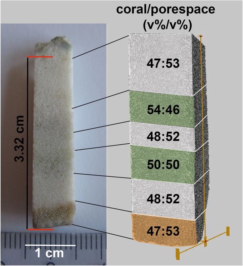
Krause et al.                                                                                                            Endolithic Algae Coral Diagenesis

                                                                                    Coral samples for wet chemistry and X-ray diffraction (XRD)
                                                                                 analyses were obtained using a handheld power drill with a
                                                                                 bediamonded milling cutter. Drilling was carried out at the lowest
                                                                                 possible speed to avoid sample warming. Sampling spots were
                                                                                 approximately 5–6 mm in diameter (see Supplementary Figure
                                                                                 S2). The obtained discrete powder samples were aliquoted for
                                                                                 light stable isotopes (δ18 O, δ13 C), element ratios (Sr/Ca, Mg/Ca,
                                                                                 U/Ca, Li/Mg), and XRD analyses.
                                                                                    Light stable isotopes were analyzed with a Thermo Scientific
                                                                                 MAT 253 stable isotope ratio mass spectrometer (SIRMS)
                                                                                 connected to an automated carbonate preparation device Kiel
                                                                                 CARBO IV at GEOMAR. The isotope values were calibrated
                                                                                 vs. NBS 19 (National Bureau of Standards) and the GEOMAR
                                                                                 in-house standard (“Standard Bremen,” Solnhofen limestone).
                                                                                 Values are reported in per mil (h) relative to the VPDB
                                                                                 (Vienna Peedee Belemnite) scale, accompanied by a typical
                                                                                 reproducibility of ±0.05h for δ18 O and ±0.07h for δ13 C (SD,
                                                                                 n = 8) for the calibrated in-house standard measured during
                                                                                 this sample set.
  FIGURE 1 | Map showing the sample location of microatolls at the western
                                                                                    Element concentrations and ratios were determined using a
  coast of Unguja island (Zanzibar).                                             quadrupole inductively coupled plasma-mass spectrometer (Q-
                                                                                 ICP-MS, Agilent 7500cx). The Coral standard JCp-1 was used as
                                                                                 a reference material and measured every fifth sample and in a
                                                                                 total of 10 times (n = 10). The average JCp-1 value and standard
were partly exposed to air when sampled at low tide (Figure 2).
                                                                                 deviation was 8.81 ± 0.04 mmol/mol for Sr/Ca, 4.19 ± 0.02 for
Instrumental records of the SST variability, observed for the
                                                                                 Mg/Ca, 1.197 ± 0.026 for U/Ca, and 1.62 ± 0.02 for Li/Mg.
region between 1982 and 2007, range between 25 and 31◦ C
                                                                                 Based on these analyses and the external precision at the 95%
with typical annual variation of ±1◦ C (McClanahan et al., 2007;
                                                                                 confidence level (2σ), the average uncertainty is 0.04 mmol/mol
LaJeunesse et al., 2010).
                                                                                 for Sr/Ca, 0.02 mmol/mol for Mg/Ca, 0.03 mmol/mol for U/Ca,
                                                                                 and 0.02 mmol/mol for Li/Mg. The error values given in the
Sample Preparation                                                               manuscript are the standard deviation of 10 measurements.
After retrieval, the microatolls were washed in running                             Additional coral subsamples for high-resolution mapping
freshwater and left to air-dry (Nothdurft and Webb, 2009). After                 were cut out under sawing conditions mentioned above and
drying, samples showed no signs of surface salt precipitation,                   embedded in resin for further analytical approaches. After
indicative for the removal of seawater remains. Subsequently,                    formatting the specimen to the appropriate dimensions, plastic
samples were wrapped in aluminum foil and shipped to the home                    rings with an outer diameter of 1 in were used to hold the samples.
laboratory. Corals were stored in the dark at approximately 20◦ C                Subsequently, the samples were embedded in Araldite 2020         R

until further use. Upon preparation, the corals were free of odor,               resin (Huntsman International LLC) and left for complete
indicating that extensive decay of organic remnants did not occur                polymerization at 50◦ C over 24 h. After being completely cured,
during storage time.                                                             the samples were formatted to the desired measures using
   The microatolls were cut into several sections using a saw                    a microtome saw (Leica SP 1600). The samples were then
with a blade cooled by running ultra-purified water (resistance                  ground down and polished with a Tegra-Pol-21 system (Struers,
18.2 M cm−1 ) to avoid the possibility of thermally induced                     Denmark) to minimize sample surface reliefs down to nano-SIMS
inversion of aragonite to low-Mg calcite (Waite and Swart, 2015)                 adequate quality.
and redistribution of abraded material. Subsequently, the sections
were left to dry at 20◦ C.                                                       Algae Culture
   After optical inspection of the coral pieces, including                       A culture of O. quekettii (strain 6.99) was purchased from the
microscopy and µCT imaging, individual spots for highly                          Culture Collection of Algae at Göttingen University (SAG). The
selective sub-sampling were defined. Prior to any imaging,                       SAG seawater medium SWES was used for algae culturing. The
coral samples were sonicated for 2 × 10 min in ultra-                            algae were kept in 250-mL glass bottles containing between 50
purified water (resistance 18.2 M cm−1 ) to remove any                          and 100 mL of medium at 20◦ C in a laminar flow box exposed to
potentially remaining sawdust from the pore volumes. To prevent                  an illuminance of 128–135 lx. The light intensity was measured
unintended dissolution of coral carbonate, the pH of the water                   with a UNITEST 93560 luxmeter, equipped with a silicon
                                                                                                    R

was set to 8.5–9.0 with ammonium solution (25%, extra pure).                     photodiode. The lux measurement accuracy is ±2%. All bottles
After the sonification procedure, samples were rinsed briefly                    were closed with autoclaved cotton plugs permitting gas exchange
with ultra-purified water and left to dry for 48 h at 28◦ C in                   between the bottle volume and ambient atmosphere. The SWES
dust-free conditions.                                                            medium was exchanged every 14 days. When the algal mass had

Frontiers in Earth Science | www.frontiersin.org                             3                                     December 2019 | Volume 7 | Article 304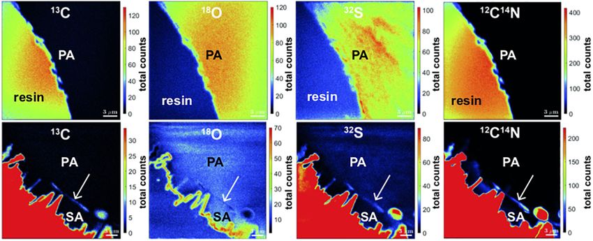
Krause et al.                                                                                                                          Endolithic Algae Coral Diagenesis

  FIGURE 2 | Original positions of the two Porites microatolls on the Buya Beach reef during low tide and corresponding coral slabs used for further analyses. (A,B)
  BB1. (C,D) BB3. Arrows indicate macroscopically visible green algae bands perpendicular to the coral surfaces.

approximately tripled after 2 months, 100 µL of algal volume was                       volume was placed directly on top of each plate. In addition, Petri
inoculated into a new bottle with medium.                                              dishes including only SWES medium and O. quekettii were also
                                                                                       prepared. Dead controls were prepared using O. quekettii fixed in
Intracellular Calcium Transport                                                        4% formalin for 24 h.
The variation of calcium concentration in O. quekettii in two                             For highlight conditions, Petri dishes with closed lid were
different illuminance conditions (high light vs. low light) was                        placed inside a clean bench exposed to an illuminance of 128–
investigated under laboratory conditions. Available skeleton                           135 lx. Low-light conditions were created in a lightproof box
parts of the scleractinian coral Stylophora pistillata were used                       with lid by cutting 1 × 1 cm holes at the bottom, which
as appropriate hard substrate. The branches were cut into                              were approximately 12 cm apart. The box was mounted with
small plates of several centimeters in length and a diameter                           a 2-cm clearance to a tabletop. Light penetrating through each
of approximately 3 mm using a handheld power drill with a                              hole inside the box had an illuminance of 0.64–0.37 lx. Each
diamond-coated saw blade. Subsequently, the plates were ultra-                         sample set for the two light conditions included living and dead
sonicated and dried as described above. In order to remove any                         O. quekettii with and without coral plates. Four replicates were
organic residue, the plates were bleached in 1% NaClO for 48 h,                        prepared for each treatment. Algae sampling was carried out
followed by thorough rinsing in ultra-purified water. Finally, the                     after 6 and 60 days.
coral plates were dried at 30◦ C under clean room conditions.                             The Fluo-4TM AM staining kit was used to image intracellular
    For the experiment, S. pistillata plates were placed individually                  variability of calcium levels of the O. quekettii cytosol. For
in transparent plastic Petri dishes filled with sterile SWES                           staining, 50 µg of desiccated dye was dissolved in 20 µL of
medium. Subsequently, approximately 50 mL of O. quekettii                              dimethyl sulfoxide (DMSO) to produce a 2.3 mM stock solution.

Frontiers in Earth Science | www.frontiersin.org                                   4                                            December 2019 | Volume 7 | Article 304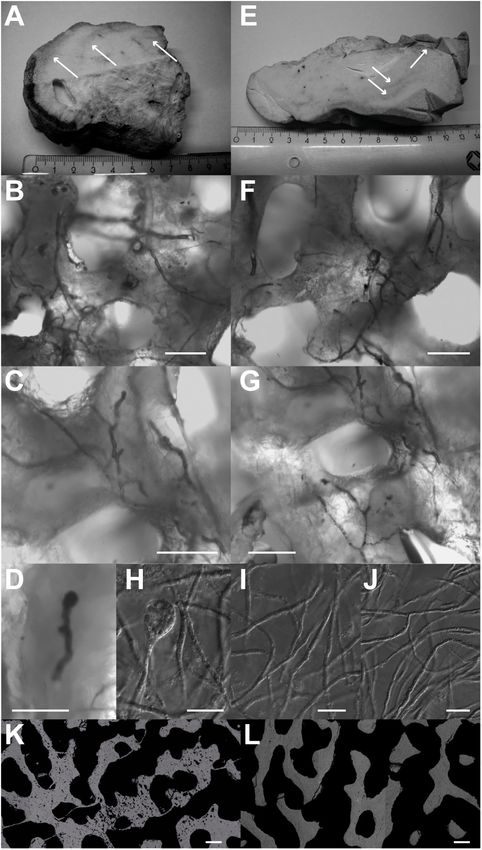
Krause et al.                                                                                                      Endolithic Algae Coral Diagenesis

For staining, approximately 50-µL algae volume was added to              average concentrations [VG-2 (Jarosewich et al., 1980), KAN-1
2295 µL of SWES medium and 5 µL of Fluo-4 AM stock                       (Reay et al., 1993), strontionite (Jarosewich and White, 1987), and
solution (final Fluo-4 concentration was 5 µM). Subsequently,            calcite (Jarosewich and MacIntyre, 1983)]. Results are illustrated
the samples were kept in the dark for 30 min. After staining,            as maps of quantitative element abundance in mass% and atomic
the algae tissue was placed shortly on WhatmanTM filter paper            Sr/Ca. The JEOL JXA 8200 was also used to generate the
and placed on a microscopy glass slide and covered with a glass          secondary electron images of the mapped area.
slip. Epifluorescence microscopy imaging was carried out using
an enhanced green fluorescent protein (EGFP) filter cube.                NanoSIMS Element Mapping
                                                                         The samples were coated with a 40-nm carbon layer using
Microscopy and SEM                                                       a Cressington carbon coater 108 carbon/A (Watford,
Coral subsamples and algae cultures were imaged using a Zeiss            United Kingdom). Areas of interest were selected based on
AxioImager.M2 and a 12-BitAxio Cam MRm Rev.3 camera at                   the approximate position based on previous microscopy analyses
GEOMAR. The software package ZEN 2.3 (Blue edition) was                  and the appearance in the NanoSIMS CCD-Camera (4×
used for image acquisition and processing. SEM was carried               magnification). SIMS imaging was performed using a NanoSIMS
out using a Zeiss Merlin Compact scanning electron microscope            50L instrument (Cameca, Paris, France). The 133 Cs+ primary
at IOW. For conductivity, samples were sputter-coated with a             ion beam was used to erode and ionize atoms of the sample. The
10-nm chromium layer. Images were obtained using secondary               electron flood gun was employed for compensation of charging
electron emission.                                                       effect in the area of the analysis. Maps of 13 C− , 18 O− , 32 S− , and
                                                                         12 C14 N− abundances were recorded simultaneously as indicators

X-Ray Diffraction                                                        for organic material within the coral. Less abundant ions (13 C− ,
                                                                         18 O− ) were employed because the signals of the main ions were
For XRD analysis, the obtained powder was ground in
an agate mortar and distributed evenly on a silicon disk.                too high for detection in parallel with the sulfur signal. Prior
Analyses were run from 4 to 75 2-theta (degree) angle at                 to the analysis, sample areas of 50 × 50 µm were sputtered
0.5/min on a Philips X-ray diffractometer PW 1710 with                   for 7 min with 600 pA to erode the carbon coating, clean the
monochromatic Co anode at GEOMAR. The XRD detection                      surface, and reach the steady state of secondary ion formation.
limit for aragonite and calcite is 2.9 and 0.9 mol.%, respectively       The primary ion beam current during the analysis was 5–20 pA,
(Kontoyannis and Vagenas, 2000).                                         depending on the signal intensities. For areas of 25 × 25 µm, 60
                                                                         planes were analyzed.
                                                                             Different spots with similar optical appearance were analyzed
Confocal Raman Microscopy                                                with the NanoSIMS Oxygen source (Duoplasmatron). Different
Confocal Raman microscopy (CRM) is an ideally suited analytical          spots were selected to avoid potential bias by the implanted Cs
method to determine mineral phases within biogenic materials             and already eroded surface. The pictures presented for Sr/Ca and
with high (micrometer range) spatial resolution (e.g., Nehrke and        Mg/Ca were derived from location at the surface of the same pore.
Nouet, 2011). CRM measurements in this study were performed              Measurements in other pores substantiated the findings (data not
at the AWI by means of a WITec alpha 300 R instrument                    shown). Prior to the analysis, sample areas of 50 × 50 µm were
connected to a diode laser having an excitation wavelength of            sputtered for 3 min with 600 pA to erode the carbon coating, 18-
488 nm. Raman spectra have been obtained using a Zeiss 20×               to 20-µm squares were analyzed, and 24 Mg+ , 39 K+ , 40 Ca+ , and
Epiplan lens (NA 0.4) and an UHTS300 ultra high-throughput               88 Sr+ ions were recorded. The primary ion beam current during
spectrometer (WITec GmbH, Ulm, Germany) equipped with                    the analysis was 50 pA. Thirty planes were analyzed.
a 1800 mm−1 grating blazed at 500 nm. Raman spectra were                     For analyses with both sources, the ions were detected with
measured and analyzed using the WITec ProjectFOUR software.              mass detectors equipped with electron multipliers (Hamamatsu).
An optical clear calcite single crystal (Iceland-spar from Mexico)       The scanning parameters were 512 × 512 pixels with a dwell
and a clear aragonite crystal (from Aragon in Spain) have been           time of 250 µs per pixel. The mass resolving power was adjusted
used as well-defined (XRD) in-house standards to obtain Raman            to be sufficient to suppress interferences at all masses allowing,
reference spectra under the same conditions the samples have             e.g., the separation of 32 S− from interfering ions such as 16 O2 − .
been measured at.                                                        The instrumental drift was checked by repeated analysis of one
                                                                         sample spot in the course of the measurements. No systematic
Electron Microprobe (EMP) Element                                        drift was revealed.
Mapping                                                                      Data analysis was performed with the Look@NanoSIMS
A JEOL JXA 8200 electron microprobe (EMP) was used at                    software package (Polerecky et al., 2012). The planes were
GEOMAR to generate element distribution maps for Ca and Sr               checked for signal instabilities, drift corrected, and accumulated.
on cross-sections of the coral skeleton. Each sample was carbon          Ratio images were produced based on the ion count pictures.
coated before the measurements. Measurements were carried                    It is worth noting that SIMS analyses are a destructive
out at an acceleration voltage of 15.0 kV, a beam current of             technique. Thus, a volume of the sample is consumed and several
20 nA, a beam diameter of 2 µm, a dwell time of 500 ms,                  layers are eroded. However, the sample erosion by the Cs source
and 10 accumulations for each image. Calibration of Sr and               was shown to be small enough to resolve cell size features
Ca concentrations was done using four standards with known               [5.7 nm/pane for a less intense primary ion beams focused to

Frontiers in Earth Science | www.frontiersin.org                     5                                       December 2019 | Volume 7 | Article 304
Krause et al.                                                                                                  Endolithic Algae Coral Diagenesis

a smaller area (Saka et al., 2014)]. In addition, the planes were       The Porites Mg/Ca–SST was calculated after Wei et al. (2000):
inspected carefully for signal changes with depth. Thus, we have
no hint that a change of the sample with depth by, e.g., reaching              SST = −14.13 + 8.846 × Mg/Ca (mmol/mol).                     (4)
edges of aragonite did occur and altered the results.                   The SST for Li/Mg was calculated using the calibration equation
                                                                        of Hathorne et al. (2013) for Porites from the Ogasawara
Micro-Computed Tomography                                               Islands (Japan):
To image two entire coral slabs (Figures 2B,D), a VivaCT 80
(Scanco Medical AG, Brüttisellen, Switzerland) µCT scanner,                 SST ◦ C = 20.9205 × (2.76 − Li/Mg (mmol/mol)). (5)


usually used for preclinical in vivo imaging, was used, which
permits a scan diameter of 80 mm with a scan length of up               The Sr partition coefficient for aragonite (DSr,a ) was calculated
to 150 mm. The used low-noise medium-resolution protocol                according to:
(70 kVp, 113 µA, 79.9 mm FOV, 125 proj./180◦ , 500 ms                              DSr,a = (Sr/Ca)aragonite /(Sr/Ca)seawater .              (6)
integration time, 6 × 6 sw-binning, standard filtered back
projection reconstruction algorithm including beam hardening            As no adequate data for modern seawater Sr/Ca are available for
correction for the density range of interest) sampled to an             the Indian Ocean, the combined mean of the Atlantic and Pacific
isotropic voxel size of 156 µm. The data were exported to               Ocean of 8.539 (mol/mol) (±0.45% SD) was used (De Villiers,
calibrated 16-bit grayscale DICOM image stacks for further              1999). As the total range of Sr/Ca of both oceans combined is
processing with the AVIZO 9.6 software. For both slabs,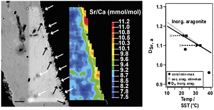
Krause et al.                                                                                                                     Endolithic Algae Coral Diagenesis

                                                                                          three concentric bands inhabited by Ostreobium were apparent
                                                                                          (Figure 3A). Three consecutive zones also with increased
                                                                                          secondary porosity, but without algae remnants, were located in
                                                                                          the inner part of the coral. These zones were termed former algae
                                                                                          bands (see Supplementary Figure S2). The BB3 section showed
                                                                                          five consecutive Ostreobium bands running perpendicular to the
                                                                                          coral’s growth direction. In addition, another band was situated
                                                                                          in close proximity to the coral surface partly overgrown by the
                                                                                          younger skeleton section (see Supplementary Figure S2).
                                                                                              Imaging of coral subsamples with SEM confirmed spatial
                                                                                          heterogeneity of the pore space surfaces. In the coral surface part,
                                                                                          as well as in the algae-free sections, pore space lining appeared as
                                                                                          smooth texture (Figure 4A). As this appearance was observed in
                                                                                          the youngest and in all algae-free sections, the smooth surface is
                                                                                          considered the pristine state.
                                                                                              In contrast, pore space surfaces from algae-band sections
                                                                                          showed an area-wide irregular surface consisting of blocky
                                                                                          crystals growing on top of the smooth surface. This mineral phase
                                                                                          showed strut bundles with blunt or flat apices (Figures 4B,C)
                                                                                          protruding between 5 and 10 µm into the pore space volume
                                                                                          (Figure 5). These infillings were present also several hundred
                                                                                          micrometers away from the cutting edge. In pristine areas,
                                                                                          smooth surfaces were present also at the saw-cut edge.
                                                                                          Consequently, mineral infilling as a consequence of sawing
                                                                                          artifacts can be excluded.
                                                                                              X-ray diffraction small-amount (10–20 mg) bulk
                                                                                          measurements as well as high-resolution Raman spectroscopy
                                                                                          performed in situ by focusing onto the blocky mineral phase
                                                                                          within the pores unambiguously identified the corals as
                                                                                          being purely aragonite {Raman peaks for the two lattice
                                                                                          modes (translation mode Ta , 152 cm−1 and librational mode
                                                                                          La , 206 cm−1 ) and the two internal modes [in-plane band
                                                                                          ν4 , ∼705 cm−1 (double peak) and symmetric stretch ν1 ,
                                                                                          1085 cm−1 ]}.

                                                                                          Coral Skeleton and Pore Space Volume
  FIGURE 3 | Algae green bands (white arrows) with Ostreobium thalli found in             Variability
  green bands of BB1 (A–D) and BB3 (E–G) and images from cultured
  O. quekettii (H–J). Different stages of sporangia development were observed
                                                                                          For comparison of the coral/pore space (v%/v%) inside and
  in the coral samples (D – premature algae sporangium from BB1) and in the               outside of algae bands, a rod-shaped subsample of the BB3 sample
  cultures (H – mature algae sporangium). Pure cultures of O. quekettii show              (Figure 6) was scanned with µ-CT. The subsample was cut
  thalli networks (I,J) similar to the ones found in coral green bands. Scale bars,       perpendicular to the surface including coral surface material with
  20 µm. Electron backscatter images of BB3 cross-section show coral                      remains of the polyp tissue followed by three alternating algae-
  skeleton parts inhabited (K) and devoid (L) of endolithic algae with different
  degrees of secondary porosity. Scale bars, 100 µm.
                                                                                          free areas with two intermittent algae bands. The voxel feed
                                                                                          size of the used scan was chosen to be 8.82 µm, thus masking
                                                                                          the vast majority of the vermiform Ostreobium burrows, as they
                                                                                          were too small in diameter to be resolved. As a result, the algae-
secondary porosity. For the image in Figure 3K, a total area                              induced secondary porosity did not add to the calculated pore
loss of 4% due to microporosity was calculated. In contrast,                              space volume. This advantage was used to constrain volume ratio
Figure 3L represents a typical region devoid of endolithic algae,                         variabilities between coral skeleton carbonate and primary pore
characterized by massive coral carbonate.                                                 space [coral/pore space (v/v)]. The youngest section of the coral,
                                                                                          defined by the presence of coral tissue remnants (Figure 6), and
Visual Diagenesis Indications                                                             the three algae-free zones showed a coral/pore space relative
Light as well as SEM revealed the presence of additional algae                            volume ratio varying between 47:53 and 48:52 (v%/v%), which
bands to the ones macroscopically visible. All of these showed                            was assumed to represent pristine conditions. In contrast, the two
increased porosity compared to the skeleton parts without algae                           algae bands were distinguished by higher ratios of coral material
bands (Figures 3K,L). At the outermost part of the BB1 section,                           of 50:50 and 54:46 (v%/v%), respectively.

Frontiers in Earth Science | www.frontiersin.org                                      7                                     December 2019 | Volume 7 | Article 304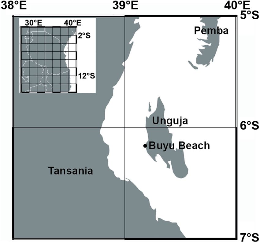
Krause et al.                                                                                                                        Endolithic Algae Coral Diagenesis

                                                                                        FIGURE 5 | Scanning electron microscopy backscattered electrons image of
                                                                                        BB1 cross-sections. (A) Pristine pore space surface. (B) With algae burrows
                                                                                        in the coral skeleton and adjacent secondary aragonite precipitates (white
                                                                                        arrows). Scale bar for A and B, 100 µm.

                                                                                        FIGURE 6 | Overview of rod-shaped subsample of BB3 (left) with coral tissue
                                                                                        (bottom-orange) and two consecutive algae bands (green). The subsample
                                                                                        was µ-CT scanned (right). Subsequently, percent ratios of coral carbonate
                                                                                        and pore space were calculated for each of the six indicated volumes.

                                                                                          The mean δ18 O values within the algae bands were more
                                                                                       negative (mean -4.66, SD 0.35, n = 6) than outside (mean -4.55,
                                                                                       SD 0.29, n = 7). The corresponding δ13 C values showed variability
  FIGURE 4 | Secondary electron emission (SEM) images of BB3 pore space                in both groups with amplitudes exceeding -5h within algae
  surfaces. (A) Original surface with smooth texture in algae-free area close to       bands and -4h outside, with most negative values found within
  the coral surface. (B,C) Secondary aragonite precipitation (black arrows)            the algae bands. The mean δ13 C and δ18 O values of samples from
  widely covering primary coral pore space surfaces (white arrows). Secondary
  precipitates appear as bundles of short and stumpy struts. Scale bars, 10 µm.
                                                                                       algae bands showed no significant differences to those obtained
                                                                                       outside the bands (δ18 O, t test unpaired, p-value = 0.53; δ13 C,
                                                                                       t-test unpaired, p-value = 0.21). The mean Sr/Ca (mmol/mol)
                                                                                       was about 2.5% higher in the bulk samples taken within the algae
Spatially Resolved Stable Isotope and                                                  bands [mean Sr/Ca (mmol/mol) 8.99, SD 0.08 (mmol/mol)],
Element Ratio Variations                                                               showing a significant difference of the group mean values (t-
In total, 13 samples were taken for the combined analysis of δ18 O,                    test unpaired, p-value = 0.01). The corresponding U/Ca values
δ13 C, and molar Sr/Ca, U/Ca, Mg/Ca, and Li/Mg (Table 1) from                          were also higher in the algae band samples, with overlapping
both corals. Six of the samples were obtained within green algae                       standard deviation. In addition, no significant difference was
bands; the remaining were from regions outside of algae bands                          observed (t-test unpaired, p-value = 0.19). The mean Mg/Ca was
(for drill sample position, see Supplementary Figure S2).                              lower in the algae bands (4.21 mmol/mol, SD 0.4) compared to

Frontiers in Earth Science | www.frontiersin.org                                   8                                           December 2019 | Volume 7 | Article 304
Krause et al. Endolithic Algae Coral Diagenesis
TABLE 1 | Bulk sample values for δ18 O, δ13 C, Sr/Ca, U/Ca, Mg/Ca, and Li/Mg of the microatolls BB1 and BB2.
Coral Location/drill position δ13C δ18O Sr/Ca mmol/mol U/Ca µmol/mol Mg/Ca mmol/mol Li/Mg mmol/mol
(VPDB (VPDB)
BB1 Algae band 2 −1.65 ± 0.07 −4.64 ± 0.05 8.96 ± 0.05 1.17 ± 0.01 4.09 ± 0.01 1.56 ± 0.03
BB1 Algae band 3 −2.24 ± 0.07 −4.33 ± 0.05 9.02 ± 0.09 1.28 ± 0.01 3.90 ± 0.01 1.66 ± 0.02
BB1 Algae band 5 −0.44 ± 0.07 −4.82 ± 0.05 9.11 ± 0.06 1.31 ± 0.01 3.76 ± 0.03 1.69 ± 0.01
BB3 Algae band 9 −5.70 ± 0.07 −4.99 ± 0.05 8.93 ± 0.02 1.07 ± 0.01 4.73 ± 0.01 1.49 ± 0.04
BB3 Algae band 10 −5.25 ± 0.07 −5.02 ± 0.05 8.92 ± 0.02 1.05 ± 0.01 4.65 ± 0.01 1.47 ± 0.02
BB3 Algae band 13 −3.35 ± 0.07 −4.18 ± 0.05 9.01 ± 0.04 1.22 ± 0.01 4.13 ± 0.02 1.5 ± 0.02
Mean
Algae band −3.11 −4.66 8.99 1.18 4.21 1.56
SD 2.07 0.35 0.07 0.11 0.40 0.09
BB1 Outside 1 −4.81 ± 0.07 −5.03 ± 0.05 8.96 ± 0.08 1.07 ± 0.01 4.74 ± 0.01 1.34 ± 0.01
BB1 Outside 4 −0.96 ± 0.07 −4.37 ± 0.05 8.79 ± 0.02 1.28 ± 0.01 4.93 ± 0.06 1.25 ± 0.01
BB3 Outside 6 −1.36 ± 0.07 −4.81 ± 0.05 8.89 ± 0.03 1.07 ± 0.01 4.97 ± 0.07 1.33 ± 0.03
BB3 Outside 7 −1.56 ± 0.07 −4.39 ± 0.05 8.99 ± 0.03 1.09 ± 0.02 4.84 ± 0.02 1.39 ± 0.02
BB3 Outside 8 −1.94 ± 0.07 −4.48 ± 0.05 8.79 ± 0.06 1.07 ± 0.01 4.69 ± 0.02 1.35 ± 0.04
BB3 Outside 11 −1.36 ± 0.07 −4.19 ± 0.05 8.81 ± 0.03 1.08 ± 0.01 4.45 ± 0.02 1.48 ± 0.01
BB3 Outside 12 −0.75 ± 0.07 −4.57 ± 0.05 8.84 ± 0.05 1.12 ± 0.01 4.55 ± 0.01 1.37 ± 0.01
Mean
Outside −1.32 −4.47 8.85 1.12 4.74 1.36
SD 0.42 0.21 0.08 0.08 0.21 0.08
t-test unpaired
p-value 0.21 0.53 0.01∗ 0.19Krause et al. Endolithic Algae Coral Diagenesis
FIGURE 8 | Left: Scanning electron microscopy backscatter image (SEM
BSE) and corresponding Sr/Ca mapping of a BB1 microatoll section with
secondary aragonite. White arrows denote secondary precipitates on primary
skeleton surface and in voids, reflected by green to orange mapping color
code. Black arrows show an optical less dense base layer, on which
secondary precipitates grow. Fine white line in color map indicates the pixel
resolution revised contour between coral (left) and embedding resin (right).
Right: Plot of DSr,a and calculated Sr/Ca–SST for primary coral material
(minimum and maximum) and secondary aragonite precipitates (minimum and
maximum); applying the vital effect including coral specific calibration of
Corrège (2006) suggests unrealistic low formation temperatures for the
secondary precipitates, whereas applying an inorganic calibration (Dietzel
et al., 2004) on the DSr,a values of the secondary precipitates results in a
temperature shift well into the SST range observed for the study area. The
black (Dietzel et al., 2004) and gray (Kinsman and Holland, 1969) lines show
FIGURE 7 | Nano-SIMS maps of 88 Sr/40 Ca and 24 Mg/40 Ca of blocky the systematic offset of temperature-dependent DSr,a for inorganic aragonite
secondary aragonite fringe (A,B) and pristine pore space surface (C,D). SA, precipitation. Values exceeding a Sr/Ca of 10 are attributed to areas of
secondary aragonite; PA, primary aragonite. Space outside corals is embedding resin. Scale bar, 10 µm.
resin-filled.
Calcium Transport Within Algae Cells DISCUSSION
In contrast to the impact on SST records of endolithic algae
activity, the fundamental mechanisms are less well constrained.
Effects of Environmental Factors on
To study the fate of elements being dissolved due to the boring Microatoll Morphology and
activity, we set up an experiment using cultures of O. quekettii, Geochemistry
belonging to a ubiquitously present group in Porites corals. Microatolls grow in the intertidal zone, causing coral vertical
Living algae were kept under two light intensities (low light, growth restriction and a dead top due to prolonged emersion
high light) on and without chips of coral skeleton for 60 days in during low tides (Scoffin and Stoddart, 1978). As a direct
seawater medium. The medium was spiked with Fluo-4 AM, a result of emersion, the microatoll top is susceptible to jointly
fluorescent dye suitable to image intracellular concentrations of mediated destruction by biological (Perry et al., 2012), chemical
dissolved calcium. (Revelle and Emery, 1957), and mechanical (Jones, 2012) erosion.
After 6 days of incubations, the algae incubated on coral The consequential increase in coral skeleton porosity at the
chips at low-light conditions showed increased intracellular microatoll top allows for the intrusion of seawater. During
calcium concentrations at the apical tips of the siphonal thallus emersion periods, increased seawater evaporation can lead to
(Figure 10A), assuming that calcium uptake was taking place the sequential precipitation of secondary minerals within a
here. In contrast, no increased concentrations were observed in microatoll starting with carbonates and followed by gypsum,
the other three treatments. After 60 days of incubation, markedly anhydrite, halite, potassium, and magnesium salts (Berner and
increased calcium concentrations were present over several 100 Berner, 1987; Boggs, 2014). In the temperature range between
mm within the algal cells incubated on coral chips (Figure 10E). 8 and 40◦ C, aragonite is likely the dominant carbonate phase
Assuming prolonged uptake of calcium ions by the apical tips, (Marion et al., 2009). As highest temperatures occur during the
an intracellular calcium transport over long distances can be summer months, it can be speculated that abiotic secondary
hypothesized. A beginning increase of intracellular calcium levels mineral precipitation takes place primarily during this time
at the apical tips was also observed in algae incubated under high- of year. Consequentially, emersed parts of a microatoll might
light conditions on coral chips after 60 days (Figure 10H). The show variable deviations in bulk geochemistry from non-
remaining treatments showed no sign of increased intracellular emersed coral sections.
calcium concentrations throughout the entire duration of the Microatolls grow under increased heat stress compared to
experiment (Figures 10B–D,F,G). their subtidal counterparts, which can slow down coral growth
Frontiers in Earth Science | www.frontiersin.org 10 December 2019 | Volume 7 | Article 304Krause et al. Endolithic Algae Coral Diagenesis
FIGURE 9 | Nano-SIMS mappings of 13 C, 18 O, 32 S, and 12 C14 N total counts in BB1 coral. Top: pristine pore space without secondary aragonite. Bottom: pore
space surface with secondary aragonite precipitates. While the primary coral skeleton is homogeneous in isotope concentrations, the interface between primary and
secondary aragonite (white arrows) is characterized by increased total counts of 13 C, 32 S, and 12 C14 N, as well as decreased total count concentrations in 18 O. The
epoxy embedding material is characterized by high C and S content (red color code).
Natural skeletal density changes cause the characteristic
annual coral banding, with a maximum skeletal density
associated with the hottest period of the year (Weber et al.,
1975). Compared to subtidal colonies, microatolls are subjected
to extended periods of high temperatures. Consequently, their
average skeletal density might generally be higher compared
to subtidal corals. This, in turn, might increase the microatoll
resilience against erosion.
Secondary Aragonite Morphology,
Geochemistry, and Mode of Precipitation
FIGURE 10 | Incubation experiments of O. quekettii with Fluor-4 AM for According to Nano-SIMS and EMP measurements, the observed
intracellular Ca imaging after 6 (A–D) and 60 days (E–H). Increased blocky aragonite has a Sr/Ca higher than observed for smooth,
fluorescence indicates increased intracellular Ca levels. (A,E) O. quekettii pristine pore space surfaces (Figure 7). If the blocky aragonite
incubated on coral chip at low-light conditions. Beginning Ca uptake after was the result of coral dissolution, a Sr-Ca exchange must have
6 days (A) and increased Ca levels within large parts of the siphonal thallus
occurred. This process, however, is highly unlikely as Ca is always
(E). (B,F) Incubation without coral chip at low-light conditions. No apparent
increase of intracellular Ca-concentration was detected during the entire preferred to Sr to be incorporated into the aragonite lattice.
incubation period. (C,G) O. quekettii incubated without coral chip at high-light Consequently, the observed blocky aragonite is a secondary
conditions. No apparent increase of intracellular Ca-concentration in algal mineral, precipitated as a result of early diagenesis.
thalli was detected during the entire incubation period. (D,H) Incubation on The occurrence of secondary carbonate minerals in tropical
coral chip at high-light conditions. After 60 days (H), beginning Ca uptake was
observed. Scale bar, 20 µm.
corals has been repeatedly reported since the early 1970s for
modern, Holocene, and fossil corals (Macintyre and Towe, 1976;
Bar-Matthews et al., 1993; Henderson et al., 1993; Obert et al.,
2019). The secondary aragonite growing on the smooth pristine
(Cantin et al., 2010). Therefore, annual bands might be spaced pore surfaces of our the studied microatolls are morphologically
narrower than in subtidal colonies. Although details regarding similar to the blunt aragonite fibers described by Ribaud-
a potential change in coral skeleton morphology as a result Laurenti et al. (2001) for Holocene Acropora corals and the
of heat stress are not constrained, several effects are plausible. aragonite laths cement reported for modern and Miocene corals
During times of highest temperature, a growth hiatus could (Griffiths et al., 2013). Living corals harbor a range of secondary
be initiated, which could occur repeatedly during the annual carbonates, often in the form of cement (Sayani et al., 2011). The
summer time or erratically during periods of exceptionally high encountered carbonate phases include high-magnesium calcite,
temperatures. Both scenarios would result in a complete loss of low-magnesium calcite (Macintyre and Towe, 1976), as well as
climate information during hiatus duration. aragonite (Hendy et al., 2007). The latter occurs in a variety
Frontiers in Earth Science | www.frontiersin.org 11 December 2019 | Volume 7 | Article 304Krause et al. Endolithic Algae Coral Diagenesis
of morphologies ranging from encrusted filaments, botryoids, secondary aragonite largely followed inorganic precipitation
rods, and needles (Nothdurft and Webb, 2009), and blocky mechanisms, which is explanatory for the observed difference
cement (Griffiths et al., 2013). Our study contributes to the in element ratios between the primary coral and the
suite of diagenetic features in tropical corals, showing that the secondary aragonite.
observed secondary aragonite blocky cement can be the result Although not statistically significantly different (t-test
of earliest diagenesis under marine conditions during a coral’s unpaired, p-value = 0.21), numerous carbonate bulk samples
life span, possibly induced by the activity of endolithic green within algae bands showed more negative δ13 C values than those
algae. However, the mechanisms inducing alternative aragonite outside the bands. As green algae have a δ13 C range between
morphologies other than acicular needles are currently not -20.3h (VPDB) and -8.8h (VPDB) (Maberly et al., 1992), it
well constrained. can be speculated that organic algal compounds might have been
An interesting feature, which, to our knowledge, has not been incorporated into the secondary aragonite.
documented before is a peculiar thin layer of approximately However, current opinions about secondary carbonate
1- to 2-µm thickness marking the transition between the precipitation in corals include precipitation from diffusing
primary coral skeleton and the secondary aragonite overgrowth. seawater as a consequence of microbial redox reactions (Tribble
This layer appears less dense than the surrounding mineral et al., 1990), the presence of intra-coral microenvironments
phases in images of electron backscattering (Figure 8, left). chemically differing from seawater (Nothdurft and Webb, 2009),
Nano-SIMS mapping of this transition area of primary and or a catalytic function of extracellular carbonic anhydrase (CA)
secondary aragonite showed that this layer differed by increased activity (Chen et al., 2018). Microbial redox reactions leading
counts of 13 C, 32 S, and 12 C14 N and a decreased 18 O signal, to pH increase and/or alkalinity production (sulfate reduction
compared to both aragonite phases (Figure 9). The opposing and denitrification) favoring carbonate precipitation require
trend in 13 C and 18 O counts and the increased counts of the hypoxic or anoxic conditions (Jørgensen, 1977; Zumft, 1997).
2 C14 N ion clearly indicate the presence of a thin organic layer These requirements are not met in endolithic algae bands as
(Remusat et al., 2012). Numerous studies have confirmed that the oxygen concentrations show considerable diurnal fluctuations,
presence of organic templates is directly involved in the microbial reaching hyperoxia during light time (Shashar and Stambler,
nucleation of non-skeletal marine carbonates (Braissant et al., 1992). Also, the absence of any iron-sulfide minerals supports
2003; Degens, 2012; Krause et al., 2012). In contrast, the the assumption of prolonged oxic conditions during secondary
role of non-coral organic compounds for secondary carbonate aragonite precipitation.
precipitation within the coral skeleton is poorly constrained. The chemical environment within algae bands is currently
Endolithic organisms such as O. quekettii are known to excrete not well characterized, and presumably the precipitation of
organic metabolic products potentially beneficial for the coral secondary aragonite is governed by more than one factor.
host tissue (Försterra and Häussermann, 2008). Consequently, Due to the combined boring and photosynthetic activity of
it can be assumed that secondary aragonitic cement with the green algae, we can assume that pH in the algae bands
morphologies other than acicular needles nucleated under shows heterogeneity, potentially promoting secondary aragonite
variable influence of present organic compounds within the precipitation in regions with elevated pH. Also, evidence exists
coral. The individual composition and concentration of organic that the enzyme CA is present within endolithic photosynthetic
molecules might then facilitate a variety of crystal morphologies. algae (Shashar and Stambler, 1992). Within coral skeletons,
In the present coral samples, aragonitic overgrowth was CO2 concentrations are generally lower than in the ambient
observed in the form of blocky cement as documented before seawater (Shashar and Stambler, 1992). In order to counteract
(Nothdurft and Webb, 2009). As the secondary aragonite CO2 limitation for endolithic photosynthesis, CA converts
was exclusively observed in green algae bands, it is plausible bicarbonate to CO2 , according to the following reaction:
that the organic compounds, potentially involved in secondary
aragonite nucleation, were excreted by the adjacent algae thalli HCO− +
3 + H ↔ CO2 + H2 O.
(Duerden, 1902; Fine and Loya, 2002). Nonetheless, other
sources of organic compounds, including coral polyp-derived Consequently, in case of the reaction driven to the right, the
skeletal organic matrix (Abelson, 1955), adsorbed organics from H+ removal increases pH, which favors carbonate precipitation.
ambient seawater (Isdale, 1984), and organic matter from bacteria Whether extracellular CA is also involved in liquid-phase pH
(DiSalvo, 1969) or fungi (Bak and Laane, 1987) cannot be modulation is currently unknown.
excluded completely. Another factor to be considered for secondary aragonite
Bulk and spatial high-resolution analyses showed that precipitation is seasonality. The stoichiometric solubility product
the secondary aragonite showed increased Sr/Ca, U/Ca, and (K∗ SP ) for aragonite is principally dependent on temperature
Li/Mg, while depleted in Mg/Ca. This result supports the (Zeebe and Wolf-Gladrow, 2001), resulting in a negative
results of previous studies for secondary aragonite (Enmar correlation between mineral solubility and temperature. As
et al., 2000; Quinn and Taylor, 2006; Griffiths et al., 2013). a consequence, secondary aragonite precipitation could be
As the strontium partition coefficient for the secondary facilitated during periods of annual peak temperatures. Although
aragonite (DSr,a ) fitted exactly on the DSr /temperature the obtained Sr/Ca values for the abiotic secondary aragonite
correlation for inorganic aragonite (Kinsman and Holland, of the present study do not indicate intensified precipitation
1969; Dietzel et al., 2004), we can safely assume that the at notably hot temperatures, further studies focused on
Frontiers in Earth Science | www.frontiersin.org 12 December 2019 | Volume 7 | Article 304Krause et al. Endolithic Algae Coral Diagenesis
seasonal resolution are required to elucidate the role of annual the presence of secondary aragonite to be responsible for pore
temperature fluctuations for microatoll secondary aragonite. space volume reduction of up to 50% in fossil Acropora corals.
The incubation experiments carried out during this study
showed uptake of dissolved calcium by the algae O. quekettii
Influence of Endolithic Green Algae on from the apical tips and subsequent increased calcium levels
Coral Skeleton throughout the siphonal thallus. This observation points to an
We showed that hot spots of diagenesis were clearly associated active uptake of calcium by the algae and subsequent transport
with skeleton regions colonized by endolithic green algae toward the distal part of the siphonal thallus. This mechanism
resulting in skeleton dissolution and potentially also the prevents increasing calcium concentrations in the borehole,
precipitation of secondary blocky aragonite within pore spaces of which might lead to spontaneous reprecipitation of carbonate
algae bands. The observed skeleton dissolution is in accordance and bore hole clogging.
with previous studies identifying endolithic green algae as Active calcium removal from the site of carbonate excavation
important agents for coral skeleton erosion (Chazottes et al., has so far, to our knowledge, only been observed for
1995; Tribollet and Golubic, 2011). Each of the microatolls cyanobacteria (Garcia-Pichel et al., 2010), but not for green algae.
showed individual macroscopically visible boreholes of 2–3 mm In our experiments, the intensity of calcium uptake was
in diameter, presumably caused by macroborers. Numerous negatively correlated with light intensity. We can therefore
studies also confirmed the important role of macroborers conclude that low-light conditions act as a trigger, initiating
for coral erosion, including lithophagine bivalves (Wizemann boring activity to avoid light limitation. In contrast, calcium
et al., 2018) and polychaetes (Davies and Hutchings, 1983). incorporation was considerably less intense under high-light
While faunal organisms generally contribute substantially to conditions. Consequently, it can be assumed that excessive
coral erosion (Zubia and Peyrot-Clausade, 2001; Tribollet and calcium uptake is an energy-demanding process only carried
Golubic, 2011), they might be less abundant in microatolls as a out when necessary. Currently, the exact boring mechanism of
consequence of increased heat stress (Przeslawski et al., 2008) endolithic green algae and the fate of the dissolved inorganic
at the water surface. Therefore, temperature-tolerant endolithic carbonate species are unknown. The mechanism could involve an
algae (Fine et al., 2005) might be largely responsible for the acid-generating metabolism as the boring tip of the thallus or the
observed microatoll internal erosion. From the analysis of two- uptake of calcium ions from the liquid phase at the boring locality
dimensional coral cross-section images, we can infer that around to decrease the local carbonate saturation state to an extent to
4% of the internal skeleton area within an algae band was lost as induce spontaneous dissolution (Garcia-Pichel et al., 2010).
a direct consequence of boring activity (Figure 3K). As calcium concentrations are usually strictly controlled in
The volumetric calculations suggested a volume gain of 3 all types of organisms (White and Broadley, 2003; Dominguez,
and 7%, respectively, within the algae bands. Although it is 2004; Brini et al., 2013) due to its toxic effects (Smith, 1995),
generally not approved to compare two- and three-dimensional passive diffusion of Ca2+ ions within the algal thallus is highly
data, it appears plausible that the values obtained for internal unlikely. Instead, once taken up, the majority of calcium ions
coral skeleton dissolution and the coral/pore volume percent might have been attached to calcium-binding proteins acting
ratio reside in the same order of magnitude. This in turn leads as concentration buffers (La Verde et al., 2018). In contrast,
to the conclusion that the majority of the dissolved primary coral calcium transport within cyanobacteria filaments is supposedly
skeleton could have reprecipitated within a short spatial distance mediated by transmembrane calcium pumps (Garcia-Pichel
inside the same algae band. Nonetheless, natural variability of et al., 2010). Once inactivated, the calcium can be transported
coral skeleton to pore space ratio (density) is known for almost intracellularly over long distances without adverse physiological
five decades (Knutson et al., 1972). In addition to annual, fine effects. Although not observed directly, from the analysis of the
monthly and irregular bands are also produced (Barnes and coral slabs, we can infer an eventual release of calcium ions
Lough, 1993). Consequently, one can argue that a diagenetic into the coral primary pore space volume, inducing secondary
density change is indistinguishable from natural coral density precipitation in case of carbonate supersaturation.
banding. Conventionally, variations in coral skeletal density are
routinely analyzed by two-dimensional X-ray radiographs of Geochemical Implications
prepared coral slices (Buddemeier et al., 1974). In order to derive Under the environmental conditions microatolls grow in,
coral density from X-ray density, elaborate calibration algorithms including higher temperature and solar radiation, compared
have to be applied involving defined standard material (Chalker to the subtidal environment, endolithic green algae might be
et al., 1985). However, a precise quantification of coral skeleton to an important driver for earliest coral skeleton diagenesis. The
pore space ratio is virtually impossible. The used µ-CT approach obtained results are not straightforward transferrable to other
has the advantage of providing such data in a non-destructive Porites morphotypes, as dome-shaped colonies, growing in
way, irrespective of individual sample mineralogy and thickness. several meters of water depth. Due to light attenuation with water
Our µ-CT approach demonstrates an increased coral/pore space depth, endolithic algae colonizing subtidal corals are subjected
in both areas inhabited by endolithic algae; whether this is to less solar energy per area, limiting the photosynthetic activity.
entirely caused by secondary aragonite precipitation, natural Although light limitation is partly compensated by an increased
coral density variability, or a mixture of both is beyond the scope ratio of Chlorophyll b to Chlorophyll a (Schlichter et al., 1997),
of this study. However, Ribaud-Laurenti et al. (2001) also found endolithic green algae generally grow slower under reduced solar
Frontiers in Earth Science | www.frontiersin.org 13 December 2019 | Volume 7 | Article 304You can also read