A DLL3-targeted antibody-drug conjugate eradicates high-grade pulmonary neuroendocrine tumor-initiating cells in vivo - Science Translational Medicine
←
→
Page content transcription
If your browser does not render page correctly, please read the page content below
RESEARCH ARTICLE

CANCER

A DLL3-targeted antibody-drug conjugate eradicates
high-grade pulmonary neuroendocrine tumor-initiating
cells in vivo
Laura R. Saunders,1 Alexander J. Bankovich,1 Wade C. Anderson,1 Monette A. Aujay,1
Sheila Bheddah,1 KristenAnn Black,1 Radhika Desai,1 Paul A. Escarpe,1 Johannes Hampl,1
Amy Laysang,1 David Liu,1 Javier Lopez-Molina,1 Milly Milton,1 Albert Park,1 Marybeth A. Pysz,1
Hui Shao,1 Brian Slingerland,1 Michael Torgov,1* Samuel A. Williams,1 Orit Foord,1
Philip Howard,2 Jacek Jassem,3 Andrzej Badzio,3 Piotr Czapiewski,3 David H. Harpole,4
Afshin Dowlati,5 Pierre P. Massion,6 William D. Travis,7 M. Catherine Pietanza,7,8 J. T. Poirier,7,8
Charles M. Rudin,7 Robert A. Stull,1 Scott J. Dylla1†

The high-grade pulmonary neuroendocrine tumors, small cell lung cancer (SCLC) and large cell neuroendocrine

                                                                                                                                                                        Downloaded from http://stm.sciencemag.org/ by guest on April 27, 2021
carcinoma (LCNEC), remain among the most deadly malignancies. Therapies that effectively target and kill tumor-
initiating cells (TICs) in these cancers should translate to improved patient survival. Patient-derived xenograft (PDX)
tumors serve as excellent models to study tumor biology and characterize TICs. Increased expression of delta-like 3
(DLL3) was discovered in SCLC and LCNEC PDX tumors and confirmed in primary SCLC and LCNEC tumors. DLL3
protein is expressed on the surface of tumor cells but not in normal adult tissues. A DLL3-targeted antibody-drug
conjugate (ADC), SC16LD6.5, comprised of a humanized anti-DLL3 monoclonal antibody conjugated to a DNA-
damaging pyrrolobenzodiazepine (PBD) dimer toxin, induced durable tumor regression in vivo across multiple
PDX models. Serial transplantation experiments executed with limiting dilutions of cells provided functional evi-
dence confirming that the lack of tumor recurrence after SC16LD6.5 exposure resulted from effective targeting
of DLL3-expressing TICs. In vivo efficacy correlated with DLL3 expression, and responses were observed in PDX
models initiated from patients with both limited and extensive-stage disease and were independent of their sen-
sitivity to standard-of-care chemotherapy regimens. SC16LD6.5 effectively targets and eradicates DLL3-expressing
TICs in SCLC and LCNEC PDX tumors and is a promising first-in-class ADC for the treatment of high-grade pulmo-
nary neuroendocrine tumors.

INTRODUCTION                                                                               sitive to chemotherapy, relapses generally occur shortly after cessation
High-grade pulmonary neuroendocrine tumors, which include small                            of SOC (1). The only widely approved second-line therapy, topotecan,
cell lung cancer (SCLC) and large cell neuroendocrine carcinoma (LCNEC),                   provides a ~17% response rate, a median progression-free survival
represent ~18% of primary lung neoplasms and predominantly devel-                          of 3 months, and an overall survival of less than 7 months (7). Although
op in older patients with a history of smoking (1, 2). Both SCLC and                       there is no clear treatment consensus for LCNEC, it is commonly treated
LCNEC remain among the most deadly malignancies because no new                             similarly to SCLC. Given the poor prognosis and lack of treatment op-
therapeutic options have emerged for these indications in more than                        tions, it is desirable to identify new therapeutic targets and treatment
30 years (3, 4). SCLC survival is measured in months, with a 5-year                        modalities to improve patient outcomes.
survival rate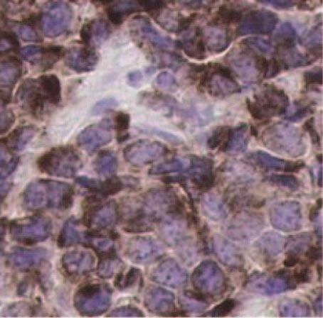
RESEARCH ARTICLE
and oxygen sensing and are abundant in the developing lung (16). one LCNEC PDX were dissociated to single-cell suspensions, and tu-
Mouse models replicating the oncogenic mutations and tumor sup- mor cells were isolated using fluorescence-activated cell sorting (FACS).
pressor losses observed in patients have implicated PNECs, or a mul- Isolated subpopulations were transplanted to evaluate their tumorige-
tipotential precursor that gives rise to PNECs, as the cell of origin for nicity, and whole transcriptome sequencing was performed in parallel
SCLC (17–19). Critical for PNEC development is the transcription factor to identify differentially expressed genes. DLL3 was identified as >100-
achaete-scute homolog-1 (ASCL1; murine ortholog Mash1). Mash1 ex- fold overexpressed in SCLC and LCNEC PDX versus seven different
pression in the developing mouse lung peaks at birth and declines in normal vital organs, including the lung (table S1), and was increased
adulthood, and mice lacking Mash1 die soon after birth because of lung in all populations of TICs (Fig. 1A).
defects (20–23). ASCL1 is also important in neuroendocrine cell fate To verify whole transcriptome data and expand analysis to additional
decisions and is highly expressed in classic SCLC and LCNEC tumors, samples, we performed quantitative reverse transcription polymerase
where it acts to maintain neuroendocrine features (21). ASCL1 expres-
sion correlates with the tumor-initiating capacity of SCLC tumors (24).
A B
The Notch pathway has likewise been implicated in regulating neuro- 100
9
10
endocrine versus epithelial cell fate decisions in the developing lung
Relative expression
RPKM_Transcript
10 7
(25). The mammalian Notch family ligands DLL1, DLL4, JAG1, and 10
JAG2 each activate Notch receptor signaling in trans (26). In contrast, 10
5
1
the related ligand delta-like 3 (DLL3) predominantly localizes to the 3
Downloaded from http://stm.sciencemag.org/ by guest on April 27, 2021
10
Golgi apparatus and is unable to activate Notch signaling (27, 28). DLL3 0.1 1
shares only 36% homology with DLL1 and differs from other delta- 10
type DSL (Delta/Serrate/LAG-2) proteins, DLL1 and DLL4, in both its 0.01 10
1
reduced number of epidermal growth factor (EGF)–like repeats and
es
LC
X
X
es
X
X
PD
PD
PD
PD
su
su
spacing of the cysteine residues within its DSL domain, which is required
SC
tis
t is
LC
EC
LC
EC
L
for Notch binding (29). Normal tissue expression of DLL3 is highest
SC
SC
N
N
L
N
N
LC
LC
in fetal brain, and DLL3 plays a key role in somitogenesis in the par-
axial mesoderm (27, 28, 30–32). Although Notch pathway activation C
108
D
LU80
acts as an oncogenic stimulus in some tumor types (33), Notch activa- 106 LU100
tion in neuroendocrine tumors suppresses tumor growth (34). In the 107
NEUROD1
105
course of normal development, DLL3 inhibits both cis- and trans-
ASCL1
acting Notch pathway activation by interacting with Notch and DLL1 10 6
104
LU80
and redirecting or retaining them to late endosomal/lysosomal com-
partments or the Golgi, respectively, thereby preventing their local- 105 103
LU100
100
ization to the cell surface (27, 35). Moreover, DLL3 is one of several 10 1 LU86 100
2 10 1
Notch ligands that appear to be direct downstream targets of ASCL1 10 10 2
10 5 10 6 10 7
10 8
10 9
105 106 107 108 109
(36, 37). Together, these observations suggest that DLL3 might be asso- DLL3
DLL3
ciated with the neuroendocrine phenotype and contributes to neuro- E F
endocrine tumorigenesis. 100 14
Normalized log2 intensity
We set out to explore heterogeneity in SCLC and LCNEC PDX by
RPKM_Transcript
10 12
characterizing gene expression in TICs from these tumors. Whole tran- 10
scriptome data from isolated populations of SCLC and LCNEC tumor 1
8
cells showed DLL3 expression to be increased relative to normal tissues,
0.1 6
including normal lung. Further analysis showed that DLL3 protein
4
was detectable at the surface of SCLC and LCNEC tumor cells, leading 0.01
2
to the hypothesis that it could make a tractable therapeutic target for
0.001 0
an antibody-drug conjugate (ADC) in these cancers (38). We developed
X
X
es
ng
LC
L
an ADC to leverage the potent activity of the cell cycle–independent
PD
PD
C
su
lu
SC
LC
LC
EC
tis
pyrrolobenzodiazepine (PBD) cytotoxin D6.5, with the expectation
L
SC
N
SC
N
L
N
LC
that it would selectively kill DLL3-expressing tumor cells and limit sys-
temic toxicities. Here, we show that the DLL3-targeted ADC, SC16LD6.5, Fig. 1. Elevated expression of DLL3 mRNA in SCLC. (A) DLL3 transcripts
effectively targets and eradicates TICs in both SCLC and LCNEC conveyed as reads per kilobase per million reads mapped to annotated exons
PDX tumors. (RPKM_Transcript) in normal tissues (NL tissues) and SCLC and LCNEC PDXs.
(B) Relative expression of DLL3 in NL tissues, primary SCLC biopsy specimens
(SCLC), and SCLC and LCNEC PDX, as measured by quantitative PCR. (C and
D) Relative expression of ASCL1 (C) and NEUROD1 (D) versus DLL3 in SCLC
RESULTS (blue diamond) and LCNEC (red triangle) PDX, as measured by quantitative
PCR. (E) DLL3 transcripts (RPKM_Transcript) in normal lung, primary SCLC
Increased DLL3 expression in high-grade pulmonary tumors, and SCLC cell lines (CL). (F) Quantile normalized log2 intensity values
neuroendocrine tumors of DLL3 mRNA in NL tissues and PDX lines assessed by microarray. Horizontal
SCLC and LCNEC PDX tumors were previously established from trans- bars represent the geometric mean. Normal tissues included in each ex-
bronchial needle aspirates or tumor resections (14). Four SCLC and pression metric are detailed in table S1.
www.ScienceTranslationalMedicine.org 26 August 2015 Vol 7 Issue 302 302ra136 2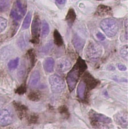
RESEARCH ARTICLE

chain reaction (qRT-PCR) in four primary SCLC tumor biopsy spec-          firmed to maintain affinity for human, cynomolgus monkey (cyno),
imens matched to established PDX models, an additional 15 SCLC            and rat DLL3 antigens. Data for a representative humanized mono-
and 2 LCNEC PDX, and 26 normal human tissues. Elevated expres-            clonal antibody, SC16, are shown in table S3. Cross-reactivity and
sion of DLL3 mRNA was confirmed in these primary SCLC tumors              equivalent SC16 binding to DLL3 from species relevant for toxicology
and low-passage SCLC and LCNEC PDX tumors (Fig. 1B). Among                studies were demonstrated using human embryonic kidney (HEK)–293T
normal tissues, mRNA expression was limited to the brain, esopha-         cells transduced and selected for expression of human, cyno, or rat DLL3,
gus, and pancreas, with the last two having 1000-fold lower levels        respectively. Flow cytometry confirmed that SC16 bound to each of these
than SCLC and LCNEC PDX tumors (table S1). Because DLL3 is                proteins on the surface of nonpermeabilized, engineered HEK-293T
thought to be a transcriptional target of ASCL1 (36), its expression      cells (Fig. 2A), establishing that DLL3 protein can localize to the cell
was also assessed and found to significantly correlate with DLL3 ex-      surface and is not necessarily confined to the Golgi (27, 28). The spec-
pression in SCLC and LCNEC PDX (Fig. 1C; Pearson r2 = 0.66, P <           ificity of SC16 for DLL3, and not its related family members DLL1 or
0.0001). Previous studies have classified SCLC into two subtypes that     DLL4, was demonstrated by ELISA (Fig. 2B). These studies establish
can be discriminated by high expression of ASCL1 (classic SCLC) or        that SC16 is specific for DLL3, that DLL3 can localize to the cell surface,
high expression of NEUROD1 (variant SCLC) (39, 40). Consistent            and that the affinity of SC16 for human, cyno, or rat antigen is in the low
with their classification as variant SCLC, LU80 and LU100 had lower       nanomolar range and within threefold across species (table S3).
ASCL1 and DLL3 expression (Fig. 1C) but higher NEUROD1 expres-
sion (Fig. 1D and fig. S1A). Notably, the cisplatin and etoposide (C/E)   Surface expression of DLL3 in SCLC and LCNEC

                                                                                                                                                        Downloaded from http://stm.sciencemag.org/ by guest on April 27, 2021
refractory PDX tumor model LU86 (14) had high NEUROD1 and                 To measure protein expression in tumor and tissue lysates, we devel-
DLL3 expression despite low ASCL1 expression. Collectively, our data      oped a sandwich ELISA using two noncompeting DLL3 monoclonal
show high expression of DLL3 in most of classic SCLC, with lower          antibodies. Analysis of total protein lysates from 28 normal tissues,
levels in variant SCLC.                                                   14 SCLC PDX, and 2 LCNEC PDX showed that DLL3 protein expres-
    To further expand our analysis of tumor and normal tissue speci-      sion in normal tissues was below the limit of quantitation of 0.37 part
mens, we examined DLL3 expression in whole transcriptome se-              per million (ppm; ng DLL3/mg total protein) in all tissues except the
quencing data sets from 29 primary SCLC biopsy specimens, 25              heart and adrenal gland (table S1), whereas an average of 3.7 ppm was
SCLC cell lines, and 25 normal lung biopsy specimens (11). This anal-     detected in SCLC PDX tumors, along with very elevated expression in
ysis confirmed our initial observations, revealing a ~35-fold elevation   the two LCNEC PDXs evaluated (Fig. 2C). Notably, there was no DLL3
in DLL3 mRNA in SCLC relative to normal lung (Fig. 1E). These             protein above the limit of detection in the brain, despite high mRNA
SCLC tumor samples were compared to transcriptome data from               expression. Furthermore, the SCLC PDXs that had lower mRNA ex-
normal tissues and other tumor types in The Cancer Genome Atlas data      pression (LU80 and LU100) were likewise found to have low levels
set, which further confirmed elevated DLL3 expression in primary SCLC     of DLL3 protein (0.6 ppm and below the limit of detection, respectively).
tumor samples, as well as low-grade glioma, glioblastoma, and melano-         To further explore protein expression and its cellular localization in
ma (fig. S1B). Illumina BeadChip data from the Clinical Lung Cancer       SCLC and LCNEC tumors, we identified a DLL3-specific monoclonal
Genome Project (10) also showed DLL3 elevation in primary SCLC            antibody for immunohistochemistry (IHC) using formalin-fixed, paraffin-
tumor specimens compared to NSCLC (fig. S1C).                             embedded samples (fig. S2). An initial assessment of two SCLC PDX
    Finally, microarray gene expression analysis of 14 SCLC and 2         (LU64 and LU149), one LCNEC PDX (LU37), and primary biopsy
LCNEC PDX models revealed ~120-fold elevation in expression of            samples from SCLC and LCNEC patients demonstrated both mem-
DLL3 mRNA compared to 12 normal tissues (Fig. 1F and tables S1            branous and cytoplasmic staining (Fig. 2D). This staining pattern is
and S2). These observations were further confirmed by publically avail-   consistent with previous observations of localization for other Notch
able microarray data sets from the Cancer Cell Line Encyclopedia (41),    ligands and receptors (27, 28). The membrane localization of DLL3
which show elevated DLL3 mRNA expression specifically in SCLC cell        staining was quantified in primary tumor specimens, using tissue micro-
lines (fig. S1D). Collectively, these expression data across numerous     arrays encompassing 9 normal lung samples, 95 non-SCLC (82 ade-
technical platforms and samples show that DLL3 mRNA is overexpressed      nocarcinoma and 13 squamous cell carcinoma), 57 LCNEC, and 187
in primary SCLC tumors, SCLC PDX, conventional SCLC cell lines, and       SCLC tumor specimens. Surface DLL3 expression was quantified by con-
LCNEC PDX, whereas mRNA expression in normal tissues appears              verting the staining intensity (range, 0 to 3) and the percentage of cells
limited primarily to the brain.                                           with expression to an H-score (range, 0 to 300) (Fig. 2E and fig. S3).
                                                                          No normal lung specimen or lung squamous cell carcinoma tumor cells
Generation and characterization of DLL3-specific                          stained positively, whereas 37 of 57 LCNEC (65%), 120 of 167 treatment-
monoclonal antibodies                                                     naïve SCLC (72%), and 17 of 20 recurrent and treatment-refractory
To assess protein expression and determine whether DLL3 is on the         (R/R) SCLC (85%) exceeded an H-score of 100. Notably, 3 of 82
surface of tumor cells, anti-DLL3 antibodies were generated and char-     (3.7%) lung adenocarcinoma tumors had DLL3 expression, suggesting
acterized. In separate immunization campaigns, recombinant DLL3-Fc        the presence of neuroendocrine components in these tumors (Fig. 2E).
(Adipogen) or DLL3-His protein purified from supernatants of trans-       This is consistent with previous observations of ASCL1 expression and
fected CHOK1 (Chinese hamster ovary–K1) cells was used as an im-          other neuroendocrine markers in ~10% of lung adenocarcinoma, ex-
munogen to produce mouse monoclonal antibodies that were confirmed        clusively in patients who smoked (42). We next explored DLL3 ex-
to bind DLL3 by enzyme-linked immunosorbent assay (ELISA) and flow        pression on the surface of cells from dissociated PDX tumors using
cytometry. Several antibodies binding to different DLL3 epitopes were     flow cytometry and a PE-conjugated DLL3 antibody. Data from repre-
humanized by CDR grafting of the murine variable regions onto the         sentative SCLC PDX (LU149) and LCNEC PDX (LU37) models
human immunoglobulin G1 (IgG1)/k constant regions and were con-           show expression of DLL3 on the cell surface (Fig. 2F). Collectively, the

                                                              www.ScienceTranslationalMedicine.org    26 August 2015   Vol 7 Issue 302 302ra136    3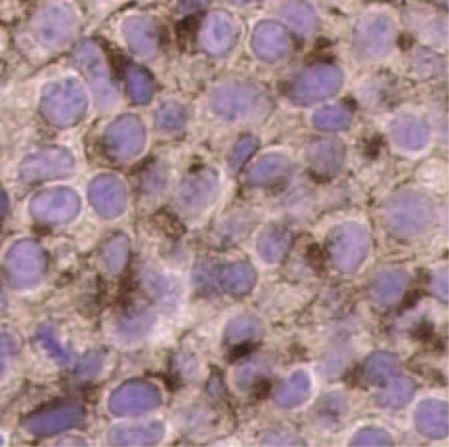
RESEARCH ARTICLE
A B C E
2.5 300
125 HEK-293T.hDLL3 DLL1 30
A647 (normalized MESF)
HEK-293T.cDLL3 DLL3 15
Membranous H-score
250
Absorbance (A450)
HEK-293T.rDLL3 2.0 DLL4 9
Avg DLL3 (ppm)
100
200
75 1.5
6 150
50 1.0 100
3
25 0.5 50
0 0 0
0.0
0.001 0.01 0.1 1 10 0.001 0.01 0.1 1 10 s X X
-A C
no
ve EC
C
LC
LC ng
ue
LC qC
R CL
PD PD
de
SC
N CN
SC lu
[Ab] (nM) [Ab] (nM) s
SC -S
S
tis EC LC
N NL
L
/R
L N SC
aï
N
LC
N
D F
SCLC LU149
Downloaded from http://stm.sciencemag.org/ by guest on April 27, 2021
LCNEC LU37
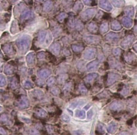
Fig. 2. Characterization of DLL3-specific and species cross-reactive primary SCLC (H-score = 170 and 200) and LCNEC (H-score = 160) tumors. Scale
monoclonal antibodies. (A) SC16 shows equivalent binding to human, bars, 20 mm. (E) DLL3 membrane expression as measured by IHC in normal lung
cyno, and rat DLL3 expressed on the surface of HEK-293T cells. (B) SC16 reacts tissue and primary tumors including lung squamous cell (NSCLC-SqCC), lung
only with DLL3 and not related family members DLL1 or DLL4. (C) DLL3 pro- adenocarcinoma (NSCLC-Adeno), LCNEC, and naïve and recurrent/refractory
tein was detected in SCLC and LCNEC PDX by ELISA. Horizontal bars repre- (R/R) SCLC. Horizontal bars represent the mean. (F) Surface DLL3 expression on
sent the mean. Normal tissue samples and the amounts of DLL3 protein SCLC LU149 and LCNEC LU37 PDX tumor cells assessed by flow cytometry
detected are detailed in table S1. (D) IHC of two SCLC (LU64, H-score = 96; with phycoerythrin (PE)–conjugated anti-DLL3 (black line) or IgG1 isotype
LU149, H-score = 134) and one LCNEC (LU37, H-score = 147) PDX, as well as control (gray-filled) antibodies. MESF, mean equivalents of soluble fluorescein.
above data show that elevated DLL3 mRNA expression translates to de- rat DLL3 (table S3). Additionally, SC16LD6.5 was incubated in human
tectable protein at the cell surface in SCLC and LCNEC tumors, but not serum and shown to be stable in physiologically relevant conditions with
normal tissue. minimal release of D6.5 over time (fig. S4).
To evaluate whether DLL3 was internalized after antibody or ADC
Internalization and toxin delivery by anti-DLL3 engagement, we evaluated its intracellular trafficking using immuno-
monoclonal antibodies fluorescence colocalization analysis. Specifically, parental HEK-293T cells
To evaluate whether anti-DLL3 antibodies can mediate internalization and cells overexpressing human DLL3 (HEK-293T.hDLL3) were infected
and delivery of a potent cytotoxin, we generated an ADC targeting DLL3, with a baculovirus expressing a fluorescently labeled protein [stomatin-
SC16LD6.5. SC16LD6.5 is comprised of the D6.5 PBD payload con- like protein-1 (SLP-1)] that localizes to late endosomes (43), a prelysoso-
jugated to cysteine residues on the SC16 antibody via a maleimide- mal endocytic compartment with low pH and abundance of cathepsin B,
containing linker with an eight-carbon polyethylene glycol spacer, which is responsible for efficient valine-alanine dipeptide cleavage (44, 45).
cathepsin B–cleavable valine-alanine dipeptide, and self-immolating group Infected cells were then exposed to SC16, SC16LD6.5, an anti-hapten human
(Fig. 3A), with a mean drug-to-antibody ratio of 2. Both SC16LD6.5 IgG1 control antibody, or an IgG1 control ADC (IgG1LD6.5). Both uncon-
and unconjugated SC16 had comparable affinity for human, cyno, and jugated SC16 and SC16LD6.5 were internalized in HEK-293T.hDLL3
www.ScienceTranslationalMedicine.org 26 August 2015 Vol 7 Issue 302 302ra136 4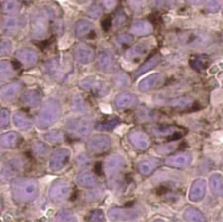
RESEARCH ARTICLE
A B
C D E
125 125 125
% Cell viability
% Cell viability
% Cell viability
100 100 100
75 75 75
SC16
50 IgG1LD6.5 50 50
D6.5
Downloaded from http://stm.sciencemag.org/ by guest on April 27, 2021
25 25 25
SC16LD6.5
0 0 0
0.1 1 10 100 1000 0.1 1 10 100 1000 0.01 0.1 1 10 100 1000
pM pM pM
F G Fig. 3. Characterization of DLL3-mediated in-
ternalization and cytotoxicity. (A) Schematic of
125 125 SC16LD6.5. (B) Demonstration of SC16LD6.5 and
% Cell viability
% Cell viability
100 100
IgG1LD6.5 localization (red) in HEK-293T.hDLL3
cells engineered to express red fluorescent protein
75 75 (RFP)–SLP-1 (false color displayed as green) in late en-
50 50 dosomes. Colocalization is indicated by yellow/
IgG1LD6.5 orange. Scale bars, 25 mm. (C to G) In vitro cyto-
25 SC16LD6.5 25 toxicity of SC16, IgG1LD6.5, D6.5, and SC16LD6.5
0 0 upon incubation with (C) HEK-293T, (D) HEK-293T.
0.01 0.1 1 10 100 1000 0.01 0.1 1 10 100 1000 hDLL3, (E) LU64 PDX, (F) LU37 PDX expressing en-
pM pM dogenous DLL3, or (G) LU37 PDX lacking DLL3 ex-
pression (LU37.D3hp) after shRNA-mediated
knockdown. mAb, monoclonal antibody.
cells and were localized to the late endosomes, as indicated by the lized and mediates cytotoxicity in an antigen-dependent manner
overlap of green and red color manifest as yellow/orange (Fig. 3B, and that sufficient endogenous DLL3 is present in SCLC PDX tumor
left panel, and fig. S5). In contrast, no overlapping signal was detected cells to mediate potent cell killing.
in HEK-293T cells (fig. S5). Neither control antibody nor control Finally, to validate the specificity of SC16LD6.5 for DLL3-expressing
ADC was internalized in HEK-293T.hDLL3 cells (Fig. 3B, right panel, cells, dissociated LU37 LCNEC PDX tumor cells were transduced with
and fig. S5). Finally, the number of cells showing localization of anti- a lentivirus expressing a DLL3-targeted short hairpin RNA (shRNA)
bodies to late endosomes was enumerated, demonstrating that SC16 and were cultured in vitro (LU37.D3hp). Successful knockdown of DLL3
and SC16LD6.5 were specifically internalized and trafficked to late en- expression in LU37.D3hp cells was confirmed by flow cytometry (fig.
dosomes in cells expressing DLL3 (fig. S5). S6). Both LU37 and LU37.D3hp cells were exposed to varying concen-
To evaluate in vitro cytotoxicity, HEK-293T or HEK-293T.hDLL3 trations of either SC16LD6.5 or IgG1LD6.5. Whereas LU37 cells were
cells were incubated with increasing concentrations of the free drug killed by SC16LD6.5 in a concentration-dependent manner (EC50,
D6.5, SC16, SC16LD6.5, or IgG1LD6.5, and cell viability was mea- 13.3 pM; Fig. 3F), LU37.D3hp cells were not (Fig. 3G), demonstrating
sured 4 days later. Exposure to D6.5 mediated equivalent killing of that the cytotoxic activity of SC16LD6.5 is dependent on the presence
HEK-293T and HEK-293T.hDLL3 cells, and neither SC16 nor IgG1LD6.5 of the DLL3 antigen on the cell surface.
mediated any cell killing (Fig. 3, C and D). SC16LD6.5 mediated po-
tent and specific killing of HEK-293T.hDLL3 cells in an antigen- and Reduction of TICs by SC16LD6.5 in vivo
concentration-dependent manner, demonstrated by sixfold greater po- To evaluate the in vivo efficacy of SC16LD6.5, nonobese diabetic–severe
tency than D6.5 alone [EC50 (median effective concentration), 7.8 versus combined immunodeficiency (NOD/SCID) mice were implanted with
46.9 pM; Fig. 3D]. We further evaluated cytotoxicity on LU64 SCLC SCLC or LCNEC PDX tumor cells and were randomized into groups of
tumor cells plated in vitro and found that SC16LD6.5 potently and five to eight mice once tumor volumes reached ~140 to 200 mm3. Each
specifically mediated cytotoxicity (EC50, 8.3 pM), whereas D6.5 was group was treated intraperitoneally with three doses of SC16LD6.5 or
unable to achieve cell killing at concentrations up to 500 pM (Fig. IgG1LD6.5 (1 mg/kg) every 4 days (Q4D×3; Fig. 4, A to C). Separate
3E). Collectively, these data demonstrate that SC16LD6.5 is interna- cohorts of SCLC tumor–bearing mice were treated with vehicle or
www.ScienceTranslationalMedicine.org 26 August 2015 Vol 7 Issue 302 302ra136 5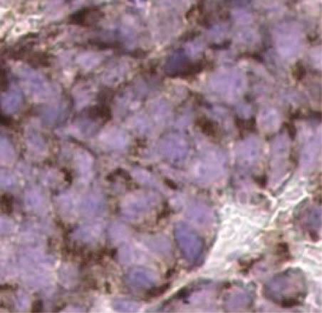
RESEARCH ARTICLE
A B C of ADC dose) or the free drug, D6.5, dosed
1500 1500 at 0.02 mg/kg Q4D×3 [equivalent to free
Tumor volume (mm 3)
Tumor volume (mm 3)
Tumor volume (mm 3)
1500
drug load on SC16LD6.5 (1 mg/kg)] showed
1000 1000 little to no inhibition of tumor growth rel-
1000
ative to controls (fig. S7). Despite the fact
500 500
that SC16LD6.5 is murine cross-reactive
500
IgG1LD6.5 and mediates antigen-dependent cytotox-
SC16LD6.5
icity in cells expressing murine DLL3, treated
0 0 0
0 30 60 90 120 150 0 30 60 90 120
mice continued to gain weight and showed
0 30 60 90 120 150
Days post-treatment Days post-treatment Days post-treatment
no signs of lethargy. In contrast, mice treated
with near maximum tolerated doses of
D E F
1500 1500 1500
SOC chemotherapeutic agents transiently
Tumor volume (mm 3)
Tumor volume (mm 3)
lost weight and showed signs of lethargy
Tumor volume (mm 3)
common with such regimens (fig. S8). Col-
1000 1000 1000
lectively, SC16LD6.5 treatment of SCLC
and LCNEC PDX resulted in effective and
500 Vehicle 500 500 durable responses that significantly corre-
Vehicle Vehicle
Downloaded from http://stm.sciencemag.org/ by guest on April 27, 2021
Cisplatin- Cisplatin- Cisplatin late with DLL3 expression (Pearson r2 =
0
etoposide
0
etoposide
0 0.58, P = 0.006; Table 1 and Fig. 4G), often
0 30 60 90 120 150 0 30 60 90 120 0 30 60 90 120 150 with greatly improved response over SOC
Days post-treatment Days post-treatment Days post-treatment chemotherapeutic regimens (Table 1).
G H I We next explored the response to
C/E ADC C/E Vehicle or C/E
250 1500 1500 SC16LD6.5 in recurrent PDX tumors.
Tumor volume (mm 3)
Tumor volume (mm 3)
200
LU64 tumors were first treated with SOC,
PDX H-score
1000 1000
to which they initially responded. Once
150
these tumors recurred 35 days later, mice
100 were re-randomized into four groups and
500 500
50 treated with SC16LD6.5, IgG1LD6.5, ve-
hicle, or a second round of C/E. All five
0 0 0
0 50 100 150 recurring LU64 tumor–bearing mice
0 30 60 90 120 150 0 30 60 90 120 150
dTTP (days) Days post-treatment Days post-treatment
treated with SC16LD6.5 showed a com-
plete response after rebounding from
Fig. 4. Demonstration of in vivo efficacy with SC16LD6.5. (A to F) Mice bearing SCLC LU64 (A and D),
first-line C/E treatment, whereas IgG1LD6.5
SCLC LU86 (B and E), or LCNEC LU37 (C and F) PDX tumors were treated with IgG1LD6.5 or SC16LD6.5
(1 mg/kg) (A to C) on a Q4D×3 regimen, or vehicle (saline) or SOC chemotherapy (D to F). (G) DLL3 surface
had no impact on tumor growth (Fig. 4H).
expression quantified by IHC (H-score) correlated with dTTP in 10 SCLC and 1 LCNEC PDX model. (H and I) A second round of C/E imparted a more
Mice bearing SCLC LU64 PDX tumors were treated with C/E and, upon tumor recurrence (35 days transient response as compared to the
after initial C/E treatment), were randomized and treated again either with (H) IgG1LD6.5 or initial response to C/E and was followed
SC16LD6.5 (1 mg/kg) on a Q4D×3 regimen or with (I) vehicle or C/E. by rapid recurrence (Fig. 4I). Cumula-
tively, the above data demonstrate that
SOC chemotherapy consisting of cisplatin (5 mg/kg) and etoposide SC16LD6.5 is efficacious in relapsed and refractory SCLC PDX tu-
(8 mg/kg) (C/E) on the day of randomization, followed by etoposide mors in vivo.
on the subsequent 2 days. LCNEC tumor–bearing mice were treated Many complete and durable responses were achieved in vivo with
with cisplatin (5 mg/kg) on the day of randomization. All five mice SC16LD6.5. To determine whether SC16LD6.5 prevents recurrence by
bearing the LU64 SCLC PDX had complete responses to SC16LD6.5 targeting TICs, we treated mice bearing SCLC PDX with C/E or a
(1 mg/kg), with no recurring tumors up to 144 days of observation (Fig. single dose (1 mg/kg) of IgG1LD6.5 or SC16LD6.5. Five days after treat-
4A). Although LU64 also had a strong initial response to C/E, tumors ment, several mice with mean tumor volumes near the average of each
recurred within 18 days (Fig. 4D). The LU86 PDX, which is refractory cohort were euthanized, their tumors were harvested, and live human
to C/E (Fig. 4E) (14), had complete and durable responses to SC16LD6.5 tumor cells were isolated by FACS. Limiting dilutions of isolated cells
in a subset of tumor-bearing mice with a delta time to tumor progression were retransplanted into at least four cohorts of mice from each orig-
(dTTP) versus IgG1LD6.5 of 32 days (Fig. 4B). SC16LD6.5 treatment inal treatment group (table S4). This serial transplantation of cells in
of LU37 LCNEC PDX showed durable responses with a dTTP of 132 days limiting dilutions from naïve or C/E-, IgG1LD6.5-, or SC16LD6.5-treated
(Fig. 4C), contrasting with cisplatin treatment that conferred a dTTP mice allowed for the estimation of residual TIC frequency using Poisson
of only 4 days (Fig. 4F). An overview of all in vivo efficacy experi- distribution statistics (Fig. 5, A and B). In these experiments, LU64 PDX
ments with these and seven additional SCLC and LCNEC PDX tumor tumors were shown to have a TIC frequency of 1:189 cells, which was
lines with varying levels of DLL3 expression is shown in Table 1. reduced to 1:1136 cells in just 5 days after a single dose of SC16LD6.5
In a further demonstration that in vivo efficacy was a result of DLL3 (Fig. 5C). In contrast, IgG1LD6.5 slightly increased, and C/E had no
target–dependent toxin delivery, SCLC PDX tumors treated with either significant impact on the frequency of TICs in LU64 (1:78 and 1:248
excess naked SC16 antibody dosed at as high as 30 mg/kg (30-fold excess cells, respectively; Fig. 5C). Similar robust impacts on the frequency of
www.ScienceTranslationalMedicine.org 26 August 2015 Vol 7 Issue 302 302ra136 6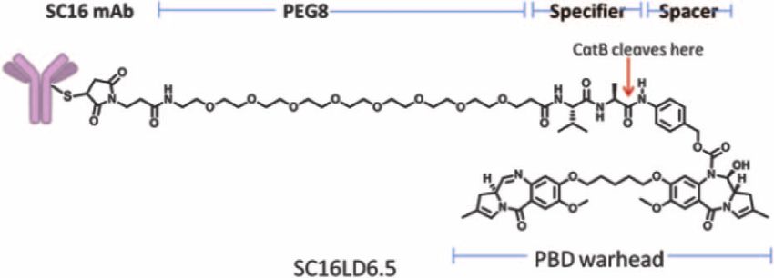
RESEARCH ARTICLE
Table 1. In vivo efficacy of SC16LD6.5 and TIC frequency in SCLC and LCNEC PDX. %TGI, percent tumor growth inhibition; Q4D×3, once every 4 days
for a total of three doses; N.D., not determined; SD, single dose. SOC for SCLC: cisplatin (5 mg/kg) SD on day 0 and etoposide (8 mg/kg) on days 0, 1, and 2;
LCNEC: cisplatin (5 mg/kg) SD. AJCC, American Joint Committee on Cancer.
SOC SC16LD6.5
DLL3
Tumor AJCC Untreated TIC
PDX %TGI protein %TGI
type stage frequency* Dose level
(dTTP; (ng/mg) Regimen (dTTP;
(mg/kg)
days) days)
SCLC LU64 IV 1:189 86 (18) 4.25 1 Q4D×3 100 (133)
LU73 IIIa 1:136 86 (28) 2.77 1 Q4D×3 77 (32)
LU80 IV 1:143 82 (14) 0.60 1 Q4D×3 55 (4)
LU86 IV 1:31 39 (0) 3.05 1 Q4D×3 80 (32)
LU95 IV 1:60 56 (14) 6.52 1 Q4D×3 95 (115)
LU100 Ia 1:166 100 (64) 0.001 1 Q4D×3 96 (3)
LU117 IV 1:388 99 (25) 2.95 1 Q4D×3 99 (137)
Downloaded from http://stm.sciencemag.org/ by guest on April 27, 2021
LU124 IIIb 1:271 85 (14) 3.23 1 Q4D×3 72 (35)
LU129 IV N.D. 84 (32) N.D. 1 Q4D×3 95 (106)
LU149 IV 1:207 93 (21) 2.71 1 Q4D×3 99 (142)†
Mean 81 (23) 87 (74)
SEM 6 (5) 5 (18)
Median 86 (17) 95 (71)
LCNEC LU37 IIb 1:16 60 (4) 5.19 1 Q4D×3 97 (132)
LU240 IIb 1:31 27 (0) 28.57 2 SD 95 (42)
Mean 44 (2) 96 (87)
SEM 17 (2) 1 (45)
Median 44 (2) 96 (87)
*Determined by implanting various cell doses (2 to 1000 cells) of dissociated cells from passage 2 to 5 PDX into mice. †IgG1.LD6.5 data not available; dTTP value represents TTP.
TICs were demonstrated with SC16LD6.5 A B C
in the LU95 SCLC PDX tumor (fig. S9, A 100 100
1.5
% Negative events
and B), in which TICs are more frequent
% Negative events
TIC frequency
80 80
than in LU64 (1:60; Table 1). Notably,
(% of tumor)
60 1.0
SC16LD6.5 administration to mice bear- 60
ing the LU80 PDX, which have low DLL3 40 40
expression, did not alter TIC frequency (fig. 0.5
20 20
S9, C and D). These data provide function-
al evidence that the tumor growth inhibi- 0 0 0.0
1 10 100 1000 10000 1 10 100 1000 10000
tion and durable responses observed
5
/E
5
ve
6.
6.
C
No. of cells No. of cells
aï
D
LD
in vivo in response to SC16LD6.5 result
N
1L
16
G
SC
Ig
from the effective targeting and eradication
of DLL3-expressing TICs. Fig. 5. Elimination of TIC by SC16LD6.5. (A) The frequency of no tumor growth after serial transplan-
tation of SCLC LU64 PDX tumor cells in limiting dilutions is shown for IgGLD6.5 (black triangles) and
Exploratory toxicology SC16LD6.5 (red circles) cohorts. (B) The frequency of no tumor growth after serial transplantation of SCLC
The preclinical safety profile of SC16LD6.5 LU64 PDX tumor cells in limiting dilutions is shown for the naïve (gray diamonds) and C/E (blue triangles)
was further characterized in repeat-dose cohorts. (C) The frequency of TICs was estimated by Poisson distribution statistics using tumor growth
studies both in rats (once every 2 weeks frequencies within each cohort.
for 2 cycles, followed by a 6-week recovery
period for a subset of the animals) and in cyno (once every 3 weeks for consisted of reversible trilineage myelosuppression, mild kidney de-
3 cycles, followed by a 6-week recovery period for a subset of the generation, and skin thickening and hyperpigmentation (fig. S10), each
animals). Both species are relevant toxicology models, given the antibody of which is attributable to off-target toxicity associated with the PBD
cross-reactivity to rat and cyno DLL3 (table S3). Observed toxicities linker drug and has been observed with PBDs (46). Together, the above
www.ScienceTranslationalMedicine.org 26 August 2015 Vol 7 Issue 302 302ra136 7RESEARCH ARTICLE
safety profile and efficacy data supported the initiation of a phase 1 clin- consideration, DLL3 may not appear to be a good ADC target because
ical trial (NCT01901653) in recurrent or refractory high-grade pulmo- murine DLL3 was reported to localize to the Golgi and cytoplasmic
nary neuroendocrine cancer patients. vesicles in the presomitic mesoderm (27, 28, 35). In contrast, our re-
sults demonstrate by IHC and flow cytometry that human DLL3 is
detectable on the surface of high-grade pulmonary neuroendocrine tu-
DISCUSSION mor cells. Furthermore, IHC analyses on primary tumors from various
sources revealed DLL3 membrane expression in ~89% of SCLC tu-
Here, we show that SC16LD6.5, a DLL3-targeted ADC, induces dura- mors and 84% of LCNEC tumors. DLL3 expression in PDX tumors
ble responses in SCLC and LCNEC PDX tumor models after a single further correlated with response to SC16LD6.5, and it is apparent that
course of therapy. Administration of high doses of naked anti-DLL3 even modest expression of DLL3 permits ADC-mediated cytotoxicity
antibody or the ADC dose equivalent of the free PBD dimer toxin showed in vitro and in vivo. This activity likely reflects the rapid internaliza-
little to no impact on tumor growth, supporting the hypothesis that tion of DLL3 in tumor cells.
SC16LD6.5 efficacy is mediated by targeted delivery of the toxin to DLL3- In addition to the specific antibody used in an ADC, the choice of
expressing tumor cells. The observed durable responses after SC16LD6.5 toxin payload influences efficacy, toxicity, and the likelihood of devel-
exposure are consistent with the effective targeting of TICs, in contrast opment of resistance. PBD dimers are a class of payloads that bind in
with the SOC C/E, which neither affected the frequency of TICs nor the minor groove of DNA, where they form covalent aminal cross-
provided durable responses. We hypothesize that the frequent and ra- links between the N2 of guanine and the C11 position of the PBD.
Downloaded from http://stm.sciencemag.org/ by guest on April 27, 2021
pid relapse observed clinically among SCLC patients despite strong The resulting PBD-DNA adducts cause replication forks to stall and
initial debulking responses to C/E is consistent with the inability of tumor cells to arrest at the G2-M boundary, ultimately resulting in ap-
SOC to affect TIC frequency (4). optosis at low nanomolar to picomolar concentrations (50, 51). PBD
One preconception that accompanies the cancer stem cell paradigm dimers are particularly potent because of their cell cycle–independent
is that these cells are rare (12). We show that TICs in SCLC are rela- activity and because their integration minimally distorts DNA, increas-
tively frequent (~1:177 cells; range, 1:31 to 1:388), and we have evidence ing the likelihood of evasion of DNA damage repair responses (51). Be-
to suggest a higher frequency of TICs in LCNEC PDX. By IHC and cause of the potency of PBD dimers and similar cell cycle–independent
flow cytometry, DLL3 expression is seen throughout the tumor, with payloads, normal tissues accessed by such potently armed ADCs must
most cells expressing the antigen at some level. The rapid tumor de- be devoid of target expression. DLL3 meets this criterion.
bulking seen with SC16LD6.5 is likely mediated by DLL3 expression The availability of a large high-grade pulmonary neuroendocrine PDX
on most tumor cells, whereas the sustained progression-free responses tumor repository facilitated the discovery and validation of DLL3 as a
are due to DLL3 expression on TICs. The sustained responses observed therapeutic target. One limitation of the PDX tumor models studied
in the single-agent efficacy studies executed here offer the promise of here is that all were initiated from treatment-naïve SCLC or LCNEC
more durable responses in the clinic. Furthermore, they suggest that patients. Patients encountered in early clinical studies will have re-
patients in the clinic may not need to be dosed until progression, but ceived at least first-line SOC chemotherapy and have recurrent or re-
rather a limited number of treatment cycles may be adequate to drive fractory disease. We show here that treatment of SCLC PDX tumors
improvements in survival endpoints—a true test of the cancer stem with SOC chemotherapy followed by second-line therapy with SC16LD6.5
cell hypothesis. upon recurrence was as effective as first-line therapy with SC16LD6.5.
Targeted cancer therapies that inhibit driver oncogene mutations Additionally, efficacy in the chemorefractory LU86 PDX tumor model
have the advantage of being highly tumor-specific, which generally trans- suggests that patients with tumors resistant to SOC will still respond
lates to a substantial safety window. However, because these tumors are to SC16LD6.5. Furthermore, IHC showed high DLL3 protein expres-
addicted to the oncogene, drug resistance frequently emerges through sion in 85% of recurrent and refractory SCLC tumors, supporting the
compensatory mutations or reactivation of the signaling pathway by hypothesis that SC16LD6.5 should be effective in patients encountered
way of mutations in other genes (47). SCLC and LCNEC tumors have in the setting of second- and third-line treatments.
elevated expression of the neuroendocrine transcription factor ASCL1, High-grade pulmonary neuroendocrine tumors metastasize through-
which is a lineage oncogene critical to neuroendocrine tumorigenesis out the lungs, lymph nodes, adrenal gland, bone, brain, and liver (2).
(24, 37, 48, 49). DLL3 appears to be transcriptionally regulated by Although PDX tumors grown subcutaneously are arguably a model of
ASCL1 (36, 37), and a strong correlation of expression is indeed observed metastasis because they represent tumors growing in a nonorthotopic
for these genes in SCLC and LCNEC PDX tumor models. The lone ex- site, a limitation of PDX models is their lack of systemic metastases. IHC
ception is LU86, which has DLL3 expression despite minimal ASCL1 data suggest equivalent expression of DLL3 in both primary and meta-
expression. The role of DLL3 in the process of SCLC tumorigenesis static sites in patients, implying that SC16LD6.5 will effectively address
is unknown. Whether DLL3 is an ASCL1-induced driver of neuroendo- ADC-accessible metastases. It is not expected that SC16LD6.5 will
crine tumors or simply a passenger in tumors that are addicted to ASCL1 cross the blood-brain barrier; however, brain metastases can be ad-
has implications for potential emergence of resistance to SC16LD6.5. If dressed with cranial irradiation. It is also unclear whether SC16LD6.5
DLL3 is elevated as a consequence of ASCL1 overexpression, it serves as an will be able to efficiently penetrate large tumors in patients compared
excellent surface protein for the ASCL1+ phenotype and can act as a Trojan to those encountered here in PDX models.
horse for toxin delivery. Moreover, if DLL3 is in fact a driver of tumori- Several lines of evidence support the hypotheses that ASCL1 drives
genesis, its down-regulation to evade SC16LD6.5 should result in slowed high-grade pulmonary neuroendocrine tumorigenesis (21, 24) and in-
tumor growth due to Notch reactivation. duces expression of Notch ligands (36, 37, 52), and that Notch pathway
ADC targets must have an extracellular epitope amenable to spe- inhibition promotes neuroendocrine cell fate decisions (25, 34). Elevated
cific antibody binding and be capable of internalization. Upon initial expression of DLL3, a protein that interferes with Notch signaling (27, 35),
www.ScienceTranslationalMedicine.org 26 August 2015 Vol 7 Issue 302 302ra136 8RESEARCH ARTICLE
in high-grade pulmonary neuroendocrine tumors may unify these ob- Whole transcriptome sequencing was performed using the Sequencing
servations. Regardless, by targeting a cytotoxic payload to DLL3-expressing by Oligo Ligation/Detection (SOLiD) 4.5 or SOLiD 5500xl system (Life
TICs with a monoclonal antibody, SC16LD6.5 offers a therapeutic ap- Technologies). Data were mapped to 34,609 genes as annotated by
proach to the treatment of high-grade pulmonary neuroendocrine tu- RefSeq version 47 using NCBI (National Center for Biotechnology In-
mors by delivering a potent cytotoxin specifically to tumor cells and formation) version hg19.2 of the published human genome, and data
avoiding normal tissues. The increased DLL3 expression in tumors for DLL3 are available (table S5).
compared to normal tissues and the observed tolerability in preclinical For qRT-PCR, cDNA was generated from 1 ng of RNA, using the
rat and cynomolgus models suggest that a clinically relevant dose can High-Capacity cDNA Reverse Transcription Kit (Life Technologies)
be safely achieved in patients. following the manufacturer’s instructions. Each cDNA sample was pre-
amplified with 0.2× TaqMan assay specific to DLL3, ASCL1, NEUROD1,
ACTB, and ALAS1 (Life Technologies) diluted in DNA suspension buf-
MATERIALS AND METHODS fer (Teknova), using the 1× TaqMan PreAmp Master Mix (Life Tech-
nologies) according to the manufacturer’s instructions. The preamplified
Study design cDNA was combined with 1× TaqMan Gene Expression Master Mix
The objective of in vivo efficacy studies was to evaluate the activity of (Life Technologies) and 1× GE Sample Loading Reagent (Fluidigm),
SC16LD6.5 in PDX tumor models. Sample size (n = 5 to 8 mice per 1× of each individual TaqMan assays was mixed with 1× Assay Load-
group) was determined on the basis of consistency and homogeneity ing Reagent (Fluidigm), and reaction mixes were run on the Fluidigm
Downloaded from http://stm.sciencemag.org/ by guest on April 27, 2021
of PDX tumor growth in the various models and was sufficient to de- BioMark according to the manufacturer’s instructions. Fluidigm Data
termine statistically significant differences in tumor response between Collection Software was used to set the threshold for each TaqMan assay,
the various treatment groups. Animals were randomized on the basis of data were normalized to the average of the endogenous controls (ACTB
tumor size so that each treatment group had average tumor volumes of and ALAS), and expression was calculated for each sample relative to
140 to 200 mm3. Tumors were measured with digital calipers in two the average expression in normal tissues (relative expression, 2−DDCt).
dimensions, long and short axis (in millimeters), and tumor volume Technical triplicates were run for qRT-PCR on two to three biological
(mm3) was calculated as the volume of a prolate ellipsoid: 0.5 × long replicates for each sample, and relative expression values were averaged.
axis × short axis2. Tumor measurements for individual mice were RNA (1 mg) from PDX lines was analyzed with the Agilent SurePrint
plotted. Data collection was stopped, and the mice were euthanized if GE Human 8x60 v2 microarray platform and the R statistical envi-
they exhibited ≥20% weight loss, inactivity, or poor body condition; ronment (v2.14.2). Microarray data are the average of two biological
when individual tumors reached ≥1000 mm3; when the average tumor replicates for each PDX. Standard industry practices were used to
volume of a given treatment group reached ≥800 mm3; or when the quantile-normalize the background-subtracted intensity values, using
study reached 150 days after randomization. Ten NOD/SCID mice in the preprocessCore Bioconductor R package (53).
the SOC group that were euthanized because of chemo-related toxicities
within 21 days after treatment were excluded from the reported data. Subcloning of DLL3 expression and lentiviral constructs and
Four individual mice (two treated with IgG1LD6.5 and two treated with cell line engineering
SC16LD6.5) that were euthanized because of non–cancer-related illness The human DLL3 extracellular domain (ECD) His fusion protein
within 40 days after treatment were excluded from the TTP calculations. was made by PCR amplification from a commercially available cDNA
No additional outliers were excluded from the data. (SC111951, Origene) followed by subcloning into pEE12.4 expression
plasmid (Lonza) modified to encode an IgK signal peptide upstream
PDX tumor model propagation of DLL3, with a C-terminal 6×His epitope tag (pEE12.4-hDLL3).
SCLC and LCNEC PDX tumor models were initiated as previously Constructs encoding soluble cyno (pEE12.4-cDLL3) and rat DLL3
described (14) and propagated in 5- to 7-week-old female NOD/SCID (pEE12.4-rDll3) ECD were similarly constructed using synthetic codon-
mice (Harlan Laboratories and Charles River Laboratories) by subcu- optimized cDLL3 and rDll3 open reading frames (ORFs; GeneWiz) as
taneous implantation of dissociated cells into a single site near the lower templates for PCR amplification. The encoded DLL3 ECD identity be-
mammary fat pad. Animal health was monitored daily, and mouse tween various species is as follows: human-cynomolgus, 96%; human-
weights and tumor volumes were measured at least weekly. All in vivo rat, 82%; human-mouse, 83%; rat-mouse, 94%. HEK-293T cells expressing
protocols were approved by the Stemcentrx Institutional Animal Care human DLL3 (HEK-293T.hDLL3) were made by transduction of HEK-
and Use Committee (protocols SCAR-3-2008 and SCAR-5-2008) and 293T cells (American Type Culture Collection) using a lentivirus made
performed in accordance with the American Association for Labora- from a commercial bicistronic lentiviral vector (Open Biosystems) that
tory Animal Science and American Veterinary Medical Association expresses both human DLL3 and green fluorescent protein (GFP) under
guidelines. All experiments described herein were performed using the control of a constitutive cytomegalovirus promoter. HEK-293T cells
PDXs from passages 1 to 4. expressing full-length cynomolgus DLL3 (HEK-293T.cDLL3) or rat Dll3
(HEK-293T.rDll3) were made by subcloning the respective ORF into a
RNA isolation and mRNA expression analysis lentiviral expression plasmid (pCDH-EF1-MCS-T2A-GFP, System Bio-
RNA was isolated with the RNeasy Mini Kit (Qiagen) following the manu- sciences). Transduced cells were single-cell sorted with FACSAria (BD
facturer’s instructions and stored at −80°C. For whole transcriptome Biosciences), and individual clones were screened by flow cytometry
analysis, complementary DNA (cDNA) was generated from 1 ng of for stable expression of DLL3 and GFP. HEK-293T.hDLL3 and paren-
RNA using the Ovation RNA-Seq System V2 (NuGEN Technologies). tal HEK-293T were cultured in Dulbecco’s modified Eagle’s medium
The resulting cDNA library was fragmented, and barcode adapters were (DMEM; Corning) containing 10% fetal bovine serum (FBS; Hyclone)
added to allow pooling of fragment libraries from different samples. in tissue culture flasks (BD Falcon) at 37°C in a humidified incubator
www.ScienceTranslationalMedicine.org 26 August 2015 Vol 7 Issue 302 302ra136 9RESEARCH ARTICLE
with 5% CO2. Cells were passaged every 2 to 4 days. Quantitation of ABC Elite (Vector Laboratories) reagents for 30 min at RT, and primary
viral particles was done with the p24 ELISA kit (Cell Biolabs). tumor samples, but not PDX, were incubated in tyramide signal ampli-
fication (PerkinElmer) diluted in amplification buffer at 1:25 and incu-
Enzyme-linked immunosorbent assay bated on the slides for 5 min at RT. After washes, the slides were
Recombinant His-tagged DLL1 and DLL4 (R&D Systems) or DLL3 incubated in streptavidin-HRP (PerkinElmer) diluted at 1:100 for
produced from pEE12.4-hDLL3 as a His fusion protein in CHO cells 30 min at RT and then incubated in Metal Enhanced DAB (Thermo
were immobilized on high protein binding 96-well ELISA plates (Greiner Fisher Scientific). The slides were then counterstained, dehydrated, and
Microlon) at 1 mg/ml in phosphate-buffered saline (PBS) overnight coverslipped. A person with more than 15 years of IHC experience scored
at 4°C. Plates were blocked with PBS plus 3% bovine serum albumin the samples, and H-scores were calculated using membranous staining
(BSA) and washed in PBS with 0.05% Tween 20 (PBST). Serial dilutions intensity and percentage of positive cells. The researcher scoring the
of SC16 or a human IgG1 isotype control in PBST containing 1% BSA samples was blinded to the corresponding pathology report and diag-
(PBSTA) were added to the plate and incubated for 2 hours at room nosis of each sample but is knowledgeable in lung cancer pathology and
temperature (RT). After washing with PBST, a 1:2000 dilution of a can distinguish SCLC, NSCLC, and normal lung pathology.
donkey anti-human IgG horseradish peroxidase (HRP) conjugate
(Jackson ImmunoResearch) in PBSTA was added to the plates for Flow cytometry
1 hour. SC16 binding was visualized using Ultra-TMB substrate (Thermo PDX tumors were disaggregated to single-cell suspensions by mincing
Fisher Scientific), and plates were read at 450-nm absorbance. with razor blades and passing through 40-mm nylon filters. The cells
Downloaded from http://stm.sciencemag.org/ by guest on April 27, 2021
were incubated with fluorophore-conjugated antibodies for 20 min,
Protein expression in tissue and tumor lysates washed three times, suspended in 4′,6′-diamidino-2-phenylindole
PDX tumors were excised from mice and flash-frozen on dry ice/ethanol, (DAPI; 2 mg/ml), then analyzed on a BD FACSAria I. The antibodies
and flash-frozen pieces of normal human tissues were purchased used were fluorescein isothiocyanate (FITC) anti-mouse CD45 (clone
(Asterand). Protein extraction buffer (Biochain Institute) was added to 30-F11, BioLegend), FITC anti-human CD45 (clone HI30, BioLegend),
thawed samples, and the samples were pulverized using the TissueLyser FITC anti-mouse H-2Kd (clone SF1-1.1, BioLegend), peridinin chlorophyll
kit (Qiagen) according to the manufacturer’s instructions. Lysates were protein (PerCP)–Cy5.5 anti-human EpCAM (clone 9C4, BioLegend), and
cleared by centrifugation (20,000g for 20 min, 4°C) and stored at −80°C. PE or Alexa Fluor 647 (AF647) anti-human DLL3. The cells were suspended
Total protein was quantified using bicinchoninic acid. Standard 384-well in DAPI (2 mg/ml) for analysis on a FACSCanto II (BD Biosciences).
plates [Meso Scale Discovery (MSD)] were coated overnight at 4°C with HEK-293T.hDLL3, HEK-293T.cDLL3, and HEK-293T.rDll3 were
15 ml of an anti-DLL3 antibody at 4 mg/ml in PBS. The next day, the plates harvested with Versene (Life Technologies), washed with PBS, and
were washed in PBST and blocked in 35 ml of PBS plus 3% BSA for 1 hour. resuspended in Hepes with 2% FBS (assay buffer) at 2.5 × 10⁶ cells/ml.
The plates were washed again in PBST. Ten microliters of 10× diluted A portion (40 ml) of this cell suspension was added per well to a 96-well
lysate in PBSTA or serially diluted recombinant DLL3 standard in plate followed by the addition of twofold serial dilutions of SC16LD6.5
PBSTA containing 10% protein extraction buffer was also added to in assay buffer. Each SC16LD6.5 concentration was tested in dupli-
the wells and incubated at RT for 2 hours. The plates were washed in cate. Cells were mixed well and incubated at 2° to 8°C for 2 hours with
PBST, and 10 ml of a second anti-DLL3 antibody recognizing a different intermittent agitation of the plate. At the end of the incubation, the cells
epitope conjugated to MSD SULFO-TAG (MSD) was added to the were washed twice with assay buffer followed by incubation with anti-
washed plates at 0.5 mg/ml in PBSTA. The plates were washed in human IgG antibody (4 mg/ml) conjugated to AF647. After 45 min at 2°
PBST, and 35 ml of 1× Read Buffer T with surfactant (MSD) was added to 8°C in the dark, the cells were washed twice with PBS followed by a
to each well. The plates were read on a SECTOR Imager 2400. Raw sig- 10-min incubation with Fixable Viability Dye eFluor 450 (E Bioscience),
nals were interpolated to a DLL3 standard curve using a Workbench which can irreversibly stain dead cells before fixation. Paraformal-
analysis program to derive DLL3 concentrations in test samples. Values dehyde (1%) in PBS was then added to fix the cells before analysis with
were then divided by total protein concentration to yield a readout of a BD FACSCanto II flow cytometer. Viable cells were selected and ana-
ppm (ng DLL3 protein/mg total protein). lyzed for AF647 fluorescence intensity. Rainbow beads (BD Biosciences)
were used as calibrators to transform mean fluorescence intensities
Immunohistochemistry into MESF in the Cy5 channel. MESF values were plotted as a func-
IHC was performed on 5-mm-thick formalin-fixed, paraffin-embedded tion of SC16LD6.5 concentration. Data were analyzed with Prism
tissue sections mounted on glass slides. The slides were deparaffinized software using a four-parameter logistic nonlinear regression model
in xylene and rehydrated through graded alcohols to water. The slides to calculate the Bmax (maximum binding) values for each cell line as
were pretreated with Target Retrieval Solution (Dako) for 20 min at a reflection of the relative DLL3 expression of each cell line. Each Bmax
99°C and treated with 0.3% hydrogen peroxide in tris-buffered saline value was set to 100% to normalize for expression level and plotted
for 8 min, followed by incubation with an avidin/biotin blocking kit against SC16LD6.5 concentration.
(Vector Laboratories). Nonspecific IgG binding was blocked with 10%
horse serum in 3% BSA in PBS. Anti-DLL3 antibody or murine IgG2a In vitro killing
isotype control at 10 mg/ml was added on the slides, followed by incu- The cytotoxicity of various antibodies was tested on HEK-293T,
bation for 60 min at RT. The competition assay used 5 M excess of HEK-293T.hDLL3, LU64, and LU37. HEK-293T cells were plated in
DLL3-His protein preincubated with the DLL3 antibody before incu- culture medium (DMEM + 10% FBS) (50 ml per well) at 500 cells per well
bating on tissue sections. The slides were rinsed and then incubated with in 96-well tissue culture–treated plates on day 1. LU64 and LU37 tumors
biotin-conjugated horse anti-mouse secondary antibody (Vector Lab- were removed from mice, dissociated to single-cell suspensions, and
oratories) for 30 min at RT. The slides were incubated with Vectastain plated under serum-free conditions at 2500 cells per well on Primaria
www.ScienceTranslationalMedicine.org 26 August 2015 Vol 7 Issue 302 302ra136 10RESEARCH ARTICLE
plates (BD Falcon) at 50 ml per well in DMEM/F12 (Mediatech). The Medical), etoposide (8 mg/kg, intraperitoneally, days 1, 2, and 3; Besse
HEK-293T plates were incubated overnight in a humidified incubator at Medical), control IgG1LD6.5 [0.3 to 1 mg/kg, intraperitoneally, days 1,
37°C containing 5% CO2, and patient-derived samples were incubated 5, and 9 (Q4D×3)], or SC16.LD6.5 [0.3 to 1 mg/kg, intraperitoneally,
with 5% CO2 and 5% O2. On day 2, D6.5, SC16, SC16LD6.5, or days 1, 5, and 9 (Q4D×3)]. Efficacy was measured by calculating the
IgG1LD6.5 serial dilutions (50 ml per well) were added to the plates, %TGI and TTP. %TGI was calculated as the tumor volume change be-
and cells were allowed to proliferate for 4 days (HEK-293T cells) and 7 days tween the arithmetic mean tumor volumes in the vehicle-treated control
(patient-derived samples). Each sample concentration was tested in trip- group on the day the first control-treated mouse was euthanized because
licate. Cell viability was measured with the CellTiter-Glo (Promega Cor- tumor volume reached ≥1000 mm3 and the arithmetic mean tumor vol-
poration) reagent according to the manufacturer’s instructions, using ume in the test cohort on that day. A TTP for each individual mouse was
the Victor plate reader (PerkinElmer Corporation). The luminescence recorded as the number of days between treatment day and the day when
values for each sample-treated well were normalized to the values ob- tumor burden reached 50 mm3 above nadir, or the study length in days
tained for untreated wells, and percent cell viability was plotted as a func- after treatment if a durable response was observed, and then the median
tion of sample concentration. Data were analyzed with GraphPad Prism TTP value was determined for each treatment group. A dTTP was calcu-
software using a four-parameter logistic nonlinear regression model. lated by subtracting the TTP for a control group (vehicle or IgG1LD6.5,
respectively) from the TTP of a treatment group (SOC or SC16.LD6.5,
Lentiviral shRNA mediated expression knockdown respectively).
Lentiviral particles containing DLL3 shRNA were generated according
Downloaded from http://stm.sciencemag.org/ by guest on April 27, 2021
to standard lentiviral production procedures (GE Dharmacon). Quan- Limiting dilution assay
titation of viral particles was done with the p24 ELISA kit (Cell Biolabs). After euthanasia of representative mice from each treatment group as-
Dissociated cells from LU37 PDX line were transduced with lentiviral sessed, live human cells were sorted on a FACSAria I (BD Biosciences),
particles at a multiplicity of infection of 3 and incubated for 72 hours on and cohorts of 8 to 10 mice were injected with decreasing numbers of
Primaria plates (Corning) in a humidified incubator at 37°C containing live human cells, ranging from 2500 down to 3 cells. Tumor-negative
5% O2 and 5% CO2. mice dying before 16 weeks after implant were excluded from the anal-
ysis. Mice were scored positive for tumor growth once their tumor size
Internalization exceeded 200 mm3. Estimates of TIC frequency were calculated using
HEK-293T.hDLL3 or HEK-293T cells were seeded (250,000 cells per the L-Calc software package (Stem Cell Technologies) to apply Poisson
well) in tissue culture–treated six-well plates (Greiner BioOne) in 2 ml distribution analysis to the frequencies of tumor-negative mice at each
of culture medium and 100 ml of the BacMam reagent (Life Technol- injected cell number.
ogies) for 48 hours. BacMam is a modified baculovirus that infects
mammalian cells and encodes for the constitutive expression of a fusion Statistical analysis
protein consisting of SLP-1 fused to RFP. Previous reports have dem- Tumor growth curves are shown for individual animals in all repre-
onstrated that SLP-1 is a marker of late endosomal intracellular com- sentative in vivo experiments. The number of biological and technical
partments (43). Cells were harvested, washed, and stained in FSM buffer replicates and the statistical tests run for various experiments are de-
containing SC16, SC16LD6.5, or control reagents (5 mg/ml) for 30 min at tailed in the corresponding Materials and Methods or Results section.
4°C. After washing, a secondary anti-human IgG AF647 conjugate A Pearson correlation was assessed in GraphPad Prism. P values reflect
(Life Technologies), diluted 1:200 in FSM buffer, was added for another two-tailed unpaired t test analyses, with an F test confirming significant
30 min at 4°C. The samples were washed in culture medium at 4°C, variance. Bars shown on vertical scatter plots represent the geometric
resuspended in cold culture medium containing 500 nM Calcein AM mean or mean for each group, as detailed in the figure legends. P values
(Life Technologies), and seeded into two 96-well flat-bottomed tissue ≤0.05 were considered statistically significant.
culture plates. One plate was strictly kept at 4°C for 3 hours, whereas
the second plate was incubated at 37°C in an incubator with 5% CO2.
After incubation, the plates were imaged with fluorescein (Calcein),
SUPPLEMENTARY MATERIALS
rhodamine (RFP), and Cy5 (AF647) filters within a 10-min period,
using an ImageXpressMicro imager (Molecular Devices). Images were www.sciencetranslationalmedicine.org/cgi/content/full/7/302/302ra136/DC1
separately analyzed for each fluorescent color, and composite images Materials and Methods
with false color were assembled in a second step to visualize fluores- Fig. S1. Elevated expression of DLL3 mRNA in SCLC.
Fig. S2. Specificity of anti-DLL3 IHC antibody.
cent colocalization (MetaXpress 4.0). The MetaXpress software was also Fig. S3. DLL3 protein expression by IHC in representative samples from tissue microarrays.
used to identify and count all viable cells (Calcein-positive) with colo- Fig. S4. In vitro plasma stability of SC16LD6.5.
calization events for SLP-1 (RFP; false color displayed as green) and hu- Fig. S5. Enumeration of cells with localization of human antibody in the late endosome.
man IgG (AF647). Fig. S6. DLL3 knockdown confirmation by flow cytometry.
Fig. S7. In vivo efficacy of SC16LD6.5, naked SC16 antibody, and free toxin, D6.5.
Fig. S8. In vivo tolerability of SC16LD6.5.
In vivo efficacy in xenografts Fig. S9. Elimination of TICs by SC16LD6.5.
Female NOD/SCID mice (Harlan Laboratories; Charles River Labora- Fig. S10. Off-target toxicities observed in nonhuman primates.
tories) were implanted with 50,000 PDX tumor cells and randomized Table S1. DLL3 normal tissue expression.
into groups of five to eight animals with average tumor volumes of Table S2. DLL3 microarray expression in PDX.
Table S3. Biacore affinity characterization of SC16 and SC16LD6.5 binding to human, cyno, and
140 to 200 mm3 per cohort, typically 5 to 8 weeks after implantation. rat DLL3.
Mice were treated with vehicle (5% glucose/saline or saline, intra- Table S4. LU64 TIC frequency determination.
peritoneally, day 1), cisplatin (5 mg/kg, intraperitoneally, day 1; Besse Table S5. DLL3 whole transcriptome metrics (provided as an Excel file).
www.ScienceTranslationalMedicine.org 26 August 2015 Vol 7 Issue 302 302ra136 11You can also read