Cutaneous melanoma dissemination is dependent on the malignant cell properties and factors of intercellular crosstalk in the cancer ...
←
→
Page content transcription
If your browser does not render page correctly, please read the page content below
INTERNATIONAL JOURNAL OF ONCOLOGY 57: 619-630, 2020

             Cutaneous melanoma dissemination is dependent on
           the malignant cell properties and factors of intercellular
             crosstalk in the cancer microenvironment (Review)
         ONDŘEJ KODET1‑3, JAN KUČERA1,2, KAROLÍNA STRNADOVÁ1,3, BARBORA DVOŘÁNKOVÁ1,3,
                       JIŘÍ ŠTORK2, LUKÁŠ LACINA1‑3 and KAREL SMETANA Jr1,3

                      1
                     Institute of Anatomy, First Faculty of Medicine, Charles University, 128 00 Prague 2;
     2
      Department of Dermatovenereology, First Faculty of Medicine, Charles University and General University Hospital,
        120 00 Prague; 3Biotechnology and Biomedicine Center of the Academy of Sciences and Charles University
             in Vestec (BIOCEV), First Faculty of Medicine, Charles University, 252 50 Vestec, Czech Republic

                                       Received December 29, 2019; Accepted June 15, 2020

                                                    DOI: 10.3892/ijo.2020.5090

Abstract. The incidence of cutaneous malignant melanoma             Contents
has been steadily increasing worldwide for several decades.
This phenomenon seems to follow the trend observed in               1. Introduction
many types of malignancies caused by multiple significant           2. Cutaneous malignant melanoma (CMM) disseminates
factors, including ageing. Despite the progress in cutaneous           extensively in the organism of the patient
malignant melanoma therapeutic options, the curability of           3. The microenvironment of CMM participates in the control
advanced disease after metastasis represents a serious chal-           of its invasive potential
lenge for further research. In this review, we summarise data       4. Differences in serum proteins between CMM patients and
on the microenvironment of cutaneous malignant melanoma                healthy individuals
with emphasis on intercellular signalling during the disease        5. Intravasation and extravasation of CMM cells and their
progression. Malignant melanocytes with features of neural             inhibition via migrastatics
crest stem cells interact with non‑malignant populations within     6. Cancer‑associated wasting and cachexia as a terminal
this microenvironment. We focus on representative bioactive            complication of CMM; clinically relevant complications
factors regulating this intercellular crosstalk. We describe the       are also associated with factors of intercellular crosstalk
possible key factors and signalling cascades responsible for the    7. Conclusion
high complexity of the melanoma microenvironment and its
premetastatic niches. Furthermore, we present the concept of
melanoma early becoming a systemic disease. This systemic           1. Introduction
effect is presented as a background for the new horizons in the
therapy of cutaneous melanoma.                                      Similarly to other malignant diseases, the incidence of cutaneous
                                                                    malignant melanoma (CMM) is increasing worldwide (1). This
                                                                    increased incidence seems to be influenced by many factors,
                                                                    including ageing of the population, behavioural habits, and
                                                                    climatic and environmental changes. Formation of CMM is
                                                                    associated with the main genetic drivers such as BRAF, NF1 and
Correspondence to: Professor Karel Smetana Jr or Dr Lukáš           NRAS mutations, also usually associated with chronic skin sun
Lacina, Institute of Anatomy, First Faculty of Medicine, Charles    damage (2). Aberrant activation of the RAS/BRAF/MEK/ERK
University, U Nemocnice 3, 128 00 Prague 2, Czech Republic          signalling pathway causes uncontrolled proliferation of
E‑mail: karel.smetana@lf1.cuni.cz
                                                                    malignant cells in the majority of CMM (3). Melanoma causes
E‑mail: lukas.lacina@lf1.cuni.cz
                                                                    most of the skin cancer‑related deaths. The patient overall
Abbreviations: CAFs, cancer‑associated fibroblasts; CMM,            survival at five years depends on the thickness of the primary
cutaneous malignant melanoma; EMT, epithelial to mesenchymal        melanoma. CMM is also known for its remarkable ability to
transition; NC, neural crest; SCs, stem cells; SMA, smooth muscle   metastasise. Despite the new therapeutic options, the curability
actin                                                               of advanced‑stage melanoma is still limited. These recent
                                                                    therapeutic approaches modulate the immune response of the
Key words: melanoma, cancer microenvironment, cancer‑associated     organism (e.g., via application of anti‑CTLA‑4 and anti‑PD‑1
fibroblast, cytokine, chemokine, growth factor                      antibodies) or target proliferation in specifically mutated
                                                                    melanomas (e.g., by application of BRAF or MEK inhibitors).
                                                                    In order to establish novel targeted melanoma therapies, it is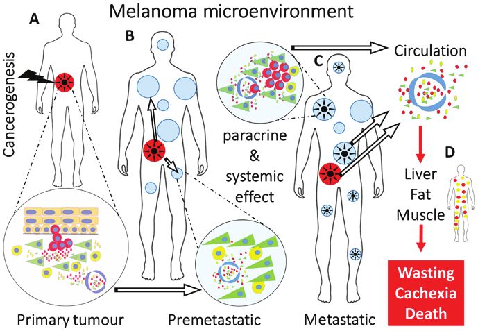
620                 KODET et al: MELANOMA DISSEMINATION IS DEPENDENT ON INTERCELLULAR CROSSTALK

of fundamental importance to understand the mechanisms                 Other common sites for melanoma metastases are the liver
activated in the permissive tumour microenvironment. In             (up to 20% of patients), bones (11-17%), or skin and subcuta-
particular, interactions between melanoma cells and the tissue      neous tissue (12).
microenvironment play key roles in the disease progression.
This article summarises data on the multifaceted roles of           CMM metastasis to the skin. Skin metastasis represents
CMM microenvironment in tumour spreading. This concept              haematogenous dissemination of melanoma cells. Specific
may be extended to the intravasation of bioactive molecules         interactions between chemokines C‑C motif chemokine
participating in the melanoma cell crosstalk with non‑malignant     receptor 10 (CCR10) and C‑C motif chemokine ligand 27
cells forming the CMM microenvironment. These molecules             (CCL27) have been determined as crucial factors in melanoma
also participate in premetastatic niche formation. Finally, their   metastasis to the skin (13). CCL27 is a chemokine expressed
role in patient wasting is also widely discussed. The concept       in the epidermis by normal keratinocytes. In addition, high
of CMM microenvironment as a complex system suitable for            expression in supratumoral epidermis is associated with
therapeutic targeting is introduced in this article.                more prolonged melanoma‑specific survival (14). Presumably,
                                                                    CCL27 interacts with the chemokine receptor CCR10, which
2. Cutaneous malignant melanoma (CMM) disseminates                  is expressed in melanoma cells. Experiments with blocking
extensively in the organism of the patient                          antibodies to CCL27 showed inhibition of development of skin
                                                                    metastasis in a mouse model (15).
The critical feature associated with melanoma is its enormous
capability to spread and form lymph node or distant visceral        CMM metastasis and somatic mutations. Despite that, no
metastases (Fig. 1). Almost any tissue in the patient's body can    specific biomarker with predictive potential to determine
host metastatic cells, and even a small and thin primary tumour     the metastatic site exists to date. In melanoma, there is also
can metastasise to the entire body, leading to the death of the     an observed lack of association between the site of visceral
patient (1). Metastatic spread is a complex multistep process,      or lymph node metastasis and either the clinicopathological
as was noted almost 200 years ago by surgeon Stephen Paget,         variant or location of the primary tumour (16). The
who coined the ‘seed and soil’ hypothesis (4). Surprisingly,        dependence on the presence of somatic mutations has been
cutaneous melanoma can spread to different organs without           reported. One study suggested that BRAF mutation is
any particular predilection, and thus differs from, e.g., uveal     associated with lymph node metastasis as the first metastasis
melanoma of similar histogenesis. However, the first predic-        and sentinel lymph node positivity. BRAF and NRAS
tive site of metastatic disease is a lymph node. The presence       mutations were associated with different metastatic patterns,
of tumour cells in this lymph node is generally investigated        with metastases more frequently affecting the central nervous
in melanoma patients with tumours thicker than 1 mm. This           system and the liver. NRAS‑mutated tumours formed lung
procedure is routinely called sentinel lymph node biopsy. The       metastases (17). This highlights an earlier‑unexpected
presence of melanoma cells in the lymph node is a powerful          internal heterogeneity of the group of tumours nowadays
predictor of melanoma recurrence, but not of survival, in           collectively called melanoma. Although intense visceral
the melanoma patients (5). VEGF‑C, which is involved in             organ‑specific surveillance may be initiated in patients with
lymphangiogenesis and promotes increased lymphatic vessel           tumours harbouring these somatic mutations, this does not
density, can also play a role in lymph node metastasis (6).         necessarily lead to a decrease in mortality. It is not easy
                                                                    to understand this metastatic potency of CMM, which
CMM metastasis to the lungs and brain and other visceral            represents the main, frequently fatal, complication in the
organs. In the case of visceral melanoma metastasis, the            treatment of patients. The complexity of these mechanisms
most predictive localisations are the lungs and pleura (7).         is also shown by the concept of pre‑malignant melanocyte
Lung metastases are also the most frequent metastases in            dissemination, suggesting that benign melanocytes may
mouse models of metastatic melanoma (8). In these mouse             exist at disseminated sites in the body and may be capable
models using the B16 model of melanoma, chloride channel            of undergoing malignant progression. It is not uncommon
accessory 2 (CLCA2), an extracellular protein expressed             to find benign melanocytic nevi in the lymph node during
predominantly in the lung, was identified as a factor mediating     sentinel lymph node biopsy or in non‑melanoma patients (up
interactions with α6β4‑integrin, which is expressed by tumour       to 7% of patients) (18). These findings support the hypothesis
cells (9). Brain metastases are associated with poor prognosis.     mentioned above. It is also critically important to identify the
Historically, melanoma patients with brain metastases have          mechanisms driving the metastatic behaviour.
had dismal outcomes and very limited treatment options.
Systemic treatment with BRAF inhibitors and immunotherapy           CMM cells are similar to neural crest‑originated stem
offers therapeutic responses in up to 55-58% of patients (10).      cells. CMM cells arise after malignant transformation from
The actual mechanism of brain metastases is not clear, but          pigment‑producing cells called melanocytes. Melanocytes
mouse models point to some factors that play a role in this         originate from the embryonic neuroectoderm structure called
process. The original model suggested a role of transferrin         the neural crest (NC). NC cells are multipotent stem cells
receptors and their interaction with their ligand, transferrin,     derived from the neuroectoderm that delaminate from the
mediating metastases of melanoma cells to the brain. Another        neural tube in early vertebrate development (in the 4th week)
study highlighted the importance of neurotrophins and neuro-        and migrate throughout the developing embryo. Consequently,
trophin receptors in the process of brain‑specific melanoma         NC cells differentiate into various cell lineages, including
metastases (11).                                                    melanocytes (19).
INTERNATIONAL JOURNAL OF ONCOLOGY 57: 619-630, 2020 621
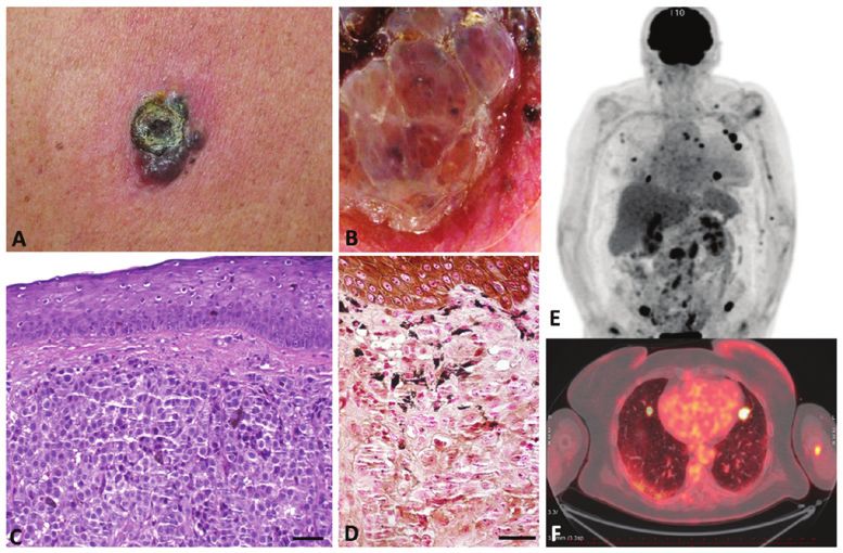
Figure 1. This figure presents the extensive capability of melanoma to form lymphatic/distant metastases in all tissues in the patient's body documented at a
single patient level. (A) Primary cutaneous melanoma (Breslow thickness 6 mm) with clinical apparent ulceration in a 69‑year‑old female, and (B) dermoscopy
of this primary cutaneous melanoma with atypical black dots, dotted vessels and erythema in regression areas. (C) The same primary tumour stained with H&E;
magnification, x200. (D) After staining with Fontana‑Masson (melanin), dark black granules of melanin in melanoma are visualised; magnification, x400.
(E) PET‑CT scan shows generalisation of the tumour to the lung, bones, lymph nodes, and soft tissue. (F) Horizontal section of PET‑CT scan demonstrates
metastasis in the lung and left humerus. The images were kindly provided by the Department of Dermatovenereology, First Faculty of Medicine, Charles
University with explicit informed consent.
NC cells are unique because of their remarkably broad This highlights the low differentiation frequently observed
differentiation potential (Table I) (20-22). Once they have in melanoma, where many cells typically have properties of
reached the final tissue niche in the skin, NC cells differ- stem cells (37). These cancer‑initiating cells of CMM have an
entiate to melanocytes by a cascade of events controlled by indispensable role in CMM resistance to therapy, progression
transcription factors such as microphthalmia‑associated tran- and generalisation (38).
scription factor (MITF) and sex‑determining region Y‑box The life‑long postnatal presence of NC cells in hair follicles
10 (Sox10). This process occurs during the prenatal period raises important questions regarding the maintenance of their
of human development (23,24). The signalling molecules and multipotency and regulation of their normal behaviour within
transcription factors that are required for NC cell specifica- this niche. There is strong evidence that the microenviron-
tion, migration and differentiation form a highly orchestrated ment is a critical condition of this steady‑state. The signalling
gene regulatory network. Every individual signalling molecule cues within the proper microenvironment, via both extrinsic
has either individual or combinatorial roles in transcriptional and intrinsic factors, orchestrate the interplay necessary for
regulation (25). The precise understanding of this mechanism healthy tissue dynamics. The importance of the normal tissue
seems to be critically important because similar pathways are microenvironment was highlighted in several studies using
activated in malignancy, and they could control the biological transplantation of malignant cells to animal embryos. In
properties of malignant CMM cells (26). The signalling path- experiments performed in the early chicken embryo, labelled
ways regulating epithelial to mesenchymal transition (EMT) CMM cells were injected into the region of the neural tube.
can be triggered by transcription factors that are active in both It was demonstrated that melanoma cells migrate to the same
NC development and cancer progression (27). regions as the autologous embryonic NC cells (39). Similar
Interestingly, both melanoblasts and NC cells also reside in experiments performed later in zebrafish embryos supported
the bulge region of the hair follicle in the outer root sheath. In these findings. Both the embryonic NC cells and the cells of
this highly specialised niche, NC cells retain their multipotency CMM in zebrafish express specific protein crestin, which is
during adult life. NC cells can be isolated and expanded in vitro absent in normal melanocytes (40).
with the remarkable features of highly multipotent stem cells Taking into account the low differentiation status of NC
(SCs). It is possible to differentiate NC cells to various special- cells and their natural migratory activity, the similarity of
ised cell types such as melanocytes, adipocytes, osteoblasts, CMM and NC cells can also explain the highly metastatic
chondroblasts, smooth muscle cells, neurons, and Schwann behaviour observed in melanoma in the clinic.
cells (28). The NC cell phenotype is defined by expression of
multiple markers, and NC cell identification cannot be based Circulating CMM cells in disease dissemination. Similarly
on a single molecule. Of note, there is a significant overlap to other types of malignant tumours, cells of CMM can also
with the marker profile of CMM [Table II based on (29-37)]. be detected in the circulation. These circulating melanoma622 KODET et al: MELANOMA DISSEMINATION IS DEPENDENT ON INTERCELLULAR CROSSTALK
Table I. Examples of cells originated from neural crest cells. Table II. Comparison of markers of hair follicle NC SCs and
CMM cells.
Cell type Specification
Factor NC SCs CMM cells
Peripheral neurons Sensory, sympathetic +
parasympathetic ganglia BMP4a + +
Glial cells Schwann cells SNAILa + +
Merkel cells Mechanoreceptor function SLUGa + +
Parafollicular cells Production of calcitonin SOX9a + +
Adrenal medullar cells Chromaffin cells TWISTa + +
Osteoblasts/odontoblasts Facial skeleton MITFb + +
Chondroblasts Facial skeleton Desminc + +/‑
Myoblasts Striated/smooth‑facial Calponinc + +/‑
region β‑III tubulind + +
Dental pulp cells Multipotent stem cell
potential
a
NC marker, bmelanocyte progenitor marker, csmooth muscle differ-
Fibroblast/mesenchymal cells Facial region entiation marker, dneuronal marker. NC, neural crest; SCs, stem cells;
CMM, cutaneous malignant melanoma; BMP3, bone morphogenetic
Cornea Stromal cells protein 3; SOX9, SRY‑box transcription factor 9; MITF, microph-
Melanocytes All parts of the body thalmia‑associated transcription factor. Based on Person et al (29),
Stasiak et al (30), Yang et al (31), Lee et al (32), Tudrej et al (33),
Iwakami et al (34), Goding and Arnheiter (35), Campbell et al (36),
Krejčí and Grim (37).
cells harbour the functional properties of cells of the primary
tumour, including their SC‑like properties (41,42). These cells
leave the primary tumour and penetrate the vessels and use
them as a highway for dissemination through the patient's CD8‑positive T lymphocytes, are attracted to the CMM site.
body to target the organ/tissue where they form metastases. Interestingly, CAFs stimulate the activity of immune cells
Using an identical vascular path, the normal adult tissue SCs supporting melanoma cells and inhibit the cancer‑suppressing
can migrate in order to facilitate body repair processes during cells (52,53).
wound healing (43,44). From this point of view and based Unlike in other epidermal tumours, keratinocytes are also
on histological/molecular similarity, cancer again resembles an important component of the CMM microenvironment.
wound healing. With a certain hyperbole, cancer can be seen Melanoma cells can stimulate surrounding keratinocytes (54).
as a distorted cascade of wound repair events (45). These data On the other hand, keratinocytes control growth and differ-
can also predict the great invasive metastatic potential of entiation of melanocytes and potentiate the invasiveness of
CMM. melanoma cells during early progression as observed in a
reconstructed skin model (55).
3. The microenvironment of CMM participates in the
control of its invasive potential Role of intercellular contact. Cell‑cell adhesion molecules
(cadherins) and cell‑extracellular matrix adhesion proteins
Melanoma is a complex ecosystem. Malignant cells define (integrins) play a critical role in the regulation of cancer invasion
the type of tumour. However, there are other non‑cancerous and metastasis. Many members of the cadherin superfamily
populations forming the tumour stroma. It is the interaction of play an important role in cancer biology. However, the most
both components of this microenvironment that finally defines significant explanation is seen in the E‑/N‑cadherin switch,
the biological behaviour of the tumour. It is truly applicable to and its role in epithelial to mesenchymal transition (EMT),
solid tumours in general, and CMM is no exception (46-48). in cancer progression. N‑cadherin expression in CMM cells
Concerning CMM, the cancer microenvironment is formed helps cancer cells to interact with fibroblasts and extracellular
by cancer‑associated fibroblasts (CAFs) and several types of matrix and stimulates the invasive potential of melanoma cells
leukocytes, as comprehensively reviewed by Lacina et al (49) and their proliferation, but also activation of PI3/AKT, mTOR,
(Fig. 2A). and ERK kinase. Inhibition of N‑cadherin represents an inter-
The origin of CAFs is not fully understood. Local normal esting possibility, with potential clinical use (56).
dermal fibroblasts, attracted mesenchymal SCs and pericytes Intercellular contacts of normal melanocytes, or malignant
are frequently mentioned as source cell populations from melanoma cells, respectively, and their non‑cancerous neigh-
which CAFs are recruited (50). However, the transition of bours within the tissue environment influence their properties
cancer cells to CAFs cannot be entirely excluded, although its mutually (62). Keratinocytes reduce expression of N‑cadherin
likeliness is not very high. This fact is difficult to prove in the not only via cell‑cell contacts, but also via cell‑derived
experimental model (51). extracellular matrix and conditioned medium with calcium
Treg lymphocytes, tumour‑associated macrophages and regulators (57). These findings support the importance of
myeloid‑derived immunosuppressive cells stimulating the the balance in communications between melanoma cells and
CMM progression, as well as NK cells, macrophages and non‑cancerous cells in the melanoma microenvironment.INTERNATIONAL JOURNAL OF ONCOLOGY 57: 619-630, 2020 623
Figure 2. The figure demonstrates the paracrine and systemic effect of CMM during progression. (A) Once initiated, the tumour growths and via paracrine
signalling influences surrounding non‑cancerous tissues (detail in the pink‑filled circle). This paracrine interaction strengthens the malignant potential of
CMM. However, signalling molecules diffuse and also leak to the circulation via capillaries. (B) Exposed to the released cytokines, chemokines and growth
factors, the normal distant tissues become activated and form premetastatic niches (detail in the blue‑filled circle). This is an early systemic effect of the
primary tumour. (C) In some of these niches, the metastasising malignant cells will harbour and initiate a metastatic tumour (detail in the blue‑filled circle).
The concentration of circulating bioactive molecules is further enhanced by their production in metastases. This represents a later systemic effect typical of
generalised malignancies. (D) Circulating bioactive molecules induce physical and functional organ changes. This consequently results in wasting, cachexia
and death. CMM, cutaneous malignant melanoma.
Integrins β1 and β3, as adhesion cell‑extracellular matrix Role of paracrine signalling in communication across the
proteins, are differentially expressed during the transforma- CMM microenvironment. The paracrine mode of signalling
tion of melanoma radial growth to the vertical invasion (58,59). between cancerous and non‑cancerous cells in CMM has been
The differentiation status of melanoma cells and the ability extensively studied (Fig. 2B and C). For example, CAFs, kera-
to invade the surrounding tissue also highlights this impact. tinocytes and infiltrating immune cells produce a variety of
For example, the expression of connexin‑43 in CMM cells growth factors/cytokines/chemokines that significantly influ-
indicates the ability of CMM cells to metastasise (52,60,61). ence the biological properties of malignant CMM cells (67,68)
Expression of desmoglein‑2, which participates in the contacts (Table III). Interestingly, many of these factors are also associ-
with keratinocytes, has an inhibitory effect on CMM cell ated with skin ageing. Collectively, these bioactive molecules
migration. are called the senescence‑associated secretome (69). As
On the other hand, the expression of desmoglein‑2 promotes expected, chemokines attract inflammatory cells to the tumour
the vasculogenic mimicry of CMM cells, which is associated with site. However, they also have multiple other functions (70).
a poor outcome of patients (62,63). Furthermore, in melanoma CMM cells express receptors for CXCL1 and interleukin
cells that do not express β3 integrins, β1 integrins instead play a (IL)8, and these factors enhance their invasiveness (71,72).
role in promoting their transendothelial migration by binding to Antagonists of CXCR1/2 receptors have a well‑documented
vascular cell adhesion molecule 1 (VCAM‑1) (64). Integrins also inhibitory effect on migration of CMM cells (73). The IL8
play an important role in connecting the extracellular matrix with chemokine also stimulates vascularisation of CMM (74,75),
the melanocyte and melanoma cell cytoskeleton. Cytoskeletal and in general, its high expression indicates poor prognosis of
rearrangements, such as the increase of the overall contrac- CMM patients (76).
tility, impact cell mechanical properties and cell deformability. Expression of chemokine CXCL16 seems to participate
These changes may then potentiate prometastatic phenotypes in the malignant transformation of a melanocytic nevus to
of melanoma cells. Expression of αvβ3 integrin increases elas- CMM (77). Activation of CXCR6 recognising this chemokine
ticity in human melanoma cells in adherent and non‑adherent induces SC‑like properties in CMM cells and initiates their
conditions (65). Intercellular contacts and molecules play an migration (78).
important role in the mechanisms of targeted therapy. Targeting IL1β is predominantly produced by macrophages. It partici-
of the CMM cell surface receptor Notch‑dependent pathway pates in the progression of CMM in collaboration with IL8 and
improves the activity of Erk inhibitors in BRAF‑V600E mutated caspase recruitment domain family member 8 (CARD8) (79).
tumours. Further, it can be combined with inhibition of ERBB3 IL6 is a crucial factor initiating the immune response. IL6
to suppress melanoma cell growth (63). On the other hand, Notch has a multifaceted role in cancer progression (80). While the
expression in CAFs reduces the growth and migration potential initial stage of CMM growth can be inhibited by IL6 (81),
of melanoma cells (66). the more advanced stages are associated with production of624 KODET et al: MELANOMA DISSEMINATION IS DEPENDENT ON INTERCELLULAR CROSSTALK
Table III. Examples of factors produced by CAFs, Kerat and melanoma cells in CMM.
Symbol Gene name CAFs Kerat CMM cells
IL8 Interleukin 8 + + +
CXCL1 Chemokine (C‑X‑C motif) ligand 1
(melanoma growth stimulating activity α) + ‑ ‑
CXCL16 Chemokine (C‑X‑C motif) ligand 16 + ‑ +
IL1B Interleukin 1β + - -
IL6 Interleukin 6 + + +
IL17D Interleukin 17D + ‑ +
ACAN Aggrecan + + +
HBEGF Heparin‑binding EGF‑like growth factor + + ‑
BDNF Brain‑derived neurotrophic factor ‑ ‑ +
TGFB2 Transforming growth factor, β2 ‑ ‑ +
IGFBP7 Insulin‑like growth factor binding protein‑7 + + ‑
GAP43 Growth associated protein 43 + + ‑
BMP2 Bone morphogenetic protein 2 + ‑ +
BMP6 Bone morphogenetic protein 6 + ‑ ‑
VEGFA Vascular endothelial factor A + + +
VEGFC Vascular endothelial factor C + + +
CTGF Connective tissue growth factor + ‑ +
PDGFRL Platelet‑derived growth factor receptor‑like + ‑ +
LEPRE1 Leucine proline‑enriched proteoglycan (Leprecan) + ‑ ‑
LEPREL1 Leprecan‑like 1 + ‑ +
KAZALD1 Kazal‑type serine peptidase inhibitor domain 1 + ‑ ‑
CAFs, cancer‑associated fibroblasts; CMM, cutaneous malignant melanoma; Kerat, keratinocytes. Based on Kodet et al (54), Jobe et al (67).
this interleukin (82,83). IL6, frequently in cooperation with tissue growth factor has a synergistic effect on VEGFA and
IL8, exhibits an additive effect on WNT5A in the stimulation stimulates the tumour site neovascularisation (96).
of CMM cell invasiveness (67,84). IL6 induces Twist and As mentioned earlier, CAFs do not support tumour
N‑cadherin expression in CMM angiogenesis in a mechanism growth and metastases exclusively. CAFs are also implicated
dependent on the p50 subunit of nuclear factor κ B (85). in the acquisition of resistance to the targeted therapy in
In general, IL17D (IL27) has an anti‑tumoral effect in BRAF‑mutated melanomas. Under the influence of BRAF
CMM (86), where it participates in generating tumour‑specific inhibitors, CAFs secrete factors that contribute to CMM cell
cytotoxic T cells (87). The effect of IL17D on CMM cells survival and melanoma resistance. CAFs release factors such
seems to be TRAIL‑dependent (88). On the other hand, it is as hepatocyte growth factor (HGF) and neuregulin 1 (NRG1),
also known as a potent inducer of the production of IL6 and which can trigger alternative cascades in MAP kinase
IL8 in endothelial cells. It is highly expressed in the initial signalling (97,98).
stages of CMM (89), stimulating the CMM growth and tumour The short overview in Table III demonstrates the complexity
vascularisation. of signalling between CMM cells and non‑cancerous cells,
Glycoprotein aggrecan is produced by cells of the CMM, where intercellular contacts, cytokines, chemokines, and
CAFs and keratinocytes. It is usually secreted during the growth factors with both the stimulatory and inhibitory effect
process of chondroblast differentiation, and it has an inhibitory influence the tumour growth and generalisation.
effect on CMM progression (90). A similar anti‑CMM effect Concluding this paragraph, paracrine signalling represents
is produced by insulin‑like growth factor‑binding protein 7 a critical aspect in the control of biological properties of
(IGFBP‑7), namely in BRAF‑mutated V600E‑positive CMM. In addition to the crosstalk between melanoma cells,
dysplastic nevi (91). On the other hand, heparin‑binding this process includes an exchange of information between
EGF‑like growth factor has a stimulatory impact on CMM CMM cells and non‑malignant cells of the microenvironment.
growth (92). The same result was described in the case of
another growth factor, neurotrophin (93,94). miRNA‑328 Role of exosomes. In addition to paracrine signalling via
controls production of TGF‑β2, and attenuation of its expression soluble products, exosomes represent another tool of CMM
has a strong inhibitory effect on CMM cell proliferation (95). cell communication with non‑cancerous partners within
VEGFA and VEGFC are generally responsible for the activa- the microenvironment. All affected cell populations of the
tion of CMM neovascularisation by blood/lymphatic vessels cancer microenvironment produce these bodies. Exosomes
that support the CMM growth and progression. Connective thus inf luence CMM cell biological properties (99).INTERNATIONAL JOURNAL OF ONCOLOGY 57: 619-630, 2020 625
Exosomes stimulate CMM cell metastasis via support of IL6 sensibly reflected the tumour burden and indicated a
the epithelial‑mesenchymal transition. Exosomes influ- relapse of the disease or insensitivity to tumour therapy in
ence vascularisation of the lymph node in order to prepare several studies (114-116).
the vascular bed for the metastasising (100,101). Exosomes A similar finding was observed in the case of serum levels
significantly participate in the regulation of local invasive- of IL8 (71,117,118). Interestingly, the levels of IL6, IL8, and
ness and also in the entrance of melanoma cells to the target VEGFA correlated with the level of Breslow index at the
organs (102). This effect is frequently associated with the time of diagnosis (117,119). Moreover, the amount of VEGFA
presence of miRNAs in CMM‑derived exosomes. It was also depended on the stage of the disease (120). As IL8 also
confirmed both in vitro and in vivo in clinical material (103). supports CMM neovascularisation, it is not surprising that the
Exosomes exert a robust immunosuppressive effect on the elevation of the serum level of both IL8 and VEGFA corre-
cancer microenvironment, where they inhibit IL2‑dependent lates with the progression of the disease and poor survival of
proliferation of CD8‑positive T lymphocytes (104). Moreover, CMM patients (121). Proteins of the TGF‑β family are also
CMM exosomal miRNA‑125b‑5p induces a tumour‑promoting elevated in the sera of CMM patients, and the prognostic
phenotype in macrophages (105). These changes can induce a relevance of these factors has been proposed (122). Elevated
mixed M1, and M2 tumour‑promoting macrophage activation concentration of factors with immunomodulatory activity
included production of CCL22, IL‑12B, IL‑1β, IL‑6, i‑NOS, such as IL6 and/IL10 influence the presence of self‑renewal
and TNF‑α (106). These data highlight exosomes as a criti- tumour‑initiating (stem) cells in CMM (123).
cally important component of the CMM microenvironment
significantly participating in its biological properties, with Serum protein imbalance influences premetastatic niche
the ability to stimulate the immune response of the melanoma formation. Based on the selected examples, it is possible to
microenvironment. demonstrate the systemic effect of CMM. The serum/plasma
of CMM patients contains numerous bioactive proteins and
4. Differences in serum proteins between CMM patients exosomes that are transported to the distant parts of the
and healthy individuals patient's body through the vessels (Fig. 2B‑D). These factors
participate in the preparation of a premetastatic niche suit-
Serological biomarkers represent a diverse group of biomol- able for cancer cell homing and metastasis formation (124).
ecules with importance in diagnosis, staging, and monitoring Under the influence of CMM‑derived exosomes, the dermal
the therapeutic response. Serum lactate dehydrogenase (LDH) fibroblasts reprogram their metabolism significantly (125).
is the only serum biomarker that has been accepted as a The distant dermal fibroblasts from CMM patients at the stage
prognostic biomarker for routine clinical use in melanoma of metastatic tumour dissemination differ from the normal
patients with a predictive therapeutic outcome and has been dermal fibroblasts from healthy donors. The phenotype of
implemented in the American Joint Committee on Cancer (8th distant dermal fibroblasts, as well as the expression profile
edition) staging system (107). Routinely used S100B (S100 and methylation profile of gene promoters, is shifted closer to
calcium binding protein B) protein is highly specific for mela- CAFs (126). Due to this activation, it is possible to hypothesise
noma patients. Its increased levels can be detected in patients that the tissue microenvironment in the distant body parts is
with advanced melanoma during melanoma prognosis (108). influenced by the released bioactive factors from the mela-
Another serological protein is MIA (melanoma inhibitory noma microenvironment. It is, therefore, likely that melanoma
activity), which interacts with extracellular matrix proteins. becomes a systemic disease very early. If so, it is the signalling
Its expression can also be detected in normal tissue such as in the primary tumour that already prepares the rest of the
cartilage. In pathological processes, its overexpression is organism to host CMM cells and facilitate metastases (114)
observed in breast cancer or colorectal cancer, in addition to (Fig. 2B).
melanoma (109).
Introduction of a new treatment strategy for advanced mela- 5. Intravasation and extravasation of CMM cells and their
noma leads to the search for new biomarkers to improve both inhibition via migrastatics
prognostic and predictive outcome. Likely, the high intensity of
molecular exchange between cancer cells and other members of In recent years, the term ‘migrastatics’ has been introduced for
the microenvironment via cytokines, chemokines and growth drugs interfering with all modes of cancer cell invasion (115).
factors can lead to leakage out from the tumour microenviron- Migrastatics inhibit local invasion and consequent metastasis.
ment, and mediators can be consequently detected in systemic This group of drugs was recently established to define and
circulations in the serum (110,111) (Fig. 2A‑D). Therapy by distinguish them from conventional cytostatic drugs that
monoclonal antibodies targeting immune checkpoint inhibi- traditionally target cell proliferation. Malignant melanoma,
tors is one of the most potent treatments of CMM patients. therefore, seems to be a tempting disease for validation of this
Measurement of current concentrations and dynamics of these concept.
mediators in the serum can have the potential of liquid biopsy. Endothelial cells of capillaries are also an important
Indeed, the serum protein signature even reflects the efficiency structure of the cancer microenvironment. Migrating CMM
of anti‑PD‑1 therapy of CMM patients and can be substantial cells adhere to the capillary endothelium, intercalate between
for therapeutic indications (112). endothelial cells, and migrate throughout the vessels in
Similarly to other types of tumours, an elevated serum both directions. From the endothelial cell perspective, this
level of IL6 in CMM has been observed (111), which has process is not passive. Endothelial cells actively participate in
reached some prognostic validity (113). Serum elevation of extravasation, where the role of N‑cadherin has been broadly626 KODET et al: MELANOMA DISSEMINATION IS DEPENDENT ON INTERCELLULAR CROSSTALK
investigated (117). It seems to be also controlled by CD146, (Fig. 2D). CAC is a highly complex and multifaceted catabolic
which cooperates with VEGFA. CD146 is also elevated in the process (135). IL6, in cooperation with TNFα and IL1β
patients' serum/plasma (127). influences the metabolism of striated muscle fibres, adipocytes
On the other hand, VE‑cadherin expressed on the and hepatocytes (136,137). The level of the mentioned factors
surface of endothelial cells prevents the migration of cancer in the serum can even predict the onset of CAC and survival
cells through the endothelium of capillaries. VE‑cadherin of cancer patients (138). The terminal stage of cancer is also
must, therefore, be eliminated from the site of malignant associated with decreased food intake in cancer patients, which
cell migration (128). P‑selectin has an essential role in the is called anorexia (139). IL6 seems to be linked to the control
recruitment of inflammatory cells to the site of inflammation, of food intake, where it inhibits the appetite and participates
so‑called homing. in the development of anorexia (140). TNFα and IL6 can cross
P‑selectin expression on endothelial cells is under the the blood‑brain barrier (141). TNFα, IL1 and IL6 can interact
control of the local microenvironment. Expression of this with hypothalamic neurons and affect the serotoninergic
molecule on endothelial cells and blood platelets seems to metabolism, which can be reflected by decreased food
be a prerequisite for successful metastasising of CMM cells intake (142). Patients with advanced cancer frequently suffer
to the target tissue (129,130). P‑selectin expression on the from depression that seems to be associated with elevation of
surface of endothelial cells is induced by STAT3 activa- IL6, IL10 and TNFα (143). On the other hand, the serum levels
tion (129). IL6 is available in the serum of CMM patients, and of IL6 (and also IL8) are significantly elevated in tumour‑free
it is known as a potent activator of STAT3. The observation patients with bipolar disease, but not with major depressive
that capsular polysaccharides from E. coli attenuate adhesion disorder (144). This section demonstrates that factors produced
of CMM cells to the endothelium via P‑selectin demonstrates by the cancer ecosystem have a strong systemic effect by which
the specificity of this interaction (130). Endogenous lectin they influence the metabolic functions of cancer patients,
galectin‑3 locally accumulates in inflamed tissues, including resulting in wasting and death.
endothelium. This lectin also enhances invasion of CMM
cells, e.g., to the lungs (131). These examples show that an 7. Conclusion
imbalance in serum proteins can participate in the process
of extravasation of CMM cells to the target tissue and metas- CMM, similarly to the majority of cancers, can be character-
tasising. ised as a genetic abnormality and a regulation failure in which
Therefore, the combination of migrastatics with other cancer cells employ predestined pathophysiological pathways
groups of traditional oncologic drugs may be possible. Beyond that are normally activated in the course of organism growth,
that, we suggest that directed therapy (biologics, small‑mole- tissue regeneration and repair. This deregulation is typically
cule receptor‑associated kinase inhibitors) against the most associated with accumulation of mutations acquired during
prominent inflammatory cytokines, namely IL6, could bring the ageing of the individual.
highly desirable synergism. However, it seems evident that The progression of CMM from tumour initiation to the
inhibition of the IL6 signalling axis is not sufficient and must, systemic effect on the patient's metabolism is organised
therefore, be accompanied by simultaneous blockade of other according to a quite uniform scenario (Fig. 2A‑D). The inter-
proteins/receptors such as IL8, VEGFA and MFGE8 (48,132). cellular crosstalk within this ecosystem is mediated either
The therapeutic blockade of IL6, in combination with check- directly by intercellular contacts, or indirectly by paracrine
point inhibitor anti‑PD1, represents an interesting possibility of secretion of numerous active molecules. This interconnec-
overcoming some immunological mechanisms of resistance. tion strengthens the malignant potential of cancer cells, and
IL6 blockade upregulated expression of PD‑L1 on melanoma it can inhibit the anticancer immune response or protect
cells in a mouse model and may sensitise melanoma to this malignant cells from the harmful effect of oncological
treatment (133). These findings underscore the importance of therapy. A plethora of bioactive factors are transported via
the IL6‑PD1/PD‑L1 crosstalk in the tumour microenviron- vessels and significantly influence even the distant tissue.
ment of melanoma. Collectively, these factors prepare a suitable microenviron-
The local microenvironment and its control by bioactive ment for the malignant cell extravasation and metastasising,
factors can be a highly relevant target in the prevention of the the premetastatic niche. The increasing mass of CMM cells
deadly complication of malignant disease (Fig. 2D). in the body of patients generates a cancer‑induced profile of
inflammatory mediators in the patients' serum. The systemic
6. Cancer‑associated wasting and cachexia as a terminal availability of these bioactive molecules triggers mental
complication of CMM; clinically relevant complications disorders, depression, and mental anorexia‑associated
are also associated with factors of intercellular crosstalk problems with food intake. Wasting leads to cancer cachexia
and death. Based on this scenario, it is evident that besides
Advanced stages of cancer, including CMM, are associated the conventional anticancer therapy, it would be necessary
with metastasising, which in the case of CMM has a character to influence the migration of CMM cells and their metas-
of extensive generalisation. The increasing burden of tumour tasising - the concept of migrastatics (115). Because the
cells generates an imbalance in growth factors, cytokines and CMM microenvironment stimulates malignant cell inva-
chemokines, among which IL6 seems to have the leading siveness (145), targeting both cancerous and non‑cancerous
position (80). This stage of the disease is usually terminated cells of the tumour ecosystem and their products seems to
by cancer‑associated cachexia (CAC), which affects be a promising approach. IL6 and its signalling pathway
approximately 16 patients per 100,000 individuals (134) influence CMM cell growth and migration, but it can alsoINTERNATIONAL JOURNAL OF ONCOLOGY 57: 619-630, 2020 627
positively affect the entire patient metabolism and mental References
status (82). Therefore, targeting the IL6/IL6R/STAT3
axis as a new therapeutic modality was enthusiastically 1. Sacchetto L, Zanetti R, Comber H, Bouchardy C, Brewster DH,
Broganelli P, Chirlaque MD, Coza D, Galceran J, Gavin A, et al:
expected, but, unfortunately, the reality did not meet this Trends in incidence of thick, thin and in situ melanoma in Europe.
expectation (146). The progress in the detection of clinically Eur J Cancer 92: 108‑118, 2018.
relevant markers using a robust omics approach that includes 2. Rabbie R, Ferguson P, Molina‑Aguilar C, Adams DJ and
Robles‑Espinoza CD: Melanoma subtypes: Genomic profiles,
stromal factors can be translated into personalised therapy prognostic molecular markers and therapeutic possibilities.
of CMM (147). For example, a combination of blocking the J Pathol 247: 539‑551, 2019.
anti‑IL6 axis with drugs blocking other signalling pathways 3. Lorentzen HF: Targeted therapy for malignant melanoma. Curr
Opin Pharmacol 46: 116‑121, 2019.
seems to be promising for future trials (48). It can be hypoth- 4. Paget S: The distribution of secondary growths in cancer of the
esised that the progress in diagnostics and therapy covering breast. Lancet 133: 571‑573, 1889.
the complex ecosystem of melanoma can bring some benefit 5. Faries MB, Thompson JF, Cochran AJ, Andtbacka RH,
Mozzillo N, Zager JS, Jahkola T, Bowles TL, Testori A,
to CMM patients. Beitsch PD, et al: Completion dissection or observation for
sentinel‑node metastasis in melanoma. N Engl J Med 376:
Acknowledgements 2211‑2222, 2017.
6. Nathanson SD: Insights into the mechanisms of lymph node
metastasis. Cancer 98: 413‑423, 2003.
Authors are grateful to Dr Šárka Takáčová for her assistance in 7. Barth A, Wanek LA and Morton DL: Prognostic factors in 1,521
English language editing of this revised version. melanoma patients with distant metastases. J Am Coll Surg 181:
193‑201, 1995.
8. Damsky WE Jr and Bosenberg M: Mouse melanoma models and
Funding cell lines. Pigment Cell Melanoma Res 23: 853‑859, 2010.
9. Abdel‑Ghany M, Cheng HC, Elble RC and Pauli BU: The breast
cancer β 4 integrin and endothelial human CLCA2 mediate lung
The project ‘Centre for Tumour Ecology-Research of the metastasis. J Biol Chem 276: 25438‑25446, 2001.
Cancer Microenvironment Supporting Cancer Growth 10. Tawbi HA, Boutros C, Kok D, Robert C and McArthur G: New
and Spread’ (reg. no. CZ.02.1.01/0.0/0.0/16_019/00007 era in the management of melanoma brain metastases. Am Soc
Clin Oncol Educ Book 38: 741‑750, 2018.
85) is supported by the Operational Programme ‘Research, 11. Menter DG, Herrmann JL and Nicolson GL: The role of
Development and Education’. Additional support was trophic factors and autocrine/paracrine growth factors in brain
received from the Ministry of Education, Youth and Sports metastasis. Clin Exp Metastasis 13: 67‑88, 1995.
12. Balch CM, Houghton AN, Sober AJ and Soong S: Cutaneous
of CR within the National Sustainability Programme II Melanoma, 4th Edition. Dermatologic Surg 31: 1715-1715, 2005.
(Project BIOCEV‑FAR, reg. no. LQ1604), project BIOCEV 13. Murakami T, Cardones AR and Hwang ST: Chemokine receptors
(CZ.1.05/1.1.00/02.0109), grant no. CZ.1.05/2.1.00/19.0400 and melanoma metastasis. J Dermatol Sci 36: 71‑78, 2004.
14. Martinez‑Rodriguez M, Thompson AK and Monteagudo C: High
supported by the Research and Development for Innovations CCL27 immunoreactivity in ‘supratumoral’ epidermis correlates
Operational Programme, co‑financed by the European with better prognosis in patients with cutaneous malignant
melanoma. J Clin Pathol 70: 15‑19, 2017.
Regional Development Fund and the state budget of the Czech 15. Ben‑Baruch A: Organ selectivity in metastasis: Regulation
Republic, from the Grant Agency of the Czech Republic, by chemokines and their receptors. Clin Exp Metastasis 25:
project no. 19‑05048S and from Charles University project 345‑356, 2008.
16. Marcoval J, Ferreres JR, Martín C, Gómez S, Penín RM,
PROGRES Q28. Ochoa de Olza M and Fabra À: Patterns of Visceral Metastasis
in Cutaneous Melanoma: A Descriptive Study. Actas
Dermosifiliog 104: 593‑597, 2013.
Availability of data and materials 17. Adler NR, Wolfe R, Kelly JW, Haydon A, McArthur GA,
McLean CA and Mar VJ: Tumour mutation status and sites of
All data and information are supported by relevant references. metastasis in patients with cutaneous melanoma. Br J Cancer 117:
1026‑1035, 2017.
18. Holt JB, Sangueza OP, Levine EA, Shen P, Bergman S,
Authors' contributions Geisinger KR and Creager AJ: Nodal melanocytic nevi in
sentinel lymph nodes. Correlation with melanoma‑associated
cutaneous nevi. Am J Clin Pathol 121: 58‑63, 2004.
Conceptualisation, data collection and manuscript preparation 19. Ji Y, Hao H, Reynolds K, McMahon M and Zhou CJ: Wnt
were carried out by KSm, OK and LL Manuscript preparation signaling in neural crest ontogenesis and oncogenesis. Cells 8:
1173, 2019.
was conducted by JK, JŠ, KSt and BD. 20. Lim J, Thiery JP, Kassem Y, Kalcheim C, Moens CB, Burden SJ
and Granato M: Epithelial‑mesenchymal transitions: Insights
Ethics approval and consent to participate from development. Development 139: 3471‑3486, 2012.
21. Mayor R and Theveneau E: The neural crest. Development 140:
2247‑2251, 2013.
Not applicable. 22. Hall BK: The neural crest and neural crest cells: Discovery and
significance for theories of embryonic organization. J Biosci 33:
781‑793, 2008.
Patient consent for publication 23. Mort RL, Jackson IJ and Patton EE: The melanocyte lineage in
development and disease. Development 142: 620‑632, 2015.
The images in Fig. 1 were kindly provided by the Department 24. Duband JL, Monier F, Delannet M and Newgreen D:
Epithelium‑mesenchyme transition during neural crest devel-
of Dermatovenereology, First Faculty of Medicine, Charles opment. Acta Anat (Basel) 154: 63‑78, 1995.
University with explicit informed consent. 25. Vega‑Lopez GA, Cerrizuela S and Aybar MJ: Trunk neural crest
cells: Formation, migration and beyond. Int J Dev Biol 61: 5‑15,
2017.
Competing interests 26. Larribère L and Utikal J: Stem cell‑derived models of neural
crest are essential to understand melanoma progression and
The authors declare no competing interests. therapy resistance. Front Mol Neurosci 12: 111, 2019.628 KODET et al: MELANOMA DISSEMINATION IS DEPENDENT ON INTERCELLULAR CROSSTALK
27. Gallik KL, Treffy RW, Nacke LM, Ahsan K, Rocha M, 50. Preisner F, Leimer U, Sandmann S, Zoernig I, Germann G and
Green‑Saxena A and Saxena A: Neural crest and cancer: Koellensperger E: Impact of human adipose tissue‑derived stem
Divergent travelers on similar paths. Mech Dev 148: 89‑99, 2017. cells on malignant melanoma cells in an in vitro co‑culture
28. Sieber‑Blum M and Grim M: The adult hair follicle: Cradle for model. Stem Cell Rev Rep 14: 125‑140, 2018.
pluripotent neural crest stem cells. Birth Defects Res C Embryo 51. Dvořánková B, Smetana K Jr, Říhová B, Kučera J, Mateu R and
Today 72: 162‑172, 2004. Szabo P: Cancer‑associated fibroblasts are not formed from
29. Person F, Wilczak W, Hube‑Magg C, Burdelski C, Möller‑Koop C, cancer cells by epithelial‑to‑mesenchymal transition in nu/nu
Simon R, Noriega M, Sauter G, Steurer S, Burdak‑Rothkamm S, et al: mice. Histochem Cell Biol 143: 463‑469, 2015.
Prevalence of βIII‑tubulin (TUBB3) expression in human normal 52. Inada M, Takita M, Yokoyama S, Watanabe K, Tominari T,
tissues and cancers. Tumour Biol 39: 1010428317712166, 2017. Matsumoto C, Hirata M, Maru Y, Maruyama T, Sugimoto Y, et al:
30. Stasiak M, Boncela J, Perreau C, Karamanou K, Chatron‑Colliet A, Direct melanoma cell contact induces stromal cell autocrine pros-
Proult I, Przygodzka P, Chak ravarti S, Maquart FX, taglandin E2‑EP4 receptor signaling that drives tumor growth,
Kowalska MA, et al: Lumican inhibits SNAIL‑induced angiogenesis, and metastasis. J Biol Chem 290: 29781‑29793,
melanoma cell migration specifically by blocking MMP‑14 2015.
activity. PLoS One 11: e0150226, 2016. 53. Ziani L, Safta‑Saadoun TB, Gourbeix J, Cavalcanti A, Robert C,
31. Yang X, Liang R, Liu C, Liu JA, Cheung MPL, Liu X, Man OY, Favre G, Chouaib S and Thiery J: Melanoma‑associated fibroblasts
Guan XY, Lung HL and Cheung M: SOX9 is a dose‑dependent decrease tumor cell susceptibility to NK cell‑mediated killing
metastatic fate determinant in melanoma. J Exp Clin Cancer through matrix‑metalloproteinases secretion. Oncotarget 8:
Res 38: 17, 2019. 19780‑19794, 2017.
32. Lee H, Torres FX, McLean SA, Chen R and Lee MW: 54. Kodet O, Lacina L, Krejčí E, Dvořánková B, Grim M, Štork J,
Immunophenotypic heterogeneity of primary sinonasal Kodetová D, Vlček Č, Šáchová J, Kolář M, et al: Melanoma cells
melanoma with aberrant expression of neuroendocrine markers influence the differentiation pattern of human epidermal kerati-
and calponin. Appl Immunohistochem Mol Morphol 19: 48‑53, nocytes. Mol Cancer 14: 1, 2015.
2011. 55. Van Kilsdonk JWJ, Bergers M, Van Kempen LCLT, Schalkwijk J
33. Tudrej KB, Czepielewska E and Kozłowska‑Wojciechowska M: and Swart GWM: Keratinocytes drive melanoma invasion in a
SOX10‑MITF pathway activity in melanoma cells. Arch Med reconstructed skin model. Melanoma Res 20: 372‑380, 2010.
Sci 13: 1493‑1503, 2017. 56. Ciołczyk‑Wierzbicka D and Laidler P: The inhibition of invasion
34. Iwakami Y, Yokoyama S, Watanabe K and Hayakawa Y: of human melanoma cells through N‑cadherin knock‑down. Med
STAM‑binding protein regulates melanoma metastasis through Oncol 35: 42, 2018.
SLUG stabilization. Biochem Biophys Res Commun 507: 57. Chung H, Jung H, Jho EH, Multhaupt HAB, Couchman JR and
484‑488, 2018. Oh ES: Keratinocytes negatively regulate the N‑cadherin levels
35. Goding CR and Arnheiter H: MITF‑the first 25 years. Genes of melanoma cells via contact‑mediated calcium regulation.
Dev 33: 983‑1007, 2019. Biochem Biophys Res Commun 503: 615‑620, 2018.
36. Campbell K, Kumarapeli AR, Gokden N, Cox RM, Hutchins L 58. Nikkola J, Vihinen P, Vlaykova T, Hahka‑Kemppinen M, Heino J
and Gardner JM: Metastatic melanoma with dedifferentiation and Pyrhönen S: Integrin chains β1 and alphav as prognostic factors
and extensive rhabdomyosarcomatous heterologous component. in human metastatic melanoma. Melanoma Res 14: 29‑37, 2004.
J Cutan Pathol 45: 360‑364, 2018. 59. Van Belle PA, Elenitsas R, Satyamoorthy K, Wolfe JT,
37. Krejčí E and Grim M: Isolation and characterization of neural Guerry D IV, Schuchter L, Van Belle TJ, Albelda S, Tahin P,
crest stem cells from adult human hair follicles. Folia Biol Herlyn M, et al: Progression‑related expression of β3 integrin in
(Praha) 56: 149‑157, 2010. melanomas and nevi. Hum Pathol 30: 562‑567, 1999.
38. Marzagalli M, Raimondi M, Fontana F, Montagnani Marelli M, 60. Li G, Satyamoorthy K, Meier F, Berking C, Bogenrieder T
Moretti RM and Limonta P: Cellular and molecular biology of and Herlyn M: Function and regulation of melanoma‑stromal
cancer stem cells in melanoma: Possible therapeutic implications. fibroblast interactions: When seeds meet soil. Oncogene 22:
Semin Cancer Biol 59: 221‑235, 2019. 3162‑3171, 2003.
39. Kasemeier‑Kulesa JC, Teddy JM, Postovit LM, Seftor EA, 61. Brandner JM and Haass NK: Melanoma's connections to the
Seftor REB, Hendrix MJC and Kulesa PM: Reprogramming tumour microenvironment. Pathology 45: 443‑452, 2013.
multipotent tumor cells with the embryonic neural crest micro- 62. Peitsch WK, Doerflinger Y, Fischer‑Colbrie R, Huck V,
environment. Dev Dyn 237: 2657‑2666, 2008. Bauer AT, Utikal J, Goerdt S and Schneider SW: Desmoglein 2
40. Kaufman CK, Mosimann C, Fan ZP, Yang S, Thomas AJ, depletion leads to increased migration and upregulation of the
Ablain J, Tan JL, Fogley RD, van Rooijen E, Hagedorn EJ, et al: chemoattractant secretoneurin in melanoma cells. PLoS One 9:
A zebrafish melanoma model reveals emergence of neural crest e89491, 2014.
identity during melanoma initiation. Science 351: aad2197, 2016. 63. Tan LY, Mintoff C, Johan MZ, Ebert BW, Fedele C, Zhang YF,
41. Agnoletto C, Corrà F, Minotti L, Baldassari F, Crudele F, Szeto P, Sheppard KE, McArthur GA, Foster‑Smith E, et al:
Cook WJJ, Di Leva G, d'Adamo AP, Gasparini P and Volinia S: Desmoglein 2 promotes vasculogenic mimicry in melanoma
Heterogeneity in circulating tumor cells: The relevance of the and is associated with poor clinical outcome. Oncotarget 7:
stem‑cell subset. Cancers (Basel) 11: 11, 2019. 46492‑46508, 2016.
42. Gkountela S and Aceto N: Stem‑like features of cancer cells on 64. Klemke M, Weschenfelder T, Konstandin MH and Samstag Y:
their way to metastasis. Biol Direct 11: 33, 2016. High affinity interaction of integrin α4β1 (VLA‑4) and vascular
43. Empringham B, Chiang KY and Krueger J: Collection of hema- cell adhesion molecule 1 (VCAM‑1) enhances migration of
topoietic stem cells and immune effector cells in small children. human melanoma cells across activated endothelial cell layers.
Transfus Apheresis Sci 57: 614‑618, 2018. J Cell Physiol 212: 368‑374, 2007.
44. Feehan J, Nurgali K, Apostolopoulos V, Al Saedi A and 65. Lacaria L, Lange JR, Goldmann WH, Rico F and Alonso JL:
Duque G: Circulating osteogenic precursor cells: Building bone αvβ3 integrin expression increases elasticity in human melanoma
from blood. EBioMedicine 39: 603‑611, 2019. cells. Biochem Biophys Res Commun 525: 836‑840, 2020.
45. Ratajczak MZ, Bujko K, Mack A, Kucia M and Ratajczak J: 66. Bedogni B: Notch signaling in melanoma: Interacting pathways
Cancer from the perspective of stem cells and misappropriated and stromal influences that enhance Notch targeting. Pigment
tissue regeneration mechanisms. Leukemia 32: 2519‑2526, 2018. Cell Melanoma Res 27: 162‑168, 2014.
46. Lacina L, Plzak J, Kodet O, Szabo P, Chovanec M, Dvorankova B 67. Jobe NP, Rösel D, Dvořánková B, Kodet O, Lacina L, Mateu R,
and Smetana K Jr: Cancer microenvironment: What can we learn Smetana K and Brábek J: Simultaneous blocking of IL‑6 and
from the stem cell niche. Int J Mol Sci 16: 24094‑24110, 2015. IL‑8 is sufficient to fully inhibit CAF‑induced human melanoma
47. Hill BS, Pelagalli A, Passaro N and Zannetti A: Tumor‑educated cell invasiveness. Histochem Cell Biol 146: 205‑217, 2016.
mesenchymal stem cells promote pro‑metastatic phenotype. 68. Jobe NP, Živicová V, Mifková A, Rösel D, Dvořánková B,
Oncotarget 8: 73296‑73311, 2017. Kodet O, Strnad H, Kolář M, Šedo A, Smetana K Jr, et al:
48. Plzák J, Bouček J, Bandúrová V, Kolář M, Hradilová M, Szabo P, Fibroblasts potentiate melanoma cells in vitro invasiveness
Lacina L, Chovanec M and Smetana K Jr: The head and neck induced by UV‑irradiated keratinocytes. Histochem Cell
squamous cell carcinoma microenvironment as a potential target Biol 149: 503‑516, 2018.
for cancer therapy. Cancers (Basel) 11: 11, 2019. 69. Strnadova K, Sandera V, Dvorankova B, Kodet O, Duskova M,
49. Lacina L, Kodet O, Dvořánková B, Szabo P and Smetana K Jr: Smetana K and Lacina L: Skin aging: The dermal perspective.
Ecology of melanoma cell. Histol Histopathol 33: 247‑254, 2018. Clin Dermatol 37: 326‑335, 2019.You can also read