Computed Tomography Imaging in the Context of Transcatheter Aortic Valve Implantation (TAVI)/Transcatheter Aortic Valve Replacement (TAVR)
←
→
Page content transcription
If your browser does not render page correctly, please read the page content below

JACC: CARDIOVASCULAR IMAGING                                                                                                    VOL. 12, NO. 1, 2019

        ª 2019 BY THE SOCIETY OF CARDIOVASCULAR COMPUTED TOMOGRAPHY

        AND THE AMERICAN COLLEGE OF CARDIOLOGY FOUNDATION

        PUBLISHED BY ELSEVIER

        GUIDELINES

        Computed Tomography Imaging in the
        Context of Transcatheter Aortic Valve
        Implantation (TAVI)/Transcatheter
        Aortic Valve Replacement (TAVR)
        An Expert Consensus Document of the
        Society of Cardiovascular Computed Tomography

        Philipp Blanke, MD,a Jonathan R. Weir-McCall, MBCHB, PHD,b Stephan Achenbach, MD,c Victoria Delgado, MD, PHD,d
        Jörg Hausleiter, MD,e Hasan Jilaihawi, MD,f Mohamed Marwan, MD,c Bjarne L. Nørgaard, MD, PHD,g
        Niccolo Piazza, MD, PHD,h Paul Schoenhagen, MD, PHD,i Jonathon A. Leipsic, MDa

        1. PREAMBLE/INTRODUCTION                                                         are ineligible for surgery or high risk surgical candi-
                                                                                         dates to also now include those at intermediate risk
        Since the publication of the first expert consensus                               for conventional surgical valve replacement (2–6).
        document on computed tomography imaging before                                      Advances in non-invasive imaging have supported
        transcatheter aortic valve implantation (TAVI)/trans-                            growth and maturation of the field. Clinical outcomes
        catheter aortic valve replacement (TAVR) by the So-                              have improved based on the thoughtful integration of
        ciety       of   Cardiovascular        Computed          Tomography              advanced non-invasive imaging into patient selec-
        (SCCT) in 2012 (1), there has been tremendous                                    tion, treatment planning, device selection, and de-
        advancement in the field. Significant technological                                vice positioning. While computed tomography (CT)
        advancements and a wealth of trial data have led to                              was initially used primarily for the assessment of
        deep integration of TAVI/TAVR and CT Imaging into                                peripheral access, the role of CT has grown substan-
        clinical practice. The indications for TAVI/TAVR as the                          tially and CT is now the gold standard tool for annular
        treatment strategy for patients with symptomatic se-                             sizing, determination of risk of annular injury and
        vere aortic stenosis (AS) has expanded from those who                            coronary      occlusion,     and     to   provide      co-planar

        From the aDepartment of Radiology, St Paul’s Hospital & University of British Columbia, Vancouver, British Columbia, Canada;
        b
            Department of Radiology, University of Cambridge, Addenbrooke’s Hospital, Cambridge, United Kingdom; cDepartment of
        Cardiology, Friedrich-Alexander-University Erlangen-Nürnberg, Erlangen, Germany; dDepartment of Cardiology, Heart Lung
                                                                                    e
        Center, Leiden University Medical Center, Leiden, the Netherlands;              Medizinische Klinik und Poliklinik I der Ludwig-
                                                                     f
        Maximilians-Universität München, Munich, Germany;                New York University Medical Center, New York, New York;
        g
            Department of Cardiology, Aarhus University Hospital, Aarhus, Denmark; hMcGill University Health Centre, Montreal, QC,
        Canada; and the iImaging Institute and Heart&Vascular Institute, Cleveland Clinic Lerner College of Medicine, Cleveland, Ohio.
        This paper will be copublished in the Journal of the Cardiovascular Computed Tomography.
        Dr. Blanke is a consultant for Edwards Lifesciences and Circle Cardiovascular Imaging; and provides CT core lab services for
        Edwards Lifesciences, Medtronic, Neovasc, and Tendyne Holdings, for which he receives no direct compensation. Dr. Delgado
        receives speaker fees from Abbott Vascular; and has received departmental unrestricted research grants from Medtronic,
        Biotronik, Edwards Lifesciences, GE Healthcare, and Boston Scientific. Dr. Hausleiter has received speaker honoraria from
        Abbott Vascular and Edwards LifeSciences. Dr. Jilaihawi has acted as a consultant for Edwards Lifesciences; and received
        research grants from Medtronic and Abbott Vascular. Dr. Marwan has received speaker honoraria from Siemens Healthcare and
        Edwards Lifesciences. Dr. Norgaard has received unrestricted institutional research grants from Edwards Lifesciences, Siemens,
        and HeartFlow. Dr. Piazza is a clinical proctor and consultant for, has received research grants from, and serves on an advisory
        board and steering committee for Medtronic. Dr. Leipsic serves as a consultant and has stock options in HeartFlow and Circle
        Cardiovascular Imaging; and receives speaking fees from GE Healthcare. All other authors have reported that they have no
        relationships relevant to the contents of this paper to disclose.

        ISSN 1936-878X/$36.00                                                                                      https://doi.org/10.1016/j.jcmg.2018.12.003
Descargado para Anonymous User (n/a) en Consejeria de Sanidad de Castilla y Leon(Sacyl) de ClinicalKey.es por Elsevier en marzo 05, 2019.
Para uso personal exclusivamente. No se permiten otros usos sin autorización. Copyright ©2019. Elsevier Inc. Todos los derechos reservados.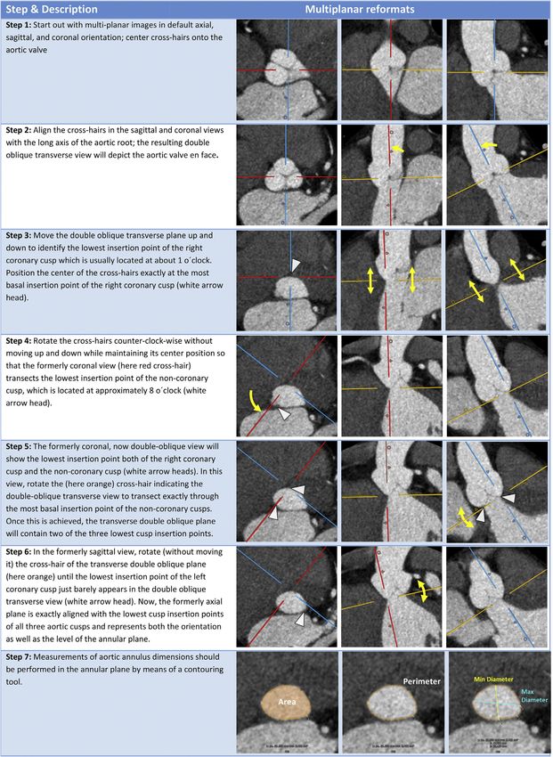
2   Blanke et al.                                                                                     JACC: CARDIOVASCULAR IMAGING, VOL. 12, NO. 1, 2019

    Guidelines                                                                                                                                JANUARY 2019:1–24

                    fluoroscopic angle prediction in advance of the pro-                        data acquisition is kept to a minimum, allowing for
                    cedure. Further benefits of cardiac CT have also                            a reduction of the overall contrast dose. Further-
                    been demonstrated in the follow-up of TAVI/TAVR                            more, limiting the ECG-synchronized data acqui-
                    for assessment of post-procedural complications                            sition     decreases      the     radiation      dose-intensive
                    including identification of leaflet thickening (7,8).                        portion of the examination, despite parts of the
                      Since the last SCCT consensus statement, there has                       scan range possibly being covered twice. The ECG-
                    been a substantial volume of new data published                            synchronized data set should at least cover the
                    describing the use of CTA in TAVI/TAVR planning and                        aortic root, but can include the entire heart.
                    post-procedural assessment. This updated consensus                      2) ECG-synchronized data acquisition of the thorax
                    statement has been written to better reflect the data                       followed by a non-ECG-synchronized CTA of the
                    now available. The content and recommendations of                          abdomen and pelvis. A disadvantage of this
                    this document reflect an expert consensus taking into                       approach is the higher radiation dose, and the
                    consideration all published literature, but is not in                      relatively long acquisition time required for the
                    itself a systematic review. For recommendations,                           entire thorax with potential need for larger
                    level of consensus was graded as strong ($9 in                             contrast dose and risk of breathing artifacts.
                    agreement out of the 11 members of the writing
                                                                                               The scanner hardware employed may further
                    group), moderate (7 to 8) and weak (6). Any recom-
                                                                                            dictate which of these approaches is employed.
                    mendations with less than 6 of the author group in
                    support of the statement were not adopted into this                     2.2. ECG-SYNCHRONIZED CTA OF THE AORTIC ROOT
                    expert consensus document.                                              AND HEART: ACQUISITION TECHNIQUE. The selected

                    2. CT DATA ACQUISITION AND                                              acquisition mode as well as exposure settings should
                    RECONSTRUCTION                                                          account for dynamic changes of aortic root geometry
                                                                                            and dimensions throughout the cardiac cycle. Given
                    Data acquisition strategies and scanning protocols                      that aortic root dimensions are commonly larger in
                    vary depending on scanner manufacturer, system,                         systole (10,11), and that most sizing algorithms for
                    and institutional preferences (9). This document                        transcatheter heart valves are based on systolic di-
                    provides recommendations for reliable CT image                          mensions, systolic scan coverage is recommended.
                    acquisition     for   TAVI/TAVR        planning.       The     key      However, diastolic information may be valuable for
                    component of all approaches is an ECG-synchronized                      evaluation of aortic valve morphology and rarely, the
                    computed tomographic angiography (CTA) data set                         largest dimensions of the annulus may occur in
                    that covers at least the aortic root in order to provide                diastole in the setting of inversed dynamism in septal
                    artifact free anatomical information of the aortic root,                hypertrophy (11–13). For that reason, image acquisi-
                    followed by a commonly non-ECG synchronized CTA                         tion covering the entire cardiac cycle should be
                    data acquisition of the aorto/ilio/femoral vasculature                  considered. This wider phase of acquisition offering
                    for assessment of the access vasculature. Ideally, both                 systolic and diastolic data may be particularly useful
                    acquisitions are combined in a comprehensive scan-                      if systolic reconstructions are degraded by artifact.
                    ning protocol with a single contrast administration.                    Scan coverage should at least include the aortic root
                    The sequence of patient preparation and the relevant                    but coverage of the entire heart is beneficial.
                    principles of CT data acquisition will be explained in                     For CT systems with limited detector coverage, i.e.,
                    brief below.                                                            systems that cannot cover the entire aortic root
                                                                                            within one rotation, retrospective ECG-gating allows
                    2.1. SCAN STRATEGY AND SCAN COVERAGE. In
                                                                                            coverage of the entire cardiac cycle, while offering
                    general, two different approaches are used to
                                                                                            most flexibility regarding data salvage by means of
                    combine the ECG-synchronized data set of the aortic
                                                                                            ECG-editing. Dose-modulation may be used to miti-
                    root   structures     and    the     non-ECG-synchronized
                                                                                            gate the high radiation exposure, with peak tube
                    computed tomography angiography (CTA) of the
                                                                                            current applied during at least systole. However, tube
                    aorto/ilio/femoral vasculature into one comprehen-
                                                                                            current outside of the peak acquisition window
                    sive acquisition protocol:
                                                                                            should be maintained to such a level as to allow for
                    1) Cardiac ECG-synchronized data set of the aortic                      characterization of the aortic annulus and valve.
                      root    and     heart     followed       by     a    non-ECG-         Prospective ECG-triggering may constitute an alter-
                      synchronized CTA of the thorax, abdomen, and                          native. However, this technique is more susceptible
                      pelvis. Although this approach can result in repeat                   to step-artifacts in the setting of increased heart rate
                      data acquisition of the aortic root and cardiac                       variability (e.g. premature contractions, atrial fibril-
                      structures, the time-intensive ECG-synchronized                       lation) with the risk of rendering images inadequate

         Descargado para Anonymous User (n/a) en Consejeria de Sanidad de Castilla y Leon(Sacyl) de ClinicalKey.es por Elsevier en marzo 05, 2019.
         Para uso personal exclusivamente. No se permiten otros usos sin autorización. Copyright ©2019. Elsevier Inc. Todos los derechos reservados.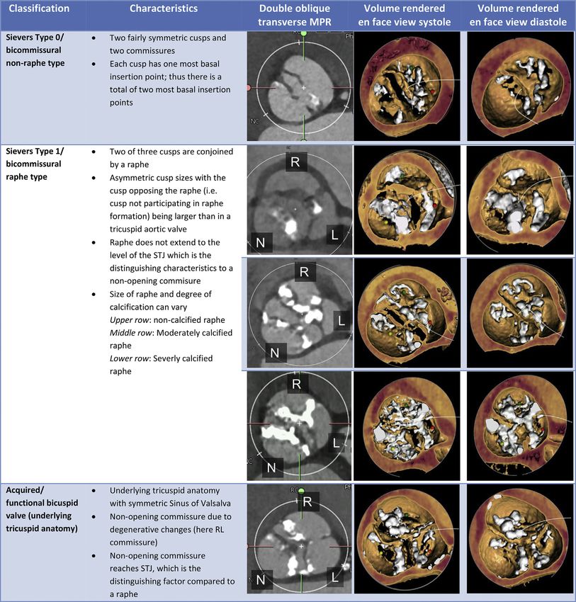
JACC: CARDIOVASCULAR IMAGING, VOL. 12, NO. 1, 2019                                                                                                    Blanke et al.   3
JANUARY 2019:1–24                                                                                                                                       Guidelines

 T A B L E 1 Preferred Mode of Acquisition for ECG-Synchronized CTA of the Aortic Root Stratified by Scanner System

 Manufacturer                    Scanner Geometry                                             Preferred Acquisition Mode

 GE                         64-row family                            Spiral/helical acquisition with retrospectively ECG-gated image reconstruction
                            Revolution (256 row)                     Prospectively ECGgated, axial one beat acquisition
 Philips                    All scanners                             Spiral/helical acquisition with retrospectively ECG-gated image reconstruction
 Siemens                    All scanners                             Spiral/helical acquisition with retrospectively ECG-gated image reconstruction
 Toshiba                    64/80-row family                         Spiral/helical acquisition with retrospectively ECG-gated image reconstruction
                            Aquilion One (320/640 row)               Prospectively ECGgated, axial one beat acquisition

for quantitative analysis, and does not allow for im-                      reconstructions. Most scanner systems require a
age salvage by means of ECG-editing. If employed,                          brief intermission between the ECG-synchronized
the acquisition window should be kept wide to cover                        and non-synchronized data acquisition to reposition
at least systole.                                                          the table and adjust scan settings. This needs to be
  For systems with whole-heart coverage (i.e., 16-cm                       accounted for by the contrast administration protocol
detector coverage), an ECG-gated one beat acquisi-                         in order to allow for sufficient contrast attenuation of
tion should be employed. Peak tube current should be                       the aorto/ilio/femoral vasculature.
applied at least during systole, with consideration for                    2.4. RADIATION PARAMETERS AND CONSIDERATIONS.
coverage of the entire cardiac cycle. Recommended                          Acquisition parameters should employ the ‘As low as
acquisition mode for employed scanner systems are                          reasonably achievable’ (ALARA) principle (9). Ac-
listed in Table 1.                                                         cording to recommendations on radiation protection
  Other acquisition settings, in particular tube                           in cardiovascular CT by the Society of Cardiovascular
voltage and tube current settings should follow                            Computed Tomography (SCCT), a tube potential of 100
institutional policies and preferences but should                          kV should be considered for patients weighing #90 kg
allow for acquisition of image data with sufficiently                       or with a body mass index (BMI) #30 kg/m 2; whereas a
low image noise to ensure fully diagnostic image                           tube potential of 120 kV is usually indicated for pa-
quality in thin-slice reconstructions. Guidance on                         tients weighing >90 kg and with a BMI > 30 kg/m 2 (9).
acquisition settings is outlined in the ‘Radiation pa-                     Tube current should be adjusted, based on individual
rameters’ section below (9). In centers with multiple                      patient’s size, to the lowest setting that guarantees
scanners, matching the most complex patients (such                         acceptable image noise (9). Expected increase in im-
as those with arrhythmias, cardiac failure, or chronic                     age noise with lower tube current can be mitigated
kidney disease) to higher specification scanners (such                      with the use of iterative image reconstruction. The
as those with whole heart coverage, or dual sources)                       majority of commercially available scanner systems
should be considered (14).                                                 provide software algorithms to automatically adjust
2.3. NON-ECG SYNCHRONIZED CTA OF THE AORTO/ILIO/                           exposure parameters to the patient’s body habitus
FEMORAL VASCULATURE: ACQUISITION TECHNIQUE.                                within defined boundaries.
The CTA of the aorto/ilio/femoral vasculature should                           To date, patients being treated with TAVI/TAVR
extend from the upper thoracic aperture to the lesser                      have predominantly been septuagenarians and octo-
trochanter to include the thoracic and abdominal                           genarians with the mean age of patients receiving
aorta, the iliac arteries and common femoral arteries,                     TAVI/TAVR being 79.8 to 83.6 years in high and in-
the latter constituting the most common vascular                           termediate risk trials, 79.1 years in the all comer
access site. For a more comprehensive evaluation the                       NOTION trial and 80.1 years in the low risk OBSER-
scan range can be extended cephalad to fully include                       VANT registry with mean age not expected to drop
the subclavian arteries for assessment of this alter-                      below 75 years of age in low risk cohorts (4–6,16,17).
native access route (15). Most commonly, this CTA is                       As a result the primary concern for the imaging pro-
performed employing a helical, non-ECG synchro-                            tocol should be to ensure diagnostic quality images,
nized data acquisition immediately following the                           and to minimize the need for repeat CTA and repeat
ECG-synchronized data set of the aortic root. Other                        contrast administration. In case of younger patients,
acquisition settings, in particular tube voltage and                       the most effective mode for reducing radiation dose is
tube current settings should follow institutional pol-                     to minimize the scan volume performed using ECG-
icies and preferences, but should allow for acquisition                    synchronization as well as limiting the peak dose
of image data with sufficiently low image noise to                          coverage when using dose modulation. Prospectively
ensure fully diagnostic image quality in thin-slice                        triggered acquisitions focusing on systole can be

                Descargado para Anonymous User (n/a) en Consejeria de Sanidad de Castilla y Leon(Sacyl) de ClinicalKey.es por Elsevier en marzo 05, 2019.
                Para uso personal exclusivamente. No se permiten otros usos sin autorización. Copyright ©2019. Elsevier Inc. Todos los derechos reservados.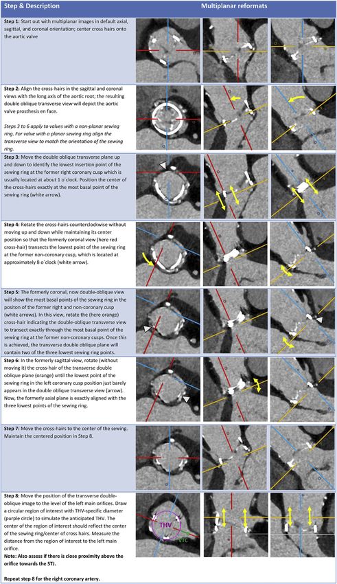
4               Blanke et al.                                                                                                   JACC: CARDIOVASCULAR IMAGING, VOL. 12, NO. 1, 2019

                Guidelines                                                                                                                                                    JANUARY 2019:1–24

                                                                                                                     scanning protocol for the scanning system employed
    T A B L E 2 Recommendations for IV Contrast Administration
                                                                                                                     allows for lower overall contrast volumes (see
         Parameter                                           Recommendation                                          Table 3).
    Iodine concentration        Iodinated contrast agent as per institutional standards                                  In patients with impaired kidney function total
    Flow rate                   4–6 ml/s
                                                                                                                     amount of contrast should be reduced to a minimum
    Volume                      As per institutional standard for routine coronary cardiac CT, commonly
                                50–100 cc
                                                                                                                     while still ensuring sufficient contrast attenuation.
    IV-access                   Antecubital vein                                                                     This can be achieved using lower flow rates as low as 3
    Timing                      Bolus tracking to allow for peak contrast in the ascending aorta (may                ml/s, low tube potential (down to 80kVp), multi-
                                vary with scanning system used)
                                                                                                                     phasic contrast injection protocols and diligent opti-
                                                                                                                     mization of the scanning protocol and timing (18,19).
                                                                                                                     Prospective high pitch imaging is an alternative use-
                                considered a reasonable alternative particularly in                                  ful adjunct to low contrast dose whilst maintaining
                                patients with lower and regular heart rates.                                         image quality (20).
                                2.5. CONTRAST ADMINISTRATION. Intravenous contrast                                   2.6. PATIENT            PREPARATION. Patients                    should be
                                administration is required for assessment of the                                     instructed to maintain an adequate fluid intake prior
                                aortic root anatomy and peripheral vasculature. In                                   to the examination. In patients with eGFR $30 ml/
                                particular accurate identification of the annular plane                               min/1.73 m 2, even in the presence of concomitant risk
                                and contour, as well as accurate evaluation of the                                   factors, a regime of no prophylaxis has been shown to
                                access route requires sufficient contrast enhance-                                    be non-inferior to IV hydration for the prevention of
                                ment. Optimal images require high intra-arterial                                     acute kidney injury (21). In cases with severe renal
                                opacification, and attenuation values should exceed                                   impairment, cautious pre-scan intravenous hydration
                                250 Hounsfield units (9).                                                             may be beneficial and should be considered according
                                     Use of dual head injector and antecubital IV access                             to institutional protocols (22).
                                is recommended. Timing of contrast administration                                        For administration of iodinated contrast, at least a
                                and the start of the ECG-synchronized data acquisi-                                  20-gauge intravenous access should be placed, pref-
                                tion should be achieved using bolus tracking with a                                  erably in an antecubital vein. Although elevated heart
                                region of interest in the ascending aorta, although                                  rates may negatively affect image quality, in partic-
                                variation occurs with the specific scanner system                                     ular when using single-source systems, it is not rec-
                                employed. Flow rates of 4 to 6 ml/s commonly result                                  ommended to perform routine heart rate control with
                                in sufficient contrast attenuation, but should be                                     beta blockade, given the risk of potential side effects
                                adjusted to both, body habitus and iodine concen-                                    in patients with severe aortic stenosis. Administra-
                                tration of the contrast agent employed (Table 2). Total                              tion of sublingual nitrates in patients with significant
                                contrast volume commonly varies between 50 and                                       AS is contraindicated.
                                100 cc, with higher volumes needed in larger patients                                2.7. NON-CONTRAST CARDIAC CT. Non-contrast eval-
                                and in older CT scanners with lower Z-axis coverage.                                 uation of the aortic root is not an essential component
                                In    general,     optimization           of    the     comprehensive                of the TAVI/TAVR work-up, but may have utility in

    T A B L E 3 Summary of Recommendations for CT Acquisition Prior to TAVI/TAVR

                                                                                                                                                                                     Grade of
    Recommendation                                                                                                                                                               Recommendation*

    The imaging volume should include the aortic root, aortic arch and ilio-femoral access                                                                                             Strong
    Imaging of the aortic root should be performed using ECG-synchronized acquisition                                                                                                  Strong
    Imaging of the aorta and iliofemoral vessels can be performed without ECG synchronization                                                                                          Strong
    Choice of acquisition mode should be tailored according to available scanner technology                                                                                            Strong
    CT acquisitions should focus on optimization of image quality while in accordance with ALARA principles                                                                            Strong
    Thin slice collimation and reconstructed slice thickness #1 mm for the root and #1.5 mm for the peripheral vasculature should be obtained and used.                                Strong
    In patients with an eGFR $30 ml/min/1.73 m2 no pre hydration is required                                                                                                            Weak
    In patients with eGFR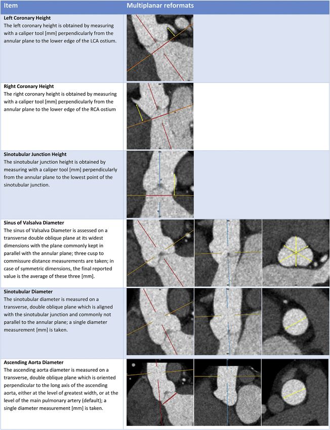
JACC: CARDIOVASCULAR IMAGING, VOL. 12, NO. 1, 2019                                                                                              Blanke et al.   5
JANUARY 2019:1–24                                                                                                                                 Guidelines

the setting of uncertain AS severity where the aortic                  axial data set with #1.5 mm slice thickness in a
valve calcium score is known to correlate well with                    contiguous or overlapping fashion, using a large field
aortic stenosis severity (23). This is of most use in                  of view and either filtered back projection or iterative
suspected low-flow low-gradient AS in those with                        reconstruction.
either a preserved or reduced ejection fraction with
no flow reserve on dobutamine stress echocardiog-                       3. AORTIC ROOT: ANATOMICAL DEFINITIONS
raphy (24). In this situation, the calcium score of the                AND ASSESSMENT
valve can be used to adjudicate the presence or
                                                                       Precise understanding and assessment of the anat-
absence of severe aortic stenosis (25). CT acquisition
                                                                       omy of the aortic valvular complex is crucial to ach-
for the assessment of aortic valve calcification should
                                                                       ieve optimal sizing of transcatheter heart valves
be performed using the same acquisition parameters
                                                                       (THV) as well as to identify patients at increased
used for assessment of the coronary artery calcium
                                                                       anatomical risk for adverse events such as coronary
score although further studies are underway to
                                                                       artery occlusion. The aortic root is an extension of
determine thresholds for post-contrast CT scans
                                                                       the left ventricular outflow tract, extending from the
(26,27). The valvular calcification is then contoured
                                                                       basal attachment of the aortic valve cusps within the
utilizing standard coronary artery calcium scoring
                                                                       LVOT to their peripheral attachment at the level of
software, with contours applied around the valvular
                                                                       the sinotubular junction. Its components are the si-
calcification, ensuring the exclusion of aortic wall,
                                                                       nuses of Valsalva, the fibrous interleaflet triangles
left ventricular outflow tract (LVOT) or coronary
                                                                       and the valvular cusps themselves (30,31).
calcification. Sex-specific thresholds are utilized,
with an Agatston score of $3,000 in men and $1,600                     3.1. AORTIC ANNULUS: DEFINITIONS AND MEASUREMENT

in women making severe aortic stenosis very likely,                    TECHNIQUES. For the purpose of anatomical sizing in

while an Agatston score of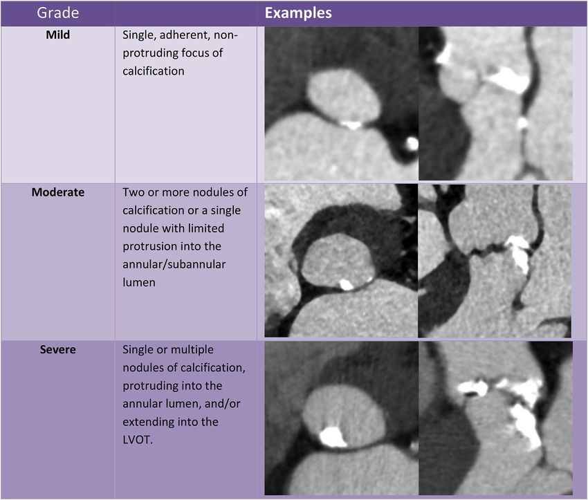
6          Blanke et al.                                                                                     JACC: CARDIOVASCULAR IMAGING, VOL. 12, NO. 1, 2019

           Guidelines                                                                                                                                JANUARY 2019:1–24

    F I G U R E 1 Identifying the Annular Plane in Aortic Valves With Tricuspid Morphology

                Descargado para Anonymous User (n/a) en Consejeria de Sanidad de Castilla y Leon(Sacyl) de ClinicalKey.es por Elsevier en marzo 05, 2019.
                Para uso personal exclusivamente. No se permiten otros usos sin autorización. Copyright ©2019. Elsevier Inc. Todos los derechos reservados.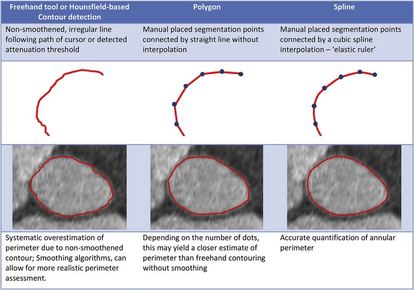
JACC: CARDIOVASCULAR IMAGING, VOL. 12, NO. 1, 2019                                                                                                Blanke et al.   7
JANUARY 2019:1–24                                                                                                                                   Guidelines

   F I G U R E 2 Contouring Tools for Annular Segmentation

2. Polygon: Manually placed segmentation points which                    between platforms (e.g., perpendicular versus non-
   are automatically connected by straight lines.                        perpendicular          orientation;        intersection        centric
3. Attenuation/Hounsfield             unit      based       contour       versus eccentric). Alternative, manual caliper mea-
   detection.                                                            surements can be performed. Overall, short and long
4. Freehand contour: The annular contour is traced                       axis dimensions inform on the eccentricity of the
   manually with the cursor.                                             annular      cross-sectional        dimensions.        Importantly,
                                                                         manual caliper measurements should not be used to
  All techniques commonly provide the annular area
                                                                         assess overall annular dimensions instead of con-
in either [mm 2] or [cm 2]. Cubic spline interpolation
                                                                         touring tools.
commonly provide also the annular perimeter in
                                                                             Independent of the measurement technique used,
[mm]. Polygon tools may underestimate the annular
                                                                         the annular contour should be drawn along the blood-
perimeter when compared to cubic spline interpola-
                                                                         pool tissue interface, carefully avoiding presence of
tion, given the straight line connection of the seg-
                                                                         any tissue within the contour and avoiding any
mentation points. Attenuation/Hounsfield unit based
                                                                         contrast outside of the contour. In case of annular
contour detection as well as freehand contour tech-
                                                                         calcification, irrespective of crescent or protruding
niques may, depending on the software platform,
                                                                         distribution, the contour should be drawn in a har-
result in jagged contours, artificially increasing the
                                                                         monic fashion as if no calcium is present, as this aids
reported perimeter. For this reason, smoothing algo-
                                                                         in measurement standardization.
rithms may be used to create a rather spline-like
contour. Perimeter values should only be reported                        3.2. ANNULAR DYNAMISM & PHASE SELECTION.
when using these measurement tools, if the contour                       During       systole,      conformational          changes        with
is smoothened.                                                           decrease in ellipticity as well as stretch of the annular
  Most contouring tools provide short and long axis                      contour commonly result in a larger annular area and
dimensions in [mm] which are automatically derived                       perimeter as compared to diastole (Fig. 3) (10,32). This
by software algorithms, although methodology differ                      annular dynamism has significant implications for

              Descargado para Anonymous User (n/a) en Consejeria de Sanidad de Castilla y Leon(Sacyl) de ClinicalKey.es por Elsevier en marzo 05, 2019.
              Para uso personal exclusivamente. No se permiten otros usos sin autorización. Copyright ©2019. Elsevier Inc. Todos los derechos reservados.
8             Blanke et al.                                                                                      JACC: CARDIOVASCULAR IMAGING, VOL. 12, NO. 1, 2019

              Guidelines                                                                                                                                 JANUARY 2019:1–24

      F I G U R E 3 Dynamic Changes of the Aortic Annulus Throughout the Cardiac Cycle

      Example with common anatomy and regular dynamism (upper row), demonstrating larger dimensions in systole than diastole, in parts due to bulging of the aortomitral
      continuity (dashed yellow line) towards the left atrium in systole and flattening in diastole. The lower row shows an example of septal hypertrophy with smaller
      annular dimensions in systole than diastole, in part due bulging of the basal septum (dashed blue line) into the annulus during systole.

                              sizing with the potential for unintended undersizing                     3.3. IMAGE         QUALITY. Reliable           quantification        of
                              if sizing is based on diastolic assessment (12,33).                      aortic annular and aortic root dimensions requires
                                  Annular measurements should be performed using                       adequate image quality defined by sharp depiction of
                              the ECG-synchronized, ideally multiphasic dataset.                       the annular contour given sufficient contrast attenu-
                              Identifying the reconstruction phase with largest                        ation, noise preposition as well as absence of motion
                              annular dimensions is important given the implica-                       artifacts, double contours, or stair step artifacts.
                              tions for device sizing. The annular anatomy should                      Image quality can be rated using a subjective, quali-
                              be reviewed throughout the available portion of the                      tative grading scale as good, fair and poor (non-
                              cardiac cycle and the phase yielding the largest di-                     diagnostic) (Table 4).
                              mensions with adequate image quality should be                           3.4. LANDING ZONE CALCIUM. The device landing
                              visually identified. Importantly, changes in the                          zone comprises the valve cusps, aortic annulus and
                              orientation of the annular plane throughout the car-                     LVOT (34,35). It has been demonstrated that the
                              diac cycle may require adjustment of the imaging                         presence of severe calcification of the LVOT and
                              plane. Care should be taken to identify a reconstruc-                    aortic valve is associated with increased risk of par-
                              tion phase with adequate image quality, ie. sharp                        avalvular regurgitation particularly when calcium
                              depiction of the annular contour. Reconstruction                         protrudes into the LVOT (36–40). In clinical practice,
                              phases with inadequate depiction of the annular                          the description of annular (within annular plane) and
                              contour, i.e., blur or double contours, should be                        sub-annular (upper 4 to 5 mm of the LVOT where
                              avoided.                                                                 device gets into contact) calcification is almost
                                                                                                       exclusively performed in a subjective, qualitative
    T A B L E 4 Grading Scale for Image Quality for Assessment of the Aortic Root
                                                                                                       fashion graded as none, mild, moderate, severe based
            Grade                                          Definition
                                                                                                       on the circumferential extent, the depth of extension
    Good (diagnostic)          Sharp depiction of the annular contour with sufficient contrast
                                                                                                       inferiorly into the LVOT and the thickness of the
                                attenuation in the absence of artifacts
    Fair (diagnostic)          Low contrast attenuation                                               calcification projecting radially into the LVOT (Fig. 4).
                               Increased image noise                                                  Annular and sub-annular calcification should be
                               Mild motion artifacts, double contours or stair-step artifacts
                                                                                                       described as crescent/flat/adherent or protruding as
    Poor (non-diagnostic)        Too low contrast attenuation
                                 Excessive image noise                                                well as its relation to the aortic cusps. The location of
                                 Excessive motion artifacts or double contours                        calcification within the LVOT varies significantly
                                 Pronounced stair-step artifact transecting the annular contour
                                                                                                       across severe aortic stenosis patients. The region

                    Descargado para Anonymous User (n/a) en Consejeria de Sanidad de Castilla y Leon(Sacyl) de ClinicalKey.es por Elsevier en marzo 05, 2019.
                    Para uso personal exclusivamente. No se permiten otros usos sin autorización. Copyright ©2019. Elsevier Inc. Todos los derechos reservados.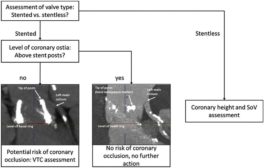
JACC: CARDIOVASCULAR IMAGING, VOL. 12, NO. 1, 2019                                                                                                                    Blanke et al.   9
JANUARY 2019:1–24                                                                                                                                                       Guidelines

 T A B L E 5 Summary of Recommendations for the Sizing and Reporting of the Aortic Valve, Annulus and Outflow Tract

                                                                                                                                                   Grade of
 Recommendation                                                                                                                                Recommendation*

 Annulus assessment and planning
 While facilitated or semi-automated workflows may be used, the interpreter analyzing the imaging must be able to confirm the                          Strong
    accuracy of the generated annular plane and perform manual corrections if required.
 Systolic measurements are preferred for measurement and calculation of device sizing                                                                Strong
 Area and perimeter measurements are preferred for sizing of the aortic annulus over isolated 2 dimensional measurements and                         Strong
    should be provided in the report
 Landing zone calcification
 Annular and subannular calcification should be qualitatively described regarding morphology and extent as well as relation to                        Strong
    the aortic valve cusps.
 Valve morphology
 Number of cusps should be stated, and if a bicuspid valve is present, its morphology should be classified.                                           Strong
 The presence of a median raphe and the absence/presence of calcification of this should be mentioned                                                 Strong
 The aortic annulus size should be measured and reported in bicuspid aortic valves as for tricuspid aortic valves.                                   Strong
 Aortic root measurement
 Pre-TAVI/TAVR CT assessment should include coronary height, mean SOV diameter, and STJ height and diameter                                          Strong
 Coronary ostial distance from aortic annulus should be measured in a perpendicular fashion from the established annular plane                       Strong

 *Based on level of consensus.
   CT ¼ computed tomography; SOV ¼ sinus of valsalva; STJ ¼ sinotubular junction; TAVI ¼ transcatheter aortic valve implantation; TAVR ¼ transcatheter aortic valve
 replacement.

below the non-coronary and the left coronary cusps,                                 (equivalent to Sievers Type 1); and 3) bicommissural
including the intervalvular fibrosa, is most frequently                              non raphe-type (equivalent to Sievers Type 0) (Fig. 5)
affected. Large protruding nodules of calcification                                  (47). Ascending aortic dilation and aneurysms are less
may increase the risk of annular rupture particularly                               common in ‘tricommissural’ BAV than ‘bicommissu-
with balloon expandable valves and should thus be                                   ral’ BAV. Within ‘bicommissural’ BAV, the non-raphe
specifically mentioned in the report (34). Finally, the                              type BAV typically exhibits larger sinus diameters
device landing zone is in close spatial relationship                                despite smaller annuli. For bicommissural raphe-type
with the conduction system which may be com-                                        BAV, raphe characteristics may also be described
pressed and damaged, causing atrioventricular block                                 qualitatively and quantitatively including raphe
and need for pacemaker implantation especially in                                   length and raphe calcium (47).
the context of severe calcification of the subannular                                    Valve morphology should be systematically char-
device landing zone, with this effect amplified in the                               acterized and reported in all pre-TAVI/TAVR CT re-
presence of pre-existent right bundle branch block                                  ports. Further, the degree of raphe calcification
(41,42). Unusual findings with regard to these vari-                                 should be reported, e.g. using a qualitative scale
ables should be included in the report.                                             (mild, moderate, severe), as severe raphe calcification
3.5. VALVE           MORPHOLOGY:                 DEFINITION             AND         may relate to higher likelihood of paravalvular
MEASUREMENT TECHNIQUES. Bicuspid aortic valves                                      regurgitation (44).
(BAV) are found in up to 6% of patients presenting for                                  Defining the annulus may be a challenge in BAV,
TAVI/TAVR, and are associated with lower device                                     particularly in Sievers Type 0 BAVs where there are
success rates and higher rates of paravalvular regur-                               only two hinge points to define the annular plane.
gitation, but similar outcomes (43,44). Numerous                                    The technique used for tricuspid aortic valves there-
classifications of BAV morphology exist including                                    fore frequently needs to be adjusted as detailed in
that of Sievers and Schmidtke (45) (which essentially                               Figure 6. The annulus size should be measured and
focuses on the number of raphes (0, 1 or 2 for the                                  reported in the same fashion for BAV as for tricuspid
respective types). More recently, a TAVI/TAVR-                                      aortic valves. The ascending aorta should also be
directed simplified BAV classification has been pro-                                  examined in BAV due to the association between BAV
posed. It distinguishes BAV morphology regarding (a)                                and aortopathy (46).
number of commissures (two vs. three) and (b) pres-                                 3.6. CORONARY OSTIAL HEIGHT AND SINUS OF
ence or absence of a raphe, yielding 3 broad mor-                                   VALSALVA ASSESSMENT. Coronary occlusion is a
phologies: 1) tricommissural, in clinical routine often                             feared complication of TAVI/TAVR which, while
referred to as ‘functional’ or ‘acquired’ BAV (not part                             relatively rare with an incidence of 0.66%, is associ-
of Sievers classification); 2) bicommissural raphe-type                              ated with a poor clinical outcome with a reported 30

                Descargado para Anonymous User (n/a) en Consejeria de Sanidad de Castilla y Leon(Sacyl) de ClinicalKey.es por Elsevier en marzo 05, 2019.
                Para uso personal exclusivamente. No se permiten otros usos sin autorización. Copyright ©2019. Elsevier Inc. Todos los derechos reservados.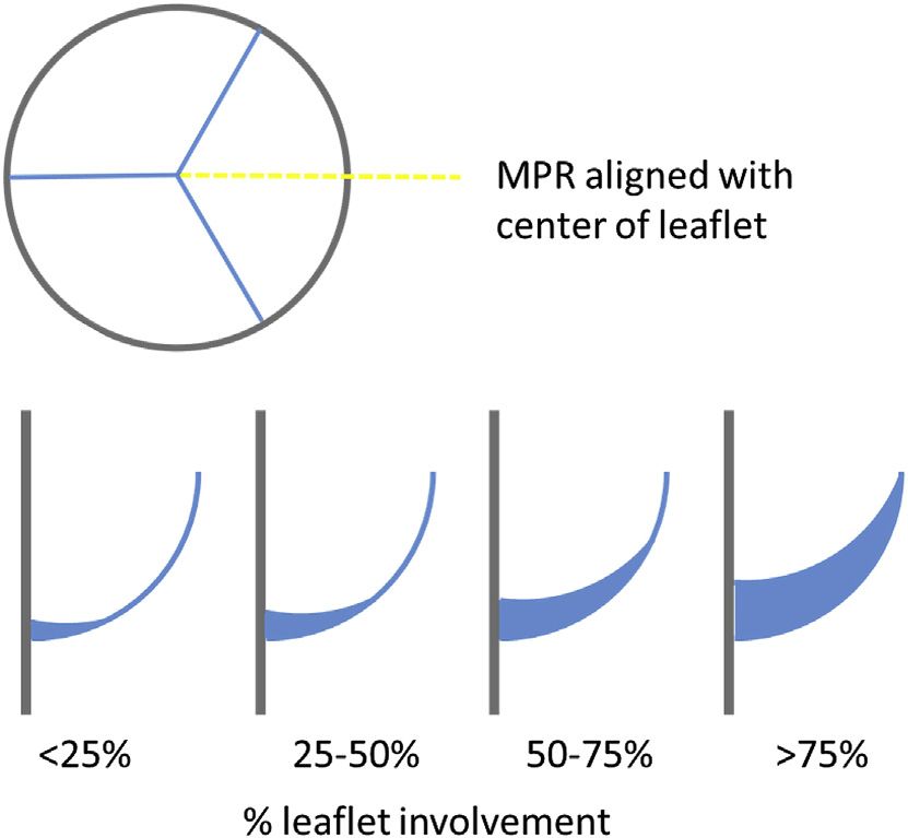
10   Blanke et al.                                                                                     JACC: CARDIOVASCULAR IMAGING, VOL. 12, NO. 1, 2019

     Guidelines                                                                                                                                JANUARY 2019:1–24

          F I G U R E 4 Qualitative Grading of Annular/Sub-Annular and Left Ventricular Outflow Tract Calcification

                     day mortality of up to 40.9% (48,49). CT is well                        from the annular plane to the lower edge of the cor-
                     established as the pre-procedural imaging gold stan-                    onary artery ostium, yielding a value in [mm] (Fig. 7).
                     dard for the determination of the risk of coronary                      Sinus of valsalva diameter should be measured cusp
                     occlusion. Low coronary ostial height (
JACC: CARDIOVASCULAR IMAGING, VOL. 12, NO. 1, 2019 Blanke et al. 11
JANUARY 2019:1–24 Guidelines
F I G U R E 5 The Heterogeneous Spectrum of Bicuspid Aortic Valve Morphology
The STJ height should be measured in a perpen- aortic dimensions should be assessed on double-
dicular fashion to the annular plane using an elec- oblique multiplanar reformats in [mm] (Fig. 7).
tronic caliper tool from the annular plane to the lowest 3.9. OPTIMAL PROJECTION CURVE. Ideally, TAVI/
edge of the STJ, yielding a value in [mm] (Fig. 7). STJ TVAR is performed with fluoroscopic angulations
diameter should be measured using a caliper tool on providing a coplanar view of the aortic annulus
an imaging plane aligned with the STJ (Fig. 7). without parallax. CT can be used to identify these
3.8. ASCENDING AORTA. The ascending aorta should patient-specific ‘optimal’ C-arm angulations (50,51).
be assessed for the presence of aortopathy. Ascending Use of these CT-derived angulations allows
Descargado para Anonymous User (n/a) en Consejeria de Sanidad de Castilla y Leon(Sacyl) de ClinicalKey.es por Elsevier en marzo 05, 2019.
Para uso personal exclusivamente. No se permiten otros usos sin autorización. Copyright ©2019. Elsevier Inc. Todos los derechos reservados.12 Blanke et al. JACC: CARDIOVASCULAR IMAGING, VOL. 12, NO. 1, 2019
Guidelines JANUARY 2019:1–24
F I G U R E 6 Identifying the Annular Plane in Bicuspid Aortic Valves With Sievers Type 0 Morphology
Descargado para Anonymous User (n/a) en Consejeria de Sanidad de Castilla y Leon(Sacyl) de ClinicalKey.es por Elsevier en marzo 05, 2019.
Para uso personal exclusivamente. No se permiten otros usos sin autorización. Copyright ©2019. Elsevier Inc. Todos los derechos reservados.JACC: CARDIOVASCULAR IMAGING, VOL. 12, NO. 1, 2019 Blanke et al. 13
JANUARY 2019:1–24 Guidelines
F I G U R E 7 Schematic for Assessment of Aortic Root Dimensions
Descargado para Anonymous User (n/a) en Consejeria de Sanidad de Castilla y Leon(Sacyl) de ClinicalKey.es por Elsevier en marzo 05, 2019.
Para uso personal exclusivamente. No se permiten otros usos sin autorización. Copyright ©2019. Elsevier Inc. Todos los derechos reservados.14 Blanke et al. JACC: CARDIOVASCULAR IMAGING, VOL. 12, NO. 1, 2019
Guidelines JANUARY 2019:1–24
T A B L E 6 Summary of Recommendations for the Reporting of Fluoroscopic Angulation
Recommendation Grade of Recommendation*
Fluoroscopic planning
Provision of optimal fluoroscopic projection angulations based on CT for each individual patient should be considered. Strong
If the patient is not positioned supine for the CT examination, this should be noted in the report and proposed fluoroscopic Strong
angles should not be provided
*Based on level of consensus.
CT ¼ computed tomography; TAVR ¼ transcatheter aortic valve replacement.
optimization of initial pre-deployment fluoroscopic formula of a circle where area ¼ p *radius2 and
angulation, reducing the need for repeat pre- circumference ¼ p*diameter, the area in-
deployment root shots thereby reducing radiation creases exponentially while perimeter increases pro-
exposure, contrast usage and procedural time (52,53). portionally with increasing diameter. The percentage
Optimal C-arm angulations should be reported as area oversizing is approximately 2-fold greater than
degrees [ ] LAO or RAO with the corresponding values the percentage perimeter when dealing with a perfect
for cranial or caudal angulation. Dependent on insti- circle but the implications are even greater than ex-
tutional preferences, typically views are either pected owing to the non-circular geometry of the
centered on the right coronary cusp, or use pre- annulus. Both annular area and perimeter are clini-
defined LAO angulation (e.g., LAO 10 ), or are opti- cally acceptable, but it is imperative that the imager
mized for visualization of the left main stem. When and implanting physician appreciate the differences
identified projections result in cranial or caudal and are aware of the parameter being used for device
angulation >25 , alternate angulations should be sizing of specific devices. Further, the optimal
provided, given the physical restraints of the C-arms. threshold for oversizing is also device specific (56,57).
However, it is essential to recognize that CT predicted Thus routine reporting of oversizing is not required,
angulations are only valid if the patient’s chest is however familiarity of the concept is beneficial for
positioned in a similar fashion on the CT table as subsequent discussion in Heart Team meetings.
during the procedure. Please refer to Table 6 for a 3.10.2. Modification of sizing based on root calcification.
summary of recommendations. CT not only provides information regarding annular
3.10. SIZING CONSIDERATIONS. CT is the non- size, but also provides important ancillary information
invasive imaging gold standard for annular sizing that is helpful in guiding sizing and THV selection.
and THV selection (1). In clinical practice, balloon Annular and sub-annular calcification, particularly
expandable devices are largely sized on the basis of when protruding into the lumen, can result in
annular area, with self-expandable devices relying on increased risk of both annular rupture and para-
perimeter. The reasons for this are partly historical but valvular regurgitation. Protruding landing zone calci-
also reflect different risk profiles of these devices and fication below the non-coronary cusp has been shown
the geometric implications of sizing with the different to be most closely-associated with annular injury, with
variables. Sizing based on manually assessed short this effect amplified when combined with aggressive
and long axis diameters have been shown to be less annular oversizing (58). Presence, location and char-
reproducible (54), and can be considered obsolete. acteristics of subannular calcification should thus be
3.10.1. Oversizing. The term ‘oversizing’ has been integrated into all pre-TAVI/TAVR CT scan reports as
introduced over the last 6 years to help describe when described in the ‘landing zone calcium’ section.
a THV is deployed that is larger than the native 3.10.3. Annular rupture. Annular rupture is an infre-
annulus. The term ‘oversizing’ is a generic term as quent adverse event but is associated with a high
oversizing can be calculated based on any measure- mortality if it occurs (58,59). Patient and procedure
ment of the annulus. In routine, oversizing is calcu- related factors increasing risk of annular rupture
lated as a percentage [%], as follows: include female sex, use of balloon-expandable valves,
Oversizing [%] ¼ (THV nominal measurement/ significant prosthesis oversizing, and prior radiation
annular measurement 1) 100 therapy. In addition, the presence of moderate/severe
It is important to recognize, that the percentage sub-annular calcification, particularly below the non-
oversizing calculated is strongly dependent on the coronary cusp on pre-procedural CTA, is associated
annular measurement used, with very different with a significantly increased risk for annular rupture
implication of oversizing area than oversizing perim- (34). In addition, the depth of calcification within the
eter or perimeter derived diameter (55). Given the LVOT is also important, as calcification immediately
Descargado para Anonymous User (n/a) en Consejeria de Sanidad de Castilla y Leon(Sacyl) de ClinicalKey.es por Elsevier en marzo 05, 2019.
Para uso personal exclusivamente. No se permiten otros usos sin autorización. Copyright ©2019. Elsevier Inc. Todos los derechos reservados.JACC: CARDIOVASCULAR IMAGING, VOL. 12, NO. 1, 2019 Blanke et al. 15
JANUARY 2019:1–24 Guidelines
T A B L E 7 Summary of Recommendations for the Reporting of Vascular Access, Coronary Artery, and Non-Cardiac, Non-Vascular Findings
Grade of
Recommendation Recommendation*
Vascular access
While facilitated or semi-automated work-flows may be used, the interpreter analyzing the imaging must be able to confirm the accuracy of the Strong
generated vessel centerline and perform manual corrections if required.
The minimal luminal diameter along both the right and left iliofemoral system should be provided including the anatomical location to the level of the Strong
expected puncture site
All areas of >270 calcification in the iliofemoral arteries should be reported Strong
Calcification located anteriorly at the site of probable puncture should be reported. Strong
The report should include a clear description of all vascular pathologies including aneurysms, dissection, and occlusions. Strong
Coronary arteries
Reporting of the coronary arteries for severity of coronary artery disease can be considered in appropriately selected patients, if image quality is of Strong
diagnostic quality
The presence and course of anomalous coronary arteries should be reported. Strong
Non-cardiac, non-vascular
CT images should be reviewed for incidental findings Strong
Extracardiac findings should be reviewed and reported in the context of the healthcare environment and health status of the patient Strong
Significant findings should be included in the dictated report and when appropriate verbally communicated to the Heart team. Strong
*Based on level of consensus.
CT ¼ computed tomography; SOV ¼ sinus of valsalva; STJ ¼ sinotubular junction; TAVI ¼ transcatheter aortic valve implantation; TAVR ¼ transcatheter aortic valve replacement.
below the annular plane connotes a higher risk than mortality after TAVI/TAVR, however the rates of
calcification lower within the LVOT. Protruding nod- complications have fallen with improved pre-
ules of calcification are considered to connote higher procedural screening with major vascular complica-
risk than flat or mural calcification. The risk of aortic tions currently occurring in 4.5% of procedures
annulus rupture increases with the degree of over- (68,69). Access sheath sizes have reduced in size,
sizing of the prosthesis (particularly with >20% however this simply shifts the threshold at which
oversizing) and with the extent of calcification of the trans-femoral access can be achieved, rather than
upper part of the LVOT (within 2 mm below the negating the importance of identification and quan-
annulus plane) particularly when immediately below tification of vascular disease and dimensions (70).
the non-coronary cusp (34,58). Given the continuous evolution of delivery systems,
3.10.4. Atrio-ventricular conduction block. An increased no reference is made to current devices and vessel
depth of implantation is the most frequently identi- diameter requirements. Please refer to Table 7 for a
fied predictor of LBBB with both balloon- and self- summary of recommendations.
expandable prostheses (60–65). Reduced implant Analysis of iliofemoral vessel size, calcification,
depth during TAVI/TAVR has been shown to signifi- and tortuosity is required to determine if trans-
cantly decrease mortality and permanent pacemaker femoral access can be achieved or whether an alter-
insertion rate (66). In addition, shorter length of the native access route is required (71,72). Due to its
semi-membranous septum has been reported to be ability to accurately quantify all these aspects, CT
associated with higher risk of conduction disturbance provides greater predictive value for vascular com-
post TAVI/TAVR, with a length of less than approx. plications than invasive angiography (73).
8mm predictive of high-degree AV-block, particularly Risk factors for vascular complications are an
when combined with a deep implant depth, or the external sheath diameter that exceeds the minimal
presence of pre-existent right bundle branch block artery diameter, moderate or severe calcification,
(41,42,67). The length of the semi-membranous and vessel tortuosity (75–77). While early reports
septum can be measured in the coronal plane, at the described an increased risk at a sheath:diameter
longest point between the annular level and the ratio $1.05, more recent work on modern access
muscular septum (67). However, routine assessment devices which are typically smaller with an
of semi-membranous septum length has not seen increasing prevalence of expanding sheath designs,
widespread adoption into clinical routine. suggests a more liberal threshold of $1.12 can be
safely used (73).
4. VASCULAR ACCESS
4.2. QUANTIFICATION OF LUMINAL DIMENSIONS.
4.1. OVERVIEW. Vascular complications are inde- The minimal diameter of the vasculature between the
pendently associated with increased morbidity and aortic valve and the right and left common femoral
Descargado para Anonymous User (n/a) en Consejeria de Sanidad de Castilla y Leon(Sacyl) de ClinicalKey.es por Elsevier en marzo 05, 2019.
Para uso personal exclusivamente. No se permiten otros usos sin autorización. Copyright ©2019. Elsevier Inc. Todos los derechos reservados.16 Blanke et al. JACC: CARDIOVASCULAR IMAGING, VOL. 12, NO. 1, 2019
Guidelines JANUARY 2019:1–24
artery should be reported. This can be performed degree of ascending aortic calcification should be
either manually on multiplanar reformats using a noted—the latter relevant for potential cross-
double-oblique technique, or using semi-automatic clamping. Further relevant pathologies include
post-processing using centerline placement and abnormal elongation and kinking, dissections, aneu-
curved multiplanar reformats. When the latter is rysms, and exophytic plaque.
used, manual verification of the center line should be 4.7. ALTERNATE ACCESS. If transfemoral access is
performed to ensure accurate vessel tracking, and not feasible, depending on local practice, subclavian
appropriate intra-luminal location of the centerline. and/or carotid arteries can be reported using the same
When using either technique, attention should be techniques and reporting parameters of the iliofe-
paid to the potential for calcium blooming artifact, moral vessels (78,79). Due to more favorable angula-
and appropriate windowing used to correct for this tion, an left-sided approach is preferred for
where necessary. It should also be ensured that all subclavian access. Finally, if a transcaval approach is
measurements are performed perpendicular to the to be considered, the presence, size and level of
long axis of the vessel at the location of the maximum calcification free windows of the aortic wall adjacent
stenosis. Transverse source images allow no more to the inferior vena cava should be reported (80).
than a preliminary assessment of vessel size. Additionally the report should also include a clear
4.3. CALCIFICATION. Extend and distribution of description of all vascular pathologies including an-
calcifications of the iliofemoral vasculature should be eurysms, dissection, and occlusions.
assessed and reported. To describe the severity, a
subjective, semi-quantitative grading scale can be 5. CORONARY ARTERIES
used: none, mild (spotty), moderate (coalescing), se-
vere (bulky, protruding, horse-shoe, circumferential). In the setting of good image quality, modest motion
Care should be taken to identify circumferential or artifact in particular, coronary CTA can rule out sig-
near-circumferential (horse-shoe) calcification nificant coronary stenosis with high negative predic-
particularly in areas of tortuosity or bifurcations, as tive value (81–85). However confident assessment of
these prevent vessel expansion when the sheath and coronary stenosis severity on TAVI/TAVR scans is
valve passes through. challenging, particularly due to a high prevalence of
coronary calcification and higher heart rates resulting
4.4. TORTUOSITY. Tortuosity of vascular structures
in motion artifact (86). Beta-blockade must be used
can be assessed on transverse source images, but
with caution and nitroglycerin is contra-indicated,
evaluation is facilitated using a volume rendered
with the absence of these associated with reduced
display from multiple viewing angles, such as anterior-
diagnostic accuracy (9). Independent from the
posterior and RAO or LAO projections. In the absence
assessment for CAD, the presence and course of
of calcification, Iliofemoral arterial tortuosity is not
anomalous coronary arteries, easily interpreted,
necessarily a contraindication for femoral access, as
should be reported.
tortuous vessel segments usually straightened with
sheath insertion. However, when calcified, tortuous
6. INCIDENTAL NON-CARDIAC
segments carry a substantial risk of access failure and
NON-VASCULAR FINDINGS
the operator should be advised about this situation.
4.5. ACCESS SITE. Careful interrogation of the com- With growing evidence and increasing clinical adop-
mon femoral artery puncture site is important to tion, TAVI/TAVR is increasingly being performed in
ensure that that there is no focal stenosis nor calcifi- younger patients with lower risk profiles. This in-
cations anteriorly that may interfere with the arterial creases the relevance of incidental findings on CT as
puncture or deployment of arterial closure devices the life expectancy of patients undergoing TAVI/
(74). Also, the anatomy should be reviewed for the TAVR will be longer than in the past. All imaging
presence of a high femoral artery bifurcation. Rele- obtained should be reviewed carefully for incidental
vant findings should be reported, ideally with findings. This is best performed by trained radiolo-
detailed location in relation to fluoroscopically iden- gists, either as a combined or separate report ac-
tifiable anatomical landmarks, such as the level of the cording to local practices. The clinical
femoral head (e.g., lower third). recommendations and downstream management of
4.6. AORTA. For transfemoral access, the thoracic incidental findings on pre-TAVI/TAVR CT will vary
and abdominal aorta should be assessed for the according to the risk profile and life expectancy of the
presences of relevant pathologies. In particular, the patients as well as local clinical practice. Please refer
presence of ascending aortic aneurysm and the to Table 7 for a summary of recommendations.
Descargado para Anonymous User (n/a) en Consejeria de Sanidad de Castilla y Leon(Sacyl) de ClinicalKey.es por Elsevier en marzo 05, 2019.
Para uso personal exclusivamente. No se permiten otros usos sin autorización. Copyright ©2019. Elsevier Inc. Todos los derechos reservados.JACC: CARDIOVASCULAR IMAGING, VOL. 12, NO. 1, 2019 Blanke et al. 17
JANUARY 2019:1–24 Guidelines
peripheral vasculature imaging. Leaflet thickening
F I G U R E 8 MPR Alignment and Semi-Quantitative Grading of
Hypo-Attenuated Leaflet Thickening
should be described based on location, extent in
length and overall thickness. Importantly, HALT takes
a meniscal shape when viewed on a long-axis MPR at
the center of the cusp (Figs. 8 and 9). In regard to
extent of HALT along the curvilinear leaflet, as sub-
jective grading scale can be employed as outlined in
Figure 8.
Restricted motion should be reported as present or
absent. Restricted valve motion without thickening
on CTA is rare and should be reported with great
caution, to avoid the risk of unnecessary treatment
(100,102,103).
8. VALVE-IN-VALVE IMPLANTATION
8.1. OVERVIEW. Valve in valve (VIV) implantation of
THVs into failing bioprosthetic valves has evolved as
a treatment alternative with high technical success
rates and promising patient outcomes (104). Broadly,
bioprosthetic valves can be categorized as stented
The dashed yellow line indicates the orientation of the long
surgical (rigid scaffold), stentless surgical (no rigid
axis views in the lower row, aligned with the center of the
cusps. The extent of leaflet thickening can be graded on a scaffold), and transcatheter heart valves.
subjective 4-tier grading scale along the curvilinear orientation The Valve-in-Valve International Data (VIVID)
of the leaflet. Typically, hypo-attenuated leaflet thickening Registry has shown that coronary occlusion is more
appears meniscal-shaped on long axis reformats, with greater
common in VIV than TAVI/TAVR in native aortic
thickness at the base than towards the center of the leaflet.
stenosis, with occlusion reported in 2.3% of VIV
compared with 0.66% in native valve TAVI/TAVR
(48,105). Factors related to the observed higher fre-
7. POST-TAVI/TAVR CT
quency are canted position of the stented surgical
bioprosthesis within the aortic root and small root
Echocardiography is the test for evaluation of trans-
dimensions, the latter in particular in patients with
catheter heart valve function and durability (87,88).
stentless surgical valves. Pre-procedural CT plays a
CT enables post-implant imaging of transcatheter
limited role in THV sizing, but a key role in the
heart valves (geometry, position, leaflets) (89–98),
discrimination of patient specific risk for coronary
There is however, no consensus regarding the clinical
occlusion. Vascular access should be reported as for
need to perform routine cardiac CT in TAVI/TAVR re-
TAVI/TAVR in native aortic valves (see Vascular Ac-
cipients. Cardiac CT is an important adjunctive mo-
cess section). Please refer to Table 8 for a summary of
dality to echocardiography in patients where there is
recommendations.
concern for valve thrombosis, infective endocarditis,
or structural degeneration (24,99). Hypoattenuated 8.2. ASSESSMENT FOR RISK OF CORONARY ARTERY
leaflet thickening (HALT) (Figs. 8 and 9) and restricted OBSTRUCTION. Implantation of a THV within a
leaflet motion (also referred to as HAM [hypoattenu- stented surgical valve is technically different from
ation affecting motion]) determined by CT often TAVR in native aortic valves, as the stented bio-
indicate leaflet thrombus formation (7,8,100–102). prosthetic valves provides the scaffold for THV
When echocardiography is indeterminate, CTA can be anchorage. The THV displaces the bioprosthetic
useful for adjudication of leaflet thrombus. When leaflets into an open position with the THV frame and
imaging in the post TAVI/TAVR setting for the overlying bioprosthetic leaflets forming a ‘covered’
assessment of HALT, full cardiac cycle imaging is cylinder. The anatomical orientation of the cylinder is
recommended to maximize image quality and to being dictated by the orientation of the surgical valve.
permit assessment of leaflet motion. IV contrast Frequently, the surgical valve is implanted in a can-
administration and timing should be performed as per ted fashion in regard to the long axis of the aortic
the routine TAVI/TAVR workup, although a smaller root, with the potential for close proximity of the
contrast dose is required due to the lack of need for bioprosthetic leaflets/THV to the coronary ostia and
Descargado para Anonymous User (n/a) en Consejeria de Sanidad de Castilla y Leon(Sacyl) de ClinicalKey.es por Elsevier en marzo 05, 2019.
Para uso personal exclusivamente. No se permiten otros usos sin autorización. Copyright ©2019. Elsevier Inc. Todos los derechos reservados.You can also read