Altered glycolysis results in drug resistant in clinical tumor therapy (Review)
←
→
Page content transcription
If your browser does not render page correctly, please read the page content below
ONCOLOGY LETTERS 21: 369, 2021

                          Altered glycolysis results in drug‑resistant
                              in clinical tumor therapy (Review)
                   JINGHUI PENG1*, YANGYANG CUI1*, SHIPENG XU2, XIAOWEI WU1, YUE HUANG1,
                              WENBIN ZHOU1, SHUI WANG1, ZIYI FU3,4 and HUI XIE1

        Departments of 1Breast Surgery, The First Affiliated Hospital, Nanjing Medical University, Nanjing, Jiangsu 210029,
        P.R. China; 2Robert H. Lurie Comprehensive Cancer Center, Northwestern University Feinberg School of Medicine,
        Chicago, IL 60611, USA; 3Nanjing Maternal and Child Health Medical Institute, Obstetrics and Gynecology Hospital
       Affiliated to Nanjing Medical University, Nanjing Maternity and Child Health Care Hospital, Nanjing, Jiangsu 210004;
     4
       Department of Oncology, The First Affiliated Hospital, Nanjing Medical University, Nanjing, Jiangsu 210029, P.R. China

                                     Received October 6, 2020; Accepted December 23, 2020

                                                     DOI: 10.3892/ol.2021.12630

Abstract. Cancer cells undergo metabolic reprogramming,               Contents
including increased glucose metabolism, fatty acid synthesis
and glutamine metabolic rates. These enhancements to                  1. Introduction
three major metabolic pathways are closely associated with            2.	Glycolysis‑related enzymes contribute to chemotherapy
glycolysis, which is considered the central component of                  resistance in cancer cells
cancer cell metabolism. Increasing evidence suggests that             3.	Glycolysis related substrates and products play roles in
dysfunctional glycolysis is commonly associated with drug                 chemotherapy resistance
resistance in cancer treatment, and aberrant glycolysis plays         4. Glycolysis is associated with immunotherapy resistance
a significant role in drug‑resistant cancer cells. Studies on the     5. Transition of glycolysis to OXPHOS enhances drug resis‑
development of drugs targeting these abnormalities have led to        tance
improvements in the efficacy of tumor treatment. The present          6. Conclusions
review discusses the changes in glycolysis targets that cause         7. Future direction and perspectives
drug resistance in cancer cells, including hexokinase, pyruvate
kinase, pyruvate dehydrogenase complex, glucose transporters,
and lactate, as well the underlying molecular mechanisms and          1. Introduction
corresponding novel therapeutic strategies. In addition, the
association between increased oxidative phosphorylation and           Metabolic disorders, particularly those concerning glucose
drug resistance is introduced, which is caused by metabolic           metabolism, play an important role in the proliferation and
plasticity. Given that aberrant glycolysis has been identified        development of tumors (1). In normal cells, the energy provided
as a common metabolic feature of drug‑resistant tumor cells,          for cell biological activity is predominantly dependent on
targeting glycolysis may be a novel strategy to develop new           changes in glucose metabolism that can transform glucose
drugs to benefit patients with drug‑resistance.                       into pyruvate after several steps. Subsequently, pyruvate is
                                                                      converted to oxaloacetate, resulting in the production of citrate
                                                                      in the mitochondrial tricarboxylic acid cycle (TCA cycle). The
                                                                      process of glucose metabolism and the mitochondrial TCA
Correspondence to: Professor Hui Xie, Department of Breast            cycle can generate energy in the form of adenosine triphos‑
Surgery, The First Affiliated Hospital, Nanjing Medical University,   phate (ATP) and other forms, such as NADPH and FADH2 (1).
300 Guangzhou Road, Nanjing, Jiangsu 210029, P.R. China               NADPH and FADH2 are subsequently committed to the elec‑
E‑mail: Hxie@njmu.edu.cn                                              tron transport chain complexes to yield ATP, which is known
                                                                      as oxidative phosphorylation (OXPHOS) (2,3).
Mr. Ziyi Fu, Department of Oncology, The First Affiliated Hospital,
Nanjing Medical University, 300 Guangzhou Road, Nanjing,
                                                                          There are significant differences between normal cells
Jiangsu 210029, P.R. China                                            and tumor cells regarding glucose metabolism. Warburg
E‑mail: fzyzzu@hotmail.com                                            suggested that glycolysis is the predominant metabolic
                                                                      mechanism that produces the most ATP in cancer cells,
*
    Contributed equally                                               which means that pyruvate derived from glucose is converted
                                                                      to lactate to exert its effects instead of being incorporated
Key words: cancer, glycolysis, drug resistance, microenvironment,     into the TCA cycle (2), and this is known as ‘the Warburg
metabolism                                                            effect’. Subsequently, the ‘reverse Warburg effect’ proposed
                                                                      that tumor‑associated fibroblasts can produce large amounts
                                                                      of lactic acid via aerobic glycolysis, which is provided to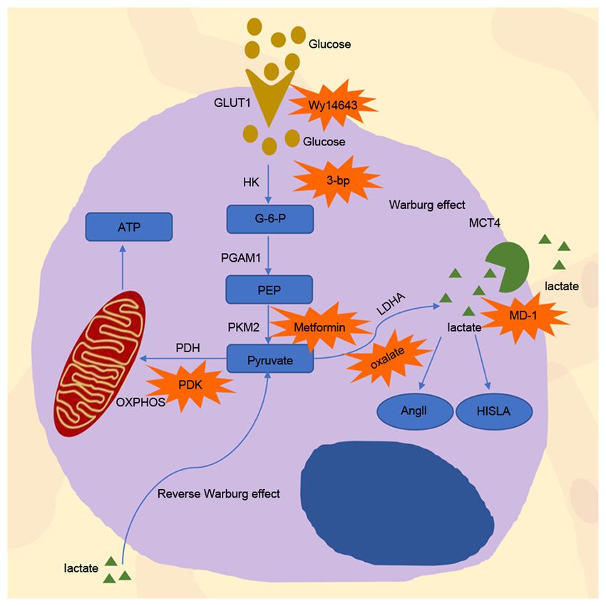
2                        PENG et al: ASSOCIATION BETWEEN GLYCOLYSIS AND THERAPY RESISTANCE

adjacent cells in a paracrine manner, causing the activation    resistance. Fan et al (18) reported that in pancreatic cancer,
of mitochondria, increasing OXPHOS in adjacent cells and        reactive oxygen species (ROS) derived from gemcitabine
promoting tumor activity (3). Generally, ‘the Warburg effect’   promote HK2 dimerization and bind to VDAC, which
and ‘reverse Warburg effect’ play an essential role in the      inhibits apoptosis by suppressing the formation of mitochon‑
development of cancer.                                          drial permeability transition pores, ultimately resulting in
    The incidence and mortality rates associated with           gemcitabine resistance (15,18).
cancer remain high, and despite the vast array of treatments        Given the vital role of HK2 in tumor resistance, it can
available, chemotherapy resistance remains a significant        be used as a valuable target in investigating chemoresis‑
challenge (4). There are several mechanisms of resistance,      tance inhibition. HK2 inhibitor 3‑bromopyruvate facilitates
such as the mutation on binding sites, the activation of        the dissociation of HK2 from the mitochondrial complex,
downstream effectors and the participation of alternative       potentiating daunorubicin‑induced apoptosis and promoting
survival pathways to bypass target inhibition (5). In addi‑     leukemia cell sensitivity to daunorubicin (19) (Fig. 1).
tion, cancer cells are resistant to immunotherapy via the       Furthermore, in ovarian cancer, the tyrosine analog, NK007,
Wnt‑β‑catenin signaling pathway, mitogen‑activated protein      can overcome taxol resistance by degrading HK2 (20). In
kinase signaling pathways, cell cycle regulation signaling      breast cancer, curcumin overcomes resistance to 4‑hydroxy‑
pathways and pathways activated based on the absence of the     tamoxifen by inhibiting snail family transcriptional repressor
tumor suppressor phosphoinositide phosphatase, PTEN (6).        2 (SLUG or SNAI 2) and subsequently downregulating
Extensive research has been performed on glycolysis, which      HK2 expression (21). In a clinical study, the combination
is considered a core process in tumor biological activity.      of docetaxel and curcumin for the treatment of patients
Currently, increasing evidence suggests that that increased     with metastatic castration tolerant prostate cancer resulted
aerobic glycolysis is closely associated with chemotherapy      in a high response rate, good tolerance and patient accept‑
resistance, even under O2‑rich conditions (7). For example,     ability (22) (Table I). In another clinical study, lonidamine
lapatinib and tamoxifen can induce resistance in breast         (LND), which inhibits aerobic glycolytic activity by influ‑
cancer cells by promoting glycolysis (8‑10). However, the       encing HK2 (23), was used with high dose epidoxorubicin
clinical application of glycolysis inhibition through medical   for refractory epithelial ovarian cancer. The results indicated
approaches is limited. Thus, it is essential to re‑emphasize    that this therapeutic strategy had an excellent second‑line
glycolysis in cancer cells to overcome therapy resistance.      therapeutic activity for patients (23). Furthermore, the addi‑
The present review summarizes some of the most common           tion of LND to the carboplatin/cisplatin‑paclitaxel standard
targets in drug‑resistant tumor cells, investigates their       regimen for advanced ovarian cancer was demonstrated to
role in the acquisition of drug resistance, and summarizes      overcome cisplatin resistance in patients (24).
corresponding drugs against these targets to suppress chemo‑
therapy resistance in tumor cells.                              Phosphoglycerate mutase (PGAM1). PGAM1 facilitates the
                                                                transformation of 3‑phosphoglycerate to 2‑phosphoglycerate
2. Glycolysis‑related enzymes contribute to chemotherapy        in glycolysis. Its metabolic activity facilitates cancer metabo‑
resistance in cancer cells                                      lism and chemotherapy resistance (25). Previous studies have
                                                                reported that PGAM1 is upregulated in different types of
Increasing evidence suggests that glycolysis in cancer is       cancer, such as hepatocellular carcinoma (26), colorectal
associated with drug resistance (11,12). Aberrant expression    cancer (27,28) and lung cancer (29). The allosteric regula‑
of glycolysis‑related enzymes, as regulators of glycolysis,     tion of PGAM1 is an important mechanism to change the
induces glycolysis dysregulation, which contributes to          activity of PGAM1 (25). HKB99, a novel allosteric inhibitor
tumorigenesis, tumor development and tumor therapy resis‑       of PGAM1, overcomes erlotinib resistance in non‑small cell
tance (13).                                                     lung cancer (NSCLC) by enhancing oxidative stress and
                                                                altering several signaling pathways, including JNK/c‑jun
Hexokinase (HK2). HK2 is a critical enzyme that promotes        activation, and AKT and ERK inhibition (25) (Fig. 1).
breast cancer progression and resistance via tumor              However, Chen et al (30) discovered that the protein and
glycolysis (14). HK2 blocks apoptosis by binding to the         mRNA expression levels of PGAM1 are downregulated in
voltage‑dependent anion channel (VDAC), which contributes       methotrexate‑resistant cells. This phenomenon indicated that
to chemoresistance (15). Upregulation of HK2 expression can     aberrant expression of PGAM1 may be associated with multi‑
also induce chemotherapy resistance. Liu et al (14) demon‑      drug resistance (MDR) in breast cancer. Further studies are
strated that by suppressing the mTOR‑S6K signaling pathway,     required to determine the molecular mechanism underlying
upregulation of HK2 promotes autophagy, subsequently            drug resistance caused by PGAM1. In addition, few clinical
conferring tamoxifen resistance to MCF‑7 breast cancer cells.   studies have emphasized on exploiting the effect of PGAM1
    In addition to upregulation of HK2 expression, its phos‑    inhibitors on tumor resistance.
phorylation on Thr473 can also induce drug resistance.
Proviral insertion in murine lymphomas 2 increases HK2          Pyruvate kinase (PKM2). As a gatekeeper of pyruvate flux (1),
enzyme activity and enhances glycolysis by phosphorylating      PKM2 plays an important role in inducing chemotherapy
HK2 on Thr473, contributing to paclitaxel resistance (16).      resistance in different types of cancer. In prostate cancer, it
Conversely, SMI‑4a can re‑sensitize paclitaxel‑resistant        has been demonstrated that PKM2 expression is upregulated
cells by dephosphorylating HK2 on Thr473 (17). In addition,     in enzalutamide‑resistant cells (31). Enhancer of zeste 2
an increase in HK2 dimers can also promote gemcitabine          polycomb repressive complex 2 subunit inhibitors or lysine
ONCOLOGY LETTERS 21: 369, 2021 3
PKM alternative splicing, which leads to resistance against
gemcitabine (38).
It has been demonstrated that a series of drugs combined
with chemotherapy drugs can increase the effectiveness of
chemotherapy. Shikonin, an inhibitor of PKM2, combined
with cisplatin exhibits a more significant cytotoxic effect
by inducing necroptosis and ROS production compared
with when either one is used alone (39). In osteosarcoma,
treatment combined with metformin leads to the inhibition
of glucose uptake, lactate production and ATP production
by downregulating PKM2 expression. It can also diminish
cisplatin resistance in osteosarcoma cancer stem cells (36,40)
(Fig. 1). Metformin increases the antitumor effect of
other chemotherapy drugs on osteosarcoma stem cells,
such as adriamycin and 5‑fluorouracil (36). Furthermore,
high expression of the ATP binding cassette subfamily B
member 1 (ABCB1) gene in patients with acute lympho‑
blastic leukemia (ALL) is associated with drug resistance
and affects prognosis (41). In a clinical study, metformin
combined with chemotherapy was particularly effective in
patients with elevated ABCB1 expression (clinicaltrials.gov,
Figure 1. Process of glycolysis inside and outside the cell. G‑6‑P, NCT03118128) (41). In addition, ROS derived from NADPH
glucose‑6‑phosphate; PEP, phosphoenol pyruvate; HK, hexokinase; PGAM, oxidase 4 (NOX4) can suppress the P300/CBP‑associated
phosphoglycerate mutase; PKM2, pyruvate kinase; PDH, pyruvate dehydro‑ factor‑dependent acetylation and lysosomal degradation of
genase; LDH, lactate dehydrogenase; MCT4, monocarboxylate transporter 4;
PKM2, leading to an increase in PKM2 expression and the
OXPHOS, oxidative phosphorylation; 3‑bp, 3‑bromopyruvate; PDK, pyru‑
vate dehydrogenase kinase; MCT, monocarboxylic transporters; GLUT1, occurrence of chemotherapy resistance (42).
glucose transporters 1; AngII, angiotensin II; HISLA, HIF‑1α‑stabilizing
long non‑coding RNA. Pyruvate dehydrogenase (PDH) complex. The PDH complex
is composed of three enzymes that serve catalytic functions,
named E1, E2 and E3. PDH is an E1 enzyme that can catalyze
demethylase 8 knockdown decrease PKM2 expression and pyruvate conversion to acetyl coenzyme A in a rate‑limiting
result in prostate cancer cell sensitivity to enzalutamide (31). reaction (43). Pyruvate dehydrogenase kinase (PDK) and
PKM2 also promotes chemotherapy resistance in ER+ breast pyruvate dehydrogenase phosphatase mainly regulate PDH
cancer by enhancing aerobic glycolysis (32). In MCF‑7 and activity (43). PDK can inhibit PDH activity by phosphorylating
T47D cells, upregulation of PKM2 hinders sensitivity to PDH, whereas pyruvate dehydrogenase phosphatase can acti‑
adriamycin amycin by enhancing glycolysis (32). Consistent vate PDH by reversing the phosphorylation of this protein (43)
with this result, 2‑deoxy‑D‑glucose (2‑DG), a PKM2 inhibitor, (Fig. 1).
can inhibit glycolysis and restore the sensitivity to adriamycin There are four subtypes of PDKs that participate in
amycin in MCF‑7 and T47D cells (32). In addition, PKM2 is glycolysis and exert their effects on chemoresistance in
positively associated with chemotherapy resistance in pancre‑ tumor response, including PDK1‑4 (43). In ovarian cancer
atic cancer (33), osteosarcoma (34), colorectal cancer (35) and cells, overexpression of PDK1 promotes cisplatin resis‑
gastric cancer (36). tance (44). Overexpression of PDK1 increases epidermal
PKM2 can translocate to the nucleus where it serves as growth factor receptor (EGFR) phosphorylation and
a transcription coactivator, and this process can induce promotes chemotherapy resistance in ovarian cancer (44).
chemotherapy resistance (37). Ge et al (37) assessed PKM2 Through the transcriptional regulation of cyclin and CBS
expression from nuclear and cytoplasmic extracts in breast domain divalent metal cation transporter 3, PDK2 promotes
cancer. The results demonstrated that PKM2 protein accumu‑ lung adenocarcinoma cell proliferation and cisplatin
lated in the nucleus instead of the cytoplasm in response to resistance (45). Several studies have demonstrated that
overexpression of nicotinamide phosphoribosyl transferase, hypoxia‑inducible factor (HIF)‑1α regulates the expres‑
and induced resistance against tamoxifen in breast cancer sion of pyruvate dehydrogenase kinase 3 (PDK3) and
cells. Furthermore, PKM2 translocation to the nucleus also further induces chemotherapy resistance under hypoxic
leads to resistance to sorafenib and enzalutamide in liver conditions (43,46). Nucleus accumbens‑1 mediates the
and prostate cancers, respectively (31,34). Polypyrimidine inhibition of mitochondrial function via HIF‑1α‑mediated
tract‑binding protein (PTBP1) is the primary regulator that PDK3 overexpression, the inhibition of pyruvate dehy‑
affects PKM2 expression, and it can influence drug resistance drogenase function and the repression of mitochondrial
in tumor cells by regulating PKM2 expression (35,38). In respiration (47). This process can protect cancer cells from
colon cancer, PTBP1 knockdown decreases PKM2 expression, apoptosis under hypoxic conditions (47). Upregulation of
inhibits glycolysis and increases cell sensitivity to vincristine PDK4 increases resistance to chemotherapy in hepatocytes
and oxaliplatin (35). In addition, upregulation of PTBP1 and colon cancer cells (48). In addition, PDK4 expression
expression in pancreatic ductal adenocarcinoma cells induces increases in tamoxifen‑resistant MCF‑7 cells, resulting in4 PENG et al: ASSOCIATION BETWEEN GLYCOLYSIS AND THERAPY RESISTANCE
augmented PDH activity and resistance to tamoxifen medi‑ cells that lack ABCC3, the blockade of LDHA signaling
ated by the phosphorylation of PDH (49). increases the sensitivity of UBC cells to cis‑diamminedichlo‑
Dichloroacetate (DCA) is a small molecule that promotes roplatinum (67).
the entry of pyruvate into the mitochondria (50). By decreasing Due to the critical role of LDHA in drug resistance, a
the expression of EGFR, DCA can sensitize MCF7 breast combination of the LDHA inhibitor oxalate and paclitaxel
cancer cells to cell death induced by tamoxifen (51). The can provide a synergistic inhibitory effect and clinical
primary molecular mechanism underlying the antitumor effect benefit against paclitaxel‑resistant breast cancer due to the
of DCA involves the conversion of glycolysis into the oxidative enhancement of apoptosis (60) (Fig. 1). Recently, galloflavin
metabolism of glucose, which decreases lactic acid production, was identified as a novel LDH inhibitor that induces human
promotes the production of cytotoxic reactive oxygen inter‑ breast cancer cell death by blocking different glycolytic
mediates (52) and stimulates the Krebs cycle, and results in pathways (68).
chemoresistance and radiotherapy resistance (53). Currently,
several studies have combined DCA with some chemotherapy 3. Glycolysis related substrates and products play roles in
drugs and achieved remarkable results. For example, DCA chemotherapy resistance
plus cetuximab notably promotes tumor regression, whereas
the use of either drug alone does not induce tumor regres‑ Glucose and glucose transporters (GLUTs). Changes in
sion (54). In addition, DCA combined with erlotinib or gefitinib glucose can significantly affect the rate of glycolysis, leading
significantly decreases EGFR activity and decreases resis‑ to the occurrence of multiple drug resistance (69). Several
tance against tamoxifen (55). Other antitumor drugs have been studies have demonstrated that high glucose intake can induce
developed based on the role of DCA. Mitaplatin, a synthetic cisplatin resistance in ovarian and bladder cancer cells, DOX
drug based on cisplatin and DCA, not only destroys nuclear resistance in breast cancer cells, and gemcitabine resistance in
DNA through the action of cisplatin but also attacks mito‑ pancreatic cancer cells (18,70,71).
chondria based on DCA in cancer cells. Under the influence of In terms of the molecular mechanism, when glucose is
mitaplatin, the mitochondrial membrane gradient potential is severely deficient, glucose regulated protein 78 (GRP78)
altered in cancer cells, resulting in the release of cytochrome c, expression is induced, leading to etoposide resistance and
translocation of apoptosis‑inducing factors from the mitochon‑ cisplatin susceptibility (71,72). GRP78 and B‑cell lymphoma
dria to the nucleus, and apoptosis (56). Due to these properties, 2 (Bcl‑2) competitively associate with Bcl‑2 interacting killer
mitaplatin can selectively kill tumor cells that are cultured (BIK), and upregulated GRP78 expression decreased the
with normal fibroblasts and partially overcome the resistance association between BIK and Bcl‑2, subsequently inhibiting
to cisplatin (56). 2,2‑Dichloro‑1‑(4‑isopropoxy‑3‑nitrophenyl) apoptosis and promoting drug resistance in breast cancer
ethan‑1‑one(Cpd64) is a novel PDK1 inhibitor that is more cells (73). Lee et al (72) performed a retrospective study
efficient and specific than DCA and enhances the anticancer and demonstrated a significant association between GRP78
effect of EGFR‑TKi (57). In a phase III clinical trial, devi‑ and recurrence time in patients, suggesting that GRP78,
mistat (CPI‑613), a PDH inhibitor, was combined with large which can predict chemotherapy outcomes, deserves further
doses of cytarabine and mitoxantrone to treat refractory acute investigation. In a high glucose microenvironment, glucose
myeloid leukemia and this combination achieved more favor‑ promotes growth factor receptor signaling through the
able results (clinicaltrials.gov, NCT03504410) (58). acetylation of acetyl‑CoA‑dependent Rictor, which activates
rapamycin complex 2 to facilitate resistance to EGFR‑, PI3K‑
Lactate dehydrogenase (LDH). LDH controls the conversion or AKT‑targeted therapy in glioblastoma (74). Furthermore,
and production of pyruvate and lactic acid (59). Previous 2‑DG combined with 5‑fluorouracil can significantly improve
studies have demonstrated that LDH is upregulated in several its therapeutic effect in a high glucose microenvironment (75).
drug‑resistant cells (60). Elevated LDH levels can mediate 18F‑fluorodeoxyglucose positron emission tomography
prostate cancer cell resistance to docetaxel (61), colorectal (18F‑FDG PET) is a metabolic imaging tool used to detect
cancer cell resistance to cetuximab (62), oral cancer and lesions with increased glycolysis based on the glucose
breast cancer cell resistance to paclitaxel (60,63), and cartilage analog, fluorine‑18 fluorodeoxyglucose (76). 18F‑FDG PET
sarcoma cell resistance to doxorubicin (DOX) (64). can predict overall tumor behavior and sensitivity to treat‑
Several factors can change the expression of LDHA and ment. In a clinical study, researchers successfully predicted
therefore affect drug resistance. When cancer cells are in the sensitivity to preoperative chemotherapy in patients with
an anoxic environment, LDH plays a considerable part in gastroesophageal cancer (77). Another clinical study demon‑
anaerobic metabolism, which is inseparable from HIF‑1α (65). strated that 18F‑FDG PET can predict treatment outcomes
It has been reported that LDH‑5, an isozyme of LDH‑1, can for 103/108 (95%) patients following two courses of conven‑
be induced by hypoxia, and the transcription of LDH‑5 is tional standard‑dose chemotherapy for advanced Hodgkin's
directly regulated by HIF1 (65). Through the upregulation of disease (78).
LDHA, HIF‑1α‑overexpressing mutants (HIF‑1α/∆ODD) are Glucose transports GLUTs primarily regulate glucose
resistant to G1 phase cell cycle arrest induced by cetuximab, flux (79). Glucose transporters are mainly required for glucose
and acquire cetuximab resistance in head and neck squamous uptake in cancer cells to promote cancer cell survival and
cell carcinoma cell (66). ATP‑binding cassette, subfamily C, resistance under hypoxic conditions. Currently, 14 GLUTs
member 3 (ABCC3), a member of the ATP‑binding cassette have been identified, which exhibit different substrate
(ABC) transporter family, is another factor that can alter specificities and tissue expression patterns. Among these,
LDHA levels (67). In human urinary bladder cancer (UBC) GLUT1 is the most widely expressed transporter, and glucoseONCOLOGY LETTERS 21: 369, 2021 5
enters cells via glucose transporters to regulate the rate of cancer, chemotherapy resistance may be associated with the
glycolysis. Thus, GLUT1 dysfunction is associated with presence of L‑ and D‑lactic acid in the cervix (93).
resistance (80). In different types of cancer, upregulation of When the production of lactic acid increases, the acidic
GLUT1 is associated with poor prognosis (81). In NSCLC, microenvironment changes. Cancer cells express several
activation of GLUT1‑mediated glucose metabolism can families of plasma membrane pH regulators to protect them
cause cells to acquire gefitinib and erlotinib resistance (82). and maintain normal physiological activities; these families
Furthermore, in colorectal cancer cells, overexpression of are co‑expressed and redundant on the plasma membrane,
GLUT1 causes chemoresistance (83). In glioma cells, GLUT1 including carbonic anhydrase IX (CAIX), sodium‑hydrogen
overexpression in paclitaxel‑resistant cells restores glucose antiporter 1 (NHE1) and the monocarboxylic transporters
metabolism, resulting in paclitaxel resistance (84). In addition, (MCT), particularly MCT1 and MCT4. These families can
in arginine deiminase‑resistant cells, GLUT1 expression is cause acidic by‑products to be leaked from the cytoplasm,
increased, consistent with an increase in glycolytic pathway resulting in the dysregulation of pH in the tumor microen‑
activation (85). vironment, that is, alkalized intracellular fluid and acidified
GLUT1 affects tumor cell resistance though several extracellular fluid (94). It has been reported that this form
pathways. Activation of the yes‑associated protein 1/TEA of metabolism can enhance resistance to radiation and
domain transcription factor 1 pathway may result in increased chemotherapy (94‑102).
GLUT1 expression, thereby increasing cell viability in cispl‑ The monocarboxylic transporters, MCT1 and MCT4, are
atin‑treated cancer cells, decreasing cell death and causing mainly involved in the transport of lactic acid (Fig. 1). MCT1
cisplatin resistance (86). The role of GLUT1 in chemotherapy is the most common monocarboxylic transporter expressed
resistance is associated with HIF‑1α. Altered expression of in p53‑deficient tumors (103), whereas MCT4 expression is
GLUT‑1 induced by HIF‑1α is associated with augmented upregulated under hypoxic conditions and elevates with HIF
proliferation, chemotherapy resistance and metastasis (87). induction (104). MCT1 is responsible for lactic acid uptake
Genistein, a natural isoflavone, can re‑sensitize aerobic via oxidative cells, and MCT4 is responsible for the release
glycolytic hepatocellular carcinoma cells to apoptosis by of lactic acid from hypoxic cells (105). Abnormal expres‑
downregulating HIF‑1α expression, inactivating GLUT1 and sion of the MCT family is associated with drug resistance.
inhibiting aerobic glycolysis, resulting in decreased resis‑ Apicella et al (12) demonstrated that intratumoral lactic acid
tance to sorafenib (87). increases caused by high MCT1 expression are associated
Given the important role of GLUT1 in drug resistance, with poor prognosis. In addition, MCT1 is a major transporter
researchers have demonstrated several ways to overcome that assists 3‑bromopyruvate (3‑BrPA) (106). Overexpression
chemotherapy resistance by inhibiting this protein. In colorectal of MCT1 in cancer cells can sensitize tumor xenografts in
cancer cells, Wy14,643, a PPARα agonist, inhibits GLUT1 response to 3‑BrPA treatment in vivo (106). Conversely, down‑
transcriptional activity, decreases glucose uptake and blocks regulation of MCT4 can overcome anti‑angiogenic therapy
the mTOR pathway, which in turn decrease tumor growth and resistance (107). Based on the effects of MCT1 and MCT4,
chemoresistance (88). GLUT1 can also react with other metab‑ it is reasonable that the monocarboxylate transporter MCT1
olites to potentiate anti‑drug resistance. AG‑PEG‑SS‑PLA, an and MCT4 dual inhibitor MD‑1 can significantly inhibit oral
aminoglucose (AG)‑conjugated, redox‑responsive nanomicelle squamous cell carcinoma, an invasive and therapeutic‑resistant
from a single disulfide bond‑bridged block polymer of poly‑ malignancy (108) (Fig. 1).
ethylene glycol and polylactic acid, can be formed by GLUT‑1 Carbonic anhydrase IX (CAIX or CA9) is a tumor‑related
and glutathione polymerization (89). Paclitaxel‑loaded metalloenzyme that can convert H2O and CO2 to HCO3− and
AG‑PEG‑SS‑PLA nanomicelles activate the caspase‑9 and H+ ions reversibly, and is also induced by HIF (109). It has been
caspase‑3 cascade by upregulating pro‑apoptotic proteins, reported that transient and long‑term exposure to an extracel‑
such as Bcl2 associated X and BH3 interacting domain lular acidic microenvironment (pH 6.7±0.1) increases CAIX
death agonist, and inhibiting Bcl‑2, leading to apoptosis and expression in melanoma, breast cancer and colorectal cancer
improvement in MDR (89). cells (109). Extracellular acidosis can cause chemotherapy
resistance, which indicates that there may be an association
Lactate and acidic microenvironment. The Warburg effect between CAIX and chemotherapy resistance (110,111). Based
implies that the main mechanism of glucose metabolism in on carbonic anhydrase CAIX staining in 188 microarray
cancer cells is aerobic glycolysis, whereas mitochondrial tumors, it was demonstrated that this protein is upregulated
OXPHOS is inhibited, which enhances lactic acid production in basal cell‑like breast tumors and is associated with chemo‑
and consequently promotes the occurrence of drug resis‑ therapy resistance (110). Simultaneously, overexpression
tance (60,90‑92). Apicella et al (12) demonstrated that MET‑ or of CAIX in tongue cancer cells can promote chemotherapy
EGFR‑addicted cancer cells exhibit increased glycolysis and resistance (111). SLC‑0111, which inhibits CAIX, enhances
lactic acid production following long‑term utilization of tyro‑ the toxic effect of temozolomide and dacarbazine, and is
sine kinase inhibitors (TKIs). In cancer cells, lactic acid can currently being used for the treatment of advanced melanoma.
promote the production of hepatocyte growth factor (HGF), SLC‑0111 also increases the response of breast cancer cells to
in a nuclear factor kb‑dependent manner. Overexpression DOX and enhances the inhibitory effects of 5‑fluorouracil on
of HGF upregulates MET expression by promoting signal colon cancer (109).
transduction, which results in continuous resistance to TKIs. NHE‑1 is a plasma membrane glycoprotein composed
Decreased lactate production attenuates TKI resistance by of 815 amino acids, and it is a member of the elevated Na/H
inducing alterations to HGF and MET activity. In cervical exchanger gene family, and is also known as a PH regulator.6 PENG et al: ASSOCIATION BETWEEN GLYCOLYSIS AND THERAPY RESISTANCE
Abnormal NHE‑1 expression has been associated with drug
resistance (112,113). For example, increased NHE1 expression
can promote T cell‑ALL resistance to DOX (113). In addition,
cariporide, a NHE1 inhibitor, significantly increases breast
cancer cell sensitivity to adriamycin by inducing apoptosis,
promoting intracellular DOX accumulation and blocking
the G 0/G1 phase (114). Recently, cariporide, zoniporide and
eniporide, which are effective and selective NHE‑1 inhibitors,
were demonstrated to be well tolerated in humans. However,
only a few clinical trials have been performed in the field of
oncology (115).
Due to the high rate of glycolysis, glucose levels within
the tumor are deficient, and additional energy is in demand
for normal physiological cancer cell activity (116); thus, other
sources of energy are required. Excess pyruvate and lactate
produced by glycolysis in cancer‑associated fibroblasts can be
delivered to adjacent cancer cells to enhance the mitochondrial
activity, resulting in cancer cell resistance to many clinically
used drugs, such as tamoxifen, used in endocrine therapy,
Herceptin, used in Her‑2‑targeted therapy, and ebithromycin,
used in chemotherapy (117). In breast cancer cells, this process
can assist cancer cell survival in the presence of a lack of Figure 2. Glycolysis in tumor and tumor microenvironment is associated
glucose for a long time, leading to PI3K/mTOR inhibitor with immunotherapy resistance. STAT1 mediates tumor resistance to
resistance (116). immunotherapy in the tumor microenvironment. Interferon regulates PD‑L1
expression via the JAK1/JAK2‑STAT1/STAT2/STAT3‑IRF1 axis in tumor
cells. STAT1 increases the expression of genes associated with glycolysis
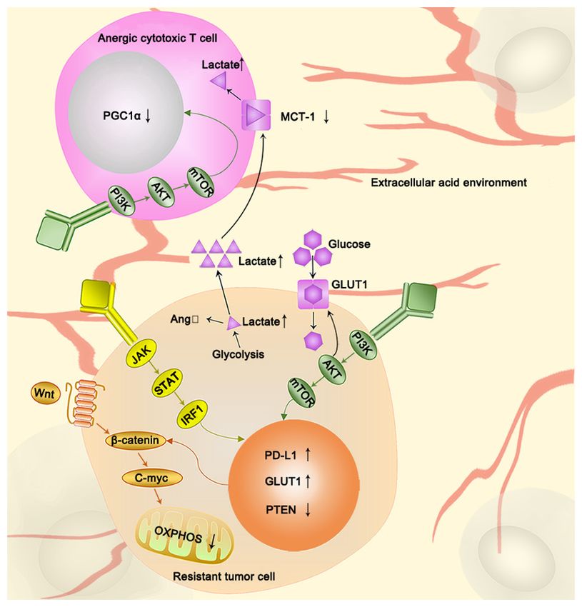
4. Glycolysis is associated with immunotherapy resistance and oxidative phosphorylation. Wnt ligand inhibits mitochondrial func‑
tion via the Wnt‑ β ‑catenin target gene, c‑myc. However, PTEN loss can
Recently, immunotherapy has been considered a milestone induce abnormal accumulation of β ‑catenin, and subsequently activate the
for cancer therapy. Programmed cell death 1 (PD1), PD1 Wnt‑β ‑catenin signaling pathway. AKT also plays key roles in regulating
immunotherapy resistance by decreasing PGC1α expression in tumor infil‑
ligand 1 (PD‑L1), and checkpoint molecules, such as cytotoxic trating lymphocytes and upregulating membrane localization of GLUT1.
T lymphocyte antigen 4, have been identified, and drugs During the lactate producing process, PTEN‑loss acts as an activator in
targeting them have been used on patients (118). Cancer immu‑ tumor cells, and increased lactate induces an extracellular acid environ‑
notherapy has achieved significant breakthroughs and success. ment that inhibits MCT‑1, which disturbs the metabolism in T‑cells. STAT,
signal transducer and activators of transcription; PD‑L1, programmed cell
However, drug resistance has deterred clinical progression for death‑ligand 1; PGC1α, peroxisome proliferator‑activated receptor gamma
several years. With a deeper understanding of immune mecha‑ coactivator 1‑α; GLUT, glucose and glucose transporters; MCT, monocar‑
nisms and immunotherapy efficacy, previous studies have boxylic transporters; OXPHOS, oxidative phosphorylation; IRF1, interferon
acknowledged that tumors are resistant to immunotherapy regulatory factor 1.
via interferon (IFN) signaling and antigen presentation, the
PI3K‑AKT‑mTOR axis, Wnt‑β‑catenin signaling, and deletion
of the tumor suppressor phosphoinositide phosphatase, PTEN, is an essential cause of tumor immunotherapy resistance (125).
which can activate different pathways (118‑121). In clinical studies, the chimeric anti‑CD20 antibody rituximab
Interferon binds IRF1 and the PD‑L1 promoter via the and IFN‑alpha 2a combined immunotherapy has been proven
Janus kinase 1 (JAK1)/JAK2‑signal transducer and activators effective (126,127). In another clinical study, the combina‑
of transcription 1 (STAT1)/STAT2/STAT3‑interferon regula‑ tion of interleukin‑2 and α‑IFN, based on a dose‑increasing
tory factor 1 (IRF1) axis, thereby regulating the expression of experiment, resulted in an increased response rate (128). These
PD‑L1 and causing resistance to immune checkpoint inhibi‑ results indicate that the IFN signaling pathway is a good
tors (119). The loss of copies of IFN‑γ‑mediated genes, such as potential target for combination therapy (Fig. 2).
IFNGR1, IRF‑1, JAK2 and IFNGR2 can cause metastatic mela‑ The metabolic reprogramming of tumor cells consumes a
noma resistance to ipilimumab (anti‑CTLA‑4 therapy) (122). lot of energy and nutrients, which means that immune cells
The loss of JAK1 and JAK2, IFN‑ γ pathway genes, is are in a state of nutrient depletion. The immune response to
associated with resistance to PD‑1 therapy (122). Notably, the tumor also has a significant negative effect (129). There
the IFN signaling pathway can also alter glycolysis to cause is considerable evidence that the Wnt‑ β ‑catenin signaling
chemotherapy resistance. Sustained STAT1 signaling causes pathway connects glycolysis to the immune response to the
chemotherapy resistance by increasing the expression of genes tumor. First, Wnt‑β ‑catenin signaling is closely associated
associated with glycolysis and OXPHOS (123) (Table II). with glycolysis (130). The Wnt‑β‑catenin target gene, c‑Myc,
STAT3 induces chemotherapy resistance by protecting regulates and controls cancer cell metabolism (130). Wnt5B,
mitochondrial oxidative phosphorylation and controlling the a Wnt ligand, has been demonstrated to inhibit mitochon‑
opening of mitochondrial permeability transition pores (124). drial functions in triple negative breast cancer cells through
In addition, IFN‑γ signaling is downregulated in highly glyco‑ c‑Myc (131). Secondly, Wnt‑β‑catenin signaling is also associ‑
lytic tumor cells, and the disruption of tumor IFN‑γ signaling ated with immunity. The Wnt ligand increases the expressionONCOLOGY LETTERS 21: 369, 2021 7
Table I. Overview of the clinical studies on the efficacy of glycolysis inhibitions in combination with chemotherapeutics.
Glycolysis Glycolysis
inhibition Chemotherapeutics target Cancer (Refs.)
Metformin Cisplatin PKM2 Osteosarcoma (36)
CPI‑613 Cytarabine/Mitoxantrone PDH Acute myeloid leukemia (58)
Lonidamine Epidoxorubicin HK2 Refractory epithelial ovarian cancer (23)
Cisplatin HK2 Ovarian cancer (24)
Curcumin Docetaxel HK2 Prostate cancer/Breast cancer (22)
PKM2, pyruvate kinase; PDH, pyruvate dehydrogenase; HK, hexokinase.
of β‑catenin in dendritic cells, and subsequently, the functions inhibitors can effectively reverse the immunotherapy resis‑
of Tregs and CD8+ T cells are activated, and antitumor immu‑ tance caused by the loss of PTEN (121). In PTEN‑deficient
nity is suppressed (120). Thus, targeting the Wnt‑β ‑catenin melanoma cells, some substrates upregulation of glycolysis,
signaling pathway has become a strategy that can both such as pyruvate and lactate, provoke overactivation, which is
target glycolysis and decrease immune resistance. Upon Wnt characterized by adoptive T cell therapy resistance (125). A
inhibition, the Wnt‑β‑catenin‑target gene, MCT1, is also down‑ clinical study has demonstrated that in PTEN‑deficient tumors,
regulated, leading to a reduction in tumor microenvironment following treatment of metastatic castration‑resistant prostate
acidity, maintaining antitumor immunity, and preventing cell cancer with the Akt inhibitor ipatasertib coupled with the
migration and metastasis (132). Accordingly, there is evidence CYP17 inhibitor abiraterone, radiographic progression‑free
that a combination of PD‑1 and Wnt inhibitors can increase survival was extended compared with tumors without PTEN
PD‑1 inhibitor efficacy (133) (Fig. 2). deficiency (139). This study further demonstrated that AKT
Overactivation of the pathological PI3K‑AKT‑mTOR inhibitors combined with antitumor drugs can play an impor‑
pathway is the primary mechanism underlying immune check‑ tant role in treating PTEN‑deficient tumors.
point inhibitor resistance (134). AKT plays a vital role in the Lactic acid levels are also associated with immune resis‑
tumor microenvironment, whereby it decreases the expression tance. Lactic acid can be derived from tumor‑associated
of peroxisome proliferator‑activated receptor gamma coacti‑ glycolysis and from activated immune cells and macro‑
vator 1‑α (PGC1α) in tumor‑infiltrating lymphocytes (TILs) phages (140). Lactate release in tumor cells also increases the
within the tumor microenvironment. PGC1α can also regulate expression of a myeloid‑specific lncRNA, HIF‑1α‑stabilizing
the biological function of mitochondria. Thus, a lack of this long non‑coding RNA (HISLA) in macrophages, and
protein can cause TIL energy exhaustion and decrease anti‑ elevated HISLA expression promotes aerobic glycolysis in
tumor immunity in the tumor microenvironment (135). Based tumor‑associated macrophages through extracellular vesicles
on in vitro experiments, the addition of PGC1 inhibits tumor (EV) transport, which forms a pre‑feedback loop (141) (Fig. 1).
growth and increases overall survival (135) (Fig. 2). Blocking EV‑mediated HISLA in vivo has been demonstrated
AKT can promote glucose uptake by increasing the to inhibit glycolysis and drug resistance in breast cancer (141).
membrane localization of facilitative GLUT1 and GLUT4. Furthermore, the excessive accumulation of lactic acid can
AKT also contributes to the phosphorylation of HK‑2 and stim‑ cause immunosuppression, leading to resistance to immu‑
ulates its translocation to the mitochondria (135). All of these notherapy. First, hypoxic tumor cells produce angiotensin II
changes can affect glycolysis and induce chemotherapy resis‑ (AngII) through an anoxia‑lactic acid‑chymase‑dependent
tance (135). Accordingly, PI3K‑AKT inhibitors have become mechanism (142). In the tumor microenvironment, local AngII
a focus of research because of their dual anti‑chemotherapy is associated with cancer cell evasion of immune surveil‑
and immunotherapy resistance effects (136). In phase I lance (143). In addition, the inhibition of AngII signaling may
clinical studies, the AKT inhibitor afuresertib combined with enhance tumor sensitivity to checkpoint immunotherapy (143)
carboplatin and paclitaxel exhibited promising results for the (Fig. 2). Secondly, lactic acid can impair the cytotoxic func‑
treatment of recurrent platinum‑resistant ovarian cancer (137). tion of T cells. The activation of T cells uses glycolysis and
However, clinical trials of AKT inhibitors for the immuno‑ relies on lactic acid secretion. The accumulation of intracel‑
therapy‑resistant disease remain to be performed (Fig. 2). lular lactic acid in the tumor cell causes an extracellular acidic
Glycolysis is also associated with immunotherapy resis‑ environment, leading to the inhibition of MCT‑1 (144). T cells
tance through PTEN‑deficiency. The abnormal accumulation cannot effectively secret lactate, and under these conditions,
of β‑catenin, caused by tumor‑specific mutations, such as the their metabolism is disordered, contributing to a significant
specific loss of PTEN from melanocytes in lung cancer, leads to reduction in cytotoxic activity, which severely affects T
activation of the Wnt‑β‑catenin signaling pathway (138) (Fig. 2). cell functionality (144) (Fig. 2). In addition, lactic acid can
PTEN‑deficiency‑associated and activated pathways can cause increase L‑arginine‑metabolizing enzyme arginase‑1 (ARG1)
immune resistance. PTEN deficiency facilitates the immune expression in macrophages and inhibit the antitumor immune
escape of melanoma by restricting T cell access to tumor cells response (145). Following treatment with DCA, the declined
and preventing T cells from killing cancer cells (121). PI3Kβ ARG1 mRNA expression can effectively reactivate the8 PENG et al: ASSOCIATION BETWEEN GLYCOLYSIS AND THERAPY RESISTANCE
Table II. Association between glycolysis and immune drug resistance.
Immunotherapy Glycolysis
resistance target target Cancer Correlation (Refs.)
IFN/STAT1 ENO1 Squamous cell carcinoma STAT1 increases the expression of ENO1 (123)
LDHA Squamous cell carcinoma STAT1 increases the expression of LDHA (123)
PKM2 Squamous cell carcinoma STAT1 increases the expression of PKM2 (123)
OXPHOS Mouse embryonal fibroblasts STAT3 protects OXPHOS (124)
Wnt‑β‑catenin PDK1 Colon cancer cells Blocking β‑catenin decreases PDK1 level (130)
OXPHOS TNBC Knockdown of WNT5B attenuates (131)
mitochondrial biogenesis and OXPHOS
PI3K‑AKT‑mTOR GLUT1/4 Mouse embryonal fibroblasts AKT increases the membrane localization (135)
pathway of facilitative GLUT1 and GLUT4
HK‑2 Mouse embryonal fibroblasts AKT contributes to the phosphorylation (135)
of HK‑2 and stimulates its translocation
to the mitochondria
PTEN‑deficiency Pyruvate/lactate Melanoma In PTEN‑deficient melanoma cells, (125)
some glycolysis substrates are upregulated,
such as pyruvate and lactate, which is
characterized by adoptive T cell therapy
resistance
IFN, interferon; STAT, signal transducer and activators of transcription; LDH, lactate dehydrogenase; PKM2, pyruvate kinase; OXPHOS,
oxidative phosphorylation; GLUT1, glucose transporters 1; HK, hexokinase.
immune state regulated by lactic acid and improve antitumor oxidative phosphorylation in cisplatin‑resistant cells were
immunotherapy benefits (145). higher than those in cisplatin‑sensitive cells (152). The
activation of OXPHOS is a typical feature of hepatocellular
5. Transition of glycolysis to OXPHOS enhances drug carcinoma cell resistance to DOX (153). Generally, increased
resistance OXPHOS can promote the occurrence of chemotherapy
resistance (150).
The Warburg effect indicates that tumor cells tend to undergo OXPHOS affects the treatment of tumors in several ways.
glycolysis regardless of aerobic and anaerobic conditions, As it results in the production of a large amount of ATP,
which implies that mitochondrial dysfunction is a feature of this is bound to stimulate the activity of some transporters,
tumor cells (146). However, recently, this statement has been one of which is drug transporters. In breast cancer cells, the
challenged (147). Several tumor cells have been reported to continuous supply of ATP derived from OXPHOS is utilized
have metabolic plasticity, indicating a transformation from by ABC transporters, leading to the outflow of DOX and the
glycolysis to mitochondrial OXPHOS, leading to the produc‑ induction of an MDR phenotype (148). Tumor stem cells are
tion of vast amounts of energy and resistance to drugs (148). also associated with drug resistance caused by OXPHOS.
Recent evidence suggests that cancer cells can obtain glycol‑ Increased OXPHOS mediated by mitochondria can stimulate
ysis/OXPHOS mixed phenotypes, in which the ATP production tumor stem cells to expand, conferring resistance to tumor
is an outcome from both glycolysis and OXPHOS to support cells (154). Furthermore, NANOG, a stem cell marker, inhibits
physiological activity of cells (149). In addition, cells with this mitochondrial OXPHOS genes and promotes sorafenib
characteristic are more likely to acquire drug resistance (150). resistance (155).
When lactic acid increases, it can be used as an energy source Several drugs inhibit the occurrence and development of
by adjacent cancer cells to activate mitochondria and stimulate tumors, and they can also affect OXPHOS. Some drugs, like
OXPHOS (151) (Fig. 1). cytarabine, 5‑fluorouracil, TKIs, MAPKi and BRAFi can
Such changes in OXPHOS can affect the drug resistance promote OXPHOS activity and increase drug resistance in the
of tumor cells. Following chemotherapy, Farge et al (150) mitochondria, whereas others, such as anthracyclines, etopo‑
described a new method to identify and study acute myeloid side, sorafenib, paclitaxel and staurosporine, significantly
leukemia (AML) cells remaining in the bone marrow. The decrease OXPHOS activity in the mitochondria (147). For
results demonstrated that OXPHOS is increased in AML cells those drugs that promote OXPHOS activity in mitochondria,
remaining in the bone marrow of mice following cytarabine it is necessary to find a better way to solve the associated drug
therapy, and that the inhibition of OXPHOS can re‑sensitize resistance. Metformin is a type of mitochondrial inhibitor and
AML cells to cytarabine. In epithelial ovarian cancer, the combining it with cisplatin can attenuate cisplatin resistance
oxygen consumption rate, mitochondrial respiration and in epithelial ovarian cancer cells (152). Metformin can alsoONCOLOGY LETTERS 21: 369, 2021 9
be combined with TKIs to overcome tumor chemotherapy 7. Future direction and perspectives
resistance (147). In addition, targeting mitochondrial respira‑
tion and HIF‑1α may reverse tumor cell chemotherapeutic Regarding these glucose metabolic processes, targeted inhibi‑
resistance (154). When the anoxic environment is destroyed tors may be used in combination with chemotherapy reagents
and the HIF1α pathway is blocked, sex‑determining region Y or immune checkpoint inhibitors in the future. However, due
(SRY)‑Box2 drives OXPHOS reprogramming, which helps to the lack of tumor drug resistance markers, targeted inhibitor
tumor cells obtain an invasive oxidative tumor phenotype prognostic indexes, and the specificity of these inhibitors, their
and enhance drug resistance and metastatic ability (156). clinical application is profoundly limited. In general, altered
glycolysis, as a ubiquitous feature of drug‑resistant tumor cells,
6. Conclusions represents a promising target, and novel strategy to overcome
drug resistance clinically.
Changes in glycolysis, particularly increases in key enzymes
and intermediates of this pathway, can affect the sensitivity Acknowledgements
of tumors to chemotherapeutic reagents, resulting in an
increase in ATP production, and providing sufficient energy Not applicable.
for the biological activity of tumor cells (14,69). This process
enhances the repair of DNA damage, increases the phosphory‑ Funding
lation, translocation into the nucleus, and autophagy‑associated
activity of enzymes, and causes drug resistance (157). In addi‑ The present review was supported by grants from the
tion, several clinical trials have been performed to investigate National Natural Science Foundation of China (grant
the therapeutic effect of glycolysis‑targeting therapy combined nos. 81672612 and 81572607) and the Project of Invigorating
with clinical first‑and second‑line chemotherapeutic drugs on Health Care through Science, Technology and Education
tumors (Table I). (The Project of Jiangsu Provincial Medical Youth Talent;
Recently, several studies have demonstrated that the role of grant no. QNRC2016095).
mitochondria in tumor metabolism is becoming essential. The
reverse Warburg effect emerged, indicating that the increase in Availability of data and materials
lactic acid as an energy material can be converted into pyruvate
in the mitochondria and enhance mitochondrial activity and Not applicable.
OXPHOS in adjacent cells (117). In addition, tumor cells also
have metabolic plasticity. Glycolysis can be moderately trans‑ Authors' contributions
formed into OXPHOS when the external environment changes
or there is plenty of oxygen around the tumor cells. This trans‑ JP and YC designed the present review and drafted the initial
formation is closely associated with chemotherapy (118,148). manuscript. XW and YH analyzed the data. SX, WZ and
Increased mitochondrial activity and OXPHOS results in the SW revised the review for important intellectual content. ZF
production of higher levels of ATP and NADPH. High levels reviewed and critiqued the review following revisions. HX
of ATP provide a vast amount of energy to tumor cells, and designed the present review, edited, reviewed and critiqued
NADPH is a key antioxidant that can decrease ROS damage the manuscript. All authors have read and approved the final
to tumor cells (158). manuscript.
With an increase in studies on tumor immunity, immune
checkpoint inhibitors have been developed for clinical appli‑ Ethics approval and consent to participate
cations (149). However, immunotherapy resistance is remains
a major challenge. Based on a broadened understanding of Not applicable.
tumor immunity, it is apparent that immunotherapy resistance
is closely associated with glucose metabolism (125). The Patient consent for publication
PI3K‑AKT‑mTOR axis, PTEN deficiency, IFN signaling and
the Wnt‑β‑catenin signaling pathway can affect corresponding Not applicable.
enzymes involved in glycolysis and enhance immunotherapy
resistance (123,125,131,135). Conversely, the release of lactic Competing interests
acid, a glycolysis intermediate metabolite, can promote the
occurrence of immunotherapy resistance. The interaction The authors declare that they have no competing interests.
between glucose metabolism and immunotherapy resistance
forms a positive feedback pathway and constitutes an impor‑ References
tant factor in tumor drug resistance (141). Currently, there are a
few studies on the effect of the PI3K‑AKT‑mTOR axis, PTEN 1. Zaal EA and Berkers CR: The influence of metabolism on drug
response in cancer. Front Oncol 8: 500, 2018.
deficiency, IFN signaling, Wnt‑β‑catenin signaling pathway 2. Warburg O, Wind F and Negelein E: The metabolism of tumors
targeting agents on immunotherapy drug resistance, and in the body. J Gen Physiol 8: 519‑530, 1927.
chemotherapy resistance. The interdisciplinary study on tumor 3. Bonuccelli G, Whitaker‑Menezes D, Castello‑Cros R, Pavlides S,
Pestell RG, Fatatis A, Witkiewicz AK, Vander Heiden MG, Migneco G,
glycolysis and immunotherapy resistance should endeavor to Chiavarina B, et al: The reverse Warburg effect: Glycolysis inhibitors
receive more attention to proceed to understand the molecular prevent the tumor promoting effects of caveolin‑1 deficient cancer
mechanisms involved. associated fibroblasts. Cell Cycle 9: 1960‑1971, 2010.10 PENG et al: ASSOCIATION BETWEEN GLYCOLYSIS AND THERAPY RESISTANCE
4. Holohan C, Van Schaeybroeck S, Longley DB and Johnston PG: 24. De Lena M, Lorusso V, Latorre A, Fanizza G, Gargano G,
Cancer drug resistance: An evolving paradigm. Nat Rev Caporusso L, Guida M, Catino A, Crucitta E, Sambiasi D and
Cancer 13: 714‑726, 2013. Mazzei A: Paclitaxel, cisplatin and lonidamine in advanced
5. Boumahdi S and de Sauvage FJ: The great escape: Tumour ovarian cancer. A phase II study. Eur J Cancer 37: 364‑368, 2001.
cell plasticity in resistance to targeted therapy. Nat Rev Drug 25. Huang K, Liang Q, Zhou Y, Jiang LL, Gu WM, Luo MY, Tang YB,
Discov 19: 39‑56, 2020. Wang Y, Lu W, Huang M, et al: A novel allosteric inhibitor of
6. Gershon O, Ezenwa NE and Osabohien R: Implications of oil price phosphoglycerate mutase 1 suppresses growth and metastasis of
shocks on net oil‑importing African countries. Heliyon 5: e02208, 2019. non‑small‑cell lung cancer. Cell Metab 30: 1107‑1119.e8, 2019.
7. Woo YM, Shin Y, Lee EJ, Lee S, Jeong SH, Kong HK, Park EY, 26. Ren F, Wu H, Lei Y, Zhang H, Liu R, Zhao Y, Chen X, Zeng D,
Kim HK, Han J, Chang M and Park JH: Inhibition of aerobic Tong A, Chen L, et al: Quantitative proteomics identification
glycolysis represses Akt/mTOR/HIF‑1α axis and restores of phosphoglycerate mutase 1 as a novel therapeutic target in
tamoxifen sensitivity in antiestrogen‑resistant breast cancer cells. hepatocellular carcinoma. Mol Cancer 9: 81, 2010.
PLoS One 10: e0132285, 2015. 27. Usuba T, Ishibashi Y, Okawa Y, Hirakawa T, Takada K and
8. Komurov K, Tseng JT, Muller M, Seviour EG, Moss TJ, Yang L, Ohkawa K: Purification and identification of monoubiq‑
Nagrath D and Ram PT: The glucose‑deprivation network coun‑ uitin‑phosphoglycerate mutase B complex from human colorectal
teracts lapatinib‑induced toxicity in resistant ErbB2‑positive cancer tissues. Int J Cancer 94: 662‑668, 2001.
breast cancer cells. Mol Syst Biol 8: 596, 2012. 28. Liu L, Wang S, Zhang Q and Ding Y: Identification of poten‑
9. Ruprecht B, Zaal EA, Zecha J, Wu W, Berkers CR, Kuster B and tial genes/proteins regulated by Tiam1 in colorectal cancer by
Lemeer S: Lapatinib resistance in breast cancer cells is accompa‑ microarray analysis and proteome analysis. Cell Biol Int 32:
nied by phosphorylation‑mediated reprogramming of glycolysis. 1215‑1222, 2008.
Cancer Res 77: 1842‑1853, 2017. 29. Chen G, Gharib TG, Wang H, Huang CC, Kuick R, Thomas DG,
10. He M, Jin Q, Chen C, Liu Y, Ye X, Jiang Y, Ji F, Qian H, Gan D, Shedden KA, Misek DE, Taylor JM, Giordano TJ, et al: Protein
Yue S, et al: The miR‑186‑3p/EREG axis orchestrates tamox‑ profiles associated with survival in lung adenocarcinoma. Proc
ifen resistance and aerobic glycolysis in breast cancer cells. Natl Acad Sci USA 100: 13537‑13542, 2003.
Oncogene 38: 5551‑5565, 2019. 30. Chen SY, Cai JX, Zhang WP, Zhang XW, Hu SS, Lu J, Xing JF
11. Icard P, Shulman S, Farhat D, Steyaert JM, Alifano M and and Dong YL: Proteomic identification of differentially expressed
Lincet H: How the Warburg effect supports aggressiveness and proteins associated with the multiple drug resistance in methotrexate-
drug resistance of cancer cells? Drug Resist Updat 38: 1‑11, 2018. resistant human breast cancer cells. Int J Oncol 45: 448-458, 2014.
12. Apicella M, Giannoni E, Fiore S, Ferrari KJ, Fernández‑Pérez D, 31. Wang HJ, Pochampalli M, Wang LY, Zou JX, Li PS, Hsu SC,
Isella C, Granchi C, Minutolo F, Sottile A, Comoglio PM, et al: Wang BJ, Huang SH, Yang P, Yang JC, et al: KDM8/JMJD5 as a
Increased lactate secretion by cancer cells sustains non-cell- dual coactivator of AR and PKM2 integrates AR/EZH2 network
autonomous adaptive resistance to MET and EGFR targeted and tumor metabolism in CRPC. Oncogene 38: 17‑32, 2019.
therapies. Cell Metab 28: 848‑865 e6, 2018. 32. Qian Y, Bi L, Yang Y and Wang D: Effect of pyruvate kinase
13. Varghese E, Samuel SM, Liskova A, Samec M, Kubatka P M2‑regulating aerobic glycolysis on chemotherapy resistance of
and Busselberg D: Targeting glucose metabolism to overcome estrogen receptor‑positive breast cancer. Anticancer Drugs 29:
resistance to anticancer chemotherapy in breast cancer. Cancers 616‑627, 2018.
(Basel) 12: 2252, 2020. 33. Tian S, Li P, Sheng S and Jin X: Upregulation of pyruvate kinase
14. Liu X, Miao W, Huang M, Li L, Dai X and Wang Y: Elevated M2 expression by fatty acid synthase contributes to gemcitabine
hexokinase II expression confers acquired resistance resistance in pancreatic cancer. Oncol Lett 15: 2211‑2217, 2018.
to 4‑hydroxytamoxifen in breast cancer cells. Mol Cell 34. Wong TL, Ng KY, Tan KV, Chan LH, Zhou L, Che N, Hoo RLC,
Proteomics 18: 2273‑2284, 2019. Lee TK, Richard S, Lo CM, et al: CRAF methylation by PRMT6
15. Krasnov GS, Dmitriev AA, Lakunina VA, Kirpiy AA and regulates aerobic glycolysis driven hepatocarcinogenesis via
Kudryavtseva AV: Targeting VDAC‑bound hexokinase II: A ERK‑dependent PKM2 nuclear relocalization and activation.
promising approach for concomitant anti‑cancer therapy. Expert Hepatology 71: 1279‑1296, 2020.
Opin Ther Targets 17: 1221‑1233, 2013. 35. Cheng C, Xie Z, Li Y, Wang J, Qin C and Zhang Y: PTBP1
16. Yang T, Ren C, Qiao P, Han X, Wang L, Lv S, Sun Y, Liu Z, Du Y knockdown overcomes the resistance to vincristine and oxali‑
and Yu Z: Correction: PIM2‑mediated phosphorylation of hexo‑ platin in drug‑resistant colon cancer cells through regulation of
kinase 2 is critical for tumor growth and paclitaxel resistance in glycolysis. Biomed Pharmacother 108: 194‑200, 2018.
breast cancer. Oncogene 39: 720‑721, 2020. 36. Shang D, Wu J, Guo L, Xu Y, Liu L and Lu J: Metformin increases
17. Yang T, Ren C, Qiao P, Han X, Wang L, Lv S, Sun Y, Liu Z, Du Y sensitivity of osteosarcoma stem cells to cisplatin by inhibiting
and Yu Z: PIM2‑mediated phosphorylation of hexokinase 2 is expression of PKM2. Int J Oncol 50: 1848‑1856, 2017.
critical for tumor growth and paclitaxel resistance in breast 37. Ge X, Zhao Y, Dong L, Seng J, Zhang X and Dou D: NAMPT
cancer. Oncogene 37: 5997‑6009, 2018. regulates PKM2 nuclear location through 14‑3‑3 ζ: Conferring
18. Fan K, Fan Z, Cheng H, Huang Q, Yang C, Jin K, Luo G, Yu X resistance to tamoxifen in breast cancer. J Cell Physiol 234:
and Liu C: Hexokinase 2 dimerization and interaction with 23409‑23420, 2019.
voltage‑dependent anion channel promoted resistance to cell 38. Calabretta S, Bielli P, Passacantilli I, Pilozzi E, Fendrich V,
apoptosis induced by gemcitabine in pancreatic cancer. Cancer Capurso G, Fave GD and Sette C: Modulation of PKM alterna‑
Med 8: 5903‑5915, 2019. tive splicing by PTBP1 promotes gemcitabine resistance in
19. Rai Y, Yadav P, Kumari N, Kalra N and Bhatt AN: Hexokinase pancreatic cancer cells. Oncogene 35: 2031‑2039, 2016.
II inhibition by 3‑bromopyruvate sensitizes myeloid leukemic 39. Wang X, Zhang F and Wu XR: Inhibition of pyruvate kinase M2
cells K‑562 to anti‑leukemic drug, daunorubicin. Biosci Rep 39: markedly reduces chemoresistance of advanced bladder cancer
BSR20190880, 2019. to cisplatin. Sci Rep 7: 45983, 2017.
20. Li Z, Tang X, Luo Y, Chen B, Zhou C, Wu X, Tang Z, Qi X, 40. Gatti M, Solari A, Pattarozzi A, Campanella C, Thellung S,
Cao G, Hao J, et al: NK007 helps in mitigating paclitaxel resis‑ Maniscalco L, De Maria R, Würth R, Corsaro A, Bajetto A, et al:
tance through p38MAPK activation and HK2 degradation in In vitro and in vivo characterization of stem‑like cells from
ovarian cancer. J Cell Physiol: Feb 20, 2019 (Epub ahead of print). canine osteosarcoma and assessment of drug sensitivity. Exp
Cell Res 363: 48‑64, 2018.
21. Geng C, Li J, Ding F, Wu G, Yang Q, Sun Y, Zhang Z, Dong T and 41. Ramos‑Penafiel C, Olarte‑Carrillo I, Ceron‑Maldonado R,
Tian X: Curcumin suppresses 4‑hydroxytamoxifen resistance in Rozen‑Fuller E, Kassack‑Ipiña JJ, Meléndez‑Mier G,
breast cancer cells by targeting SLUG/Hexokinase 2 pathway. Collazo‑Jaloma J and Martínez‑Tovar A: Effect of metformin on
Biochem Biophys Res Commun 473: 147‑153, 2016. the survival of patients with ALL who express high levels of the
22. Mahammedi H, Planchat E, Pouget M, Durando X, Curé H, Guy L, ABCB1 drug resistance gene. J Transl Med 16: 245, 2018.
Van‑Praagh I, Savareux L, Atger M, Bayet‑Robert M, et al: The 42. Shanmugasundaram K, Nayak BK, Friedrichs WE, Kaushik D,
new combination docetaxel, prednisone and curcumin in patients Rodriguez R and Block K: NOX4 functions as a mitochondrial
with castration‑resistant prostate cancer: A pilot phase II study. energetic sensor coupling cancer metabolic reprogramming to
Oncology 90: 69‑78, 2016. drug resistance. Nat Commun 8: 997, 2017.
23. Gadducci A, Brunetti I, Muttini MP, Fanucchi A, Dargenio F, 43. Lu CW, Lin SC, Chen KF, Lai YY and Tsai SJ: Induction of
Giannessi PG and Conte PF: Epidoxorubicin and lonidamine pyruvate dehydrogenase kinase‑3 by hypoxia‑inducible factor‑1
in refractory or recurrent epithelial ovarian cancer. Eur J promotes metabolic switch and drug resistance. J Biol Chem 283:
Cancer 30A: 1432‑1435, 1994. 28106‑28114, 2008.You can also read