Neutrophil Extracellular Traps (NETs) in Cancer Invasion, Evasion and Metastasis
←
→
Page content transcription
If your browser does not render page correctly, please read the page content below
cancers
Review
Neutrophil Extracellular Traps (NETs) in Cancer Invasion,
Evasion and Metastasis
Urszula Demkow

                                          Department of Laboratory Diagnostics and Clinical Immunology of Developmental Age,
                                          Medical University of Warsaw, 02-091 Warsaw, Poland; urszula.demkow@wum.edu.pl

                                          Simple Summary: This review focuses on the pro-tumorigenic action of neutrophil extracellular
                                          traps (NETs). NETs were found in various samples of human and animal tumors. The role of the
                                          NETs in tumor development increasingly includes cancer immunoediting and interactions between
                                          immune system and cancer cells. NETs awake dormant cancer cells, play a key regulatory role in
                                          the tumor microenvironment, and exacerbate tumor aggressiveness by enhancing cancer migration
                                          and invasion capacity. Furthermore, NETs induce the epithelial to mesenchymal transition in tumor
                                          cells. NET proteinases can also degrade the extracellular matrix, promoting cancer cell extravasation.
                                          Moreover, NETs can entrap circulating cancer cells and, in that way, facilitate metastasis. A better
                                          understanding of the crosstalk between cancer and NETs can help to devise novel approaches to the
                                          therapeutic interventions that block cancer evasion mechanisms and prevent metastatic spread.

                                          Abstract: The present review highlights the complex interactions between cancer and neutrophil
                                          extracellular traps (NETs). Neutrophils constitute the first line of defense against foreign invaders
                                          using major effector mechanisms: phagocytosis, degranulation, and NETs formation. NETs are
                                composed from decondensed nuclear or mitochondrial DNA decorated with proteases and various

                                          inflammatory mediators. Although NETs play a crucial role in defense against systemic infections,
Citation: Demkow, U. Neutrophil           they also participate in non-infectious conditions, such as inflammation, autoimmune disorders, and
Extracellular Traps (NETs) in Cancer      cancer. Cancer cells recruit neutrophils (tumor-associated neutrophils, TANs), releasing NETs to the
Invasion, Evasion and Metastasis.         tumor microenvironment. NETs were found in various samples of human and animal tumors, such
Cancers 2021, 13, 4495. https://          as pancreatic, breast, liver, and gastric cancers and around metastatic tumors. The role of the NETs
doi.org/10.3390/cancers13174495
                                          in tumor development increasingly includes cancer immunoediting and interactions between the
                                          immune system and cancer cells. According to the accumulated evidence, NETs awake dormant
Academic Editor:
                                          cancer cells, causing tumor relapse, as well as its unconstrained growth and spread. NETs play a
Hendrik Ungefroren
                                          key regulatory role in the tumor microenvironment, such as the development of distant metastases
Received: 26 July 2021
                                          through the secretion of proteases, i.e., matrix metalloproteinases and proinflammatory cytokines.
Accepted: 2 September 2021                NETs, furthermore, directly exacerbate tumor aggressiveness by enhancing cancer migration and
Published: 6 September 2021               invasion capacity. The collected evidence also states that through the induction of the high-mobility
                                          group box 1, NETs induce the epithelial to mesenchymal transition in tumor cells and, thereby,
Publisher’s Note: MDPI stays neutral      potentiate their invasiveness. NET proteinases can also degrade the extracellular matrix, promoting
with regard to jurisdictional claims in   cancer cell extravasation. Moreover, NETs can entrap circulating cancer cells and, in that way, facilitate
published maps and institutional affil-   metastasis. NETs directly trigger tumor cell proliferation through their proteases or activating signals.
iations.                                  This review focused on the pro-tumorigenic action of NETs, in spite of its potential to also exhibit
                                          an antitumor effect. NET components, such as myeloperoxidase or histones, have been shown to
                                          directly kill cancer cells. A better understanding of the crosstalk between cancer and NETs can help
                                          to devise novel approaches to the therapeutic interventions that block cancer evasion mechanisms
Copyright: © 2021 by the author.          and prevent metastatic spread. This review sought to provide the most recent knowledge on the
Licensee MDPI, Basel, Switzerland.        crosstalk between NETs and cancer, and bring more profound ideas for future scientists exploring
This article is an open access article    this field.
distributed under the terms and
conditions of the Creative Commons
                                          Keywords: cancer; neutrophil extracellular traps; metastasis; evasion
Attribution (CC BY) license (https://
creativecommons.org/licenses/by/
4.0/).

Cancers 2021, 13, 4495. https://doi.org/10.3390/cancers13174495                                            https://www.mdpi.com/journal/cancers
Cancers 2021, 13, 4495 2 of 17
1. Neutrophils and NETs
Polymorphonuclear neutrophils (PMNs), the most abundant white blood cells, are
frontline fighters against invading microorganisms. PMNs destroys pathogens, or other
endogenous or exogenous factors, using a combination of mechanisms, including phago-
cytosis, oxidative bursts, the release of antimicrobial mediators, and the production of
neutrophil extracellular traps (NETs) [1]. NETs are web-like structures built from nuclear or
mitochondrial DNA fibers, decorated with anti-microbial enzymes and histones, which are
released to entrap and kill pathogens [2]. Besides their role as an anti-microbial weapon,
NETs create a physical barrier for both pathogens and immune cells. The process of NET
formation in its classical form is called NETosis and has been defined as a type of regulated
cell death distinguished from apoptosis and necrosis [3]. Further studies have described an
alternative pathway of a non-cell-death NETs generation, named vital NETosis. NET release
is initiated by an oxidative burst via raf-MEK-ERK activation of NADPH oxidase. Subse-
quently, neutrophil elastase (NE) translocates from azurophil granules into the nucleus,
where it instigates chromatin breakdown through histone hydrolysis. Further observations
have suggested that myeloperoxidase (MPO) has also been implicated in chromatin decon-
densation and the rupturing of the nuclear envelope. Chromatin decompaction is further
supported by peptidyl arginine deiminase 4 (PAD4)—a protein-citrullinating enzyme that
enters the nucleus to deiminate specific arginine residues on histones, resulting in the
loss of positive charge from the transformed arginine residues and the disassembling of
nucleosome structure [4]. Crucial steps in NET formation include nuclear swelling, nuclear
envelope disintegration, the mixing of nucleic acids and granule proteins within a large
intracellular vacuole, the spilling of nuclear content into the cytoplasm, and, finally, cell
membrane breakdown [5].
2. NETs—Friend or Foe?
NETs not only act as a host defense mechanism, but also play a pivotal role in infec-
tious and non-infectious conditions [5–7]. While the beneficial effects of NETs in fighting
pathogens have already been largely described, the detrimental role of NETs is rapidly be-
ginning to emerge. Netting neutrophils play a significant role in the pathogenesis of various
diseases, such as systemic lupus erythematosus, small vessel vasculitis, rheumatoid arthri-
tis, preeclampsia, cystic fibrosis, psoriasis, and, as recently described, in Covid-19 [5–7].
NET generation and degradation in patients with granulomatosis with polyangiitis and
systemic lupus erythematosus is impaired [6,7]. NETs are also implicated in various other
pathological processes, such as coagulation disorders, diabetes, atherosclerosis, wound
healing, and periodontitis [8].
2.1. Heterogeneity of Neutrophils
Although neutrophils have long been considered as a terminally differentiated, ho-
mogenous cell population of the innate immune response, different studies started to
highlight the heterogeneity of their phenotypes and the versatility of their functions [5].
The phenotype and function of a resident neutrophil is the result of a specific maturation
program and/or inflammatory signals from surrounding milieu (cytokines, chemokines,
enzymes, growth factors, various lipids and proteins) translating various environmental
signals into specific gene and transcription factor programs. This paradigm is supported
by the presence of distinct neutrophil precursors at different stages of development con-
tributing to the diversity of mature neutrophils [9].
2.2. Tumor-Infiltrating Neutrophils
The tumor microenvironment (TME) comprises different non-malignant cell types
and an extracellular matrix (ECM), altogether named the stroma. The stroma consists of the
basement membrane, immune cells, cancer-associated fibroblasts (CAFs), pericytes, and
vascular endothelial cells [10]. Tumor cell proliferation, the evasion of immune surveillance,
and the spread and metastasis are affected by the changes in the composition, function,Cancers 2021, 13, 4495 3 of 17
and communication between all stromal components [10,11]. Amongst various immune
cells within the TME, such as dendritic cells, lymphocytes, macrophages, granulocytes,
and fibroblasts, infiltrating neutrophils, in concert with other cell types, play a prominent
role in cancer development [11]. However, the pro-tumor functions of tumor-infiltrating
neutrophils have only recently come to the light. Consistently, various mediators pro-
duced by tumor or stromal cells stimulate granulopoiesis, neutrophil release from the bone
marrow, and the migration of these cells [11]. These mediators include growth factors:
G-CSF, GM-CSF and CXC chemokines, and CCL3 [11]. Recently, different studies started
to highlight that cancer cells release chemokines attracting neutrophils to tumor microenvi-
ronments [12,13]. In the recent past, tumor-associated neutrophils (TANs) have emerged as
important contributors to the tumor biology. However, consistent and continuous evidence
has confirmed that these cells appear to play an important role in the entire process of
cancerogenesis, followed by the metastatic spread to distant organs [12]. TANs are capable
of polarization into two populations (N1 and N2) according to cytokine production patterns
and effector functions. These two populations present either an anti-tumorigenic “N1”
phenotype or, fed by TGFβ, a pro-tumorigenic “N2” phenotype [12]. Both N1 and N2
cells bear similar surface markers to peripheral blood neutrophils, i.e., CD66b+, CD11b+,
CD15+, CD16+, HLA-DR−, and arginase-1+ [13]. In fact, due to the often-shared cell mor-
phology and the overlap of the expression of these surface markers between the different
functional groups, it is difficult to clearly distinguish between the subtypes N1 and N2 [13].
N1 neutrophils can effectively eliminate tumor cells via lysis, indirect cytotoxicity or the
induction of tumor cell apoptosis. N1 cells exhibit increased cytotoxicity and a reduced
immunosuppressive ability due to the increased release of TNFα, Fas, ICAM-1, and ROS,
and through a decreased arginase expression [14]. On the other hand, N2 cells promote
immunosuppression, support tumor growth, invasion, epithelial–mesenchymal transition
(EMT), angiogenesis and the metastasis of cancer cells [15]. N2 neutrophils express high
levels of arginase, MMP-9 VEGF, and numerous chemokines (for example CXCL4, CCL2,
and CCL5). The affluence of these cells corresponds with poor clinical outcomes [15]. The
tumor-secreted TGF-β was shown to transform N1 TANs (tumor-suppressive phenotype)
into N2 TANs (tumor-promoting phenotype) [15]. Infiltrating neutrophils continue to pro-
mote tumor development by secreting pro-inflammatory and pro-angiogenic chemokines
and cytokines, such as matrix metallopeptidase 9 (MMP9) and interleukin 6 (IL-6) [14,15].
Circulating tumor cells shed from the primary tumor sites are disseminated via blood
or lymphatic vessels and reach distant organs. In a recent study, neutrophils emerged
as important players supporting circulating tumor cells survival during hematogenous
dissemination [16]. Furthermore, it was confirmed that neutrophils escort circulating tumor
cells, increasing the dynamics of cell cycle progression [16]. Wculek et al. have identified
neutrophils as the main drivers in establishing the pre-metastatic microenvironment in
different murine breast cancer models [17].
3. NETs Are Present in Tumor Microenvironment
The discovery of NETs has created a completely new field of investigation in oncology.
The first evidence of NET formation by tumor-associated neutrophils in human tissues
came from a histopathological analysis of diagnostic biopsies from Ewing sarcoma. Out
of eight tissue samples, TANs were found in six specimens and NETs in two patients. In
this study, NET formation was associated with relapse and metastatic disease, despite
chemotherapy treatment [18]. Several further studies revealed the presence of NETs in
peripheral blood and tumor specimens from animals and cancer patients. NETs were
found in tumor samples from primary and metastatic sites. Murine neutrophils from
animals with leukemia, mammary, and lung cancer were more prone to release NETs
compared to granulocytes of healthy mice. Overproduction of NETs went in parallel with
activation of intravascular coagulation and the presence microvascular thrombosis in these
animals [19,20]. Currently, there are scarce published data regarding the occurrence of
NETs in clinical samples from patients with hematological malignancies [21,22]. Nie et al.Cancers 2021, 13, 4495 4 of 17
reported that neutrophils are prone to produce NETs in hematological malignancies, such
as chronic lymphocytic leukemia, and participate in disease progression via TLR9 sig-
naling [21]. Cedervall et al. discovered that the number of netting neutrophils in the
kidneys and hearts of tumor-bearing animals (MMTV-PyMT—breast cancer and RIP1-
Tag2—insulinoma) is increased. The kidney involvement in these animals is accompanied
with concomitant kidney insufficiency. DNase (NET-degrading enzyme) treatment recov-
ered renal function in experimental animals, pointing to the pathogenic role of NETs in
acute kidney damage [23]. In another in vitro study, it was demonstrated that extracellular
RNAs from Lewis lung carcinoma cells induced the release of NETs [24]. TANs were active
in the low-oxygen environment with the presence of proinflammatory cytokines, such as
IL-8, IL-1β, and G-CSF [25,26]. The molecular mechanism of NET formation in TME is also
dependent on the nuclear factor high mobility group box 1 (HMGB1), which, by binding to
TLR4, induce activation of p38 MAPK/ERK signaling pathways, further contributing to
the excessive release of inflammatory cytokines [27].
4. Circulating NET Markers in Cancer Patients
The plasma NET markers include citrullinated histones (H3Cit-DNA), cell-free DNA
(cfDNA), neutrophil elastase (NE), and nucleosomes [28]. All circulating markers can be
easily measured in human plasma. NET marker concentrations in the plasma of different
cancer patients, including lung, pancreatic, and bladder cancer, were found to be higher
than in healthy controls [29]. In lung cancer patients, Li et al. demonstrated the presence of
NETs in lung tissues, peripheral blood, and sputum [24]. The circulating levels of NETs
(DNA-histone complex, double-stranded DNA, NE) were measured in the peripheral
blood of liver cancer patients, along with contact system activation markers. Both NETs
and contact system activation markers were higher in cancer patients than in healthy
volunteers [30]. In accord with the above-mentioned observations, Rosell et al. confirmed
the presence of circulating markers of neutrophil activation and NET formation (NE,
H3Cit-DNA) in 106 patients with terminal cancer with concomitant hypercoagulation
and hyperfibrinolysis. They found that NET markers had a prognostic value in terminal
cancer patients. NE and H3Cit-DNA were both associated with a poor clinical outcome.
Interestingly, although the markers of coagulation and fibrinolysis were elevated, they did
not have a prognostic significance in the patients of this study. Moreover, the correlations
between NETs and coagulation/fibrinolysis markers were weak or non-existing. This
observation suggests that NETs contribute to poor prognosis in terminal cancer through
mechanisms independent of thrombosis [31]. Consistently Oklu et al. [29] detected high
levels of nucleosomes, cfDNA, DNase-1, the thrombin-antithrombin III (TAT) complex, as
well as endonuclease-G and its activity in plasma from cancer patients. Additionally, NETs
were found and quantified by fluorescent immunohistochemistry in tumor tissue samples
and venous thrombi of cancer patients. These authors have found that plasma samples
from cancer patients contained higher levels of nucleosomes and free-circulating DNA
compared to the non-cancer group. A Western blot analysis revealed a significantly lower
level of DNase-1 protein that paralleled a lower nuclease activity in plasma samples from
cancer patients compared to non-cancer subjects. Venous thrombi from cancer patients and
tumor tissue from liver and lung cancer also showed increased presence of NETs. However,
high levels of NETs in cancer patients did not correlate with TAT complex activation or the
incidence of venous thrombosis in these patients [29]. The objectively measured diagnostic,
prognostic, and predictive biomarkers of tumors are desperately needed in clinical practice.
The assays quantifying the circulating NET markers should be developed into commercially
available laboratory tests validated in human plasma samples that are easily accessible.
This will allow for the potential clinical implementation of such tests as prognostic tools, or
as guides to the decision-making process necessary in cancer therapy.Cancers 2021, 13, 4495 5 of 17
5. NETs Fuel Cancer Progression and Indicate Poor Prognosis
Theoretically, NETs might have potential anti-tumorigenic effects through the direct
killing of cancer cells or the activation of the immune system. Like histones, NE, and MPO
in vitro, NET components destroy tumor cells and block tumor growth and metastasis
formation [32–34]. Surprisingly, accumulating evidence suggests that NETs exert multi-
faceted protumorigenic effects. Different studies have highlighted the prominent role of
this structure in the progression and enhancement of metastatic potential of animal and
human tumors. Richardson et al. confirmed the association between in vitro NET release
by stimulated neutrophils and the poor prognosis in colorectal cancer patients [35]. The
role of NETs in tumor immuno-editing has been investigated in the previously mentioned
pediatric Ewing sarcoma study of Berger-Achituv et al. [18]. These authors demonstrated
the presence of NETs in tissue samples of Ewing sarcoma pediatric patients with an early
relapse after high doses of chemotherapy, suggesting a possible role of NETs in Ewing
sarcoma progression [18]. Similarly, in histopathological specimens of colorectal liver
metastases from patients who underwent selective curative resection, Tohme et al. found
an abundance of TANs and NETs in comparison to normal liver tissue [36]. Citrullinated
histones were also differently expressed in tumor samples compared to normal tissue.
Furthermore, preoperative levels of MPO-DNA, a well-known marker for systemic NET
release, were higher in patient serum than in healthy controls and were associated with
poor disease-free survival and overall survival. Thus, MPO-DNA serum levels could
represent a possible prognostic biomarker in these patients [36,37]. Kanamaru et al. [38]
found that CD66-positive mature light-density neutrophils (a subpopulation of neutrophils
with enhanced capability of producing NETs) were clustering in the peritoneal cavity of
patients who underwent laparotomy due to gastric cancer. NET presence was found to be
related to abdominal recurrence of cancer [38]. Moreover, neutrophils from cancer patients
showed a higher amount of H3Cit than normal cells. Additionally, higher levels of plasma
H3Cit were observed in more advanced stages of cancer [12]. This study confirmed that
light-density neutrophils play a critical role in tumor invasiveness [12]. Surprisingly, H3Cit
in the plasma of cancer patients did correlate with activators or products of NETs, such as
MPO, NE, IL-8, and IL-6 [39]. Interactions of NETs with coagulation systems have become
increasingly apparent in cancer. NETs induce the intravascular activation of the blood
clotting cascade (cancer-associated thrombosis) that contributes to primary tumor growth,
cancer aggressiveness, progression, and metastasis [40]. According to Lima et al. [40], there
is a significant correlation between the incidence of thromboembolic events and a worse
prognosis of neoplastic disease. These authors suggested that the NETs assembled on a
scaffold with thrombus and may play an important role in cancer pathogenesis in concert
with the hemostatic system [40]. A large body of evidence has indicated that both circulat-
ing NET-derived and hemostatic factors play a key role in tumor development, such as the
angiogenesis, metastasis, and modulation of innate immune responses [40]. Consistently,
Jung and al. showed that NETs stimulate cancer-associated thrombosis correlated with
a worse outcome [41]. It is well known that the incidence of thromboembolic disease
markedly depends on cancer type. For instance, patients with breast cancer have a low rate
of thromboembolic events, whereas patients with pancreatic cancer have a high rate [42].
Pancreatic cancer patients are at high risk of developing venous thrombosis attributed to
NET production, as confirmed in an orthotopic cancer model in mice and patients [43].
5.1. How Do NETs Awaken Dormant Cancer Cells?
Cancer cells from a primary tumor can migrate to other tissues, remaining dormant
and clinically silent for a long time. The concept of tumor cell dormancy has been described
for most common solid cancers, including breast, prostate, lung, colon, and kidney cancers,
as well as melanoma. Hematological malignancies, such as multiple myeloma, lymphoma,
and leukemia, were included as well [44]. The slow-cycling cancer cells can disseminate
early and seed secondary organs where they wait to be awakened, thus causing cancer to
recur. Dormant cancer cells settle in specific niches. For example, breast cancer cells inhabitCancers 2021, 13, 4495 6 of 17
the perivascular regions of the lung [44]. The exact mechanisms causing the awakening,
the restart of proliferation, and the metastasis of the slow-cycling cells overlooked by the
immune system (immune evasion) are largely unknown. It has been reported that NETs
possess the ability to wake dormant cancer cells, and are thus responsible for tumor relapse
and metastatic spread [45]. Consistently, NETs formed in the course of the inflammatory
process have awakened malignant cells in experimental tumor models. In an excellent
study Albrengues et al. proved that NETs released in the course of chronic pulmonary
inflammation awaken dormant breast cancer cells and promote metastatic spread [45].
The chronic lung inflammation in this model was induced by infection or cigarette smoke.
Using a cell cycle reporter to measure dormancy against the reactivation of cancer cells,
these authors found that prolonged inflammation induced by repeated lipopolysaccharide
(LPS) inhalation caused dormant cancer cells to restart proliferation, and this process was
dependent on the presence of intact neutrophils. The dormant malignant cells could be
awakened by LPS even a month after they had inhabited the lungs. The effect of NETs on
the cancer was exerted indirectly via extracellular matrix (ECM) remodeling. The analysis
revealed that NET proteinases, NE and MMP9, cleaved laminin, revealing new epitopes of
this molecule. Such modified laminin activated integrin α3β1, which in turn re-initiated
cancer cell proliferation. The researchers confirmed the presence of cycling cells close to
remodeled laminin, and on the contrary, the cells near intact laminin remained dormant.
Blocking the new epitope of laminin with dedicated antibodies hindered the awakening
of cancer cells, both in vitro and in vivo. Furthermore PAD 4 inhibitor or DNAase treat-
ment impeded the formation of NETs and prevented the activation of quiescent cells and
metastasis formation [45]. Recent discoveries have suggested that NE and MMP9 block-
ades in vitro prevent cancer from re-entering cell cycle and block LPS-mediated cancer
progression in vivo. Furthermore, inhibiting NET formation further prevented neutrophil
accumulation, thus breaking the vicious cycle of self-perpetuating inflammation. These
effects were also reported by studies of Orgaz et al., who revealed that NET proteases, such
as MMP9, are associated with metastatic dissemination [46]. Continuing this experimental
work, Albrengues et al. found that not only laminin, but also thrombospondin-1 (TSP-1),
was disintegrated by NE and MMP9 [45]. TSP-1 upregulates integrin 6 subunit expression,
thus promoting tumor cell adhesion to laminin, and subsequently supporting malignant
cell invasion [47]. The observations of Albrengues et al. suggest that TSP-1 abolished the
effect exerted by cleaved laminin-111 on cell proliferation, thus, TSP-1 prevented metastatic
relapse by proteolytic remodelling of laminin-111 [45]. Albrengues et al. thus concluded
that both TSP-1 degradation and laminin remodeling are necessary to awake quiescent cells
in their niches. Integrin β1 accounts for the activation of FAK-ERK-MLC2-YAP signaling
pathway, contributing to proliferation and survival of malignant cells. In accord with this
observation, NET-induced activation of the same pathway, requiring NE and MMP9 activ-
ity, awakes slow-cycling cancer cells. Whatever the precise mechanistic basis of this process
may be, experiments with RNAi silencing suggest that α3β1 integrin and transcriptional
regulator YAP in cancer cells are necessary for NET-dependent awakening of dormant
cancer cells. The study of Albrengues et al. confirmed the hypothesis of “seed and soil”,
i.e., the predilection for metastasis to specific organs where the local microenvironment is
favorable [45,48]. Amongst the many components of the tumor microenvironment (soil),
neutrophils, and their products, all play a prominent role in tumor (seeds) progression, the
evasion of the immune system, and metastasis [45].
5.2. How NETs Promote Cancer Invasion, Evasion, Its Spread, and Metastasis Formation
The systemic spread and formation of metastases in distant organs is responsible
for the majority of cancer deaths. A multi-step process of metastasis formation includes
local invasion, intravasation, and the survival of tumor cells in the circulation, which is
followed by extravasation from blood or lymphatic vessels, the colonization of distant sites,
the awakening from dormancy, and the metastatic spread. At each step of this complex
process, malignant cells must also resist attacks from the host’s immune system. There5.2. How NETs Promote Cancer Invasion, Evasion, Its Spread, and Metastasis Formation
The systemic spread and formation of metastases in distant organs is responsible for
the majority of cancer deaths. A multi-step process of metastasis formation includes local
invasion, intravasation, and the survival of tumor cells in the circulation, which is fol-
Cancers 2021, 13, 4495 7 of 17
lowed by extravasation from blood or lymphatic vessels, the colonization of distant sites,
the awakening from dormancy, and the metastatic spread. At each step of this complex
process, malignant cells must also resist attacks from the host’s immune system. There is
experimental evidence
is experimental evidencesuggesting that
suggesting NETs
that NETsparticipate
participateatatevery
everystage
stageof
of this
this process,
process,
given
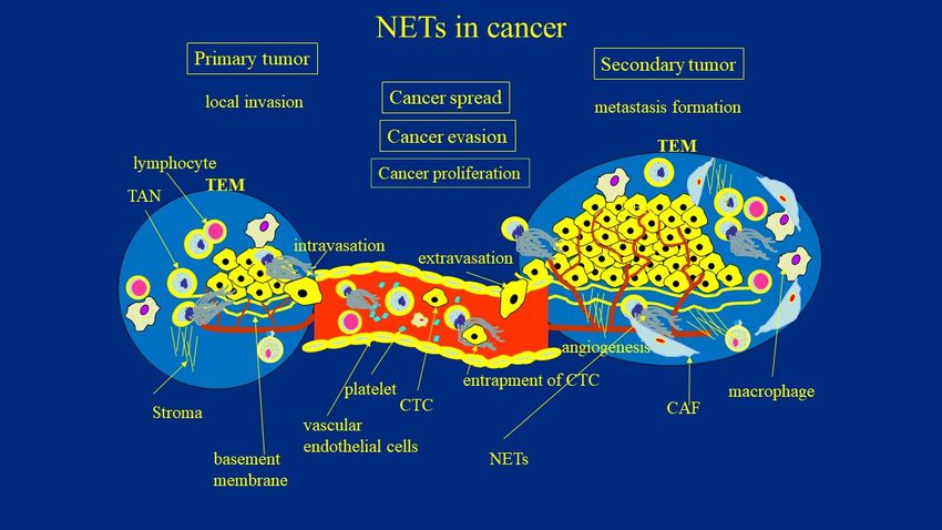
given their versatile role in the metastatic cascade. (Figure 1).
Figure 1. The role of NETs in cancer development. TAN—tumor associate neutrophils; TEM—tumor environment; CAF—
Figure 1. The role of NETs in cancer development. TAN—tumor associate neutrophils; TEM—tumor environment;
cancer associated fibroblasts; NETs—neutrophil extracellular traps; CTC—circulating tumor cells.
CAF—cancer associated fibroblasts; NETs—neutrophil extracellular traps; CTC—circulating tumor cells.
5.3.
5.3. NETs
NETs Supports
Supports thethe Cancer
Cancer Evasion Strategies
Evasion Strategies
The
The evasion
evasion of of tumor
tumor cells
cells from
from immunosurveillance
immunosurveillance depends depends on on the
the interplay
interplay be-be-
tween various infiltrating immune cells and tumor cells. Whatever
tween various infiltrating immune cells and tumor cells. Whatever the mechanistic basis, the mechanistic basis,
it
it appears
appears that
that immunosurveillance
immunosurveillance ofof tumors
tumors is is canonically
canonically dependent
dependent ononthethe presence
presence of
of
thethe major
major histocompatibility
histocompatibility complex
complex 1 (MHC1)
1 (MHC1) antigens
antigens on on cancer
cancer cellscells enabling
enabling lym-
lympho-
phocytes T (both CD4+ and CD8+) to discriminate tumor cells
cytes T (both CD4+ and CD8+) to discriminate tumor cells from normal cells, as well from normal cells, as well
as
as
to control the tumor cell survival [49,50]. Various proteolytic enzymes (proteinases) are
to control the tumor cell survival [49,50]. Various proteolytic enzymes (proteinases) are
able
able to
to modulate
modulate the cell-surface-associated presentation
the cell-surface-associated presentation of MHC molecules.
of MHC molecules. MMP9,MMP9, for for
example,
example, isisresponsible
responsiblefor forthe shedding
the shedding of of
MHCMHC class I antigen
class fromfrom
I antigen cancer cellscells
cancer [51]. [51].
The
selected subclones
The selected of malignant
subclones of malignantcells cells
achieve the capability
achieve to hide
the capability fromfrom
to hide the immune
the immune sys-
tem by losing
system the ability
by losing to present
the ability cancer
to present antigens
cancer to T-cells.
antigens Various
to T-cells. components
Various componentsof ECM of
have been recognized as sources of signals for the immune system
ECM have been recognized as sources of signals for the immune system to slow down to slow down immune
reactions, for example
immune reactions, through the
for example expression
through of checkpoint
the expression molecules.
of checkpoint The ECM
molecules. Theis aECM
res-
ervoir of immunomodulatory
is a reservoir of immunomodulatory cytokines and growth
cytokines factorsfactors
and growth that are
thatreleased upon upon
are released their
proteolytic degradation.
their proteolytic Metalloproteinases
degradation. Metalloproteinases and NEand canNE modulate immuneimmune
can modulate and inflam-and
matory responses
inflammatory through
responses the degradation
through the degradationof the ofECM. The cleavage
the ECM. products
The cleavage of the of
products ECM
the
ECM (e.g., matrikines) can, by themselves, affect immune surveillance. NET proteases can
impede the immune response and, thus, ensure the best possibility of cancer cell survival
by enabling the metastatic process [45]. According to Albrengues et al. [45], the degrada-
tion of matrix proteins is one of the mechanisms of tumor evasion that silences the host’s
immune system. NET proteinases stimulate the production of IL-8, IL-1β, and TNF-α with
tumor-associated macrophages through the activation of several MMPs. This process is
dependent on Src kinase activation, highlighting the fact that NE also impacts integrins
and integrin-mediated intracellular signaling [22,52]. As another example, the inhibition of
hyaluronic acid (a major component of the ECM) synthesis by 4-methylumbelliferone in a
mesothelioma xenograft has led to a significant increase in the expression of both immune
checkpoint molecules, PD-1 and PD-L1 [53]. Although the mechanisms involved in ECMCancers 2021, 13, 4495 8 of 17
modification by NET components are not fully elucidated, the clear connection between
the ECM composition and proteinases, as well as the immune escape, strongly support
the existence of such an effect. Onuma et al. [54] confirmed that the blockade of NETs, in
combination with immune checkpoint PD-1 inhibition, improved the response rates of
colorectal cancer metastases to immune checkpoint inhibitors as a single therapy. This was
achieved through the improving of the function of exhausted CD8+ T-cells [54].
5.4. NETs Enhance Invasion Capacity of Cancer Cells
A crucial event at the first stage of metastatic colonization is the formation of a
favorable niche for tumor engraftment attributable to tumor–stroma crosstalk [55]. The
process of metastatic spread began from proteolytic remodeling of ECM and the release
of ECM metabolites necessary, or even mandatory, for the dissemination of cancer cells.
As mentioned above, the ECM is digested by MMPs, disintegrin, metalloproteinases
with thrombospondin motifs (ADAMTS), and proteases that specifically cleave at cysteine,
serine, and threonine residues [56]. Several components of mature NETs cause an imbalance
in the microenvironments, as well as the emergence of metastatic niches. For example,
NET-derived NE and MMP-9 degraded ECM to actively induce tumor invasion [57].
Accordingly, it was shown that matrix metalloproteinase catalytic activity modulated the
invasiveness and provided a route for the malignant cells to metastasize via modulation
of the integrins–FAK signaling pathway [58]. In an experimental model using Boyden
transwell invasion assay, Park et al. [59] focused on the neutrophil-mediated invasion
of tumor cells. The applied model confirmed that tumor invasion through the filter in
the transwell system can be promoted by the mutual interaction between tumor cells in
the upper chamber and neutrophils in the lower chamber. Furthermore, the blockade of
NE and matrix metalloproteinases impeded tumor invasion [59]. In agreement with this
observation, DNase I treatment downregulated NE and NET activities and reduced the
invasive and metastatic potential of malignant cells [59]. Other investigators were able to
confirm significant correlation between NETs and liver metastases of patients with breast
and colon cancers, thus confirming increased binding activity of transmembrane protein
CCDC25 on primary cancer cells to NET DNA. These authors proved that CCDC25 senses
extracellular DNA and, subsequently, activates the ILK-β-parvin pathway to attract cancer
cells. NET-mediated metastasis was abrogated in CCDC25-knockout cells. Moreover, the
expression of CCDC25 was associated with a poor outcome of the disease [60]. Although
the detailed mechanism of tumor invasion and metastasis via NET molecules is still not
completely understood, it would be interesting to investigate the role of TANs in the
regulation of NET-mediated tumor invasion. Signaling is an integral process in controlling
invasive and metastatic potential of tumor cells. The signaling between various structures
in TME, including NETs fragments, is crucial in controlling the invasive potential of the
tumor. Thus, in silico studies modelling these critical interactions and their effects are
warranted to discern alternative explanations of these processes and pave the way for the
development of new therapeutic strategies [14].
5.5. NETs Enhance Systemic Spread and Tumor-Associated Angiogenesis
Tumor cells can migrate and intravasate the blood or lymph vasculature. They can
survive within the circulation, then extravasate at distant sites. The factors determining
adhesion strength, which might influence the ability of cells to transmigrate through an
endothelial cell monolayer and the basement membrane, are poorly understood. Recent
studies have highlighted that these processes are driven not only by signals from cancer
cells, but are also modified by signals from components of the TME [55]. Current evi-
dence suggests that NETs may play a crucial role in the hematogenous spread of tumors.
Jung et al. [41] showed that NETs promoted tumor growth, metastasis, and angiogenesis
of the pancreatic cancer cell line (AsPC-1). NETs used as chemoattractans stimulated
AsPC-1 cell migration (in a Matrigel-coated invasion chamber) better then intact neu-
trophils. These effects were abrogated by histone-binding agents (heparin, polysialic acid),Cancers 2021, 13, 4495 9 of 17
DNAse I, and Toll-like receptor neutralizing antibodies. Antibodies against both TLR2
and TLR4 significantly inhibited NET-mediated AsPC-1 cell migration. Although not
unexpectedly, these results support the opinion that TLR2 and TLR4 participate in tumor
transmigration. In patients with pancreatobiliary malignancy, elevated NET markers cor-
related with hypercoagulability makers. Histone–DNA complexes were used as markers
of NETs. Another component of NETs, histones, significantly increased the endothelial
cell proliferation and the formation of new blood vessels in a dose-dependent manner.
Application of histone-binding agents abrogated histone-induced angiogenesis [41]. The
same directionality of the effect was observed by Tohme et al., who reported that the
chemotactic factor released during NET formation may stimulate proliferation and mi-
gration of cancer cells [36]. Finally, the transmigration mechanisms were explained by
Kołaczkowska et al., who observed the adherence of circulating NETs to blood, resulting
in increased cancer extravasation efficiency, which would enable cancer cells to cross the
endothelial barrier [61]. On the other hand, the previously mentioned report of Park et al.
suggested that not DNA itself, but rather NET-related proteases are responsible for this
effect [59]. Such a discrepancy may be explained by the fact that such a structure as com-
plex as the one between NETs and the locally concentrated enzymes, must be taken as an
inseparable assembly, rather than a conglomerate of individual components. Once in the
circulation, tumor cells become entrapped by NETs DNA threads. Through the use of cecal
ligation, Cools-Lartigue et al. [62] demonstrated the presence of circulating lung carcinoma
cells wrapped in NET DNA conglomerates in a murine model of infection. Consequently,
circulating “packages” were seeded in the liver, forming micrometastases within 48 h and
secondary liver cancer 2 weeks after the cancer cell injection. DNAse or NE inhibitors
abrogated the effects [62]. Evidence consistent with these observations was provided by
Najmeh et al. from the same group, who found a significant association between upreg-
ulation of β1-integrin and NET-related entrapment of circulating lung carcinoma cells,
further facilitating metastasis formation and cancer spread [63]. Whatever the precise basis
of this mechanism is, it appears that inflammatory mediators harbored by neutrophils
may be responsible for insufficient clearance of circulating cells [64]. NETs’ entrapping
abilities can be, at least partially, attributed to the ability to adhere to DNA mesh carried
by the variety of integrins expressed on the surface of cancer cells. Such interaction was
completely abrogated by DNase 1 [65]. Furthermore, the TAN-CTC adhesion process
facilitates cancer cell extravasation through the breaking of the transendothelial barrier [66].
The proposed adhesive interaction between circulating neoplastic cells and TANs leads
to the increased endothelial cell contraction, permeability, and malignant cell extravasa-
tion [66]. A multi-level model shed new light on the fundamental processes elucidating the
role of NETs in cancer invasions, transport, and transendothelial migration, thus taking
into account specific NET–cell adhesion, ECM–tumor–NET interaction, and intracellular
signaling [67–71]. Further studies, however, are still warranted to explore these issues.
5.6. How NETs and Tumor Communicate
The interaction between the tumor and NETs is reciprocal. In their excellent paper,
Demeters et al. compiled initial reports showing that TANs are a potent source of NETs
and, on the other hand, cancer cells can stimulate neutrophils to release NETs as shown
in various animal models of cancer [19]. NETs enhance the gathering and proliferation
of single cancer cells, contributing to tumor metastasis by releasing MMP and NE, which
through the degradation of ECM, paves a way for tumor cells to leave the primary niche
and to migrate to other organs. Conversely, inflammatory cytokines, such as IL-8 and gran-
ulocyte colony-stimulating factor, as well as various soluble factors, i.e., exosomes released
from cancer cells, stimulate neutrophils to release NETs [72]. Metastatic cancer cells possess
the ability to stimulate the release of NETs directly and without the engagement of inflam-
matory mechanisms [33]. According to the model of a vicious circle proposed by Park et al.,
the metastatic breast cancer cells induced neutrophils to form NETs, which further en-
hanced tumor cell growth in target organs [59]. McInturff et al. demonstrated that cancerCancers 2021, 13, 4495 10 of 17
cells themselves are able to stimulate neutrophils to form NETs in a hypoxic environment
where solid tumor growth is enhanced by the higher expression of HIF-1α [73]. Another
mechanism by which cancer cells may stimulate neutrophils to form NETs depends on
the production of IL-8 and the release of exosomes which require additional priming with
granulocyte colony-stimulating factors. Leal et al. found that tumor-derived exosomes of
cancer patients in a hypercoagulable state can induce NET release, and that NETs can serve
as a scaffold for coagulation factors, platelets, and exosomes carrying prothrombotic media-
tors, altogether promoting the development of thrombo-embolic complications and cancer
progression [72]. In an excellent review, Yousefi et al. summarized various experimental
evidence that lung, colon, ovarian, and anaplastic thyroid cancer (ATC) cells induce the
release of mitochondrial extracellular DNA traps by viable neutrophils [74]. Furthermore,
tumor cells have been demonstrated to produce IL-8, attracting myeloid-derived suppres-
sor cells and activating neutrophil precursors to release NETs [75]. Similarly, liver ischemia
reperfusion in a murine model resulted in NET extrusion in parallel with the progression of
metastatic disease, while the pre-treatment of mice with topical DNase or a PAD4 inhibitor
abrogated these effects [36]. Consistently with these observations in mice, an increased
postoperative NET formation inversely correlated with the disease-free survival in patients
undergoing liver resection for metastatic colorectal cancer [36]. However, the limitation
of this study manifested in the use of NET plasma markers (MPO–DNA complexes) as
surrogates of netting capacities of neutrophils rather than a direct analysis of NET presence
in the examined tissues.
5.7. NETs in the Formation of Metastatic Niche
Tumors metastasize to distant organs with tissue-specific microenvironments, which
are very different from that of a primary tumor. The precondition of distinct microenviron-
ments involving ECM remodeling and the creation of a favorable pre-metastatic niche is
necessary for the seeding of new tumor colonies [56]. The most common modification of
the ECM in the primary TME is increased collagen deposition. On the contrary, fibronectin
dominates along with glycoproteins and proteoglycans such as tenascin C, osteopontin,
and versican in a pre-metastatic niche [76]. The primary niche is mainly formatted by
mediators released by growing tumor cells, further acting on various components of TME,
which in turn release a second generation of molecules, directly creating a favorable mi-
croenvironment. NETs participate in this process, conferring the effect on the electrostatic
charge and conformation of fibronectin and collagens in the process of citrullination. This
effect is mediated by the enzyme PAD4, derived from NETs during pre-metastatic niche
formation [23,77]. Moreover, NETs equipped with proteases are highly associated with
aggressive tumor growth and invasion, but this high metastatic potential is abrogated by
DNase I treatment [14,59]. Recently, different studies began to highlight that epithelial–
mesenchymal transition (EMT), a process by which epithelial cells acquire mesenchymal
properties endowing cancer cells with invasive and metastatic potential, is driven by
NETs [78,79]. Martins-Cardoso et al. recently described the association between NETs and
the pro-metastatic phenotype of human breast cancer cells [78]. Co-cultures of tumor cells
treated with isolated NETs underwent several experiments, including migration assay,
quantitative RT-PCR, Western blotting, immunofluorescence, and flow cytometry assays.
RNA-seq data from The Cancer Genome Atlas (TCGA) database were also assessed [79].
NET components changed the epithelial into mesenchymal phenotype (upregulated ex-
pression of N-cadherin and fibronectin, and downregulation of E-cadherin). The effect was
accompanied by the increased motility of cells. RNA-seq revealed pro-inflammatory and
pro-metastatic signatures. Accordingly, TCGA data analysis of samples from breast cancer
patients showed a significant correlation between neutrophil and the pro-tumoral signature
of gene expression [78]. Further studies have shed light on the crosstalk between glioma
progression and NETs in TME. The tumor growth was mediated via the HMGB1/RAGE/IL-
8 axis [80]. Covid-era discoveries also led to the conclusion that lung inflammation and aCancers 2021, 13, 4495 11 of 17
cytokine storm accompanied by NET formation in the course of COVID-19 contributes to
dormant cancer cells awakening and the formation of a pro-metastatic niche [81].
5.8. NETs Is Physically Blocking T-Cell Infiltration to the TME
It has been demonstrated that PMNs and their products are engaged in multiple inter-
actions with T-lymphocytes and a molecular basis of these associations is being explored.
A number of important links between NETs and functions of T-lymphocytes have been
discovered [82–84]. The communication between T-cells and NETs occurs either via direct
contact of lymphocytes with the NET backbone or depends on released mediators, includ-
ing enzymes, cytokines, and radical oxygen species. Tillack et al. [83] showed that NETs can
directly reduce the T-cell activation threshold in response to specific stimuli. Both NET/cell
contact and TCR signaling are necessary for T-cell priming [83]. Bilyy et al. demonstrated
that NETs form a barrier between necrotic and viable areas in acute abdominal inflamma-
tion [84]. In a very recent study, Surashri Shinde-Jadhav et al. [85] discovered a direct link
between intratumoral NETs and T-cell cytotoxicity. These authors demonstrated that NETs
formed a barrier between the irradiated tumor and stroma, blocking the invasion of CD8
T-cells to TME [85]. NETs were found to be surrounding CD8 T-cells but not colocalizing
with them. Moreover, increased intratumoral CD8 T-cell infiltration was noted in tumors of
mice treated with DNAse I [85]. These authors claimed that NETs may play a role in tumor
radioresistance by blocking intratumoral CD8 T-cell infiltration post-RT. This effect was
related to the clinical effect of the RT [85]. A higher intratumoral PMN to CD8 ratio was
observed in RT non-responders compared to RT responders. Additionally, these authors
found that patients with persistent disease had a high pre-treatment intratumoral PMN to
CD8 ratio. In line with this experimental data is the clinical observation that a high PMN to
CD8 ratio was associated with worse overall survival [85]. The NET barrier at the interface
of tumor cells and necrotic tissue was also noted by the previously mentioned study of
Berger-Achituv et al. in Ewing sarcoma biopsy samples [18]. These authors proposed,
for the first time, that this NET barrier may enable tumor immune escape [18]. These
observations are in accord with the report of Teijeira et al., who show that intratumoral
NETs block the contact between tumor cells and cytotoxic cells [86]. Summarizing, NETs
provide a physical barrier protecting from the spread of infectious agents, thus localizing
the infection. On the other hand, this mechanism is not beneficial in the course of tumor
development as it contributes to the immune evasion mechanisms by blocking the access
of cytotoxic cells to the growing tumor.
6. Potential Anti-NETs Therapy of Cancer
Targeting NETs is a relatively new option with significant potential for the treatment
of PMN-mediated disorders. This review focused on the pro-tumorigenic activity of NETs,
highlighting their ability to serve as an appealing therapeutic target for cancer. An in-
teresting option is also the combination of anti-cancer and anti-NET intervention. With
the recent advances in the knowledge of how NETs are generated or how to dismantle
their structure, several approaches can be considered to develop strategies to prevent the
awakening of dormant cancer cells and to inhibit the spreading of tumors, as well as the
formation of metastases. The detection of NETs in tumor biopsies or the presence of NET
markers in the circulation may stand for the most accurate method of identifying patients
who could benefit from NET-targeting therapy. Recently, a number of different researchers
have presented emerging and promising concepts for cancer treatment based on the anti-
NETs strategy. Park et al. showed that inhibiting NET formation or dismantling NETs
with DNase I-coated nanoparticles markedly reduced lung metastases of breast cancer in
mice [59]. The effectiveness of such an approach was confirmed by the experimental evi-
dence of the inhibitory effect of DNase-I on the invasion and migration of breast cancer cells
in vitro concomitantly with NET degradation [59]. Other groups of scientists have stated
that cannabinoids, which act through the cannabinoid receptor, suppress PMN functions,
including cell migration, production of ROS, and TNF-α production followed by NET re-Cancers 2021, 13, 4495 12 of 17
lease [87,88]. Very recently, Munir et al. reported that cancer-associated fibroblasts secreted
amyloid β, modulating tumor-associated NET release through CD11b in a ROS-dependent
manner [89]. This effect was observed both within the TME and at systemic levels in the
blood and bone marrow. The prevention of amyloid β release abrogated tumor growth
and restored an anti-tumor status in TANs, suggesting a potential therapeutic strategy
on various cancer types [89]. In the previously discussed paper, Albrengues et al. [45]
provided compelling evidence that antibodies against NET-remodeled laminin prevented
awakening of dormant cells. These results provide a rationale for targeting this pathway to
treat metastatic cancer and to prevent the disease relapse [45]. A comprehensive analysis
of the signaling pathways regulating PAD4 activation may result in the generation of
pharmaceuticals that target NET-related disorders. Such a strategy could be applied to
prevent chromatin decondensation and the expulsion of chromosomal DNA and, what is
more, to decrease metastatic behavior of cancer. A large panel of available PAD4 selective
inhibitors have been developed recently [90]. It was demonstrated that PAD4 knockout
inhibited tumor growth and metastasis of colorectal cancer by preventing the citrullination
of the ECM in the liver and impeding the subsequent epithelial-to-mesenchymal transi-
tion [90]. Moreover, inhibition of PAD4 activity by a novel therapeutic, BMS-P5, abolished
citrullination of histone H3 and NET releases, thus improving the disease prognosis in
patients and mice with myeloma [91]. The PAD4 inhibitor Cl-amidine significantly reduced
NET formation, the number of breast cancer cells that extravasated into the lung tissue,
however, was not altered [59]. Moreover, another PAD4 inhibitor, GSK484, was recently
shown to prevent tumor-associated renal dysfunction in mice and the effect was deter-
mined to be NET-mediated [23]. Targeting PAD4 has been well acknowledged to have
anti-NET capacity and an anti-tumor effect, although the exact molecular mechanisms of
long-term therapy with PAD4 inhibitors and their long-term effects need further studies.
Alternatively, the disruption of NET formation can be achieved by targeting the receptor for
G-CSF (G-CSFR). A recent report by Wang et al. [92] underlined the ability of anti-G-CSFR
monoclonal antibodies to inhibit NET release, as well as to downregulate hyperinflam-
matory reactions in the course of infections with no impact on pathogen clearance. Thus,
blocking the G-CSFR receptor might represent a promising option to treat NET-dependent
conditions without compromising the immune response against pathogens [92]. Another
therapeutic intervention may be based on the above-described targeting of transmembrane
DNA receptor CCDC25, which thus decreases cancer invasiveness [60].
The specialists agree that Ca2+ signaling constitutes an important component of
the process of NET formation [2,3]. In addition, several authors have claimed that Ca2+
influxes have pro-oncogenic impact [49–51]. Ca2+ influxes in physiological processes
can come from two sources: intracellular Ca2+ stores and external Ca2+ entering across
the plasma membrane through cell membrane channels [22]. Another therapeutic option
in cancer is targeting the ability of NETs to secrete the Ca2+-binding proteins S100A8
and S100A9 [93]. Both proteins S100A8/A9 are able to recruit tumor cells, to maintain
inflammatory milieu, promote tumor progression, and create a favorable environment
for metastatic niche formation, although the molecular mechanisms underlying their
involvement in these processes remain unknown [22,94–96]. Schenten et al. demonstrated
that S100A8 and S100A9 are key players in the cancer progression and proposed further
investigations, enabling the development of an appropriate therapeutic intervention [93].
Animal studies have shown that DNase [36,62,97], cathepsin C (CTSC) inhibitors [98],
PAD4 inhibitors [59], and NE inhibitors [62,97] displayed certain anti-metastasis effects
by abrogation of NET formation. DNase I treatment suppressed the development of
gross metastases and the growth of established liver micrometastases in colorectal cancer
animal models [36]. The previously mentioned interventions hindering NET formation,
such as DNase, NE inhibitors, and PAD4 knockout, reduced spontaneous lung and liver
metastasis of lung carcinoma cells in NET-deficient mice [97]. Inhibiting CTSC by a second-
generation inhibitor AZD7986, a potential drug for neutrophil-mediated inflammatory
diseases, effectively wrecked NETs and abrogated lung metastasis of breast cancer inCancers 2021, 13, 4495 13 of 17
murine model, though without significant influence on primary tumor growth [98,99]. In
addition, metformin, a well know anti-diabetic drug, is also proposed for cancer treatment,
but its mechanism of action is not completely understood. A recent clinical study uncovered
anti-NET properties of metformin, predominantly dependent on the inhibitory effect of
metformin on the protein kinase C (PKC)-NADPH oxidase pathway [100]. Moreover,
hydroxychloroquine can impede NET formation, potentially modulating the upstream
signaling pathway for autophagy [101]. Clinical data from pancreatic cancer patients
showed that hydroxychloroquine downregulates hypercoagulability and decreases the rate
of thromboembolic complications typically attributed to overproduction of NETs [43].
The above-mentioned findings support the potential of NET-targeting approaches
for the decreasing of metastatic behavior of cancer cells and the boosting of efficiency of
anti-cancer therapy. Targeting NETs is a tempting opportunity and worthwhile strategy,
however the risk of severe infections in NET-depleted patients may limit its clinical ap-
plications and further studies are warranted to investigate this issue. Nevertheless, the
potential benefits of blocking pro-tumorigenic TAN properties encourage further research.
Both options—either to dismantle formed NETs or to block their production—require
further testing. Such strategies and underlying molecular mechanisms are in their infancy
and further data to explore their therapeutic potential and lack of severe side-effects are
awaited [22].
7. Conclusions
The NETs exert numerous pro-tumorigenic effects at various steps of tumor devel-
opment. A better understanding of the crosstalk between cancer and NETs can help to
elucidate basic aspects of the immune response to cancer and to devise novel therapeutic
interventions that can block cancer evasion mechanisms and prevent metastatic spread.
Funding: This research received no external funding.
Data Availability Statement: No new data were created or analyzed in this study.
Conflicts of Interest: The author declares no conflict of interest.
References
1. Teng, T.S.; Ji, A.L.; Ji, X.Y.; Li, Y.Z. Neutrophils and Immunity: From Bactericidal Action to Being Conquered. J. Immunol. Res.
2017, 2017, 9671604. [CrossRef]
2. Pruchniak, M.P.; Demkow, U. Potent NETosis inducers do not show synergistic effects in vitro. Cent. Eur. J. Immunol. 2019, 44,
51–58. [CrossRef]
3. Manda-Handzlik, A.; Bystrzycka, W.; Cieloch, A.; Glodkowska-Mrowka, E.; Jankowska-Steifer, E.; Heropolitanska-Pliszka, E.;
Skrobot, A.; Muchowicz, A.; Ciepiela, O.; Wachowska, M.; et al. Nitric oxide and peroxynitrite trigger and enhance release of
neutrophil extracellular traps. Cell Mol. Life Sci. 2020, 77, 3059–3075. [CrossRef]
4. Manda-Handzlik, A.; Fiok, K.; Cieloch, A.; Heropolitanska-Pliszka, E.; Demkow, U. Convolutional Neural Networks-Based
Image Analysis for the Detection and Quantification of Neutrophil Extracellular Traps. Cells 2020, 9, 508. [CrossRef]
5. Leliefeld, P.H.C.; Koenderman, L.; Pillay, J. How Neutrophils Shape Adaptive Immune Responses. Front. Immunol. 2015, 6, 471. [CrossRef]
6. Pruchniak, M.P.; Ostafin, M.; Wachowska, M.; Jakubaszek, M.; Kwiatkowska, B.; Olesinska, M.; Zycinska, K.; Demkow, U.
Neutrophil extracellular traps generation and degradation in patients with granulomatosis with polyangiitis and systemic lupus
erythematosus. Autoimmunity 2019, 52, 126–135. [CrossRef] [PubMed]
7. Becker, R.C. COVID-19-associated vasculitis and vasculopathy. J. Thromb. Thrombolysis 2020, 50, 499–511. [CrossRef] [PubMed]
8. Gomez-Moreno, D.; Adrover, J.M.; Hidalgo, A. Neutrophils as effectors of vascular inflammation. Eur. J. Clin. Investig. 2018, 48,
e12940. [CrossRef] [PubMed]
9. Velten, L.; Haas, S.F.; Raffel, S.; Blaszkiewicz, S.; Islam, S.; Hennig, B.P.; Hirche, C.; Lutz, C.; Buss, E.C.; Nowak, D.; et al. Human
haematopoietic stem cell lineage commitment is a continuous process. Nat. Cell Biol. 2017, 19, 271–281. [CrossRef]
10. Bremnes, R.M.; Dønnem, T.; Al-Saad, S.; Al-Shibli, K.; Andersen, S.; Sirera, R.; Camps, C.; Marinez, I.; Busund, L.T. The Role of
Tumor Stroma in Cancer Progression and Prognosis: Emphasis on Carcinoma-Associated Fibroblasts and Non-small Cell Lung
Cancer. J. Thorac. Oncol. 2011, 6, 209–217. [CrossRef]
11. Mishalian, I.; Granot, Z.; Fridlender, Z.G. The diversity of circulating neutrophils in cancer. Immunobiology 2017, 222, 82–88. [CrossRef]
12. Masucci, M.T.; Minopoli, M.; Carriero, M.V. Tumor Associated Neutrophils. Their Role in Tumorigenesis, Metastasis, Prognosis
and Therapy. Front. Oncol. 2019, 9, 1146. [CrossRef]You can also read