Post-translational Acetylation Control of Cardiac Energy Metabolism
←
→
Page content transcription
If your browser does not render page correctly, please read the page content below
REVIEW
                                                                                                                                          published: 02 August 2021
                                                                                                                                    doi: 10.3389/fcvm.2021.723996

                                            Post-translational Acetylation
                                            Control of Cardiac Energy
                                            Metabolism
                                            Ezra B. Ketema and Gary D. Lopaschuk*

                                            Department of Pediatrics, Cardiovascular Research Centre, University of Alberta, Edmonton, AB, Canada

                                            Perturbations in myocardial energy substrate metabolism are key contributors to the
                                            pathogenesis of heart diseases. However, the underlying causes of these metabolic
                                            alterations remain poorly understood. Recently, post-translational acetylation-mediated
                                            modification of metabolic enzymes has emerged as one of the important regulatory
                                            mechanisms for these metabolic changes. Nevertheless, despite the growing reports of a
                                            large number of acetylated cardiac mitochondrial proteins involved in energy metabolism,
                                            the functional consequences of these acetylation changes and how they correlate to
                                            metabolic alterations and myocardial dysfunction are not clearly defined. This review
                                            summarizes the evidence for a role of cardiac mitochondrial protein acetylation in altering
                                            the function of major metabolic enzymes and myocardial energy metabolism in various
                           Edited by:       cardiovascular disease conditions.
                        Kunhua Song,
                                            Keywords: mitochondria, fatty acid oxidation, succinylation, sirtuins, lysine acetylation, glucose oxidation
      University of Colorado Anschutz
      Medical Campus, United States
                        Reviewed by:
                Bradley S. Ferguson,
                                            INTRODUCTION
          University of Nevada, Reno,
                         United States
                                            Following the discovery of histone acetylation and its regulatory effect on RNA synthesis by Allfrey
                         Matt Stratton,     et al. (1), it has been established that alterations in the level of histone acetylation can modulate gene
           The Ohio State University,       expressions via chromatin remodeling and epigenetic modifications (2, 3). Indeed, dysregulation
                         United States      of histone acetylation level has been strongly linked with the development and progression of
                  *Correspondence:          cancer and other human diseases (4, 5). This was further reinforced by the identification of histone
                   Gary D. Lopaschuk        acetyltransferases (HAT) and histone deacetylases (HDACs), enzymes that mediate the addition
          gary.lopaschuk@ualberta.ca        or removal of an acetyl group to and from a lysine residue of histone proteins (6, 7), and the
                                            development of several HDAC inhibitors to treat cancer and heart diseases (8–10).
                    Specialty section:          In addition to nuclear histone acetylation, the potential for non-histone protein acetylation
          This article was submitted to     of non-nuclear proteins has also recently generated considerable interest. The first acetylation of
           Cardiovascular Metabolism,
                                            cytoplasmic proteins was described in microtubules (α-tubulin) in 1987 by Piperno et al. (11).
                a section of the journal
                                            The involvement of acetylation of non-nuclear proteins was further confirmed by the isolation
  Frontiers in Cardiovascular Medicine
                                            of other acetylated proteins in both the cytosol and mitochondria, as well as the presence of
            Received: 11 June 2021
                                            deacetylase enzymes, such as SIRT2 and SIRT3 outside the nucleus (12–15). Since then, a number
            Accepted: 30 June 2021
                                            of acetylases and deacetylases have been identified outside the nucleus (Figure 1) (16). However,
          Published: 02 August 2021
                                            it is the advancements in protein acetylome quantitative methods, and the identification of
                              Citation:
                                            several thousands of cytosolic and mitochondrial acetylated proteins using these techniques, that
Ketema EB and Lopaschuk GD (2021)
 Post-translational Acetylation Control
                                            have recently helped advance our understanding of non-histone acetylation dynamics and its
       of Cardiac Energy Metabolism.        biological implications.
   Front. Cardiovasc. Med. 8:723996.            Kim et al. reported the first large acetyl proteomic data profile by identifying 388 acetylation
     doi: 10.3389/fcvm.2021.723996          sites in 195 proteins in HeLa cells and mouse liver using immunoprecipitation of lysine-acetylated

Frontiers in Cardiovascular Medicine | www.frontiersin.org                       1                                          August 2021 | Volume 8 | Article 723996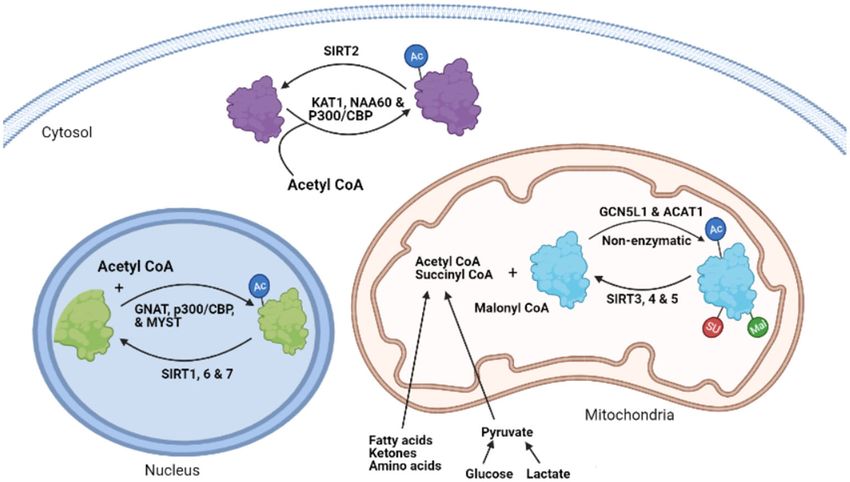
Ketema and Lopaschuk                                                                                                 Acetylation Control of Cardiac Energy Metabolism

 FIGURE 1 | The process of protein lysine acetylation/acylation and deacetylation/deacylation. KAT1, lysine acetyltransferase; NAA60, N-α-acetyltransferase 60; CBP,
 CREB-binding protein; SIRT, sirtuin, GNAT, GCN5-related N-acetyltransferases; MYST, MYST family acetyltransferase; GCN5L1, General control of amino acid
 synthesis 5 (GCN5) like-1; ACAT1, acetyl-CoA acetyltransferase; AC, lysine Acetylation; SU, succinylation; MU, malonylation.

peptides and mass spectrometry analysis (17). Of these,                               poorly understood. Understanding these post-translational
133 proteins with 277 lysine acetylation sites were from                              modifications is particularly important in heart failure, where
mitochondria, showing for the first time the abundance of                             significant alterations in mitochondrial metabolism are seen, and
the acetylation process in mitochondria. Subsequently, several                        disturbances in the metabolites used as a substrate for these
hundred acetylated proteins were also identified in other studies                     post-translational modifications occur.
using either nutritional or acetylase/deacetylase enzymatic                               Impairments in myocardial energy substrate metabolism
manipulations (18, 19). For example, in response to caloric                           are a major contributor to the pathogenesis of heart failure
restriction in mice, Schwer et al. reported acetylation of                            (28–30). However, the underlying causes of these metabolic
several proteins involved in metabolic pathways in the liver                          perturbations remain unclear. In the failing heart, changes in the
(19). Another study by Zhao et al. revealed the prevalence                            transcription of genes encoding metabolic enzymes, particularly
of acetylation of several metabolic enzymes and its possible                          fatty acid metabolic enzymes, have been suggested as one of the
regulatory role using both HDAC and non-histone acetylation                           mechanisms for altered energy substrate metabolism (31–34).
inhibitors (20). Similarly, Lombard et al. demonstrated extensive                     Nevertheless, transcriptional regulation alone is not sufficient to
mitochondrial protein acetylation in SIRT3 knockout (KO) mice                         explain all the metabolite and enzyme activity changes observed
(21), a finding confirmed by Hirschey et al. (22). Strikingly, these                  in the failing heart (35, 36). Several post-transcriptional and post-
studies described the exceptional susceptibility of mitochondrial                     translational processes have been shown to modulate the original
proteins to acetylation modification in response to various                           transcription products, and thus can significantly alter energy
stressors, with up to 60% of mitochondrial proteins reported                          metabolism in the failing heart. Post-translational acetylation-
to being acetylated (23). Of importance, the majority of these                        mediated modification of metabolic proteins is thought to have
acetylated proteins were enzymes catalyzing energy metabolic                          a regulatory role in these energy metabolic changes (37–39).
processes (19–21, 24). Similar to acetylation modifications,                              In addition to acetylation, many other post-translational
several studies have demonstrated increased succinylation and                         modifications including phosphorylation, malonylation,
malonylation of a large number of cytosolic and mitochondrial                         succinylation, glutarylation, ubiquitination, SUMOylation,
proteins in response to SIRT5 deletion (25–27). For instance,                         O-GlcNAcylation, N-glycosylation, methylation, citrullination,
in liver mitochondria isolated from SIRT5 KO mice, Rardin                             and S-nitrosylation play important roles in cardiac disease
et al. identified 1190 succinylation sites, of which 386 sites                        pathogenesis, including metabolic perturbations (40–42).
on 140 proteins were seen in enzymes involved in energy                               However, the focus of this review will be on the role of acetylation
metabolism, including fatty acid β-oxidation and ketogenesis                          (and some acylation) modifications of energy metabolic enzymes
(26). Despite these studies, that have revealed widespread                            and their contributions to altering cardiac energy metabolism.
acyl modifications on proteins in most of the metabolic                               Although the relevance of protein acetylation changes have
pathways, its biological significance and regulation are still                        also been reported in other pathological processes, including

Frontiers in Cardiovascular Medicine | www.frontiersin.org                        2                                           August 2021 | Volume 8 | Article 723996
Ketema and Lopaschuk                                                                                  Acetylation Control of Cardiac Energy Metabolism

inflammation, oxidative stress, and apoptosis, this review will            of calorie restriction on health and aging, including metabolic
mainly discuss the connections between acetylation imbalances              reprogramming and stress tolerance. In support of this, caloric
and cardiac energy metabolic changes.                                      restriction is also shown to increase the expression of sirtuins
                                                                           (21, 22, 65, 66).
                                                                               There are seven mammalian sirtuin proteins (SIRT1–SIRT7)
THE PROCESS OF PROTEIN                                                     with variation in their tissue specificity, subcellular localization,
ACETYLATION                                                                enzymatic activity, and targets (16). SIRT1, 6, and 7 are
                                                                           mainly localized in the nucleus (16, 67, 68), while SIRT2 is
Lysine acetylation of proteins occurs through the covalent                 predominantly localized in the cytoplasm (12). However, SIRT1
attachment of an acetyl group to the lysine residues of proteins.          and SIRT2 can shuttle between the nucleus and the cytoplasm
This acylation modification causes important changes to the                and acetylate proteins in both compartments (67, 69). SIRT1 can
protein at its lysine residue, which includes altering its charge          regulate the acetylation state of diverse cellular proteins in the
status and adding an extra structural moiety (43, 44). These               nucleus (70). In contrast, SIRT 3, 4, and 5 are mainly localized
changes impact the proteins’ native structure, its interactions            in the mitochondria (13, 16, 71), although some studies have
with other proteins or regulatory molecules, its stability, and            reported cytosolic localization of SIRT5 (25). In terms of their
its function (45). Similar to acetylation, lysine succinylation,           enzymatic activity, SIRT 1–3 possess potent deacetylase activity,
and malonylation have also emerged as functionally important               regulating protein acetylation status in the respective organelles
acyl group modifications. These acyl modifications occur by the            (21, 23, 72). The other sirtuins, SIRT 4–7, have weak or no
addition of malonyl and succinyl groups to the same or different           detectable deacetylase activity or either have very protein specific
lysine residues modified by acetylation (25, 26, 46). As discussed         deacetylation activity (73) or mediate other deacylation processes
in the following section, cellular protein acetylation dynamics            (21). Of importance, SIRT5 has potent lysine demalonylation and
are regulated by various factors including pathological stressors,         desuccinylation activity (25, 56, 74). Additionally, SIRT4 and 6
substrate availability, and the balances between acylation and             have been shown to possess ADP-ribosyltransferase activity in
deacylation enzymes (47).                                                  the mitochondria and nucleus, respectively (71, 75). Also, SIRT7
    While several acyltransferases have been characterized and             has been described to have desuccinylase activity on nuclear
shown to catalyze histone and other nuclear protein acetylation            histones (76). Combined, deacylation by sirtuins regulates diverse
processes (48, 49), the involvement of these acyltransferases in the       processes including, metabolism, gene expression, cell survival,
transfer of acetyl (acyl) group during cytosolic and mitochondrial         and several other processes in the heart (77). In addition to
protein acetylation (acylation) modifications remains to be                sirtuins, recent studies have also suggested the participation
clearly defined. A few studies suggest that some of the nuclear            of non-sirtuin HDACs in the regulation of mitochondrial
acetyltransferases, such as p300/CBP, may shuttle between the              acetylation dynamics (78, 79). In support of this, HDAC1 and
nucleus and cytoplasm and participate in the acetylation of                HDAC2 have been detected in the mitochondrial isolates from
cytosolic proteins (44, 50, 51). Type B lysine acetyltransferases          mouse hearts (79).
(KATs), which include KAT1 and NAA60, are also cytoplasmic
enzymes (48). The GNAT family, ATAT1 and general control
of amino acid synthesis 5-like 1 (GCN5L1) acetyltransferase,               MYOCARDIAL CONTROL OF
also contribute to mitochondrial protein acetylation changes               ACETYLATION/ACYLATION
(Figure 1) (52, 53). However, it has also been suggested that
mitochondrial protein acetylation can occur through non-                   Lysine acylation in the heart can be driven and affected by
enzymatic modifications (54, 55). Although widespread protein              several factors including the altered level and function of
malonylation and succinylation have been described in the                  acetyltransferases (such as GCN5L1) and deacylation enzymes
mitochondria (25, 56), no specific succinyltransferases or                 (sirtuins), the levels of acetyl-CoA and short-chain acyl-CoAs,
malonytransferases have been identified to date. As a result, some         the levels of NAD+ , and the underlying disease state (39,
researchers have proposed that non-enzymatic mechanisms may                80, 81). However, it is not yet clear how these individual
be responsible for such acyl modifications (55), while others              factors contribute to the degree of mitochondrial protein
suggest that some nuclear acetyltransferases, such as histone              acetylation/acylation, and whether their contribution varies
acetyltransferase 1 (HAT1), may be involved in nuclear protein             according to variable conditions. As a result, despite the increased
lysine succinylation (57).                                                 recognition of excessive protein acetylation and acylation in
    Deacetylation of cytoplasmic and mitochondrial proteins                various forms of heart failure, there is a need to better understand
mainly involves the actions of sirtuin enzymes. Sirtuins are               the actual mechanism that is responsible for these protein post-
class III NAD+ -dependent protein deacetylases, which are                  translational modifications (PTMs).
considered as orthologs of silent information regulator 2 (SIR2)
in yeast (58, 59). SIR2 regulates the transcription of silencing           Altered Acyl-CoA Levels
of mating-type loci, telomeres, and ribosomal DNA, thereby                 Short-chain acyl-CoA species such as acetyl-CoA, malonyl-
prolonging the yeast’s lifespan (60, 61). Sirtuins can also regulate       CoA, and succinyl-CoA are important metabolite intermediates
mammalian lifespan (62–64). This effect of sirtuins has led to             generated during catabolism of various energy fuels. They are also
the suggestion that sirtuins are mediators of the favorable effects        donors of acetyl, malonyl, and succinyl groups for protein lysine

Frontiers in Cardiovascular Medicine | www.frontiersin.org             3                                      August 2021 | Volume 8 | Article 723996
Ketema and Lopaschuk Acetylation Control of Cardiac Energy Metabolism
acetylation, malonylation, and succinylation, respectively. Thus, Altered Expression of Acyltransferases
the levels and distribution of these short acyl-CoA species can In contrast to the well-characterized role of multiple
significantly affect cellular PTMs patterns. acetyltransferases for histone or nuclear protein acetylation,
Previous studies have suggested that increased acetylation less is known regarding the role of acetyltransferases in
largely arises from the non-enzymatic reaction of high levels cytosolic and mitochondrial lysine acetylation. Enzymatic
of acetyl-CoA generated during a high-fat diet (HFD), obesity, acetylation of mitochondrial or cytosolic proteins may involve
and diabetes (82–86). Myocardial fatty acid ß-oxidation increases the GNAT family of acetyltransferases, including acetyl-CoA
with a HFD, diabetes, and obesity, leading to an increase in acetyltransferase (ACAT1) (98) and GCN5L1 (52, 53). Studies
acetyl-CoA generation (84, 87, 88). Compromised mitochondrial by Thapa et al. demonstrated a correlation between an excess
tricarboxylic acid (TCA) cycle activity, such as during ischemia nutrient (i.e., a HFD), upregulation of GCN5L1 expression,
and severe heart failure, can also increase mitochondrial acetyl- and increased mitochondrial lysine acetylation (53), although
CoA levels. The mitochondrial acetyl-CoA production in these we observed no changes in GCN5L1 expression under similar
conditions may exceed the oxidative capacity of the TCA experimental conditions (39). Reduced mitochondrial protein
cycle and therefore increase the mitochondrial acetyl-CoA acetylation in GCN5L1 cardiac-specific KO mice subjected to
pool size. As acetyl-CoA is a substrate for acetylation, this a HFD has also been reported (99). We have also shown an
excess acetyl-CoA has the potential to drive acetylation of increased expression of GCN5L1 in association with increased
mitochondrial proteins. In agreement with this, Pougovkina lysine acetylation in the newborn heart (100).
et al., using radioactively labeled palmitate, showed that acetyl- As discussed, protein lysine malonylation and succinylation
CoA generated by fatty acid ß-oxidation in cultured liver modifications are highly prevalent in enzymes of mitochondrial
cells is sufficient to drive global protein hyperacetylation (89). metabolism and the TCA cycle (25, 56, 74). However, it is still
Similarly, in a recent study, Deng et al. observed a high unknown whether these processes are catalyzed by succinyl or
incorporation of fatty acid-derived 13 C isotope onto acetylated malonyl transferases or whether they occur passively. Therefore,
peptides in failing mouse hearts. The authors also demonstrated it remains unclear how these protein acylation modifications are
a significant elevation in the levels of protein acetylation in H9c2 regulated during pathological conditions.
cells when incubated with palmitate, suggesting an association
between fatty acid ß-oxidation and protein hyperacetylation (90). Altered Expression of Sirtuins
Wagner and Payne also demonstrated that widespread protein SIRT3 is a major mitochondrial deacetylase. Studies have
acetylation in the mitochondria may be facilitated by alkaline shown an association between SIRT3 deletion and mitochondrial
pH and high concentrations of reactive acyl-CoAs independent protein hyperacetylation, supporting its critical deacetylating role
of any enzymatic action (55). Although these studies suggest (21, 22). Many key enzymes in fatty acid and carbohydrate
that elevations in acetyl-CoA levels during increased fatty acid metabolism are substrates for SIRT3 deacetylation (72, 101,
utilization enhances protein acetylation events, it has not yet been 102). Downregulation of SIRT3 occurs in response to stressors
demonstrated whether an increased acetyl-CoA production from such as a HFD or various heart diseases (81, 103). Decreased
other fuels also contributes to protein acetylation modification in SIRT3 has been also implicated in various cardiac pathologies
the mitochondria. in association with hyperacetylation of mitochondrial proteins
Unlike acetyl-CoA, the association between malonyl-CoA (54, 104). For instance, a change in the expression of SIRT3
and succinyl-CoA levels and corresponding changes in lysine isoforms (long and short forms) is seen in mice hearts subjected
acylation in the heart has not been examined. However, in to different hypertrophic stimuli (105). However, there is a lack
contrast to acetyl-CoA levels, malonyl-CoA levels are reduced of understanding as to how SIRT3 gene expression is affected by
under conditions of increased fatty acid ß-oxidation as a result altered metabolic (nutrient) state or heart failure. Moreover, most
of increased malonyl-CoA decarboxylase (MCD) enzymatic previous studies have focused on the expression levels of SIRT3,
activity, the enzyme that degrades malonyl-CoA (91, 92). Others as opposed to actual SIRT3 enzymatic activity.
have also suggested that malonyl-CoA levels are unchanged Cardiac SIRT1, a deacetylase enzyme in the nucleus and
during obesity or with a HFD (93, 94). High levels of fatty cytosol, is also downregulated in advanced heart failure (106).
acids seen in these conditions also increase myocardial MCD Similar findings in other heart failure studies have also
expression, contributing to a decrease in malonyl-CoA levels been observed, which demonstrated an association between
(95). In contrast, increased malonyl-CoA levels in MCD deficient decreased SIRT1 expression and increased oxidative stress
human fibroblast cells resulted in a two-fold increase in the (107). Paradoxically, other researchers have shown a correlation
levels malonylation, suggesting that malonyl-CoA levels may between constitutive overexpression of SIRT1 and impaired
impact malonylation status (96). Although succinyl-CoA is one cardiac function, as well as disturbed cardiac energy metabolism
of the most abundant acyl-CoAs in the heart (46), it is not in response to acute pressure overload (108). SIRT1 protein is also
known if succinyl-CoA levels alter succinylation status in the negatively regulated by HFD, which induces its cleavage by the
heart. It is known that protein lysine succinylation is increased inflammation-activated caspase-1 in adipose tissue (109).
in mice hearts lacking SIRT5 (46), and that many of these Some studies have indicated a high level of SIRT5 expression
proteins participate in metabolic pathways that include oxidative in normal hearts (16). However, the pattern of changes in
phosphorylation, fatty acid ß-oxidation, ketogenesis, branched- SIRT5 levels under stress conditions is not well-characterized.
chain amino acid (BCAAs) catabolism, and the TCA cycle (97). A previous study on mouse primary hepatocytes have suggested
Frontiers in Cardiovascular Medicine | www.frontiersin.org 4 August 2021 | Volume 8 | Article 723996Ketema and Lopaschuk Acetylation Control of Cardiac Energy Metabolism
upregulation of SIRT5 by peroxisome proliferator-activated insignificant effect on sirtuin regulation (117, 121). Accordingly,
receptor coactivator-1α (PGC-1α), and downregulation by AMP- NADH has a very poor binding affinity to sirtuins (117), and
activated protein kinase (AMPK) (110). Unlike SIRT1 and 3, sirtuins are insensitive to NADH inhibition at the concentration
the absence of SIRT5 does not affect the development of HFD- of NADH found in the cell (121). As a result, changes in NAD+ ,
induced metabolic abnormalities and insulin resistance (111). as opposed to commonly reported changes in the NAD+ /NADH
HDACs are known to modulate histone acetylation status and ratio, should be used for assessing NAD+ regulation of sirtuin
thus affect its interaction with DNA, which results in chromatin activity (122). Both NADH & NAD+ , as well as NAD+ /NADH
remodeling and transcriptional changes (2). However, recent ratio, also significantly varies across cellular compartments and in
studies have also implicated a role for HDAC in modifying response to various disease or metabolic states, making it difficult
the mitochondrial acetylome directly in a non-transcriptional to interpret the implication of NAD+ /NADH ratio in controlling
manner using various HDAC inhibitors (78, 79, 112). Both sirtuin activity (122). In addition, the NAD+ /NADH ratio alone
hyperacetylation and hypoacetylation of mitochondrial proteins does not also provide specific information on the direction of
was observed in response to a pan-HDAC inhibitor in a feline changes to the individual nucleotides. Thus, measurement of free
model of heart failure (78). However, the effects of these NAD+ levels is most relevant when it comes to the regulation of
acetylation modifications was not investigated in this study. sirtuins and perturbations in protein acetylation.
Moreover, though a positive association has been made between Previous studies have demonstrated changes in the activity of
HDAC inhibition and improved cardiac function in relation to sirtuins and protein acetylation levels by modulating both NAD+
acetylation changes, it has not yet been explored how HDAC synthetic and catabolic pathways (41, 115, 123). Accordingly,
inhibition affects sirtuin functions or whether the patterns of Lee et al. observed a significant decrease in the mitochondrial
acetylation regulated by HDAC inhibition is different from those proteins acetylome in response to NAMPT overexpression or
regulated by mitochondrial sirtuins. NAD+ supplementation (80). Similarly, other studies have also
shown increased NAD+ levels accompanied by enhanced SIRT1
Altered NAD+ Levels and SIRT3 activities in responses to NR supplementation, which
NAD+ is an important cofactor for sirtuins, and as such was accompanied by an increase in oxidative metabolism and
fluctuation in NAD+ levels may be one of the contributing protection against HFD-induced metabolic abnormalities (123).
factors for altered protein acetylation levels (58). Through Supporting this, producing a CD38 deficiency (which is a
NAD+ , sirtuin activity is directly linked to the energy status NAD+ degrading enzyme) protects the heart from HFD-induced
of the cell. NAD+ is synthesized from different biosynthetic oxidative stress by increasing NAD+ availability and activating
precursors. In the salvage pathway, the major NAD+ generating SIRT3 mediated protein deacetylation (115). NAD+ depletion
pathway, nicotinamide riboside (NR) and nicotinamide (NAM) occurs in many cardiac pathologies, such as during ischemia-
are converted into nicotinamide mononucleotide (NMN) reperfusion (I/R) injury, and several therapeutic strategies to
by nicotinamide riboside kinase (NRK) and nicotinamide increase NAD+ levels have been proposed (124, 125). However,
phosphoribosyltransferase (NAMPT) enzymes, respectively. the mechanisms which mediate the favorable effects of increasing
Nicotinamide mononucleotide adenyltransferases (NMNAT) NAD+ levels are not completely understood, although emerging
then converts NMN to NAD+ . In the de novo pathway, NAD+ is data suggests activation of sirtuins and decreasing protein
generated from the amino acid tryptophan, which is ultimately acylation modifications as key effectors (126–129).
converted into the biosynthetic intermediate, nicotinic acid
mononucleotide (NaMN) through multiple enzymatic steps.
Nicotinic acid mononucleotide is then converted to nicotinic acid ACETYLATION/ACYLATION OF ENERGY
dinucleotide (NaAD+ ) by NMN/NaMN adenylyltransferases METABOLIC ENZYMES AND
(NMNATs) and then converted to NAD+ by NAD+ synthetase MYOCARDIAL METABOLIC ALTERATIONS
through deamination (113). Intracellular NAD+ levels can
also be altered by rates of glycolytic and mitochondrial Alterations in myocardial energy metabolism, both in terms
metabolic pathways using NAD+ to produce NADH, rates of of changes in energy substrate preference, and decreased
mitochondrial electron transport chain activity that produce mitochondrial oxidative metabolism and ATP production, are
NAD+ from NADH, and by enzymes that consume or catabolize key contributors to heart failure development (28, 130–133).
NAD+ , such as the poly ADP-ribosyltransferases (PARPs) and Various injury or stress signals, including ischemia, hypertrophy
the NAD+ cyclases (CD38) (113, 114). or neurohormonal changes, are thought to mediate these
Alterations in the NAD+ biosynthetic or degradation metabolic derangements (130). While these disturbances result
pathways may directly affect the activity of sirtuins and thus in an unbalanced use of glucose and fatty acids and a decreased
protein acetylation status (115, 116). Kinetics studies on sirtuins contractile efficiency during heart failure, it remains controversial
and NAD+ metabolites, have demonstrated the sensitivity of whether the shifts occur toward increased glucose use or
sirtuins to changes in nicotinamide and NAD+ levels, which increased fatty acid use, and whether these shifts are adaptive
inhibits and activates its enzymatic activity, respectively (58, 117, or pathological (30, 33, 134–139). Our limited understanding of
118). While both the NADH and NAD+ /NADH ratio have been the underlying mechanisms regulating these metabolic changes
previously suggested to impact lysine acetylation status (80, 119, is a major challenge to better characterizing these alterations for
120), recent studies indicated that alterations in NADH have potential therapeutic interventions.
Frontiers in Cardiovascular Medicine | www.frontiersin.org 5 August 2021 | Volume 8 | Article 723996Ketema and Lopaschuk Acetylation Control of Cardiac Energy Metabolism
Changes in metabolic gene expressions, predominantly down- occurs, accompanied by an increase in fatty acid ß-oxidation.
regulation of fatty acid transporting and metabolizing proteins, In association with this, we have shown increased acetylation
have been described as one of the contributors to the metabolic of LCAD and β-HAD, accompanied by an increase in their
changes seen in heart failure (31–34, 139). Recently, apart activities, during the maturation of fatty oxidation in neonatal
from transcription regulation, several post-transcriptional and rabbit hearts (100). The increased acetylation of these enzymes
post-translational processes have been shown to modulate is accompanied by an up-regulation of the acetyltransferase,
transcriptional and protein products (41, 42). There is also an GCN5L1. In a separate study, we also found a decreased LCAD
inconsistency in these transcriptional changes, within both the and βHAD activities and a decrease in fatty acid ß-oxidation
metabolic pathways and across species (31). For instance, while rates in hypertrophied newborn human and rabbit hearts in
downregulation of fatty acid metabolic enzymes expression is association with decreased acetylation status of these enzymes
observed in various animal models of heart failure, no significant (154). Similarly, Thapa et al. revealed a positive association
alterations are observed in genes regulating glucose metabolism, between increased acetylation and activities of several cardiac
or changes are largely inconsistent, in human heart failure fatty acid ß-oxidation enzymes, including LCAD, β-HAD, and
samples (31, 35). Even within the fatty acid metabolic genes, short-chain acyl-CoA dehydrogenase in chronic HFD mice,
transcriptional downregulation has been observed only in a along with GCN5L1 upregulation (53). Furthermore, decreasing
few of them compared to the widespread post-translational acetylation by GCN5L1 knockdown leads to diminished fatty
modification seen in most of these enzymes (34, 35). In support acid ß-oxidation in cultured H9C2 cells, supporting the idea
to this, Sack et al. observed, a mismatch between mRNA levels that lysine acetylation promotes fatty acid ß-oxidation in the
and activities of some of the fatty acid metabolic enzymes in the heart (53).
failing heart (139), suggesting a role for post-transcriptional and Additional evidence for a positive correlation between
post-translational changes. increased acetylation and increased activities of fatty acid ß-
Growing evidence suggests that post-translational acetylation oxidation enzymes have been reported from studies in diabetic
modification may play a significant role in altering myocardial animals. In streptozotocin-induced type 2 diabetic rat hearts,
metabolism during heart failure by modifying the function Vazquez et al. found a significant increase in mitochondrial lysine
and structure of major metabolic proteins (39, 104, 140). acetylation compared to the controls (143). Increased activities
Hyperacylation of key metabolic enzymes involved in fatty acid of medium- and long-chain acyl-CoA dehydrogenases (MCAD,
and glucose metabolism has been shown in heart failure as well LCAD) and fatty acid ß-oxidation rates were observed in the
as response to excess nutrition or sirtuin deletions (81, 83, 97, hearts of diabetic animals. Analysis of substrate preference in
141, 142). However, despite the modification of many of these these hearts also revealed an increase in state 3 respiration
enzymes by acetylation or other acylations, the actual impact of using palmitoylcarnitine as a substrate (146). Similarly, in type
these PTMs on individual pathways and enzymes remains poorly 1 diabetic mice, a 2.5-fold increase in total acetylation levels
understood. In this section, we summarize recent evidence on the compared to age-matched controls was observed in the heart. In
impact of hyperacetylation on selected metabolic enzyme activity this study, the maximal rate of respiration remained unchanged
in the heart as well as in other organs or cells (Figure 2). only when palmitoylcarnitine or a fatty acid-based substrate was
used (85). Furthermore, data from the two most recent studies
Fatty Acid ß-Oxidation also indicated that fatty acid utilization in the heart is either
The enzymes catalyzing the cyclic reactions of fatty acid β- unaffected or proceed in harmony with increased acetylation
oxidation (which converts fatty acid carbons into acetyl-CoA state (90, 155).
moeities) includes long-chain acyl CoA dehydrogenase (LCAD), Activation of fatty acid ß-oxidation by acetylation is also
enoyl-CoA hydratase, L-3-hydroxy acyl-CoA dehydrogenase reported in other tissues/cells. A direct relationship between
(β-HAD), and 3-ketoacyl-CoA thiolase (KAT) (143, 144). hyperacetylation and activity of enoyl-coenzyme A hydratase/3-
Acetylation of these enzymes has been widely reported in various hydroxy acyl-coenzyme A was demonstrated in the presence of
studies (Table 1) (20, 22, 83). The functional consequences of high fat and deacetylase inhibitors in cultured Chang human liver
acetylation are relatively well-studied for LCAD and β-HAD. cells (20). Other investigators have also found a link between
However, most of these studies were conducted in the liver HDAC3 mediated deacetylation of 3-hydroxy acyl-coenzyme
and skeletal muscle, and only a few studies examined the direct A and its decreased activity in macrophages, while HDAC3
impact of acetylation on fatty acid metabolism in the heart. depletion reversed this effect (112). Moreover, in SIRT3 KO mice
The exact effect of acetylation on fatty acid metabolizing and SIRT3 deficient skeletal muscle cells, Jing et al. showed an
enzymes’ activity remains controversial, and there are two increase in palmitate oxidation rates in the presence of excessive
opposing views. Using both HFD and SIRT3 KO mice, acetylation (155). In the presence of high palmitate, oxygen
we demonstrated a positive correlation between increased consumption rates are significantly higher in SIRT3 lacking
acetylation of myocardial LCAD and β-HAD and their enzymatic myoblasts, which is lost in the presence of etomoxir, a fatty acid
activities as well as increased fatty acid ß-oxidation rates in ß-oxidation inhibitor (156). Together, these studies demonstrate
the heart (83). A similar relationship between acetylation and that increased acetylation of myocardial fatty acid ß-oxidation
increased fatty acid ß-oxidation was also seen in newborn rabbits enzymes is associated with their enhanced activities. This is
and human hearts. In the early newborn period, a dramatic further supported by the fact that both myocardial fatty acid
maturational change in myocardial energy substrate metabolism utilization and mitochondrial acetylation are enhanced during a
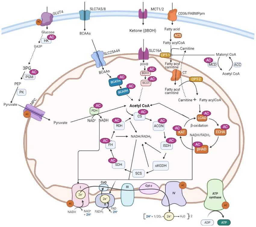
Frontiers in Cardiovascular Medicine | www.frontiersin.org 6 August 2021 | Volume 8 | Article 723996Ketema and Lopaschuk Acetylation Control of Cardiac Energy Metabolism FIGURE 2 | Metabolic proteins subjected to acetylation control in the heart. AC, lysine acetylation; GLUT4, glucose transporter isoform 4; SLC7A5/8, solute carrier family-7; SLC25A44, solute carrier family-25; SLC16, solute carrier family-16; MCT, monocarboxylate transporter 1; CD36, cluster of differentiation 36; FABPpm, plasma membrane fatty acid-binding protein; MCD, malonyl CoA decarboxylase; ACC, acetyl CoA carboxylase; PDH, pyruvate dehydrogenase; LCAD, long-chain acyl CoA dehydrogenase; β-HAD, β-hydroxyacyl CoA dehydrogenase; KAT, 3-ketoacyl-coa thiolase; ECH, enoyl-CoA hydratase; FAS, fatty acyl CoA synthase; CPT-1, carnitine palmitoyltransferase 1; CPT-2, carnitine palmitoyltransferase-2; CT, carnitine acyl translocase; BCAA, branched-chain amino acids; BCATm, mitochondrial branched-chain aminotransferase; BCADH, branched-chain amino acid dehydrogenase; β-OHB, β-hydroxybutyrate; BDH-1, 3-hydroxybutyrate dehydrogenase 1; SCOT, 3-ketoacid CoA transferase; CS, citrate synthase; ISDH, iso-citrate dehydrogenase; α-KGDH, alpha-ketoglutarate dehydrogenase; SCS, succinate CoA synthetase; MDH, malate dehydrogenase; SDH, succinate dehydrogenase; FH, fumarate hydratase; OAA, oxaloacetate; MCD, malonyl CoA decarboxylase; ACON, aconitase; HK, hexose kinase; GA3P, glyceraldehyde 3-phosphate; 3PG: 3-phosphoglycerate; PGM, Phosphoglycerate mutase; PK, pyruvate kinase; CoQ, coenzyme Q; Cytc: cytochrome C; FAD/FADH2 , flavin adenine dinucleotide; NAD/NADH2, nicotinamide adenine dinucleotide. HFD, obesity, and diabetes (157). The high fatty acid ß-oxidation In contrast to the scenarios discussed above, a study by rate seen in these conditions may also lead to the increased Koentges et al., in isolated working hearts, found a negative production of acetyl-CoA that can serve as a substrate for correlation between hyperacetylation of LCAD and its activity acetylation (82). Thus, it is reasonable to assume that increased along with reduced palmitate oxidation in SIRT3 deficient acetylation of fatty acid oxidative enzymes can further trigger transverse aortic constriction (TAC) mice (158). However, the enzyme activity and led to the continuation of fatty acid these hearts were perfused with a buffer that contained ß-oxidation in the heart in these circumstances. an ultra-physiological high concentration of glucose (11 mM) Frontiers in Cardiovascular Medicine | www.frontiersin.org 7 August 2021 | Volume 8 | Article 723996
Ketema and Lopaschuk                                                                                                     Acetylation Control of Cardiac Energy Metabolism

TABLE 1 | Effect of lysine acetylation on major metabolic enzymes in the heart.               fatty acid ß-oxidation. From the eight acetylated lysine residues
                                                                                              detected on LCAD, lysine residue 42 was identified as a critical
Metabolic pathway Enzymes             Effect on enzyme activity References
                                                                                              regulation site for acetylation (22). Furthermore, analysis of the
Fatty acid oxidation    LCAD                       ↑, ↓               (53, 83, 145)           rate of conversion of radiolabeled palmitate revealed lowered
                        β-HAD                       ↑                 (53, 83)                oxidizing capacity and ATP production in tissue homogenates
                        MCAD                        ↑                 (146)                   from SIRT3–/– compared to wild-type tissue at a high substrate
                        MCD                         ↑                 (73)
                                                                                              concentration. Reduced activities of fatty acid ß-oxidation
Glucose oxidation       PDH                         ↓                 (53, 83, 147)
                                                                                              enzymes in the liver by acetylation were also reported in other
                        MPC                         ↓                 (148)
                                                                                              studies (160). In addition to LCAD and β-HAD, hyperacetylation
Glycolysis              HK                          ↓                 (100)
                                                                                              of hydroxy acyl-CoA dehydrogenase, another important enzyme
                                                                                              in fatty acid ß-oxidation, led to its decreased activity and
                        PGM                         ↓                 (100)
                                                                                              decline in overall fatty acid oxidation rate in the mouse liver.
                        GLUT4                       ↓                 (149)
                                                                                              Deletion of GCN5L1 acetylase enzyme or overexpression of
Insulin signaling       Akt                         ↓                 (147, 150)
                                                                                              SIRT3 reduced hydroxy acyl-CoA dehydrogenase acetylation
TCA cycle               ICDH                       ↓,⇔                (151)
                                                                                              and increased its activity as well as fatty acid ß-oxidation in
                        SDH                         ↓                 (81)
                                                                                              the liver (161, 162). However, there is presently no agreement
                        ACON                        ↑                 (152)
                                                                                              on the functionally significant acetylation sites or SIRT3 target
ETC                     Complex I                   ↓                 (24, 153)
                                                                                              sites in these studies. While Hirschey et al. noted lysine 42
                        Complex III                 ↓                 (24)
                                                                                              residue on LCAD as an important regulation site (22), using
                        Complex V                   ↓                 (24, 153)
                                                                                              chemically acetylated recombinant proteins, Bharathi et al.
GLUT4, glucose transporter isoform 4; PDH, pyruvate dehydrogenase; LCAD, long-chain           identified Lys-318 and Lys-322 residues as an important site
acyl CoA dehydrogenase; β-HAD, β-hydroxyacyl CoA dehydrogenase; ICDH, iso-citrate             for LCAD acetylation/sirt3 deacetylation (160). To date, a
dehydrogenase; MCAD, medium-chain acyl CoA-dehydrogenase; MCD, malonyl CoA
                                                                                              detailed analysis of lysine residue modification and functional
decarboxylase; MPC, mitochondrial pyruvate carrier; SDH, succinate dehydrogenase;
ETC, electron transport chain; Akt, protein kinase B; PGM, phosphoglucomutase; ACON,          acetylation/deacetylation target sites for LCAD and β-HAD is
aconitase; HK, hexokinase; TCA, tricarboxylic acid; ↑, increased activity; ↓, decreased       lacking in the heart. Not all lysine residues within LCAD that
activity; ⇔, no change in the activity of the enzyme.                                         are acetylated are expected to impact LCAD activity in the
                                                                                              same manner. Understanding the acetylation status of different
                                                                                              acetylation sites, and their effect on the enzyme function in
and a lower fatty acid to albumin ratio (1.5%) where both                                     multiple tissues will help to characterize the tissues specific effects
conditions may contribute to decreased fatty acid ß-oxidation                                 of acetylation dynamics.
rates. Likewise, Chen et al. also reported an abnormal lipid                                      Compared to acetylation, the effect of succinylation and
accumulation and decreased palmitate ß-oxidation rates in                                     malonylation modification on fatty acid ß-oxidation enzymes is
TAC-induced hypertrophic hearts, with a further decline in                                    not clear. Some studies have shown an impaired β-oxidation and
SIRT3 KO mice hearts in association with hyperacetylation                                     accumulation of medium- and long-chain acylcarnitines in the
of LCAD (145). A recent study by Davidson et al. showed                                       liver and muscles of SIRT5 KO mice (26). Most of the acyl-CoA
reduced expression of genes of fatty acid catabolism despite                                  dehydrogenase enzymes, including very long-chain acyl-CoA
no overt abnormalities in mitochondrial respiration in mice                                   dehydrogenase (VLCAD), LCAD, and MCAD, were found to be
deficient for cardiac carnitine acetyltransferase and SIRT3,                                  hypersuccinylated, suggesting a suppressive effect of excessive
despite the fact that the hearts exhibited extreme acetylation                                succinylation on fatty acid oxidizing enzymes (26). In contrast,
(159). In common, these three studies were done in mice with                                  cardiac ECH is desuccinylated and activated by SIRT5 (46, 97).
TAC-induced heart failure, while the last two did not assess                                  But the effects of similar modifications on other enzymes in this
directly the acetylation status of the enzymes. Overall, it is                                pathway have yet to be determined.
not clear if TAC alters the dynamics of acetylation on LCAD                                       Overall, from all these data it is possible to suggest that the
differently, such as through distinctive sites or multiple site                               effect of lysine acetylation on fatty acid ß-oxidative enzymes
modifications. However, several acyl modifications may likely                                 may not be similar between different tissues, at least between
coexist under these circumstances, which possibly compete                                     the liver and heart. Reasonably, these differences may account
with acetylation for the same lysine residue (104). However,                                  for the specialization of these tissues in regulating fatty acid ß-
experimental data are lacking regarding these interactions in                                 oxidation differently in line with their physiological functions.
heart failure.                                                                                The liver has a high capacity for both synthesizing and oxidizing
   Unlike the heart, studies conducted in the liver reported                                  fatty acids and the two processes are regulated reciprocally during
an inhibitory effect of acetylation on fatty acid metabolism.                                 a fed or fasting state as well in disease conditions such as in
Hirschey et al. described a decreased activity of LCAD enzyme                                 obesity or diabetes. On the contrary, the heart continually uses
and reduced fatty acid ß-oxidation following hyperacetylation of                              fatty acids as a source of energy, which accounts for up 60–
these enzymes in SIRT3 KO mice. The authors further reported                                  90% of the total energy requirements for its normal contractile
an accumulation of long-chain acylcarnitine species, fatty acid                               function irrespective of the fed state or disease conditions.
ß-oxidation intermediate products, and triacylglycerol in livers                              While acetylation may serve as a feedback regulation in the
from SIRT3 KO mice that could suggest a decreased rate of                                     liver as suggested by Bharathi et al. (160), the same mechanism

Frontiers in Cardiovascular Medicine | www.frontiersin.org                                8                                      August 2021 | Volume 8 | Article 723996
Ketema and Lopaschuk Acetylation Control of Cardiac Energy Metabolism
would potentially compromise the heart’s ability to produce that lysine acetylation at lysine 202 inhibits PDP1 by dissociating
energy if fatty acid ß-oxidation enzymes were down-regulated by it from PDHA1, thus promoting its phosphorylation (97).
acetylation induced by excess fatty acid ß-oxidation. Thus, future Hypersuccinylation of PDH accompanied by a decrease in
studies are needed to determine the differences in acetylome SIRT5 expression is also associated with a decrease in PDH
between the two tissues, and if acetylation-mediated regulation activity as the heart maturates in postnatal rabbits (100).
of fatty acid ß-oxidation is tissue- or site-specific. Unexpectedly, Park et al. observed the suppressive effect of
SIRT5 catalyzed desuccinylation on PDH in SIRT5 KO mouse
Glucose Metabolism embryonic fibroblasts (MEFs), while SIRT5 deletion led to
Glucose Oxidation an increase in PDH activity (25). In contrast, Zhang et al.
In contrast to fatty acid metabolism, the effects of acetylation found significantly reduced malate/pyruvate-driven respiration
on glucose metabolism have received less attention, especially in SIRT5-deficient HEK293 cells, as well as in homogenates
in the heart. However, recent studies reported the acetylation prepared from SIRT5 KO livers (166). While these discrepancies
of several proteins involved in glucose transport, glycolysis, and need further investigation, tissue/cell-specific variation in these
glucose oxidation (83, 149, 156). The pyruvate dehydrogenase modifications may contribute to these differences.
complex (PDH) is one of the most widely investigated glucose
metabolizing enzymes in relation to acetylation. It is a key Glycolysis
enzyme that catalyzes the irreversible and rate-limiting step Acetylation of glycolytic enzymes has been reported in hearts
in glucose oxidation that links glycolysis to the TCA cycle. as well as various other cells. In newborn rabbit hearts, we
PDH is regulated by several mechanisms, but the change in its showed a significant decline in glycolysis rates in line with
phosphorylation status is critical to its activity. It is inhibited hyperacetylation of its enzymes, including hexokinase (HK-
by phosphorylation on the E1 subunit by a specific PDH kinase 1) and phosphoglycerate mutase (PGM) (100). In contrast,
(PDK), PDK4, and is activated when dephosphorylated by PDH Hallows et al. have shown a negative regulation of PGM
phosphatase (163, 164). by SIRT1 (165). Acetylated PGM displayed enhanced activity,
Modification of PDH by acetylation has been demonstrated while Sirt1-mediated deacetylation reduced its activity in
in several studies (53, 83, 156). In HFD induced obese mice human embryonic kidney (HEK293) cells (167). Glyceraldehyde-
subjected to a TAC, we showed a significant increase in PDH 3-phosphate dehydrogenase (GAPDH) is another glycolytic
acetylation with a decrease in glucose oxidation rates (147). enzyme subjected to acetylation modification. Similar to PGM,
Similarly, Thapa et al. reported increased acetylation of the acetylation of GAPDH at lysine 254 increases its enzymatic
α-subunit of PDH in the heart after HFD feeding, and its activity in response to glucose in HEK293T cells (168).
hyperacetylation was shown to inhibit its activity (53). Reduced In contrast, GAPDH acetylation enhances its translocation
activity of PDH in association with its increased acetylation and from the cytoplasm to the nucleus in NIH3T3 cells, thereby
decreased SIRT3 level has been also demonstrated in mice with inhibiting downstream glycolysis and accumulation of glycolytic
angiotensin II-induced cardiac hypertrophy (165). intermediates (169). In another study, Xiong et al. showed
In addition, increased acetylation has also been implicated in an inhibitory effect of acetylation of pyruvate kinase (PK),
a reduced transport of pyruvate into the mitochondria. Akita which catalyzes the last step of glycolysis (170). In addition
type 1 diabetic mice hearts exhibit a significant hyperacetylation to acetylation, other acyl modifications can regulate glycolysis.
state, along with a 70% decrease in the rate of mitochondrial GAPDH, PGK, and enolase are hypermalonylated in livers of
pyruvate transport that occurs without any changes in the protein db/db mice (142), although the effects of this hypermalonylation
level of the mitochondrial pyruvate carriers 1 and 2 (MPC1 and on the activities of these enzymes has not been investigated.
MPC2). Mass spectrometry analysis revealed that acetylation of Another study in hepatocytes demonstrated SIRT5 mediated
lysines 19 and 26 of MPC2 were increased in Akita mice heart demalonylation of GAPDH and increased activity, suggesting
mitochondria, and that acetylation at these sites is associated with that malonylation decreases glycolytic flux (27). Similarly,
impaired pyruvate metabolism in the heart (148). desuccinylation of PK by SIRT5 increases its kinase activity
The impact of acetylation on PDH has also been examined in (171). In contrast, Xiangyun et al. reported that desuccinylation
skeletal muscle by Jing et al. Deletion of SIRT3, both in vivo in of PKM2 by SIRT5 inhibits its activity in tumor cells (172).
SIRT KO mice and in vitro in myoblasts, lead to a significant Unfortunately, there is a lack of data on the effect of malonylation
increase in acetylation associated with decreased PDH activity and succinylation modifications on glycolysis in the heart.
along with a reduced glucose oxidation rate and accumulation At the transcriptional level, glycolysis is regulated by the level
of pyruvate and lactate metabolites. Six acetylation sites have of hypoxia-inducible factor-1α (HIF-1α), a master transcriptional
been identified on the PDH E1α subunit, with lysine 336 being regulator of glycolytic enzymes (173). Studies by Geng et al.
significantly altered by SIRT3 deletion (156). Interestingly, it have shown a positive correlation between increased acetylation
was also shown that hyperacetylation of the PDH E1α at lysine of HIF-1α by p300 at lysine 709 and its stability, or decreased
336 enhances its phosphorylation leading to suppressed PDH polyubiquitination, in HEK293 cells (174). Similarly, other
enzymatic activity. Additionally, Ozden et al. and Fan et al. also studies also showed an inhibition of HIF-1α by SIRT1 mediated
demonstrated the inhibitory effect of acetylation at lysine 321 deacetylation at lysine 674, in HT1080 and HEK293 cells (175).
on PDH activity in cancer cells, which is completely reversed by These results were also found in SIRT6 deficient embryonic
SIRT3 activation (97, 164). In the latter study, it was also shown stem cells and MEFs cells (174). Interestingly, these cells exhibit
Frontiers in Cardiovascular Medicine | www.frontiersin.org 9 August 2021 | Volume 8 | Article 723996Ketema and Lopaschuk Acetylation Control of Cardiac Energy Metabolism
increased glucose uptake with up-regulation of glycolysis in acetylation status and its impact on enzymes involved in ketone
association with increased HIF-1α activity. and BCCA metabolism.
The impact of acetylation on glucose uptake and its In hepatic mitochondria of SIRT3 KO mice, hydroxymethyl-
transporters has also been described. In both cultured glutaryl (HMG)-CoA synthase (HMGCS2), the rate-limiting
cardiomyocytes and perfused hearts, Renguet et al. found enzyme in ketogenesis, is hyperacetylated and its enzymatic
that acetylation of GLUT 4 inhibits glucose uptake in adult activity reduced, leading to a decrease in β-hydroxybutyrate
cardiomyocytes, as well as in perfused hearts, by decreasing synthesis. Deacetylation of HMGCS2 by SIRT3 increases its
its translocation to the plasma membrane (173). Strikingly, enzymatic activity and β-hydroxybutyrate levels (183). Similar to
treatment with inhibitors of acetyltransferases prevents the the acetylation effect, loss of SIRT5 results in hypersuccinylation
increase in protein acetylation and reverses the inhibition of of HMGCS2 and reduces its activity both in vivo and in vitro
glucose uptake and GLUT4 translocation (149). Unfortunately, in liver mitochondria, which leads to reduced β-hydroxybutyrate
the direct acetylation status of GLUT4 was not analyzed in this levels during fasting (26).
study. However, the inhibitory effect of acetylation on glucose With regard to ketone oxidation, succinyl-CoA:3-ketoacid-
uptake is supported by other studies. Using SIRT3 KO mice and CoA transferase (SCOT), a key enzyme of ketone oxidation, is
hyperinsulinemic-euglycemic clamp experiments, Lantier et al. hyperacetylated in the brain and heart at multiple sites in SIRT3
showed that increased acetylation leads to insulin resistance and KO mice (184). In vitro biochemical analysis of recombinant
reduced muscle glucose uptake that is associated with decreased SCOT demonstrates that acetylation at lysine 451 residues results
hexokinase II (HKII) binding to the mitochondria in HFD-fed in decreased enzyme activity that is reversed by SIRT3 activation.
SIRT3 KO mice (176). This suggests a reduced HKII activity and Moreover, in brain homogenates from WT and SIRT3 KO mice,
translocation as a result of increased acetylation. Similar to the acetoacetate-dependent acetyl-CoA production is decreased by
above study, unfortunately, there was no direct analysis of the three-fold in SIRT3 KO mice, suggesting decreased ketone
acetylation status of the proteins involved in glucose uptake or oxidation rates upon increased acetylation (180). In contrast, a
glucose phosphorylation. decrease in ketogenesis capacity was noted in the liver of mice
lacking SIRT3 (184). However, similar studies are lacking in
Insulin Signaling the heart.
Insulin resistance in type 2 diabetes and obesity as well as in other Similarly, enzymes in BCAA (isoleucine, leucine, and valine)
heart conditions contributes to a number of adverse changes in catabolic pathways are among the proteins regulated by
the heart that includes alterations in cardiac energy metabolism, acetylation and SIRT3 in the liver. Some acetylation sites
lipotoxicity, and hypertrophy, and is associated with an increased were detected in branched-chain alpha-keto acid dehydrogenase
risk of heart failure (84, 177, 178). Cardiac insulin signaling is (BCKDH), a key enzyme catalyzing the breakdown of BCAAs,
impaired in heart failure, diabetes, and obesity (147, 178, 179). in SIRT3 KO mice (181, 182). As BCAA levels were raised,
Recent studies have shown that several proteins in the insulin the authors suggested that acetylation may have an inhibitory
signaling pathway are targets for acetylation modification, which effect on branched-chain ketoacid dehydrogenase (BCKDH)
therefore may impact insulin signaling. Akt is an important activity (23, 185). Other investigators have also suggested
component of the insulin signaling pathway. Akt activation that acetylation of BCAA aminotransferase (BCAT) promotes
requires binding with phosphatidylinositol 3,4,5-trisphosphate its degradation in the ubiquitin-proteasome pathway, thereby
[PIP (3)], which promotes its membrane localization and decreasing BCAA catabolism in the pancreas (186). cAMP-
phosphorylation by the upstream kinase, phosphoinositide- responsive element-binding (CREB)-binding protein (CBP) and
dependent protein kinase 1 (PDK1). Previously, we have shown SIRT4 were identified as the acetyltransferase and deacetylase for
a negative association between acetylation of insulin signaling BCAT at lysine 44 (K44), respectively (186).
mediators, such as Akt and PDK1, and their decreased activation
as a result of changes in their phosphorylation status due to Acetylation and TCA Cycle Enzymes
acetylation (147). In support of this, Sundaresan et al. showed Acetyl-CoA is the final common product in the oxidative
that acetylation of Akt and PDK1 occurs in their pleckstrin metabolism of various fuels, and is a substrate for the TCA
homology (PH) domains, which blocks PIP (3) binding, and cycle. While acetylation of all TCA cycle enzymes have been
that this is reversed by SIRT1 deacetylation (150). SIRT2 also reported in the liver (20), 6 of the 8 enzymes were found
binds and activates Akt in insulin-responsive cells, through to be acetylated in the heart (151). However, examination
its interaction with the PH domain, whereas SIRT2 inhibition of the effect of acetylation on the TCA cycle has produced
impairs AKT activation by insulin (180). mixed results. Increased acetylation of malate dehydrogenase
(MDH) in Chang liver cells enhances its enzyme activity. When
cells were treated with deacetylase inhibitors, trichostatin A
Acetylation and Metabolism of Other Fuel (TSA) and NAM, MDH acetylation doubled the endogenous
Substrates MDH activity, while in vitro deacetylation of purified MDH
Ketone body and BCAA oxidation can impact cardiac energy decreased its activity (20). Similarly, significant acetylation-
metabolism and heart failure progression (179, 181, 182). Both dependent activation of aconitase was found in both isolated
pathways may also contribute to mitochondrial acetylation heart mitochondria subjected to in vitro chemical acetylation,
changes. However, only a few studies have characterized the and in hearts of HFD fed obese mice (152). Increased aconitase
Frontiers in Cardiovascular Medicine | www.frontiersin.org 10 August 2021 | Volume 8 | Article 723996You can also read