Metabolic Flexibility and Mitochondrial Bioenergetics in the Failing Heart. Therapeutic Approaches - Romanian Journal of Cardiology
←
→
Page content transcription
If your browser does not render page correctly, please read the page content below
Romanian Journal of Cardiology | Vol. 31, No. 2, 2021

REVIEW

Metabolic Flexibility and Mitochondrial Bioenergetics in
the Failing Heart. Therapeutic Approaches
Mariana G. ROSCA1

            ABSTRACT
            Objectives. We will review current concepts regarding bioenergetic decline in heart failure (HF). In the heart,
            the high energy demand must be met by continuous ATP generation. Cardiac energetic machinery orchestrates
            the ATP production by using oxidation of multiple energetic substrates including fatty acids (FA), glucose, amino
            acids and ketone bodies. The normal heart is metabolically flexible and able to use different energetic fuels during
            physiologic or pathologic circumstances to better match the energy demand. Mitochondria have critical role in
            maintaining cardiac metabolic flexibility.
            Methods. We analyzed the scientific literature pertinent to HF and mitochondrial dysfunction.
            Results. The general consent is that metabolic flexibility is lost in HF with either preserved or reduced ejection
            fraction (HFpEF and HFrEF, respectively). The prototype of HFpEF is the metabolic heart disease that is characterized
            by increased reliance on FA oxidation for ATP production and decreased glucose oxidation, while HFrEF presents
            a decreased FA oxidation. Both types of HF are associated with a decline in mitochondrial function leading to
            increased oxidative stress, abnormalities in the redox status and energy deficit.
            Conclusion. Current research is committed to find novel metabolically targeted therapeutic approaches to improve
            energetic metabolism and alleviate HF progression.
            Keywords: mitochondria, heart failure, energy.

            REZUMAT
            Objective. Lucrarea reprezintă o revizuire a conceptelor curente referitoare la declinul bioenergetic în insuficiența
            cardiacă (IC). În cord, consumul mare de energie trebuie compensat prin generare continuă de ATP. Sistemul
            energetic cardiac orchestrează producția de ATP prin folosirea de substraturi energetice multiple incluzând acizi
            grași (AG), glucoză, aminoacizi și corpi cetonici. Inima sănătoasă este metabolic flexibilă și capabilă să utilizeze
            substraturi energetice variate în diferite circumstanțe fiziologice și patologice pentru a răspunde cerinței energetice.
            Mitocondriile au un rol critic în menținerea flexibilității metabolice cardiace. Metode. Am analizat literatura științifică
            referitoare la IC și disfuncția mitocondrială.
            Rezultate. Consensul general este că flexibilitatea metabolică este pierdută în ambele forme de IC, IC cu fracția
            de ejecție prezervată sau redusă (ICpFE și ICrFE, respectiv). Prototipul de ICpFE este boala cardiacă metabolică
            care este caracterizată prin creșterea oxidării AG pentru producerea de ATP și scăderea oxidării glucozei, în timp
            ce ICrFE prezintă scăderea oxidării AG. Ambele tipuri de IC sunt asociate cu declinul funcției mitocondriale care
            determină creșterea stresului oxidativ, anomalii în statusul redox, și deficit energetic.
            Concluzie. Cercetarea curentă este determinată să găsească noi abordări terapeutice orientate spre metabolism
            cu intenția de a îmbunătăți metabolismul energetic și atenua evoluția în IC.
            Cuvinte cheie: mitocondrii, insuficiență cardiacă, energie.

INTRODUCTION                                                               re worldwide2. There are two major types of HF, HF
Heart Failure (HF) is a growing public health concern,                     with reduced ejection fraction (HFrEF) and HF with
and a leading cause of morbidity and mortality in in-                      preserved ejection fraction (HFpEF), which share simi-
dustrialized countries worldwide. HHF is a frequent                        lar prevalence and poor prognosis with mortality rates
disease with a prevalence of approximately 37.7 milli-                     of 50% 5 years after diagnosis3. HFrEF is defined by
on globally1 and accounting for 2-3% of total healthca-                    the presence of systolic dysfunction with an ejection

1
    Department of Foundational Sciences, Central Michigan University          Contact address:
    College of Medicine, Mount Pleasant Michigan, USA                      Mariana G. ROSCA, Central Michigan University College of Medicine,
                                                                           2630 Denison Drive, Research Building Room 105, Mount Pleasant MI
                                                                           48858, USA.

                                                                                                                                          269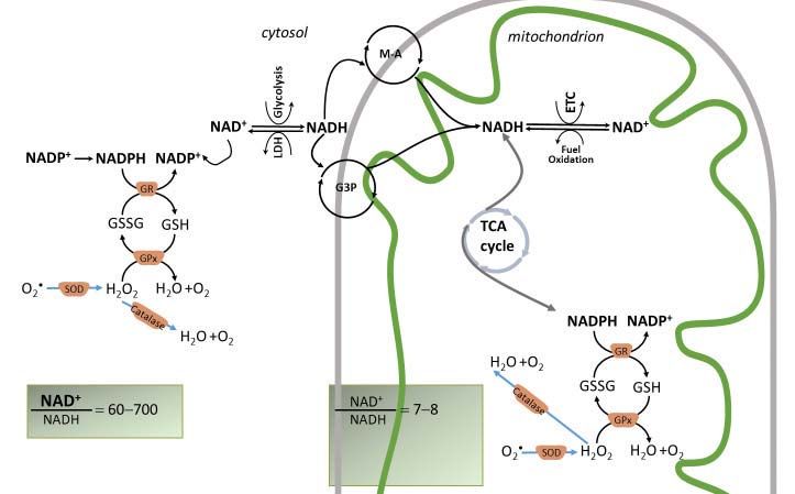
Mariana G. ROSCA et al.                                                                                            Romanian Journal of Cardiology
Bioenergetics as a Therapeutic Target in Heart Failure                                                                       Vol. 31, No. 2, 2021

fraction lower than 45% disregarding the diastolic dys-                          The impairment of bioenergetics is considered a
function. HFpEF is characterized by an increased left                         key pathogenic mechanism in HF. The heart needs
ventricle (LV) filling pressure without LV dilation, and                      energy in the form of ATP in both systolic and di-
with ejection fraction higher than 50%4. Disregarding                         astolic periods to sustain the excitation contraction
the type, effective therapeutic strategies to preserve                        coupling and myosin-actin cross-bridge cycles, as well
the remaining functional myocardium and delay the                             as termination of contraction supported by energy de-
progression of HF are yet to be determined. In additi-                        pendent processes including calcium sequestration in
on, although both types of HF have different features,                        the sarcoplasmic reticulum and its extrusion from car-
they are treated with similar traditional drugs5 with                         diomyocytes. During maximal exercise cardiac muscle
little success.                                                               uses 90% of its oxidative capacity indicating that the
    As a complex clinical syndrome induced by impai-                          heart lacks an excess capacity for energy production
red contractile and/or relaxation performances of                             over energy utilization. There is no significant energy
the myocardium, HF leads to inability of the heart to                         deposit, and the coupling between energy supply and
supply adequate amounts of blood to meet the pe-                              consumption follows a “pay as you go” basis. This
ripheral tissues metabolic needs. Cardiac ischemia,                           means that the energy demand dictates the intensity
increased preload and afterload, neurohormonal dys-                           of energy production.
regulation, and intrinsic abnormalities of the myo-                              Ninety percent of cardiac energy requirement is
cardium are common etiologic factors of HF2. Major                            provided by mitochondrial oxidative phosphorylation,
pathogenic mechanisms responsible for HF progressi-                           which is finely tuned to the energy demand. An opti-
on are abnormalities of calcium homeostasis and bioe-                         mal energy balance is achieved when energy producti-
nergetics, alterations of the cardiac contractile appara-                     on matches the energy consumption. HF is associated
tus with impaired mechanics, and increased oxidative                          with altered mitochondrial bioenergetics2, which may
stress 2 (Figure 1).                                                          induce a state of energy starvation and is correlated

Figure 1. Major structural, functional, metabolic and bioenergetic features of the failing heart. Heart failure (HF) with preserved ejection fraction
and HF with reduced ejection fraction may both evolve to congestive HF. These functional abnormalities are progressively induced by either primary
stiffness of the ventricular wall (defect in cardiomyocyte relaxation, interstitial fibrosis and cardiomyocyte hypertrophy) or contractile dysfunction,
cardiomyocyte death and eventually chamber dilation. The heart relies on constant ATP supplied by oxidative metabolism. Heart failure is considered
a disease of the myocardial energetic metabolism induced by mitochondrial dysfunction leading to energy deficit, increased oxidative stress and altered
redox status.

270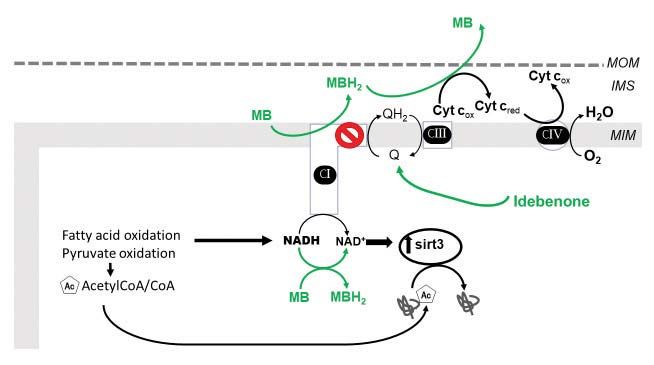
Romanian Journal of Cardiology Mariana G. ROSCA et al.
Vol. 31, No. 2, 2021 Bioenergetics as a Therapeutic Target in Heart Failure
with hemodynamic markers of severity in human sub-
jects with HF6,7.
Current therapies are mostly focused on decrea-
sing myocardial oxygen consumption and energy de-
mand, and aimed to decrease heart rate and afterload.
These therapies are limited by their own effects that
include hypotension and bradycardia. The results of
the majority of phase III clinical trials with cardiopro-
tective agents performed in the last decade have been
largely negative8. Current inotropic therapy is also
limited by its disadvantage of increasing oxygen con-
sumption by the less efficient failing heart. Therefore,
there is a need for therapies to act on activating signals
Figure 2. Electron microscopy image of the mouse heart.
to increase energy production. Mitochondria is central
for cardiac bioenergetics, and the major site of ATP
production. This review focuses on alterations in mi- pyruvate, is either converted to lactate or transported
tochondrial bioenergetics in HF, and novel therapeutic into mitochondria via the mitochondrial pyruvate car-
strategies aimed to correct mitochondrial dysfunction rier, and converted by pyruvate dehydrogenase (PDH)
in order to balance the bioenergetics and improve the to acetyl-CoA for the tricarboxylic acid (TCA) cycle,
HF outcome. also known as Krebs cycle (Figure 3).
After entry into cardiomyocytes, long chain FAs
MITOCHONDRIAL ENERGY (i.e., palmitate) are activated to FA-CoA that is either
METABOLISM IN THE NORMAL HEART esterified as triacylglycerol or enter the mitochondria
The heart weights only approximately 0.5% of the via carnitine palmitoyltransferases (CPT1 and 2) to be
human body and consumes 8% of the 65 Kg of ATP oxidized via FA -oxidation. The end products of each
produced by the whole body per day. Therefore, the FA -oxidation cycle are NADH, FADH2 and acetyl-
heart is the highest metabolically active tissue in the CoA, which are further oxidized by electron transport
human body. Approximately 95% of cardiac ATP re- chain (ETC) complexes or Krebs cycle, respectively,
sults from mitochondrial oxidative metabolism with ultimately leading to ATP synthesis via mitochondrial
the rest deriving from glycolysis6. Cardiomyocytes are oxidative phosphorylation. FA -oxidation is control-
rich in mitochondria that are located both beneath the led at different steps including the inhibitory effect of
plasma membrane (subsarcolemmal) and within the in- malonylCoA (formed from AcCoA via AcCoA carbo-
terfibrillar regions of cardiomyocytes (Figure 2). xylase, ACC), FADH2/FAD+ and NADH/NAD+ redox
In order to accomplish their energetic mission, ratios, and acetyl-CoA/CoA ratio, all unfavorable to
cardiac mitochondria transform the chemical energy FA oxidation12. MalonylCoA is degraded by malonyl-
stored in fuel substrates into ATP through oxidative CoA decarboxylase (MCD) thus releasing its inhibito-
phosphorylation. The normal adult heart obtains 60% ry effect on CPT1 (Figure 3).
of ATP from fatty acids (FA) oxidation with the re- HB is produced by the liver at rates proportional
maining 40% originating from the oxidation of other to FA oxidation and NADH/NAD+ ratio, and repre-
fuel substrates including glucose, lactate, amino acids sents the main ketone body utilized by the heart as
and ketones (mainly -hydroxybutyrate, HB) (Figure an energy fuel. Within mitochondria, HB is sequen-
3). tially converted to acetoacetate, acetoacetyl-CoA and
While glucose uptake into cardiomyocytes is de- acetyl-CoA for the Krebs cycle10 (Figure 3). Cardiac
pendent on insulin activity, the uptake of FA and HB mitochondria can also fully metabolize branched chain
is not hormonally regulated9,10. Glucose enters cardi- amino acids (leucine, isoleucine and valine) providing
omyocytes mostly via the insulin-dependent glucose acetyl-CoA for the Krebs cycle and succinyl-CoA for
transporter4 (GLUT4)11 and is directed through mul- anaplerosis. Krebs cycle is a source of reducing equi-
tiple metabolic pathways such as glycolysis, glycogen valents in the form of NADH and NADPH.
synthesis, polyol, hexosamine biosynthetic or pentose While electrons are transferred from the reducing
phosphate pathways. The end product of glycolysis, equivalents, NADH and FADH2, to oxygen by the ETC
271Mariana G. ROSCA et al. Romanian Journal of Cardiology
Bioenergetics as a Therapeutic Target in Heart Failure Vol. 31, No. 2, 2021
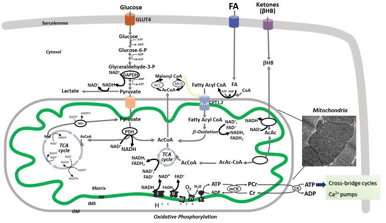
Figure 3. Cardiac oxidative metabolism. Normal adult heart obtains ATP mostly from fatty acid (FA) oxidation with the remaining delivered from
glucose, amino acids and ketones (mainly -hydroxybutyrate, HB)5. Glucose uptake is mediated by the glucose transporter4 (GLUT4), and follows
multiple metabolic pathways including glycolysis and mitochondrial glucose oxidation. For simplicity, other metabolic pathways are not depicted in this
figure. The end product of extramitochondrial glycolysis, pyruvate, is converted by mitochondrial pyruvate dehydrogenase (PDH) to acetyl-CoA (Ac-
CoA) that enters the tricarboxylic acid (TCA) cycle (Krebs cycle). Long chain FAs are activated to FA-CoAs that enter the mitochondria via carnitine
palmitoyltransferases (CPT1 and 2) and are oxidized via FA -oxidation. The end products of pyruvate and FA -oxidation spiral are NADH, FADH 2
and acetyl-CoA, which are further oxidized by electron transport chain (ETC) complexes or Krebs cycle, respectively, ultimately leading to ATP syn-
thesis via mitochondrial oxidative phosphorylation. FA -oxidation is inhibited by malonylCoA (formed from AcCoA via AcCoA carboxylase, ACC),
FADH 2 /FAD+ and NADH/NAD+ redox ratios, and acetyl-CoA/CoA ratio. MalonylCoA is degraded by malonylCoA decarboxylase (MCD) thus releas-
ing its inhibitory effect on CPT1. HB is oxidized within cardiac mitochondria to acetoacetate (AcAc) that is converted to acetyl-CoA for Krebs cycle.
Mitochondrial oxidative phosphorylation provides more than 95% of the cardiac ATP, with the remainder derived from glycolysis. While electrons are
transferred from the reducing equivalents, NADH and FADH 2 , to oxygen by the ETC complexes, an electrochemical gradient is developed across the
mitochondrial inner membrane (IM), which is used by the ATP synthase (complex V) to form ATP. Mitochondrial generated ATP is transferred to the
cytosol by the mitochondrial and cytosolic creatine kinases (CK) for contractile apparatus, sarcoplasmic reticulum Ca2+-ATPase and other ion pumps.
The inset represents an electron micrograph of mouse cardiac muscle showing interfibrillar mitochondria.
complexes, an electrochemical gradient is developed mal heart to respond properly to the energy demand.
across the mitochondrial inner membrane (IM), whi- Energetic substrates have different energy efficiency,
ch is used by the ATP synthase (complex V) to pho- which is defined by the amount of ATP produced for
sphorylate ADP and form ATP. Mitochondrial ATP is the oxygen consumed and expressed as P/O ratio.
transferred to the cytosol by phosphate exchange ne- While FA oxidation gives the greatest ATP yield, it
tworks including mitochondrial and cytosolic creatine also uses the highest amount of oxygen with a P/O
kinases (CK) for contractile apparatus, sarcoplasmic ~2.3. Glucose is the most efficient energy substrate
reticulum Ca2+-ATPase and other ion pumps. with a P/O ratio of 2.58. 5. HB oxidation has an in-
termediate efficiency with a P/O~2.5.
CARDIAC METABOLIC FLEXIBILITY HB is oxidized by the normal heart in proportion
Although the heart is enzymatically equipped to simul- to its availability at the expense of FA and glucose10. It
taneously utilize multiple fuels to produce energy, it is is reported that HFpEF associated with diabetes acqui-
also able to change the relative contribution of these res the ability to shift the acetyl-CoA towards ketone
substrates to cardiac ATP in an effort to better adjust body synthesis, a characteristic of the fetal heart13. A
to different physiological and pathological conditions5. decrease in glucose oxidation induces HFpEF indica-
This characteristic is vital for the ability of the nor- ting that maintaining proper glucose metabolism is re-
272Romanian Journal of Cardiology Mariana G. ROSCA et al.
Vol. 31, No. 2, 2021 Bioenergetics as a Therapeutic Target in Heart Failure
Figure 4. The main redox couples governing the redox balance in cardiac mitochondria (NAD+/NADH, NADP+/NADPH, and GSH/GSSG). Normal
cardiomyocytes maintain a constant NAD pool. Both oxidized forms, NAD+ and NADP+, are hybrid acceptors, and are converted to the reduced
forms, NADH and NADPH. NADH is oxidized by complex I, and therefore, the NADH/NAD+ couple is important for ATP generation. The NADPH/
NADP+ redox couple is central to the antioxidant defense by donating electrons to glutathione (GSH/GSSG) that scavenges the hydrogen peroxide
(H 2O2 , a Reactive Oxygen Species, ROS) via the enzymes glutathione reductase (GR), glutathione peroxidase (GPx). H 2O2 is generated from superox-
ide, O• 2 , by dismutation via the enzyme, superoxide dismutase (SOD). For simplicity, the thioredoxin antioxidant system is not shown. Mitochondrial
antioxidant system is mirrored by a similar scavenging mechanism in the cytosol. In these reactions, the reduced and oxidized members of the redox
couples interconvert but are not consumed. Catalase also scavenges H 2O2 .
Mitochondrial NADH/NAD+ and NADPH/NADP+ redox couples are linked by the enzyme nicotinamide nucleotide transhydrogenase (NNT) that
reduces NADP+ at the expense of NADH oxidation and utilizing the mitochondrial inner membrane proton motive force to drive this process. NNT
is a physiologically relevant source of NADPH to drive the enzymatic degradation of H 2O2 . The figure shows that the mitochondrial redox state of the
NADH/NAD+ and NADPH/NADP+ redox couples are maintained different as these nucleotides have different metabolic roles. The NADH/NAD+
pool supports the divergent transfer of electrons from fuel substrates to both the ETC and antioxidant system via NNT, and thus is only partially re-
duced in comparison to NADPH/NADP+. The cytosolic NADH is imported in mitochondria by redox shuttles, most commonly the malate-aspartate
(M-A) and glycerol 3 phosphate shuttles (G3P).
quired for cardiac metabolic health14,15. An excessive to the collapse of mitochondrial function. In terms of
dependence on FA oxidation occurs in the heart ex- ATP production, one molecule of palmitate yields far
posed to an excess in energy fuels (overfeeding-indu- more ATP than does glucose. Therefore, to maintain
ced obesity, metabolic syndrome and diabetes). a constant ATP content, a pronounced increase in glu-
In contrast, a reversal back to a fetal metabolic state cose oxidation must accompany a relatively modest
with overreliance on glucose oxidation and decreased decrease in FA oxidation. Most studies report that the
FA oxidation occurs in the failing heart, and is asso- decrease in FA oxidation is not compensated for by an
ciated with a state of “energy starvation” as glucose, increase in glucose oxidation20,21.
although a low oxygen consuming substrate, is also a The decrease in mitochondrial oxidative metabo-
low ATP-yield when calculated per mole5. Most cli- lism is associated with an increase in cytosolic glycoly-
nical16,17 and experimental18 studies confirm this type tic rates5. Although glycolysis is an alternate source of
of cardiac metabolic inflexibility, and show that the energy, producing 2 ATP molecules from one glucose
decrease in mitochondrial FA oxidation predicts the molecule, this is insufficient to compensate for energy
onset of contractile dysfunction in pressure overload- deficit because the complete glucose oxidation would
challenged rats19. In overt HF, disregarding the etio- produce 31 ATP molecules. The general conclusion is
logy, the severe decrease in FA oxidation may be due that there is no true metabolic switch characterized
273Mariana G. ROSCA et al. Romanian Journal of Cardiology
Bioenergetics as a Therapeutic Target in Heart Failure Vol. 31, No. 2, 2021
Figure 5. Mitochondrial redox modulators. Increasing the efficiency of the electron transport chain. The figure shows a proposed mechanism for the
NAD+ enhancing and lysine deacetylating effect of methylene blue (MB). A complex I defect causes a decrease in NADH oxidation. In experimental
models of complex I defect MB accepts electrons from catalytic subunits of complex I and become reduced (MBH2) whereas cytochrome c reoxidizes
MBH2 to MB. Therefore, MB provides an alternative electron route within complex I-deficient cardiac mitochondria and favors NADH oxidation thus
increasing NAD+ and SIRT3 activity. The administration of exogenous NAD or precursors (+) improved the mitochondrial NAD pool and cardiac
function (discussed in the main text). Idebenone increases the coenzyme Q pool.
by a decrease in FA oxidation and a corresponding enzymes, and is downregulated in both animal models
increase in glucose oxidation, and that the failing heart and humans with HFrEF26 but is increased in HFpEF
is an energy-compromised (starved) organ20. associated with metabolic syndrome, which is associa-
ted with an increase in FA oxidation19.
CARDIAC MITOCHONDRIA IN HF
ETC abnormalities
The ATP amount in the failing heart is reported de- There is plethora of evidence that specific activities
creased compared with the normal heart, suggesting of individual ETC complexes are decreased in HF20.
a decrease in mitochondrial oxidative phosphorylati- ETC complexes aggregate into functional supercom-
on. The decrease in mitochondrial oxidative capacity plexes27, and this form of organization provides a
is multifactorial and may be induced by 1) decreased more efficient electron transport and is protective
mitochondrial biogenesis pathway; 2) specific defects against excessive mitochondrial reactive oxygen spe-
in the ETC complexes. cies (ROS) generation27. A decrease in mitochondrial
Mitochondrial biogenesis supercomplexes has been reported in HF28.
The formation of new mitochondria (mitochondrial Cardiolipin (CL) is an anionic phospholipid with
biogenesis) is supported by synthesis of mitochon- four acyl chains that are enriched in linoleic acid
drial proteins and replication of mitochondrial DNA, ((C18:2)4-CL), which resides in the inner mitochon-
both processes under the control of the transcripti- drial membrane. CL provides structural and functio-
on factor peroxisome proliferator-activated receptor nal support to ETC components29, and its depletion
(PPAR ) and its co-activator (PGC1). PGC1 results in reduced activities of ETC complexes. CL
is considered the master regulator of mitochondrial is also proposed to maintain the structural integrity
biogenesis due to the activation of nuclear respiratory of ETC supercomplexes, as it may act as a molecular
factors 1 and 2, (NRF1 and 2) as well as mitochondri- ‘glue’ to hold the complex protein subunits together in
al transcription factor A (TFAM), all targeting genes a supramolecular organization30,31. In humans, reduced
encoding for mitochondrial proteins and mtDNA22,23. (C18:2)4-CL, due to defective CL remodeling (Barth
PGC1 is downregulated in humans with HF lea- syndrome), causes dilated cardiomyopathy associated
ding to decreased mitochondrial density24,25. PPAR with destabilization of all supercomplexes containing
is an isoform predominantly regulating FA oxidation complex IV, loss of complex I from the supermolecu-
274Romanian Journal of Cardiology Mariana G. ROSCA et al.
Vol. 31, No. 2, 2021 Bioenergetics as a Therapeutic Target in Heart Failure
lar assembly and decrease in the individual enzymatic between reduced and oxidized subunits within the mi-
activities of complexes I, III and IV31, indicating that tochondrial ETC according to their redox potential.
CL is essential for the function of cardiac mitochon- Mitochondria have multiple redox couples (redox pla-
dria. Myocardial ischemia32 and HF33-35 are associated yers)44 including NAD+ (oxidized)/NADH (reduced),
with mitochondrial dysfunction and CL peroxidation, NADP+/NADPH, GSSG (glutathione disulfide)/GSH
loss of total CL content and decrease in (C18:2)4- (glutathione) (Figure 3). Energized mitochondria have
CL. Approaches that target cardiolipin are likely to a high NADH concentration to provide electrons for
improve electron transport across the ETC and, by oxidative phosphorylation45. In contrast, in the extra-
correcting mitochondrial function, might be beneficial mitochondrial space, the NADPH/NADP+ ratio is ma-
in treating HF. intained in a reduced state (the reduced NADPH >
the oxidized NADP) via several enzymatic reactions
CONSEQUENCES OF ALTERATIONS IN in order to drive reductive biosynthesis and maintain
MITOCHONDRIAL BIOENERGETICS IN antioxidant defense. The cytosolic GSH/GSSG couple
is also maintained in a reduced state that is needed
HF
for ROS detoxification. These redox couples are in-
Oxidative stress is defined by an increase in re- terconnected (Figure 4). In mitochondria, the inner
active oxygen species (ROS) related to the antioxi- membrane nicotinamide nucleotide transhydrogena-
dant mechanisms. The ROS-generating sources in the se (NNT) reduces NADP+ at the expense of NADH
heart are both extramitochondrial and mitochondrial. oxidation, utilizing the mitochondrial inner membra-
Defects in the ETC complexes lead to an impaired ne protonmotive force to drive this process. NNT
electron flow with accumulation of electrons at ETC is a physiologically relevant source of mitochondrial
sites that, according to their redox potential, can do- NADPH46. The NADPH/NADP+ couple supplies elec-
nate electrons and univalently reduce the molecular trons to keep mitochondrial GSH pool in order to
oxygen to form superoxide, a strong ROS. Similarly, scavenge H2O2, a strong ROS47. In conclusion, redox
an increase in mitochondrial proton gradient (mito- signaling regulates metabolism while metabolic state
chondrial hyperpolarization) also slows down the influences redox signaling. The NAD+/NADH redox
electron transport and increases ROS generation36. couple is a critical node integrating metabolic and sig-
A mild generation of reactive oxygen species has naling events.
beneficial effects on the heart by facilitating physiolo- The redox signaling network linked to the NAD+/
gical adaptive responses such as adaptation to physical NADH couple depends on the total mitochondrial
exercise37. In addition, exercise training causes be- NAD pools. NAD is a substrate for enzymes including
neficial adaptation in the heart such as an increase in the SIRT family, which continuously converts NAD+
endogenous ROS-scavenging mechanisms37, restores to nicotinamide. As NAD is degraded, cardiomyocytes
bioenergetics in porcine models of HFpEF38, and al- must maintain a constant pool by de novo synthesis
leviates symptomology in patients with HFrEF39,40 and or recycle nicotinamide to replenish NAD. In car-
HFpEF41. diomyocytes, mitochondrial NAD pool is relatively
Uncoupling proteins dissipate the electrochemical high matching its critical role in mitochondrial Krebs
gradient by allowing proton translocation back into cycle and ETC48. Pathological cardiac hypertrophy, the
the mitochondrial membrane, thus uncoupling the prerequisite of HF, is associated with a decrease in
oxidation and phosphorylation processes. The obser- the cardiomyocyte NAD pool49. Similar observation
ved increased expression of mitochondrial uncoupling was reported in diabetic cardiomyopathy, a model of
proteins in HF42 might be a compensatory mechanism HFpEF50.
to reduce ROS by inducing a mild decrease in the mi- The oxidized form, NAD+, is an electron acceptor
tochondrial inner membrane electrochemical gradi- in the redox reactions. Therefore, NAD+ and NADH
ent, a process called “mild uncoupling”43. However, interconvert but are not irreversibly consumed. NAD+
the decrease in ROS production by uncoupling may participates in all major energetic pathways including
be an efficient ROS decreasing mechanism in absence glycolysis, Krebs cycle, FA oxidation, ketone body me-
of ADP, a state that is unlikely to occur in the heart tabolism, and ETC (Figure 2). NAD+ is a potent acti-
in vivo. vator of the Krebs cycle enzymes whereas NADH is a
Krebs cycle allosteric inhibitor, and increases in ETC
Alterations in the redox state. Classic exam- defects45,51,52. For example, Complex I45 and IV53 defects
ples of redox reactions are the transfer of electrons lead to increased mitochondrial NADH content. The
275Mariana G. ROSCA et al. Romanian Journal of Cardiology
Bioenergetics as a Therapeutic Target in Heart Failure Vol. 31, No. 2, 2021
deficiency of frataxin, a mitochondrial protein integral Energy deficit. There is a large variability regarding
to the assembly and function of iron-sulfur proteins the reported mitochondrial ETC defects in HF. The
in ETC complexes I, II and III and aconitase (Krebs causal relationship between these defects and the de-
cycle), is associated with an 85-fold decrease in cardi- crease in ATP has not been defined. For example, a
omyocyte NAD+/NADH ratio and pathologic cardiac severe murine complex I defect did not cause energy
hypertrophy52. Approaches to correct mitochondrial deficit45 suggesting that in the murine heart ATP pro-
ETC defects increased NAD+ content51. In conclusion, duction is not directly related to complex 1 activity.
the disruption of the electron flow to oxygen by ETC In contrast, most studies report bioenergetic impair-
defects increases NADH causing a highly-reduced re- ment in human subjects diagnosed with HF, which
dox environment within mitochondria. The cardiac manifest as decrease in cardiac ATP, phosphocreatine
amount of the oxidized form, NAD+, is reported re- (PCr)73,74, or, most common, a decline in the pCr/ATP
duced in HFrEF54, and either unchanged55 or altered56 ratio73,75,76.
in HFpEF.
While NADH/NAD+ redox ratio determines the THERAPEUTIC APPROACHES
production of mitochondrial ROS, the NADPH/
NADP ratio is key to antioxidant defense. They are Change the metabolic substrate preference
linked by the NNT enzyme that transfers electrons Metabolic inflexibility with an excessive increase in
from NADH to NADP+ (Figure 3). FA oxidation seems detrimental in HFpEF77. In this re-
Sirtuins (SIRTs) remove an acetyl group from lysine gard, the -adrenergic receptor antagonist, carvedilol,
residues in an NAD+-dependent manner by cleaving used in HF to reduce cardiac workload and decrease
NAD+ to nicotinamide57, and are reported to prolong oxygen consumption, also inhibited mitochondrial FA
lifespan in mammals58. There are seven mammalian uptake, increased glucose oxidation and limited the in-
SIRTs that differ in their cellular localization. Although farct size after ischemia78 indicating that balancing the
all SIRTs are NAD+-dependent, the extramitochon- metabolic health is beneficial for the heart.
drial SIRT 1 and mitochondrial SIRT3 are well-known Malonyl-CoA inhibits carnitine palmitoyltransferase
players in the heart. SIRT 1 protects against pathologic (CPT)1, the rate-limiting enzyme in mitochondrial FA
cardiac hypertrophy, and SIRT 1 knockout mice ex- uptake (Figure 2), and its amount is dependent on the
hibit developmental cardiac defects59. Sustained SIRT balance between the synthesis via acetyl-CoA carbo-
1 overexpression causes cardiomyopathy whereas xylase and degradation via malonyl-CoA decarboxyla-
moderate SIRT 1 expression ameliorates age-induced se. Inhibiting malonyl-CoA decarboxylase in animal
cardiac hypertrophy and dysfunction60, suggesting its models improved ischemic-induced cardiac dysfuncti-
effect is dose-dependent. SIRT 1 also protects mito- on, reduced cardiac FA oxidation, and increased the
chondrial function by activating PGC-1a61 to increase glycolytic flux79. Studies of malonyl-CoA decarboxyla-
mitochondrial FA oxidation62. Overall, NAD+, via SIRT se inhibitors are yet to be performed in human pati-
1, regulates pathological hypertrophy and mitochon- ents with HF.
drial metabolism. Due to the observed and possibly incomplete me-
SIRT3 is the major mitochondrial NAD+-dependent tabolic switch towards increased glucose use in both
deacetylase63. SIRT3 knockout causes cardiac hyper- animal models and humans with HF, it is proposed
trophy and failure under stress64 while overexpression that stimulating glucose oxidation may be an attractive
protects against pathological hypertrophy via activa- therapeutic strategy to compensate for the energe-
ting antioxidant mechanisms65. tically ‘starved’ failing heart80. Ketone body metabo-
SIRT 1 and SIRT3 regulate bioenergetic metabolism lism is altered in HF. There is an increased ketone
during energetic crises. SIRT3-mediated deacetylation utilization in the severely failing heart in humans81,82.
activates enzymes involved in glycolysis66,67, FA oxida- Further research is needed to understand the role of
tion68-70, Krebs cycle cycle71, and the ETC72. By upregu- ketone oxidation in the failing heart, and to determine
lating metabolic machinery during states of decreased whether targeting ketone metabolism is an efficient
fuel availability, SIRT3 appears to be a critical metabo- approach to improve energetics in HF.
lic regulator of coupling substrate oxidation with the Normalize the increased oxidative stress
formation of reducing equivalents to ATP production Although the increased oxidative stress is an accep-
thus maximizing efficiency. ted pathogenic mechanism in HF, clinical trials yielded
276Romanian Journal of Cardiology Mariana G. ROSCA et al.
Vol. 31, No. 2, 2021 Bioenergetics as a Therapeutic Target in Heart Failure
negative results to support the long term role of ROS tron transport, which bypass the ETC defect, rescued
scavengers to alleviate HF83,84. The lack of long-term mitochondrial function in ETC defects. For example,
benefits may be related to the inability to reach effec- methylene blue (MB), an FDA approved pharmaco-
tive therapeutic doses to stoichiometrically scavenge logical drug used to treat various ailments in human
the ROS due to poor absorption, decreased cellular subjects98-109 and rodents110-114, may provide such an
uptake or lack of strategy to match the ROS gene- electron route. MB has a low redox potential that
ration which is a continuous process. Mitochondrial allows the compound to receive electrons from com-
targeted antioxidants have been tested on experimen- plex I114-116 and become reduced (MBH2) while be-
tal models of cardiac disease and HF. For example, ing able to be re-oxidized by cytochrome c back to
XJB-5-131, a mitochondria-targeted ROS scavenger is MB114,117. Therefore, MB is protective because it helps
reported to decrease ROS generation and maintain the electrons to bypass the complex I and III defects
mitochondrial and cardiac functions in rats subjected and still maintain oxidative phosphorylation (Figure
to ischemia-reperfusion injury85. Similar effects were 5). We recently reported that MB protected retinal
obtained with another mitochondrial antioxidant, photoreceptors in a murine model of mitochondrial
MitoTEMPO86. The compound EUK-8, a mimetic of complex I defect118, and improved cardiac function by
two major mitochondrial antioxidant enzymes (supe- shifting electron away from NADH in diabetic cardi-
roxide dismutase and catalase, Figure 4), suppressed omyopathy, a model of HFpEF119.
the progression of cardiac dysfunction and diminished
Increase the efficiency of the electron
ROS production and oxidative damage in dilated car-
transport within the mitochondrial ETC
diomyopathy in mice87. These compounds are yet to
Coenzyme Q (CoQ) pool is composed by two redox
be tested in human subjects with HF.
coenzymes, the reduced uniquinol and the oxidized
Mitochondrial redox therapy ubiquinone. CoQ is endogenously synthesized, con-
As cardiac54 and circulating88 NAD pool are reported verted to ubiquinol by two-electron reduction from
decreased in HF, NAD-boosting strategies are expec- energetic substrates fed into complexes I and II (i.e.,
ted to be beneficial to the cardiac metabolic health. pyrucate, acetylCoA), which is then oxidized back to
For example, the food supplementation with nicotina- ubiquinone by donating electrons to complex III (Fi-
mide riboside, the most energy efficient NAD precur- gure 2). Incomplete, one-electron reduction of CoQ
sor was found beneficial in a murine model of dilated produces semiquinone, which is a highly reactive radi-
cardiomyopathy and transverse aorta constriction by cal. An increase in the reduced CoQ pool (ubiquinol)
stabilizing myocardial NAD+ levels in the failing heart89. causes a reverse electrons flow back to complex I re-
The oral supplementation with nicotinamide riboside sulting in ROS generation120. Circulating CoQ is de-
in patients with advanced HF decreased systemic in- creased in patients with HF121, which correlated with
flammation by normalizing mitochondrial function in poor clinical outcome and increased mortality122. Q-
peripheral blood mononuclear cells90. Elevating the SYMBIO clinical trial123 revealed a reduction in mor-
NAD level suppressed mitochondrial protein hyper- tality after 2 years of treatment with CoQ. Recently,
acetylation and cardiac hypertrophy, and improved CoQ analogues with more efficient penetrability into
cardiac function in responses to stresses91. the mitochondria have been developed. The deli-
NAD+-dependent SIRTs have been investigated as very to mitochondria was improved by novel quino-
therapeutic targets in HFpEF induced by diabetes. For ne conjugates that are tethered to lipophilic, cationic
example, resveratrol, a polyphenol and well-known triphenyl-phosphonium moieties, such as MitoQ and
SIRT 1 activator, alleviated diabetic cardiomyopathy SkQ124, which have proved efficient in experimental
via activating SIRT 1, 2, 3 and 592,93, improved glucose models of HF125. The administration of idebenone, a
metabolism in human subjects94,95 and decreased oxi- short-chain synthetic CoQ analogue126, has had promi-
dative stress in cultured cardiomyocytes96. In a rodent sing benefits in small clinical trial of genetic mitochon-
model of genetic obesity, resveratrol decreased cardi- drial defects127 and HF in experimental models128 and
ac fibrosis and improved FA metabolism97. human subjects129.
In the mitochondrial ETC, electrons are passed
Protection of cardiolipin
from donors to acceptors according to their redox
Cardiolipin is decreased of oxidized in HF. Maintaining
potential. As ETC defects delay or reverse the elec-
the amount and integrity of cardiolipin may be an effi-
tron transport, providing alternative paths for elec-
cient therapeutic approach to improve mitochondrial
277Mariana G. ROSCA et al. Romanian Journal of Cardiology
Bioenergetics as a Therapeutic Target in Heart Failure Vol. 31, No. 2, 2021
bioenergetics in HF. The cell-permeable tetrapeptide used to discover novel therapeutic targets and mito-
MTP-131 (Bendavia or Elamipretide) localizes to the chondrial modulator to mitigate HF.
mitochondrial inner membrane130, protects against
cardiolipin oxidation and improves mitochondrial func- Compliance with ethics requirements:
tion131,132. MTP-131 reduced pathologic hypertrophy The authors declare no conflict of interest regarding this
and cardiac remodeling, and improved mitochondrial article. The authors declare that all the procedures and ex-
and cardiac function in murine133-135, porcine136 and ca- periments of this study respect the ethical standards in the
Helsinki Declaration of 1975, as revised in 2008(5), as well
nine137 models of HF. In human subjects with HFrEF,
as the national law. Informed consent was obtained from all
elamipretide is safe and well tolerated, and improved the patients included in the study.
left ventricular function138.
Financial support: This research has been funded by Cen-
Calcium homeostasis
tral Michigan University College of Medicine and Ameri-
Altered Ca2+ homeostasis leading to impaired excitati- can Heart Association Institutional Research Enhancement
on–contraction coupling occurs in many types of HF139. Award.
Mitochondria are critical in regulating cardiomyocyte
calcium dynamics because all membrane-bound Ca2+
References
pumps are ATP- dependent. In the myocardium, Ca2+ 1. Ziaeian B and Fonarow GC. Epidemiology and aetiology of heart fail-
necessary for the cross-bridge actin-myosin cycles de- ure. Nat Rev Cardiol. 2016;13:368-78.
rives mostly from the extracellular space via the volta- 2. Brown DA, Perry JB, Allen ME, Sabbah HN, Stauffer BL, Shaikh SR,
Cleland JG, Colucci WS, Butler J, Voors AA, Anker SD, Pitt B, Pieske
ge-dependent Ca2+ channels with a lower contribution B, Filippatos G, Greene SJ and Gheorghiade M. Expert consensus
from the sarcoplasmic reticulum (SR) via the SR Ca2+ document: Mitochondrial function as a therapeutic target in heart
failure. Nat Rev Cardiol. 2017;14:238-250.
release channels, also called ryanodine receptors. Di- 3. Writing Group M, Mozaffarian D, Benjamin EJ, Go AS, Arnett DK,
astolic relaxation depends upon the Ca2+ sequestrati- Blaha MJ, Cushman M, Das SR, de Ferranti S, Despres JP, Fullerton
on within the SR via the SR Ca2+ ATPase (SERCA2a) HJ, Howard VJ, Huffman MD, Isasi CR, Jimenez MC, Judd SE, Kissela
BM, Lichtman JH, Lisabeth LD, Liu S, Mackey RH, Magid DJ, McGuire
and expulsion out of cardiomyocyte versus a Ca2+ DK, Mohler ER, 3rd, Moy CS, Muntner P, Mussolino ME, Nasir K,
pump and Na-Ca exchanger. Neumar RW, Nichol G, Palaniappan L, Pandey DK, Reeves MJ, Ro-
HFpEF is defined by impaired diastolic relaxation. driguez CJ, Rosamond W, Sorlie PD, Stein J, Towfighi A, Turan TN,
Virani SS, Woo D, Yeh RW, Turner MB, American Heart Associa-
Increasing the duration of the diastole by decreasing tion Statistics C and Stroke Statistics S. Heart Disease and Stroke
heart rate led to modest benefits in patients with Statistics-2016 Update: A Report From the American Heart Associa-
tion. Circulation. 2016;133:e38-360.
HFpEF140 potentially by providing more time for Ca2+ 4. Nagueh SF, Smiseth OA, Appleton CP, Byrd BF, 3rd, Dokainish H,
to move to the SR via SERCA2a. However, SERCA2a Edvardsen T, Flachskampf FA, Gillebert TC, Klein AL, Lancellotti
is reported downregulated in HF141, and SERCA2a P, Marino P, Oh JK, Alexandru Popescu B, Waggoner AD, Hous-
ton T, Oslo N, Phoenix A, Nashville T, Hamilton OC, Uppsala S,
overexpression improved cardiac function in experi- Ghent, Liege B, Cleveland O, Novara I, Rochester M, Bucharest R
mental models of HF142,143. Gene therapy through infu- and St. Louis M. Recommendations for the Evaluation of Left Ven-
sion of adeno-associated virus 1/SERCA2a in patients tricular Diastolic Function by Echocardiography: An Update from
the American Society of Echocardiography and the European As-
with HFrEF did not improved the clinical course144 sociation of Cardiovascular Imaging. Eur Heart J Cardiovasc Imaging.
suggesting that it is the SERCA2a ATP-dependent acti- 2016;17:1321-1360.
5. De Jong KA and Lopaschuk GD. Complex Energy Metabolic Chang-
vity rather than amount that is impaired in human HF. es in Heart Failure With Preserved Ejection Fraction and Heart Fail-
ure With Reduced Ejection Fraction. Can J Cardiol. 2017;33:860-
CONCLUSION 871.
6. Stanley WC, Recchia FA and Lopaschuk GD. Myocardial sub-
Heart failure is the unfortunate outcome of many car- strate metabolism in the normal and failing heart. Physiol Rev.
diac diseases. Disregarding the etiology, bioenergetic 2005;85:1093-129.
7. Barth AS, Kumordzie A, Frangakis C, Margulies KB, Cappola TP and
collapse is reported by most studies in human HF. Tomaselli GF. Reciprocal transcriptional regulation of metabolic and
Before the heart becomes an energy starved organ, signaling pathways correlates with disease severity in heart failure.
cardiac tissue suffers multiple consequences induced Circ Cardiovasc Genet. 2011;4:475-83.
8. Senni M, Gavazzi A, Gheorghiade M and Butler J. Heart failure at the
by mitochondrial defects including increased oxidative crossroads: moving beyond blaming stakeholders to targeting the
stress and changes in the redox status, both detrimen- heart. Eur J Heart Fail. 2015;17:760-3.
9. Bayeva M, Sawicki KT and Ardehali H. Taking diabetes to heart—de-
tal to the contractile apparatus and leading to poor regulation of myocardial lipid metabolism in diabetic cardiomyopa-
mechanics. Research conducted on experimental mo- thy. Journal of the American Heart Association. 2013;2:e000433.
dels of HF have contributed to our understanding of 10. Puchalska P and Crawford PA. Multi-dimensional Roles of Ketone
Bodies in Fuel Metabolism, Signaling, and Therapeutics. Cell metabo-
multiple aspects of bioenergetic impairment, and are lism. 2017;25:262-284.
278Romanian Journal of Cardiology Mariana G. ROSCA et al.
Vol. 31, No. 2, 2021 Bioenergetics as a Therapeutic Target in Heart Failure
11. Abel ED. Glucose transport in the heart. Front Biosci. 2004;9:201- 32. Lesnefsky EJ, Chen Q, Slabe TJ, Stoll MS, Minkler PE, Hassan MO,
15. Tandler B and Hoppel CL. Ischemia, rather than reperfusion, inhib-
12. Kerner J and Hoppel C. Fatty acid import into mitochondria. Bio- its respiration through cytochrome oxidase in the isolated, perfused
chim Biophys Acta. 2000;1486:1-17. rabbit heart: role of cardiolipin. Am J Physiol Heart Circ Physiol.
13. Cook GA, Lavrentyev EN, Pham K and Park EA. Streptozotocin dia- 2004;287:H258-67.
betes increases mRNA expression of ketogenic enzymes in the rat 33. Sparagna GC, Chicco AJ, Murphy RC, Bristow MR, Johnson CA,
heart. Biochimica et biophysica acta. 2017;1861:307-312. Rees ML, Maxey ML, McCune SA and Moore RL. Loss of cardiac
14. Sun W, Quan N, Wang L, Yang H, Chu D, Liu Q, Zhao X, Leng J and tetralinoleoyl cardiolipin in human and experimental heart failure. J
Li J. Cardiac-Specific Deletion of the Pdha1 Gene Sensitizes Heart to Lipid Res. 2007;48:1559-70.
Toxicological Actions of Ischemic Stress. Toxicol Sci. 2016;153:411. 34. Chatfield KC, Sparagna GC, Sucharov CC, Miyamoto SD, Gru-
15. Abel ED, Kaulbach HC, Tian R, Hopkins JC, Duffy J, Doetschman T, dis JE, Sobus RD, Hijmans J and Stauffer BL. Dysregulation of car-
Minnemann T, Boers ME, Hadro E, Oberste-Berghaus C, Quist W, diolipin biosynthesis in pediatric heart failure. J Mol Cell Cardiol.
Lowell BB, Ingwall JS and Kahn BB. Cardiac hypertrophy with pre- 2014;74:251-9.
served contractile function after selective deletion of GLUT4 from 35. Saini-Chohan HK, Holmes MG, Chicco AJ, Taylor WA, Moore RL,
the heart. The Journal of clinical investigation. 1999;104:1703-14. McCune SA, Hickson-Bick DL, Hatch GM and Sparagna GC. Cardio-
16. Anderson EJ, Kypson AP, Rodriguez E, Anderson CA, Lehr EJ and lipin biosynthesis and remodeling enzymes are altered during devel-
Neufer PD. Substrate-specific derangements in mitochondrial me- opment of heart failure. J Lipid Res. 2009;50:1600-8.
tabolism and redox balance in the atrium of the type 2 diabetic hu- 36. Korshunov SS, Skulachev VP and Starkov AA. High protonic poten-
man heart. J Am Coll Cardiol. 2009;54:1891-8. tial actuates a mechanism of production of reactive oxygen species
17. Anderson EJ, Lustig ME, Boyle KE, Woodlief TL, Kane DA, Lin CT, in mitochondria. FEBS Lett. 1997;416:15-8.
Price JW, 3rd, Kang L, Rabinovitch PS, Szeto HH, Houmard JA, Cor-
37. Frasier CR, Moore RL and Brown DA. Exercise-induced cardiac pre-
tright RN, Wasserman DH and Neufer PD. Mitochondrial H2O2
conditioning: how exercise protects your achy-breaky heart. J Appl
emission and cellular redox state link excess fat intake to insulin re-
Physiol (1985). 2011;111:905-15.
sistance in both rodents and humans. J Clin Invest. 2009;119:573-81.
38. Marshall KD, Muller BN, Krenz M, Hanft LM, McDonald KS, Dell-
18. Aon MA, Tocchetti CG, Bhatt N, Paolocci N and Cortassa S. Pro-
sperger KC and Emter CA. Heart failure with preserved ejection
tective mechanisms of mitochondria and heart function in diabetes.
fraction: chronic low-intensity interval exercise training preserves
Antioxid Redox Signal. 2015;22:1563-86.
myocardial O2 balance and diastolic function. J Appl Physiol (1985).
19. Bayeva M, Sawicki KT and Ardehali H. Taking diabetes to heart--de-
regulation of myocardial lipid metabolism in diabetic cardiomyopa- 2013;114:131-47.
thy. J Am Heart Assoc. 2013;2:e000433. 39. Flynn KE, Pina IL, Whellan DJ, Lin L, Blumenthal JA, Ellis SJ, Fine
20. Rosca MG, Tandler B and Hoppel CL. Mitochondria in cardiac hy- LJ, Howlett JG, Keteyian SJ, Kitzman DW, Kraus WE, Miller NH,
pertrophy and heart failure. J Mol Cell Cardiol. 2013;55:31-41. Schulman KA, Spertus JA, O’Connor CM, Weinfurt KP and Investi-
21. Rosca MG and Hoppel CL. Mitochondrial dysfunction in heart fail- gators H-A. Effects of exercise training on health status in patients
ure. Heart Fail Rev. 2013;18:607-22. with chronic heart failure: HF-ACTION randomized controlled trial.
22. Lai L, Leone TC, Zechner C, Schaeffer PJ, Kelly SM, Flanagan DP, JAMA. 2009;301:1451-9.
Medeiros DM, Kovacs A and Kelly DP. Transcriptional coactivators 40. O’Connor CM, Whellan DJ, Lee KL, Keteyian SJ, Cooper LS, Ellis
PGC-1alpha and PGC-lbeta control overlapping programs required SJ, Leifer ES, Kraus WE, Kitzman DW, Blumenthal JA, Rendall DS,
for perinatal maturation of the heart. Genes Dev. 2008;22:1948-61. Miller NH, Fleg JL, Schulman KA, McKelvie RS, Zannad F, Pina IL and
23. Lehman JJ, Barger PM, Kovacs A, Saffitz JE, Medeiros DM and Kel- Investigators H-A. Efficacy and safety of exercise training in patients
ly DP. Peroxisome proliferator-activated receptor gamma coacti- with chronic heart failure: HF-ACTION randomized controlled trial.
vator-1 promotes cardiac mitochondrial biogenesis. J Clin Invest. JAMA. 2009;301:1439-50.
2000;106:847-56. 41. Edelmann F, Gelbrich G, Dungen HD, Frohling S, Wachter R,
24. Sack MN, Rader TA, Park S, Bastin J, McCune SA and Kelly DP. Fatty Stahrenberg R, Binder L, Topper A, Lashki DJ, Schwarz S, Her-
acid oxidation enzyme gene expression is downregulated in the fail- rmann-Lingen C, Loffler M, Hasenfuss G, Halle M and Pieske B. Ex-
ing heart. Circulation. 1996;94:2837-42. ercise training improves exercise capacity and diastolic function in
25. Sihag S, Cresci S, Li AY, Sucharov CC and Lehman JJ. PGC-1alpha patients with heart failure with preserved ejection fraction: results of
and ERRalpha target gene downregulation is a signature of the failing the Ex-DHF (Exercise training in Diastolic Heart Failure) pilot study.
human heart. J Mol Cell Cardiol. 2009;46:201-12. J Am Coll Cardiol. 2011;58:1780-91.
26. Goikoetxea MJ, Beaumont J, Gonzalez A, Lopez B, Querejeta R, Lar- 42. Akhmedov AT, Rybin V and Marin-Garcia J. Mitochondrial oxidative
man M and Diez J. Altered cardiac expression of peroxisome pro- metabolism and uncoupling proteins in the failing heart. Heart Fail
liferator-activated receptor-isoforms in patients with hypertensive Rev. 2015;20:227-49.
heart disease. Cardiovasc Res. 2006;69:899-907. 43. Brand MD. Uncoupling to survive? The role of mitochondrial inef-
27. Lapuente-Brun E, Moreno-Loshuertos R, Acin-Perez R, Latorre-Pel- ficiency in ageing. Exp Gerontol. 2000;35:811-20.
licer A, Colas C, Balsa E, Perales-Clemente E, Quiros PM, Calvo E, 44. Jones DP and Sies H. The Redox Code. Antioxidants & redox signal-
Rodriguez-Hernandez MA, Navas P, Cruz R, Carracedo A, Lopez- ing. 2015;23:734-46.
Otin C, Perez-Martos A, Fernandez-Silva P, Fernandez-Vizarra E and 45. Karamanlidis G, Lee CF, Garcia-Menendez L, Kolwicz SC, Jr., Sut-
Enriquez JA. Supercomplex assembly determines electron flux in the hammarak W, Gong G, Sedensky MM, Morgan PG, Wang W and
mitochondrial electron transport chain. Science. 2013;340:1567-70. Tian R. Mitochondrial complex I deficiency increases protein acety-
28. Rosca MG, Vazquez EJ, Kerner J, Parland W, Chandler MP, Stanley lation and accelerates heart failure. Cell Metab. 2013;18:239-50.
W, Sabbah HN and Hoppel CL. Cardiac mitochondria in heart fail- 46. Ronchi JA, Francisco A, Passos LA, Figueira TR and Castilho RF.
ure: decrease in respirasomes and oxidative phosphorylation. Car- The Contribution of Nicotinamide Nucleotide Transhydrogenase to
diovasc Res. 2008;80:30-9. Peroxide Detoxification Is Dependent on the Respiratory State and
29. Rosca M, Minkler P and Hoppel CL. Cardiac mitochondria in heart Counterbalanced by Other Sources of NADPH in Liver Mitochon-
failure: normal cardiolipin profile and increased threonine phosphor- dria. The Journal of biological chemistry. 2016;291:20173-87.
ylation of complex IV. Biochim Biophys Acta. 2011;1807:1373-82. 47. Rydstrom J. Mitochondrial NADPH, transhydrogenase and disease.
30. Pfeiffer K, Gohil V, Stuart RA, Hunte C, Brandt U, Greenberg ML Biochimica et biophysica acta. 2006;1757:721-6.
and Schagger H. Cardiolipin stabilizes respiratory chain supercom- 48. Alano CC, Tran A, Tao R, Ying W, Karliner JS and Swanson RA.
plexes. J Biol Chem. 2003;278:52873-80. Differences among cell types in NAD(+) compartmentalization: a
31. McKenzie M, Lazarou M, Thorburn DR and Ryan MT. Mitochondri- comparison of neurons, astrocytes, and cardiac myocytes. Journal of
al respiratory chain supercomplexes are destabilized in Barth Syn-
neuroscience research. 2007;85:3378-85.
drome patients. J Mol Biol. 2006;361:462-9.
279Mariana G. ROSCA et al. Romanian Journal of Cardiology
Bioenergetics as a Therapeutic Target in Heart Failure Vol. 31, No. 2, 2021
49. Pillai VB, Sundaresan NR, Kim G, Gupta M, Rajamohan SB, Pillai JB, impairs mitochondrial and contractile function in the heart. Basic re-
Samant S, Ravindra PV, Isbatan A and Gupta MP. Exogenous NAD search in cardiology. 2015;110:36.
blocks cardiac hypertrophic response via activation of the SIRT3- 65. Sundaresan NR, Gupta M, Kim G, Rajamohan SB, Isbatan A and Gup-
LKB1-AMP-activated kinase pathway. The Journal of biological ta MP. Sirt3 blocks the cardiac hypertrophic response by augment-
chemistry. 2010;285:3133-44. ing Foxo3a-dependent antioxidant defense mechanisms in mice. The
50. Khanra R, Dewanjee S, T KD, Sahu R, Gangopadhyay M, De Feo V Journal of clinical investigation. 2009;119:2758-71.
and Zia-Ul-Haq M. Abroma augusta L. (Malvaceae) leaf extract at- 66. Jing E, O’Neill BT, Rardin MJ, Kleinridders A, Ilkeyeva OR, Ussar S,
tenuates diabetes induced nephropathy and cardiomyopathy via in- Bain JR, Lee KY, Verdin EM, Newgard CB, Gibson BW and Kahn
hibition of oxidative stress and inflammatory response. Journal of CR. Sirt3 regulates metabolic flexibility of skeletal muscle through
translational medicine. 2015;13:6. reversible enzymatic deacetylation. Diabetes. 2013;62:3404-17.
51. Akie TE, Liu L, Nam M, Lei S and Cooper MP. OXPHOS-Medi- 67. Ozden O, Park SH, Wagner BA, Yong Song H, Zhu Y, Vassilopoulos
ated Induction of NAD+ Promotes Complete Oxidation of Fatty A, Jung B, Buettner GR and Gius D. SIRT3 deacetylates and increases
Acids and Interdicts Non-Alcoholic Fatty Liver Disease. PloS one. pyruvate dehydrogenase activity in cancer cells. Free radical biology
2015;10:e0125617. & medicine. 2014;76:163-72.
52. Wagner GR, Pride PM, Babbey CM and Payne RM. Friedreich’s atax- 68. Hirschey MD, Shimazu T, Goetzman E, Jing E, Schwer B, Lombard
ia reveals a mechanism for coordinate regulation of oxidative me- DB, Grueter CA, Harris C, Biddinger S, Ilkayeva OR, Stevens RD,
tabolism via feedback inhibition of the SIRT3 deacetylase. Human Li Y, Saha AK, Ruderman NB, Bain JR, Newgard CB, Farese RV,
molecular genetics. 2012;21:2688-97. Jr., Alt FW, Kahn CR and Verdin E. SIRT3 regulates mitochondri-
53. Sung HJ, Ma W, Wang PY, Hynes J, O’Riordan TC, Combs CA, Mc- al fatty-acid oxidation by reversible enzyme deacetylation. Nature.
Coy JP, Jr., Bunz F, Kang JG and Hwang PM. Mitochondrial respira- 2010;464:121-5.
tion protects against oxygen-associated DNA damage. Nature com- 69. Hirschey MD, Shimazu T, Jing E, Grueter CA, Collins AM, Aouiz-
munications. 2010;1:5. erat B, Stancakova A, Goetzman E, Lam MM, Schwer B, Stevens RD,
54. Horton JL, Martin OJ, Lai L, Riley NM, Richards AL, Vega RB, Leone Muehlbauer MJ, Kakar S, Bass NM, Kuusisto J, Laakso M, Alt FW,
TC, Pagliarini DJ, Muoio DM, Bedi KC, Jr., Margulies KB, Coon JJ and Newgard CB, Farese RV, Jr., Kahn CR and Verdin E. SIRT3 deficiency
Kelly DP. Mitochondrial protein hyperacetylation in the failing heart. and mitochondrial protein hyperacetylation accelerate the develop-
JCI insight. 2016;2. ment of the metabolic syndrome. Molecular cell. 2011;44:177-90.
55. Kouzu H, Miki T, Tanno M, Kuno A, Yano T, Itoh T, Sato T, Sunaga 70. Bharathi SS, Zhang Y, Mohsen AW, Uppala R, Balasubramani M, Sch-
D, Murase H, Tobisawa T, Ogasawara M, Ishikawa S and Miura T. reiber E, Uechi G, Beck ME, Rardin MJ, Vockley J, Verdin E, Gib-
Excessive degradation of adenine nucleotides by up-regulated AMP son BW, Hirschey MD and Goetzman ES. Sirtuin 3 (SIRT3) protein
deaminase underlies afterload-induced diastolic dysfunction in the regulates long-chain acyl-CoA dehydrogenase by deacetylating con-
type 2 diabetic heart. Journal of molecular and cellular cardiology. served lysines near the active site. The Journal of biological chemis-
2015;80:136-45. try. 2013;288:33837-47.
56. Bhatt NM, Aon MA, Tocchetti CG, Shen X, Dey S, Ramirez-Correa 71. Someya S, Yu W, Hallows WC, Xu J, Vann JM, Leeuwenburgh C,
G, O’Rourke B, Gao WD and Cortassa S. Restoring redox balance Tanokura M, Denu JM and Prolla TA. Sirt3 mediates reduction of
enhances contractility in heart trabeculae from type 2 diabetic rats oxidative damage and prevention of age-related hearing loss under
exposed to high glucose. American journal of physiology Heart and caloric restriction. Cell. 2010;143:802-12.
circulatory physiology. 2015;308:H291-302. 72. Ahn BH, Kim HS, Song S, Lee IH, Liu J, Vassilopoulos A, Deng CX
57. Imai S, Armstrong CM, Kaeberlein M and Guarente L. Transcription- and Finkel T. A role for the mitochondrial deacetylase Sirt3 in regu-
al silencing and longevity protein Sir2 is an NAD-dependent histone lating energy homeostasis. Proceedings of the National Academy of
deacetylase. Nature. 2000;403:795-800. Sciences of the United States of America. 2008;105:14447-52.
58. Satoh A, Brace CS, Rensing N, Cliften P, Wozniak DF, Herzog ED, 73. Smith CS, Bottomley PA, Schulman SP, Gerstenblith G and Weiss
Yamada KA and Imai S. SIRT 1 extends life span and delays aging in RG. Altered creatine kinase adenosine triphosphate kinetics in failing
mice through the regulation of Nk2 homeobox 1 in the DMH and hypertrophied human myocardium. Circulation. 2006;114:1151-8.
LH. Cell metabolism. 2013;18:416-30. 74. Weiss RG, Gerstenblith G and Bottomley PA. ATP flux through cre-
59. Cheng HL, Mostoslavsky R, Saito S, Manis JP, Gu Y, Patel P, Bronson atine kinase in the normal, stressed, and failing human heart. Proc
R, Appella E, Alt FW and Chua KF. Developmental defects and p53 Natl Acad Sci U S A. 2005;102:808-13.
hyperacetylation in Sir2 homolog (SIRT 1)-deficient mice. Proceed- 75. Phan TT, Abozguia K, Nallur Shivu G, Mahadevan G, Ahmed I, Wil-
ings of the National Academy of Sciences of the United States of liams L, Dwivedi G, Patel K, Steendijk P, Ashrafian H, Henning A and
America. 2003;100:10794-9. Frenneaux M. Heart failure with preserved ejection fraction is char-
60. Alcendor RR, Gao S, Zhai P, Zablocki D, Holle E, Yu X, Tian B, acterized by dynamic impairment of active relaxation and contrac-
Wagner T, Vatner SF and Sadoshima J. SIRT 1 regulates aging and tion of the left ventricle on exercise and associated with myocardial
resistance to oxidative stress in the heart. Circulation research. energy deficiency. J Am Coll Cardiol. 2009;54:402-9.
2007;100:1512-21. 76. Esposito A, De Cobelli F, Perseghin G, Pieroni M, Belloni E, Mel-
61. Nemoto S, Fergusson MM and Finkel T. SIRT 1 functionally interacts lone R, Canu T, Gentinetta F, Scifo P, Chimenti C, Frustaci A, Luzi
with the metabolic regulator and transcriptional coactivator PGC- L, Maseri A and Maschio AD. Impaired left ventricular energy me-
1{alpha}. The Journal of biological chemistry. 2005;280:16456-60. tabolism in patients with hypertrophic cardiomyopathy is related to
62. Gerhart-Hines Z, Rodgers JT, Bare O, Lerin C, Kim SH, Mosto- the extension of fibrosis at delayed gadolinium-enhanced magnetic
slavsky R, Alt FW, Wu Z and Puigserver P. Metabolic control of resonance imaging. Heart. 2009;95:228-33.
muscle mitochondrial function and fatty acid oxidation through SIRT 77. Berthiaume JM, Kurdys JG, Muntean DM and Rosca MG. Mitochon-
1/PGC-1alpha. The EMBO journal. 2007;26:1913-23. drial NAD(+)/NADH Redox State and Diabetic Cardiomyopathy.
63. Lombard DB, Alt FW, Cheng HL, Bunkenborg J, Streeper RS, Mosto- Antioxid Redox Signal. 2019;30:375-398.
slavsky R, Kim J, Yancopoulos G, Valenzuela D, Murphy A, Yang Y, 78. Igarashi N, Nozawa T, Fujii N, Suzuki T, Matsuki A, Nakadate T,
Chen Y, Hirschey MD, Bronson RT, Haigis M, Guarente LP, Farese Igawa A and Inoue H. Influence of beta-adrenoceptor blockade on
RV, Jr., Weissman S, Verdin E and Schwer B. Mammalian Sir2 homo- the myocardial accumulation of fatty acid tracer and its intracellu-
log SIRT3 regulates global mitochondrial lysine acetylation. Molecu- lar metabolism in the heart after ischemia-reperfusion injury. Circ J.
lar and cellular biology. 2007;27:8807-14. 2006;70:1509-14.
64. Koentges C, Pfeil K, Schnick T, Wiese S, Dahlbock R, Cimolai MC, 79. Stanley WC, Morgan EE, Huang H, McElfresh TA, Sterk JP, Okere
Meyer-Steenbuck M, Cenkerova K, Hoffmann MM, Jaeger C, Oden- IC, Chandler MP, Cheng J, Dyck JR and Lopaschuk GD. Malonyl-CoA
ing KE, Kammerer B, Hein L, Bode C and Bugger H. SIRT3 deficiency decarboxylase inhibition suppresses fatty acid oxidation and reduces
280You can also read