Fiji Guidelines for Acute Rheumatic Fever and Rheumatic Heart Disease Diagnosis, Management and Prevention - Evidence Based Best Practice ...
←
→
Page content transcription
If your browser does not render page correctly, please read the page content below
Fiji Guidelines for
       Acute Rheumatic Fever
                   and
      Rheumatic Heart Disease
Diagnosis, Management and Prevention

     Evidence Based Best Practice

             Guidelines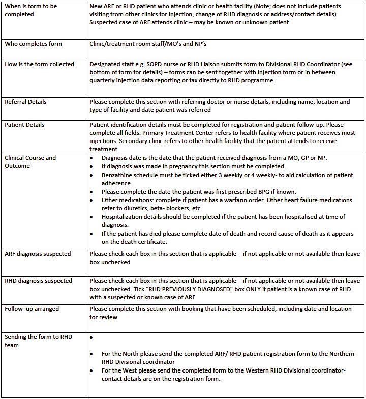
Scope and Purpose
These guidelines have been developed by an expert writing group (which
included infectious disease, cardiology, epidemiology and public health
practitioners) to aid the diagnosis and clinical management of Acute Rheumatic
Fever (ARF) and Rheumatic Heart Disease (RHD) in Fiji. The guidelines have
been based on international best practice guidelines for ARF/ RHD including: the
Jones criteria update 2015[1], The New Zealand Guidelines for Rheumatic Fever
2014[2], The Australian Guidelines for Prevention, Diagnosis and Management of
Rheumatic Fever and Rheumatic Heart Disease 2012[3], the Fiji Cardiovascular
Guidelines 2015, the Fiji Obstetrics and Gynaecology Clinical Practice Guidelines
2015, the Fiji National RHD Policy 2015, the Fiji Ministry of Health and Medical
Services National Strategic Plan 2016-20, the World Heart Federation Diagnosis
and Management of Acute Rheumatic Fever Rheumatic Heart Disease 2008[4]
and the WHO Expert Consultation Technical Report: Rheumatic Fever and
Rheumatic Heart Disease 2003.[5]
The purpose of these guidelines is to:
identify the evidence for best practice in ARF and RHD diagnosis,
identify the standard of care that should be available for people in Fiji; and
provide an evidence based protocol for diagnosis, treatment and prevention of
ARF and RHD in Fiji.
Disclaimer
The authors do not warrant the accuracy of the information contained in these
guidelines and do not take responsibility for any deaths, loss, dam-age or injury
caused by using the information contained herein. While every effort has been
made to ensure that the information contained in these guidelines is correct and
in accordance with current evidence based clinical practice, the dynamic nature
of medicine requires that users in all cases employ independent professional
judgment when using these guidelines.
Funding support
Funding to develop and review these guidelines was made available through
the NZ Aid Programme MFAT grant 2014-18.
i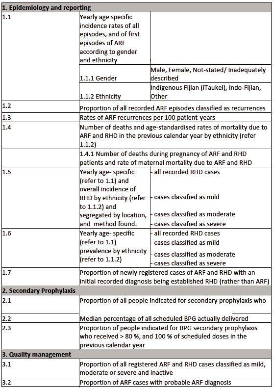
Foreword
The Fiji RHD Prevention and Control Program is established within the
Wellness Centre of the Ministry and Medical Services. The program works to
improve the diagnosis, management and reporting of Acute Rheumatic
Fever (ARF) and Rheumatic Heart Disease (RHD) for the people of Fiji.
This set of ARF and RHD guidelines have been developed using evidence
based resources locally and internationally, in collaboration with key
stakeholders and experts. The guidelines are intended to be used by nurses,
doctors and allied health professionals to assist in the provision of
standardised care and optimal patient management. The guidelines are
accompanied by a summary guideline and ARF clinical diagnostic posters.
The Ministry will continue to engage with other ministries and donor
partners, in promoting healthy lifestyles and behaviour changes in
communities to reduce the burden of ARF and RHD and at the same time
consolidate knowledge and practice of health professionals. It is our hope
that these guidelines will be used effectively by Fijians to gain knowledge
and skills to equip them to perform their roles very well in the communities
they serve.

Dr Luisa Cikamatana Rauto
Chairperson National Medicines and Therapeutic Committee
Ministry of Health and Medical Services.
Suva, Fiji

                                                                           ii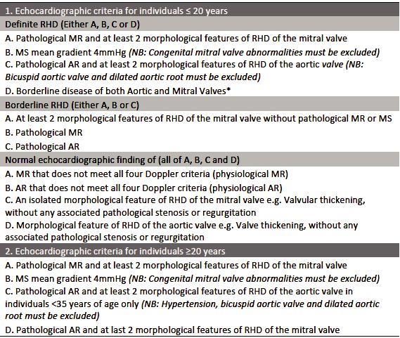
Fiji ARF/ RHD Guidelines: Writing Group
Dr Samantha Colquhoun
International Health Systems Advisor, NHMRC Research Fellow Fiji
GrASP & Centre for International Child Health & Group A
Streptococcal Research Group, University of Melbourne and
Murdoch Childrens Research Institute
Associate Professor Dr Joseph Kado
Chair Fiji RHD TAG, Paediatrician
Fiji National University
Dr Reapi Mataika
Paediatrician
Fiji Ministry of Health & Medical Services
Clinical Associate Professor Dr. Nigel Wilson
University of Auckland
Paediatric Cardiologist
Starship Children’s Hospital, Auckland, New Zealand
Associate Professor Internal Medicine Dr William May
Physician/Cardiologist
Acting Dean
College of Medicine, Nursing and Health Sciences
Fiji National University
Dr Sukafa Tevita
Physician
Fiji Ministry of Health & Medical Services
Dr Lisi Tikoduadua
Paediatrician
Fiji Ministry of Health & Medical Services
Professor Andrew Steer
Paediatrician, Infectious Diseases Specialist &
Director Group A Streptococcal research Group
Centre for International Child Health, University of Melbourne and Murdoch
Childrens Research Institute

                                                                             iii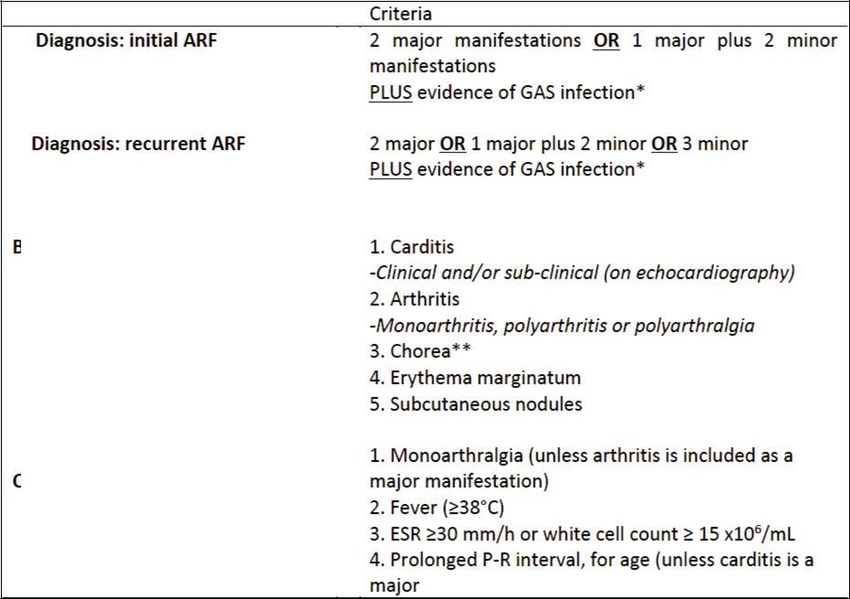
Dr Daniel Engelman
Paediatrician and International Health Research Fellow
Centre for International Child Health & Group A Streptococcal Research
Group, University of Melbourne and Murdoch Childrens Research Institute
Dr Joan Lal
National Officer Oral Health
Fiji Ministry of Health & Medical Services
Dr James Fong
Chair Obstetrics and Gynaecology Clinical Services
Network Fiji Ministry of Health & Medical Services
Ms Laisiana Matatolu
RHD Scientific Technical Support Officer
Wellness Unit
Fiji Ministry of Health & Medical Services
Mrs Emele Naiceru
RHD National Coordinator
Wellness Unit
Fiji Ministry of Health & Medical Services
Mrs Meresini Kamunaga
RHD Divisional Coordinator Central and Eastern
Fiji Ministry of Health & Medical Services
Mrs Ana Ramaka
RHD Divisional Coordinator Western
Fiji Ministry of Health & Medical Services
Dr Sainimere Boladuadua
Technical Advisor Fiji RHD Programme
Cure Kids New Zealand & Fiji Ministry of Health & Medical Services
Ms Liz Kennedy
Fiji RHD Project Lead
Cure Kids New Zealand & Wellness Unit,
Fiji Ministry of Health & Medical Services

                                                                          iv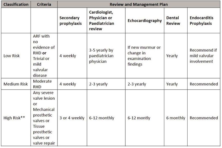
Fiji RHD Technical Advisory Committee members
Dr Isoa Bakani, Dr Eric Rafai, Dr Lisi Tikoduadua, Dr Tevita Baravilala, Dr
Dave Whippy, Dr Rigamoto Taito, Dr Joseph Kado, Dr Isimeli Tukana, Dr
Reapi Mataika, Dr Josaia Samuela, Dr Shahin Nusair, Dr Ilsapeci Vereti,
Mrs Emele Naiceru, Mrs Ana Ramaka, Mrs Resina Lailai

                                                                              v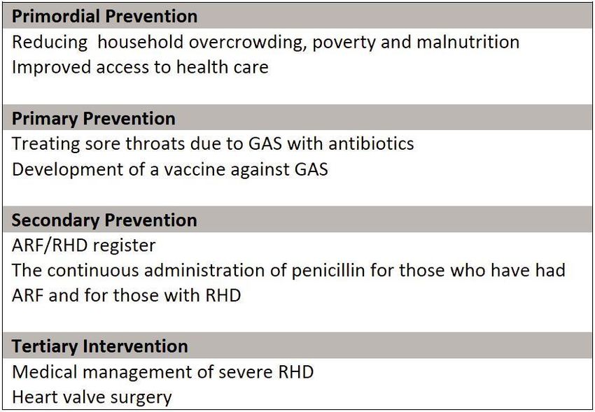
Table of Contents
Scope and Purpose………………………………………………………………………....i
Disclaimer…………………………………………………………………………………....i
Funding Support…………………………………………………………………………..…i
Foreword………………………………………………………………………………….….ii
Fiji ARF/RHD Guidelines: writing groups………………………………………………...iii
Fiji RHD TAG members………………………………………………………………….…v

Section 1: Guidelines and clinical pathways for ARF ........ 1
Acute Rheumatic Fever Key Messages .......................................................................................................... 1
What is Acute Rheumatic Fever?..................................................................................................................... 2
1.1 Epidemiology.................................................................... 3
Global Epidemiology .......................................................................................................................................... 3
Who gets ARF?................................................................................................................................................... 3
Fiji Epidemiology................................................................................................................................................. 3
ARF incidence ..................................................................................................................................................... 3
RHD prevalence ................................................................................................................................................. 4
RHD mortality ...................................................................................................................................................... 4
Cost to Fiji ............................................................................................................................................................ 4
1.2 Rheumatic Fever Prevention .......................................... 5
Primordial prevention ......................................................................................................................................... 5
Primary Prevention ............................................................................................................................................. 6
Antibiotic treatment of sore throats .................................................................................................................. 6
Secondary Prevention ....................................................................................................................................... 7
Tertiary prevention ............................................................................................................................................. 8
1.3 Acute Rheumatic Fever Diagnosis ................................. 9
Summary of key diagnostic requirements ....................................................................................................... 9
Problems with diagnosis and management .................................................................................................. 10
Referral pathways for ARF .............................................................................................................................. 10
Protocol for referral........................................................................................................................................... 11
Diagnostic criteria for ARF .............................................................................................................................. 12
ARF categories ................................................................................................................................................. 13
Evidence of preceding Group A Streptococcal infection ............................................................................ 15
1.4 Clinical Features of Acute Rheumatic Fever ............... 16
1.41 Major manifestations ............................................................................................................................... 16
Joint pain............................................................................................................................................................ 16
Classic polyarthritis of ARF ............................................................................................................................. 16
Carditis ............................................................................................................................................................... 18
Sydenham’s chorea ......................................................................................................................................... 20
Subcutaneous Nodules ................................................................................................................................... 21
Erythema Marginatum ..................................................................................................................................... 21
Differential diagnosis of common major manifestations ............................................................................. 22
1.4.2 Minor Manifestations .............................................................................................................................. 22
Monoarthralgia .................................................................................................................................................. 22
Fever .................................................................................................................................................................. 22
Elevated acute phase reactants (ESR or WCC) .......................................................................................... 23
Prolonged PR interval ...................................................................................................................................... 23
Echocardiography and diagnosis of ARF ..................................................................................................... 23
1.4.3 Management of ARF .............................................................................................................................. 26
Guidelines for general in-hospital care .......................................................................................................... 26
Notification ......................................................................................................................................................... 28
Secondary prophylaxis of ARF ....................................................................................................................... 28
Regular secondary prophylaxis: ..................................................................................................................... 28
Adherence to secondary prophylaxis ............................................................................................................ 29
Strategies to promote continuing adherence include: ................................................................................. 29
Duration of Secondary Prophylaxis ............................................................................................................... 30
Secondary prophylaxis and pregnant women .............................................................................................. 32
Secondary Prophylaxis While Breastfeeding ............................................................................................... 32
Management plan when the ARF episode is controlled ............................................................................. 33
Long-term Management of ARF ..................................................................................................................... 33
Echocardiogram following each episode of ARF, and routine echocardiogram: .................................... 33
Role of RHD divisional nurse coordinators in monitoring and reporting secondary prophylaxis .......... 34
Role of RHD liaison nurses ............................................................................................................................. 34
1.4.4 Protocol for Secondary Prophylaxis Delivery ..................................................................................... 35
Assessment and Preparation .......................................................................................................................... 35
Injection Procedure .......................................................................................................................................... 35
Prepare Benzathine penicillin G solution as directed by the product information:.................................. 35
Documentation .................................................................................................................................................. 36
Pain Reduction .................................................................................................................................................. 36
Anaphylaxis ....................................................................................................................................................... 37
Discharge from hospital ................................................................................................................................... 38
Dental referral ................................................................................................................................................... 38
Reproductive health referral............................................................................................................................ 39
Prevention of Infective Endocarditis .............................................................................................................. 40
Section 2: Guidelines and clinical pathways for Rheumatic
Heart Disease ....................................................................... 42
Introduction ........................................................................................................................................................ 42
2.1 Diagnosis of RHD ........................................................... 43
Signs and symptoms ........................................................................................................................................ 43
Physical Examination ....................................................................................................................................... 44
Diagnostic tests................................................................................................................................................. 45
2.2 Management of RHD ...................................................... 46
Management of RHD depends on the severity of disease. ........................................................................ 46
Clinical management ....................................................................................................................................... 46
Dental care ........................................................................................................................................................ 47
Clinical review ................................................................................................................................................... 47
Evidence for optimal delivery of Benzathine Penicillin G for Acute Rheumatic Fever and Rheumatic Heart
Disease .............................................................................................................................................................. 49
Secondary prevention ...................................................................................................................................... 49
Anticoagulation therapy ................................................................................................................................... 49
RHD and Pregnancy ........................................................................................................................................ 50
Clinical Management during Pregnancy ....................................................................................................... 51
Complications of RHD ..................................................................................................................................... 54
Surgery for rheumatic heart disease ............................................................................................................. 54
Contra-indications to surgery .......................................................................................................................... 56
Cardiac surgical referral in Fiji ........................................................................................................................ 57
Long-term complications ................................................................................................................................. 58
Long-term postoperative management ......................................................................................................... 58
Notification ......................................................................................................................................................... 59
Screening for Rheumatic Heart Disease ....................................................................................................... 59
Appendices ........................................................................... 61
References ............................................................................ 65
List of Tables
Table 1 Minimal list of tests at Sub-divisional hospital level ....................... 12
Table 2 Fiji Criteria for ARF Diagnosis [1]...................................................... 13
Table 3 Upper limits for serum antibody titres ............................................... 16
Table 4 Differential Diagnoses of Arthritis, Carditis and Chorea................ 22
        to be considered in Fiji
Table 5 Upper limits of normal P-R interval ................................................... 23
Table 6 Summary of uses of echocardiography in ARF diagnosis ............ 24
Table 7 Diagnostic and clinical utility of sub-clinical carditis in................... 25
        managing ARF
Table 8 Minimal echocardiographic diagnostic criteria for the ................... 25
        diagnosis of pathological valvular regurgitation
Table 9 Recommended secondary prophylaxis regimens .......................... 31
Table 10 Antibiotic regimens for secondary prophylaxis for ARF/ RHD….36
Table 11 Adrenaline dosage for anaphylaxis ................................................. 37
Table 12 Procedures for which Endocarditis prevention is
         recommended………………………………………………………..41
Table 13 Suggested prophylactic antibiotic regimens for dental, oral
         respiratory tract and other surgical
         procedures……………................................................................ 41
Table 14 Recommended routine clinical review and management ............ 48
         plan for RHD [24]
Table 15 Indications for surgery in Adults [24] ............................................... 55
Table 16 Indications for surgery in children [24] ............................................ 56
Table 17 Review and management plan for RHD patients .......................... 57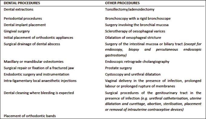
List of Boxes
Box 1 Causal pathways for ARF and RHD ..................................................... 2
Box 2 Intervention opportunities for ARF/ RHD ............................................. 5
Box 3 Investigations in Suspected ARF…………………………………….15
Box 4 Treatment of Acute Rheumatic Fever ……………………………….27
Box 5 Acute Clinical Management of ARF………………………………….27
Box 6 Initial symptoms of RHD are the symptoms of early heart
failure…………………………………………………………………………...43
Box 7 Recommended therapeutic INR ranges……………………………..50

List of Figures
Figure 1: Referral pathways for suspected ARF cases ................................ 11
Figure 2 Referral pathway for RHD patients .................................................. 45
List of Abbreviations
AF          Atrial Fibrillation
AHA         American Heart Association
AMVL        Anterior Mitral Valve Leaflet
ARF         Acute Rheumatic Fever
ASOT        Anti-streptolysin Otitre
AV          Aortic Valve
BPG         Benzathine Penicillin G
CHD         Congenital Heart Disease
CWMH        Colonial War Memorial Hospital
CXR         Chest X ray
ECG         Electrocardiogram
EPI         Expanded Programme for Immunisation
ESD         End Systolic Dimension
ESR         Erythrocyte Sedimentation Rate
FBC         Full Blood Count
GAS         Group A Streptococcus
GBD         Global Burden of Disease
GrASP       Group A Streptococcal Project (research group)
HIU/HIS     Health Information Unit/ Health Information System
IM          Intramuscular
IU          International Units
JVP         Jugular Venous Pressure
LFT         Liver Function Tests
LVEDD       Left Ventricular End Diastolic Dimension
LVEF        Left Ventricular Ejection Fraction
LVESD       Left Ventricular End Systolic Dimension
LVESV       Left Ventricular End Systolic Volume
LMWH        Low Molecular Weight Heparin
MoHMS       Ministry of Health and Medical Services
MFAT        Ministry of Foreign Affairs and Trade, New Zealand
MV          Mitral Valve
NYHA        New York Heart Association
PHIS        Public Health Information System
PMVL        Posterior Mitral Valve Leaflet
RFIS        Rheumatic Fever Information System
RHD         Rheumatic Heart Disease
U and E     Urea and Electrolytes
UFH         Unfractionated Heparin
WHF         World Heart Federation
WHO         World Health Organization
YLL         Years of Life Lost
Section 1: Guidelines and clinical pathways for ARF
Acute Rheumatic Fever Key Messages
ARF occurs following a group A streptococcal (GAS) infection
The incidence of ARF in Pacific Islanders is amongst the highest in the
world
ARF predominantly affects children aged 5 to 15 years
ARF and RHD largely affect disadvantaged populations
Crowding in the household and poverty are associated with an
increased risk of developing rheumatic fever
There is no convincing evidence of a genetic cause of rheumatic fever
and no reliable genetic markers of susceptibility to the disease.
Historically ARF has affected all races in all parts of the world.
Accurate diagnosis of ARF requires a combination of clinical criteria,
laboratory and echocardiographic investigation
Diagnosis ideally requires hospital admission for accurate diagnosis
and optimal management
A first attack of acute rheumatic fever is potentially preventable if the
individual presents with GAS pharyngitis and receives effective
antibiotic treatment
There is some limited evidence that rheumatic fever is caused by skin
infections, so treating skin infections is also considered important in
the prevention of acute rheumatic fever
ARF patients are at 10 times the risk for subsequent episodes of ARF
Delivery of regular Benzathine penicillin G injections to ARF cases is
the mainstay of secondary prevention, preventing recurrence of ARF
and progression of RHD.
1What is Acute Rheumatic Fever?
Acute rheumatic fever (ARF) is an illness caused by a reaction to a bacterial
infection by group A streptococcus (GAS). It causes an acute, generalised
inflammatory response and an illness that targets specific parts of the body,
including the heart, joints, brain and skin. Individuals with ARF are often
unwell, have significant joint pain and require hospitalisation. Despite the
dramatic nature of the acute episode, ARF generally leaves no lasting
damage to the brain, joints or skin, but can cause persisting and life
threatening heart damage, which is then called rheumatic heart disease
(RHD). (Box 1)
Box 1 Causal pathways for ARF and RHD
Exposure to bacteria
Group A Streptococcus (GAS)
Bacterial (GAS) infection
Sore throat*
Acute Rheumatic Fever
(ARF)
GAS infection &
recurrences of
acute rheumatic fever
Rheumatic Heart Disease
(RHD)
Heart Failure and other
complications
(Stroke, heart rhythm
disturbance, heart valve
infections)
21.1 Epidemiology
Global Epidemiology
It is well established that during the 20th century, the incidence of ARF
and the prevalence of RHD declined substantially in Europe, North
America, and other developed nations. This decline has been attributed to
improved hygiene, improved access to antibiotic drugs and medical care,
reduced household crowding, and other social and economic changes.[5]
Although sporadic cases of ARF continue to be seen in affluent nations,
the major burden is currently found in low- and middle-income countries
and in selected Indigenous populations.[6]
Who gets ARF?
Acute rheumatic fever is seen predominantly in children aged 5-14 years,
although recurrent episodes may occur in people aged into their 40s.
Rheumatic Heart Disease represents the cumulative heart damage
caused by previous ARF episodes. The prevalence of RHD peaks in the
third and fourth decades of life.
Fiji Epidemiology
Data on ARF and RHD in Fiji were not routinely recorded until the
establishment of a national RHD Register and commencement of the
World Heart Federation RHD control programme in 2005. Since this time,
activity around RHD has increased and a number of epidemiological and
clinical research studies undertaken by the Fiji Group A Streptococcal
Project (Fiji GrASP) have provided an insight into the burden of ARF and
RHD in Fiji.
ARF incidence
In Fiji estimates of the incidence of first episodes of ARF in children and
young adults range from 15 to 25 per 100,000 per year.[7, 8]Many
children present late with valve damage (RHD) because many ARF
presentations are not recognized in Fiji.[9]The researchers found,
“patients presenting with potential features of ARF seldom had a
diagnostic evaluation sufficient to exclude its diagnosis” suggesting that
many clinical staff working in the high incidence setting of Fiji are not
familiar with the symptoms of ARF.[7]
3For example, if no murmur is heard on auscultation or if auscultation is not
performed, diagnosis of ARF or RHD may be dismissed.
Similarly, if there is no presenting joint involvement with ARF, a diagnosis
can be dismissed.[10] In Fiji patients frequently present late with advanced
RHD and significant valvular damage.
RHD prevalence
Nearly 1% of all Fijians have evidence of RHD (10,11) with
echocardiographic confirmed prevalence of RHD in school-aged children
in Fiji estimated at 8.4 per 1000[11] and the Global Burden of Disease
2010 study estimated an all age prevalence of RHD of 9.8 per
1000.[12]The Indigenous I-Taukei Fijians are more likely to have RHD
than the Indo-Fijian and RHD is more prevalent in women.[12]
RHD mortality
Recent studies focusing on RHD mortality have found that death from
RHD is common (2.4% of autopsy cases audited)[13]and that significant
numbers of young people die from RHD in Fiji with a mean age of death
of38 years.[12] Age-standardised death rates from RHD are more than
twice those reported in current global estimates. In a study using record
linkage in Fiji it was found that between 2008 and 2012 there were 378
deaths attributable to RHD, with over half in occurring in people aged less
than 40 years.[14]In this study RHD was the second most common cause
of death in people aged 5 – 29 years, behind drowning. RHD leads to 9.9
deaths (95% CI 9.8–10.0) and 331 years of life-lost (YLL, 95% CI 330.4–
331.5) per 100,000 person-years.[15]Thus, RHD is a leading cause of
premature death in Fiji.[14]
Cost to Fiji
RHD is costly to the Fiji health system. The estimated annual total cost of
RHD in Fiji is US$6,077, 431. The largest cost item comprised productivity
losses from premature mortality (77% of total cost of illness). The total
indirect costs of RHD were 20 times larger than the total direct costs of
health care.[14]
41.2 Rheumatic Fever Prevention
Interventions on the causal pathway from Group A Streptococcal infection
to chronic RHD are typically considered in four areas of intervention. (Box
2)
Box 2: Intervention opportunities for ARF/ RHD
Primordial prevention
Primordial prevention is key to reducing the incidence of ARF and RHD.
Household overcrowding, poor sanitation and reduced nutrition and living
conditions have all been shown to contribute to increasing the risk of ARF/
RHD.[16-19]Over the past century ARF has largely disappeared from middle
to high-income countries and now exists primarily in resource-poor settings.
Improving social determinants of health can lead to reduction in ARF/RHD
incidence. Targeted measures undertaken at an individual household or com-
munity level may also have an impact in reducing the risk of recurrence of
ARF.
5Primary Prevention
Primary prevention is a strategy that seeks to prevent disease occurring in
the first instance rather than treating it once it has developed. In the case of
ARF this means treating Group A streptococcus bacterial throat infections
before they can initiate ARF. When an individual is exposed to GAS, the
organism attaches to and colonises the pharyngeal mucosa. A process of
infection incorporating an immune response is initiated, and an episode of
ARF may occur 2–3 weeks later. The aim of primary prevention is to identify
symptomatic GAS pharyngitis (sore throats) in those individuals most at risk
of ARF (typically children aged 5–14 years), and treat and eradicate the
bacteria with antibiotics. Studies show that ARF associated with GAS
pharyngitis can be prevented if treatment is commenced within 9 days of
symptoms appearing. While the association between GAS pharyngitis and
ARF is well described, the role of GAS-associated skin infection remains
unclear.[20] Most experts would advise treatment of GAS skin infections
with antibiotic therapy, to achieve cure of the infection and to potentially
reduce the risk of subsequent ARF.
Antibiotic treatment of sore throats
Prevention of an initial attack of ARF requires the prompt and accurate
diagnosis and adequate antibiotic treatment of GAS throat infections. ARF
can be prevented if the preceding throat infection is treated in a timely and
effective way. Recommended treatment of streptococcal throat infection is
intramuscular (IM) benzathine penicillin or a ten-day course of oral (twice a
day) phenoxymethyl penicillin or once a day amoxicill in which eradicates
the streptococci from the pharynx. The role of the treatment of impetigo
(skin sores) in the control of rheumatic fever is less well established,
however Fiji has a very high incidence of skin disease and treatment for
skin disease has been included in the accompanying Fiji Ministry of Health
and Medical Services (MoHMS) Sore Throat and Skin Sore Guidelines.
6People who have had an episode of acute rheumatic fever are at high risk
of a subsequent episode. The Fiji RHD programme technical Advisory
Group suggests a housing assessment and targeted education for
individuals who have had an episode of ARF or have RHD. This should
include an assessment of hygiene, nutrition and sanitation. For example
the availability of soap, water, washing facilities for bodies and clothes –
‘clean house clean body message’ and good nutrition. Patient and family
education is important, with a focus on reducing risk of recurrence and
increasing adherence to secondary prophylaxis:
Reduce numbers of people sleeping in same room
Treat suspected bacterial sore throats, scabies and skin sores
Present early to clinic to prevent complications
Importance of regular BPG for those individuals with a history of ARF or
RHD
Detailed guidelines for Primary Prevention and the
management of acute sore throat and skin disease in Fiji are
provided in a separate guideline.
This guideline provides clinical guidance for clinicians faced with the
patient presenting with acute sore throat as their primary complaint
including around the role of clinical decision rules, the role of throat
swabbing, choice of antibiotic treatment and appropriate follow-up. [21-23]
Secondary Prevention
Secondary prevention of further episodes of ARF is of
the highest priority for best practice care.
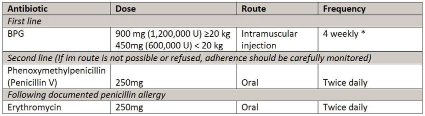
Secondary prophylaxis with regular benzathine penicillin G (BPG) is the
only RHD control strategy shown to be effective and cost-effective at both
community and population levels. The appropriate duration of secondary
prophylaxis is determined by age, time since the last episode of ARF and
potential harm from recurrent ARF, but is likely to be 10 years or more.
While secondary prophylaxis is a proven strategy for controlling RHD, and is
also simple, cheap and cost-effective, it must be adequately implemented.
Persistent high rates of recurrent ARF in high-risk populations highlight the
continued barriers to secondary prevention.
7These factors relate to overcrowded housing, poor access to health services,
limited educational opportunities and poor environmental conditions.
Communities with the highest rates of ARF and RHD are often the least
equipped to deal with the problem. Secondary prevention should include:
BPG injections to provide continuous delivery of penicillin
Strategies aimed at optimizing the delivery of secondary prophylaxis
and patient care
Nursing and medical staff to deliver the BPG on time
A well-coordinated register based system for monitoring and reporting
secured availability of penicillin
The provision of education for, and coordination of available health
services
Community advocacy and health promotion activities
Appropriate clinical and diagnostic resources and guidelines
Tertiary prevention
Tertiary prevention activities aim to avoid early mortality and include the
medical management of symptomatic RHD, anti-coagulation, triage of
candidates for cardiac surgery and delivery of cardiac surgery. Tertiary
prevention does not have an impact of the incidence of disease and will not
assist to control ARF/ RHD at a population level. However, optimal care for
those with RHD reduces the burden of living with RHD, by controlling
symptoms and extending life. Improving clinical care will maximize the
benefit of surgery (if access to cardiac surgery is available) by ensuring the
most suitable candidates are selected for intervention and capacity is
strengthened for the delivery of secondary prevention and anti-coagulation
medications. Surgical intervention is tremendously expensive, does not
‘cure’ the underlying disease, nor prevent the development of new cases in
high risk communities. While there is a clear role for cardiac surgery in RHD
to alleviate symptoms and prevent heart failure, the best approach to
minimise surgical risk and maximise social benefit is yet to be clearly
defined in the Pacific region.
81.3 Acute Rheumatic Fever Diagnosis
Summary of key diagnostic requirements
- The diagnosis of ARF requires health professionals to have a high index of
suspicion and to be aware of the diagnostic criteria. Hospital referral where
expertise is available for accurate diagnosis particularly echocardiography,
is usual.
- It is important that an accurate diagnosis of ARF is made as:
A misdiagnosis may result in the individual receiving benzathine
penicillin G (BPG) injections unnecessarily every four weeks for a
minimum of 10 years.
Under-diagnosis of ARF may lead to the individual suffering a further
attack of ARF, further cardiac damage and premature death.
- There is no single laboratory test specifically diagnostic for ARF;
diagnosis is based on full clinical assessment and assessment of the
probability of ARF. Clinicians experienced with ARF can play a key role to
help make this decision. The probability for a diagnosis of ARF varies
according to location and ethnicity. e.g. in a region with high incidence of
ARF, such as Fiji, a person with fever and arthritis is more likely to have
ARF than one in a low incidence region.
- The American Heart Association Jones diagnostic criteria for ARF have
evolved over many years. The 2015 Revision incorporates Doppler
echocardiography and brings the AHA into closer alignment with other
international guidelines (that have been using Doppler echocardiography to
its fullest extent for over 20 years). [1]
- In all cases, every attempt should be made to follow the criteria, including
demonstrating evidence of GAS infection and using echocardiogram, but
where this is not possible, a clinical diagnosis of probable ARF may be
made by an experienced clinician.
- All patients with suspected or definite ARF (first episode or recurrence)
should undergo echocardiography within 6 months (if not immediately
available) to identify evidence of carditis and categorised as Probable ARF,
until echocardiography confirmation is available to accurately classify the
case.
9Where possible all patients with suspected ARF (first episode or
recurrence) should be hospitalised as soon as possible after the onset of
symptoms. This ensures that all investigations are performed, and if
necessary, the patient should be observed to confirm the diagnosis
before commencing treatment. Hospitalisation also provides opportunity
for patient education and counselling especially regarding the need for
secondary prophylaxis.
Problems with diagnosis and management
A number of factors contribute to the barriers in diagnosis and
management of ARF in Fiji. These include:
The timely and accurate diagnosis of first ARF episode
Health seeking behaviour
Delivery of secondary prophylaxis medication to reduce risk for
subsequent ARF episodes
Many clinicians may have not seen cases of ARF
Patients face transportation cost issues in accessing specialist
diagnosis and care; and
Access to laboratory tests and echocardiography is limited outside
the major population centres.
Referral pathways for ARF
Labasa Hospital and a number of sub-divisional hospitals are serviced by
visiting clinicians from Suva. Therefore referral pathways for diagnosis for
ARF vary across the country. The two main referral pathways in Fiji are:
1. Health clinic referral to sub-divisional hospital for laboratory and Senior
Medical Officer confirmation (echocardiography to be booked with visiting
echo team on rotation)
2. Health clinic/ sub-divisional hospital to Divisional Hospital Urban and peri-
urban i.e. near CWM, Labasa and Lautoka Hospitals – Medical Officer
refers to cardiologist/paediatrician and/or echo clinic. See Figure 1.
10Figure 1: Referral pathways for suspected ARF cases
Divisional hospitals
have echo capacity
Admit patient if ARFconfirmed
for education and counselling
Protocol for referral
- At the health centre or nursing station level the medical officer or nurse
practitioner should refer the patient with suspected ARF to the
nearest sub-divisional hospital for investigation. Investigations should be
for all:
- Laboratory tests: FBC, LFT, U and E, ESR, ASOT
- Cardiac tests: ECG and CXR
- Throat swab (if possible).
- Ideally the patient remains in hospital until the results are back i.e. ARF
cannot be diagnosed until ASOT is back.
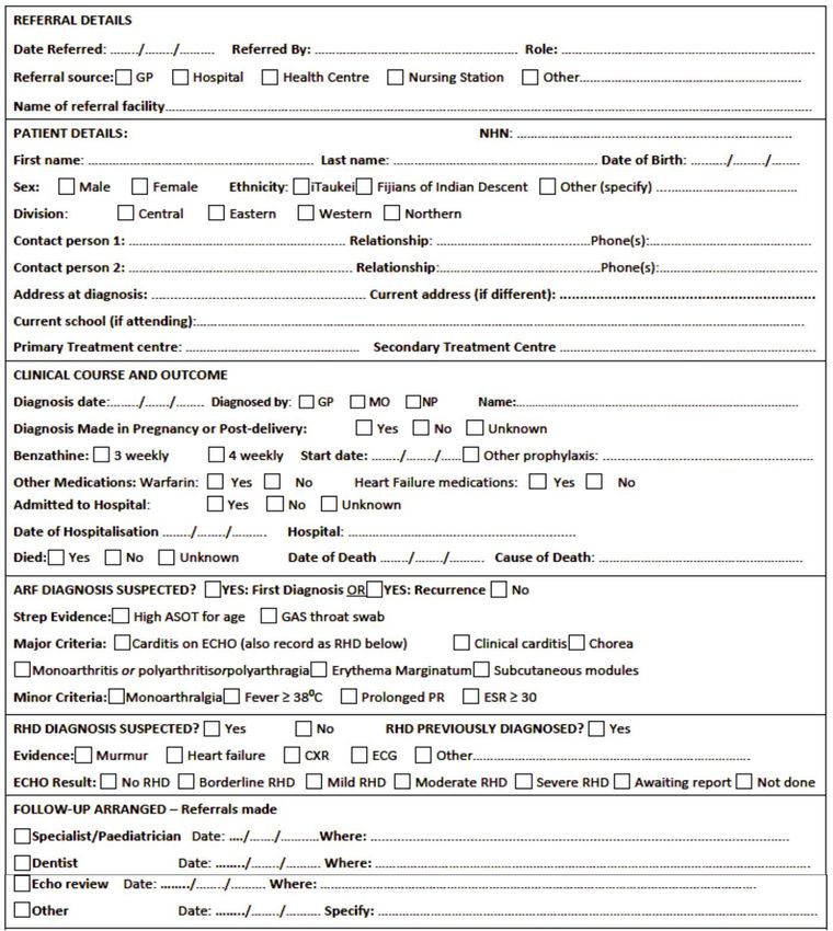
- A specific “ARF referral form” should be completed and the senior
medical officer at the sub-divisional hospital contacted by phone to
discuss the referral.
- All patients with suspected ARF should be discussed with someone
expert in ARF (a paediatrician/physician/ cardiologist) and a plan for
referral and admission should be arranged.
11- All unwell patients presenting in heart failure* or with carditis, must be
admitted and evacuated to a Divisional Hospital. This may involve
consultation with SMO/Cardiologist/Paediatrician/ Physician.
- Patients presenting at Divisional hospitals will be reviewed by a specialist/
Cardiologist as early as possible and investigated accordingly.
*Heart failure is defined by breathlessness walking or at rest, resting tachycardia, as
well as classical signs of raised jugular venous pressure (JVP), hepatomegaly and
crackles.
The minimal list of tests for diagnosis that should be available at a Sub-
divisional hospital is included in Table 1below.
Table 1: Minimal list of tests at Sub-divisional hospital level
Refer to divisional hospital or
*ASOT may not be currently available at all sub-divisional hospitals. If ASOT
not available blood sample should be sent to the nearest Divisional hospital
Diagnostic criteria for ARF
Diagnosis of ARF is based on a combination of clinical criteria laboratory
investigation and echocardiography. As of 2015, some sub-divisional
hospitals in Fiji are equipped to undertake laboratory investigations, such as
ASOT, however the majority do not have this facility. The diagnosis for ARF
in Fiji is guided by the 2015 Revision of the Jones Criteria.[1]The Fiji Criteria
for the diagnosis of ARF are presented in Table 2 and investigative tests are
presented in Box 3. Echocardiogram is available by referral at all divisional
hospitals.
12Table 2: Fiji Criteria for ARF Diagnosis[1]
Major manifestations
Minor manifestations
manifestation
* Evidence of preceding group A streptococcal infection is defined by at least one of
the following:
1. Elevated ASOT above cut-offs by age determined for Fiji (see Table 3)
2. A rising ASOT defined as a twofold or greater difference between titres
measured at presentation and when convalescent (2-4 weeks later generally)
3. A positive throat swab for group A streptococcus at presentation
** Note that ARF can be diagnosed on the basis of chorea without other
manifestations or evidence of GAS infection.
ARF categories
1. ARF
Fulfils Fiji diagnostic criteria (Table 2)
Note that ARF can be diagnosed on the basis of chorea without other
manifestations or evidence of GAS infection.
132. Probable ARF:
In some circumstances, a clinical presentation may not fulfil the full ARF
criteria (Table 2), yet the clinician may still have good reason to suspect that
ARF is the diagnosis. In such situations, clinicians should use their discretion
and clinical acumen to make the diagnosis that they consider most likely and
manage the patient accordingly. A senior clinician should be involved in in
this decision.
This may occur where, for example:
Testing for evidence of GAS infection (ASOT or throat swab) is
unavailable or results pending
Testing for ESR and WCC is unavailable
Echocardiography is unavailable
The history is not considered reliable or documentation of clinical
features is not clear.
Specific clinical scenarios where a diagnosis of “Probable ARF” could
be considered
Carditis on echocardiography with only 1 minor manifestation
Arthritis or polyarthralgia with 0 or 1 minor manifestations. Note
that migratory polyarthritis or migratory polyarthralgia affecting the
large joints is highly suggestive of ARF.
14Box 3 : Investigations in Suspected ARF
Recommended for all cases
White blood cell count
Erythrocyte sedimentation rate (repeat weekly once diagnosis
Blood cultures if febrile (and where available)
Electrocardiogram (ECG) repeat as necessary if conduction abnormality more
than first degree)
Chest x-ray
Echocardiogram (repeat as necessary in 2-4 weeks if equivocal, or if severe
carditis
Throat swab (preferably before giving antibiotics) culture for group A
streptococcus
Anti-streptococcal serology: anti-streptolysin O, if available (repeat 10-14 days
later if first test not confirmatory
Tests for alternative diagnosis depending on clinical features:
Repeated blood cultures if possible to exclude endocarditis or septic arthritis
Joint aspirate (microscopy and culture) for possible septic arthritis*
Joint X-ray
Dengue Fever rapid antigen and Elisa tests, arboviral testing,
Chickungunya, Zika Virus (if available)
Serology and autoimmune markers for autoimmune or reactive arthritis
(including ANA - Anti Nuclear Antibody) - initial screening can be done at CWMH
* Typically, the synovial fluid in joints affected by ARF contains 10,000 to 100,000 white
blood cells/mm3. In septic arthritis the white cell count may be higher, bacteria may be seen
on Gram stain and the culture for bacteria (usually Staphylcoccus aureus) is positive. ASOT
testing is not widely available in Fiji outside the Divisional hospitals. Patients presenting to
sub-divisional hospitals will require a blood sample for ASOT to be taken and sent to the
nearest Divisional hospital.
Evidence of preceding Group A Streptococcal infection
In Fiji, evidence of preceding group A streptococcal infection is defined by
at least one of the following:
- Elevated ASOT above cut-offs by age determined for Fiji (see Table 3)
- A rising ASOT defined as a fourfold or greater difference between titres
measured at presentation and when convalescent (2-4 weeks later
generally)
- A positive throat swab for Group A streptococcus at presentation
If the initial ASOT level is above the age specific cut-off this is sufficient to
confirm diagnosis. If the initial ASOT level is below the age specific cut-off,
ASOT should be repeated in 2 weeks.
15Table 3: Upper limits for serum antibody titres
Age group UNL (U/ml)
(years) ASO Titre
1-4 170
5-14 276
15-24 238
25-34 177
> 35 127
1.4 Clinical Features of Acute Rheumatic Fever
1.41 Major manifestations
Joint pain
- Joint pain is the most common presenting symptom of ARF (occurring in
up to 75% of first attacks).
Classic polyarthritis of ARF
- Arthritis is defined as swelling of the joint in the presence of two or more of
the following: limitation of movement, hotness of the joint, pain in the joint
or localized tenderness.
- The classic arthritis of ARF is asymmetrical large joint (especially knees
and ankles), polyarthritis (multiple joints) that is migratory (one joint
becoming inflamed as another subsides) or additive (multiple joints
progressively becoming inflamed without waning).
- Classic ARF arthritis as described above can be considered as ARF in
high incidence settings unless proven otherwise.
- The arthritis of ARF is extremely painful, often out of proportion to the
clinical signs. The arthritis of ARF is especially susceptible to aspirin and
other non-steroidal anti-inflammatory drugs, usually responding within 24
hours of commencement of treatment and almost always within 48-72
hours.
- Because of the migratory nature of the arthritis, a definite history of
arthritis, rather than documentation by the clinician, is sufficient to satisfy
this criterion.
- History of a painful hip too painful to walk is accepted as evidence of
arthritis of a hip.
16Mono-arthritis and polyarthralgia
- In high RHD prevalence regions such as the Pacific Islands, the classic
polyarthritis of ARF is not always observed, and mono-arthritis (arthritis of
a single join) or polyarthralgia (pain in multiple joints without evidence of
inflammation) may be the presenting feature of ARF.
- As such, either mono-arthritis or polyarthralgia can be considered as a
major manifestation of ARF in Fiji.
- In patients with possible ARF that present with arthritis of a single joint
only, some experts recommend initial treatment with paracetamol or
codeine for pain relief until a second joint is involved so that the classic
pattern of ARF arthritis is observed, thereby facilitating the diagnosis of
ARF.
- It is important to rule out septic arthritis in the patient who looks toxic and
has a mono-arthritis. In the hospital setting, physicians and surgeons
should collaborate when the diagnosis of arthritis is unclear. The diagnosis
of a single swollen joint with a high fever is septic arthritis unless joint
aspirate negative. Patients with sterile joint aspirates should never be
treated speculatively for septic arthritis without further investigation,
particularly in areas with a high ARF/RHD prevalence, such as Fiji.
(18-20).
- Arthralgia differs from arthritis in that there is pain on joint movement
without evidence of swelling or heat. It is a non-specific symptom.
- Arthralgia involving multiple joints may suggest ARF if it appears in the
same pattern as rheumatic polyarthritis: that is, asymmetrical, affecting
large joints, and migratory or additive.
- Mono-arthritis and polyarthralgia are often associated with overt or
subclinical carditis. [24]
- ARF should always be considered in the differential diagnosis of patients
presenting with joint pain in high-risk populations (Algorithm 1).
17Algorithm 1: Diagnosis of ARF in patients presenting with joint pain
Carditis
- Cardiac inflammation (carditis) in ARF almost always affects the valves
(valvulitis), especially the mitral and aortic valves; although pericarditis and
myocarditis may also occur.
- Valvulitis observed clinically as an apical holosystolic murmur (mitral
regurgitation) with or without mid-diastolic flow murmur (mitral stenosis) or
an early diastolic murmur at the base (2nd or 3rd left intercostal space) of
the heart (aortic regurgitation).[25]
- Early disease leads to valvular regurgitation, whereas prolonged or recurrent
disease may lead to increased valvular regurgitation with impaired
ventricular function or mitral stenosis.
- The rheumatic aetiology can usually be confirmed on echocardiography (see
Appendix 1)
- Echocardiographic evidence of sub-clinical carditis can also be accepted as
a major criteria(Table 2).
- The incidence of carditis in the initial attack of ARF varies between 50% and
82%. [26]
- Diagnosis of ARF in patients presenting with clinical cardititis shown in
Algorithm 2.
18Algorithm 2: Diagnosis of ARF in patients presenting with
clinical carditis, without arthritis
1. For patients with arthritis, use arthristis algorithm instead
2. Echocardiography is a higher level of evidence than clinical assessment
19Sydenham’s chorea
This manifestation predominantly affects females, particularly in adolescence.
[27, 28]
Chorea consists of jerky, uncoordinated movements, especially affecting the
hands, feet, tongue and face. The movements disappear during sleep. They
may affect one side only (hemichorea) or both sides of the body.
Useful signs include:
- The ‘milkmaid’s grip’ (rhythmic squeezing when the patient grasps the
examiner’s fingers)
- ‘Spooning’ (flexion of the wrists and extension of the fingers when the
hands are extended)
- the ‘pronator sign’ (turning outwards of the arms and palms when held
above the head)
- Inability to maintain protrusion of the tongue.
Because chorea may occur after a prolonged latent period following GAS
infection, the diagnosis of ARF under these conditions does not require the
presence of other manifestations or evidence of a recent GAS infection.
Patients with pure chorea may have a mildly elevated ESR (approximately 40
mm/h), but have a normal serum CRP level and white cell count.
Echocardiography is important for cases of chorea without clinical carditis.
Most studies show > 50% of cases of chorea have subclinical carditis, which
makes the diagnosis of ARF certain. If echo is normal, it is necessary to
investigate for the differential diagnoses of chorea (Table 4). Specialist
paediatrician or physician involvement is recommended.
Chorea is the ARF manifestation most likely to recur, and may be associated
with pregnancy or oral contraceptive use. The vast majority of cases resolve
within 6 months (usually within 6 weeks), although rare cases lasting as long
as 3 years have been documented. Chorea patients have a higher-than-
expected prevalence of attention-deficit hyperactivity disorder, anxiety,
depression and cognitive dysfunction after they have recovered from the
movement disorder, although there is some evidence that attention-deficit
hyperactivity disorder and anxiety features are often present before the onset
of chorea, suggesting that they may be risk factors, rather than long-term
complications.[29-32]
20You can also read