Report of the International Emergency Fish Disease Investigation Mission on a Suspected Outbreak of Epizootic Ulcerative Syndrome (EUS) in the ...
←
→
Page content transcription
If your browser does not render page correctly, please read the page content below
Report of the International Emergency
Fish Disease Investigation Mission on a Suspected
 Outbreak of Epizootic Ulcerative Syndrome (EUS)
          in the Democratic Republic of the Congo
                                   13−19 March 2015
All photographs were contributed by Dr David Huchzermeyer (field photos) and
Dr Bernard Mudenda Hangómbe (histopathology and PCR photos).
Report of the International Emergency
Fish Disease Investigation Mission on a Suspected
 Outbreak of Epizootic Ulcerative Syndrome (EUS)
          in the Democratic Republic of the Congo
                                                    13−19 March 2015

      FOOD AND AGRICULTURE ORGANIZATION OF THE UNITED NATIONS
                            Rome, 2017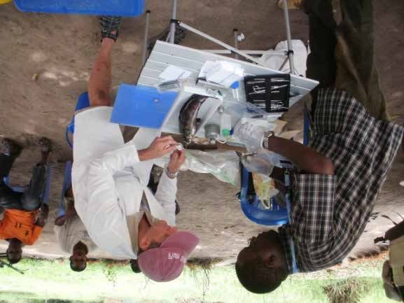
FAO. 2017. Report of the International Emergency Fish Disease Investigation Mission
on a Suspected Outbreak of Epizootic Ulcerative Syndrome (EUS) in the Democratic
Republic of the Congo, 13 to 19 March 2015. Rome, FAO. 58 pp.

 The designations employed and the presentation of material in this information
 product do not imply the expression of any opinion whatsoever on the part of
 the Food and Agriculture Organization of the United Nations (FAO) concerning
 the legal or development status of any country, territory, city or area or of its
 authorities, or concerning the delimitation of its frontiers or boundaries. The
 mention of specific companies or products of manufacturers, whether or not
 these have been patented, does not imply that these have been endorsed or
 recommended by FAO in preference to others of a similar nature that are not
 mentioned.

 The views expressed in this information product are those of the author(s) and do
 not necessarily reflect the views or policies of FAO.

 ISBN 978-92-5-109555-3

 © FAO, 2017

 FAO encourages the use, reproduction and dissemination of material in this
 information product. Except where otherwise indicated, material may be copied,
 downloaded and printed for private study, research and teaching purposes,
 or for use in non-commercial products or services, provided that appropriate
 acknowledgement of FAO as the source and copyright holder is given and that
 FAO’s endorsement of users’ views, products or services is not implied in any way.

 All requests for translation and adaptation rights, and for resale and other
 commercial use rights should be made via www.fao.org/contact-us/licence-
 request or addressed to copyright@fao.org.

 FAO information products are available on the FAO website (www.fao.org/
 publications) and can be purchased through publications-sales@fao.org
iii

Preparation of this document
This is the final report of the work carried out by the International Emergency Fish Disease
Investigation Mission on a Suspected Outbreak of Epizootic Ulcerative Syndrome (EUS) in the
Democratic Republic of the Congo, organized and funded by the Food and Agriculture Organization
of the United Nations (FAO) through the Special Fund for Emergency and Rehabilitation Activities
and funds from the FAO Representation in the Democratic Republic of the Congo and jointly
implemented with the Government of the Democratic Republic of the Congo.

This document is based on the results of the work carried out by the Field Investigation Team
to the Democratic Republic of the Congo conducted from 13 to 19 March 2015 and composed
of two international consultants, Dr Bernard Mudenda Hang’ombe from Zambia and Dr David
Huchzermeyer from the Republic of South Africa, with assistance from a local task force led by Dr
Leopold K. Mulumba-Mfumu, Director of the Central Veterinary Laboratory (CVL) in Kinshasa, the
Democratic Republic of the Congo and staff and officials of FAO Representation in the Democratic
Republic of the Congo and the subsequent outcomes of laboratory analysis of field samples conducted
by the team. All photographs were contributed by Dr David Huchzermeyer (field photos) and
Dr Bernard Mudenda Hangómbe (histopathology and PCR photos).

The finalization of this report was under the technical oversight of Dr Melba B. Reantaso,
Aquaculture Officer, Aquaculture Branch (FIAA), Fisheries and Aquaculture Department of FAO.
Technical and English grammar editing was provided by Dr James Richard Arthur.
iv

     Abstract
     In response to a request for an emergency technical assistance from the Government of the
     Democratic Republic of the Congo in connection with a serious disease affecting fish in Lokame
     River in Loko and in Mbanza Oton, 60 km from Gbadolite, The Food and Agriculture Organization
     of the United Nations (FAO) formed an International Emergency Disease Investigation Task Force.
     The overall objective of the Task Force was to: (1) confirm that an outbreak was happening; establish
     a case definition and make a presumptive diagnosis of the causative agent; (2) collect and process
     fish samples for relevant laboratory tests; (3) identify risk factors, confirm diagnosis and define
     further investigation or follow-up work; (4) recommend border/cross-border control measures to
     prevent further spread of the disease; (5) identify specific short-term and medium-term biosecurity
     action plans that the government may undertake; and (6) provide further recommendations to
     FAO on how to prevent the further spread of the disease.

     Some members of the Task Force travelled to the Democratic Republic of the Congo from
     13 to 19 March 2015, where they conducted field investigations. Subsequent laboratory tests have
     confirmed the presence of epizootic ulcerative syndrome (EUS) using two confirmatory tests
     recommended by the World Organisation for Animal Health (OIE), i.e.: (1) histopathology, which
     demonstrated granulomas in Parachanna obscura (snakehead), Protopterus annectens (lungfish)
     and Clarias theodorae (snake catfish) and (2) polymerase chain reaction (PCR), which confirmed
     the presence of genomic DNA of Aphanomyces invadans in collected specimens.

     The Task Force concluded that permissive factors that favoured the propagation, infectivity and
     disease occurrence of EUS occur in the rivers and streams investigated in the Equateur Province
     of the Democratic Republic of the Congo. The findings also showed that environmental, climatic,
     water quality and human demographic conditions in the Congo River basin support the possibility
     of pandemic spread of the disease.

     The Task Force suggested several actions which need to be undertaken to curb the spread of
     the outbreak. These include: active surveillance and monitoring of fish markets and other food
     channels used in the movement of live fish; capacity building for involved government personnel
     to strengthen knowledge and expertise in the identification and control of the disease through
     biosecurity measures; continued dialogue among the Democratic Republic of the Congo,
     neighbouring countries and FAO about EUS status, including subregional disease surveillance,
     monitoring, and response programmes; and the formulation of a national aquatic biosecurity
     strategy for the Democratic Republic of the Congo.
v

Contents
PREPARATION OF THIS DOCUMENT		                                                                             iii
ABSTRACT                                                                                                   iv
CONTENTS                                                                                                     v
TABLES                                                                                                      vi
FIGURES                                                                                                    vii
ACKNOWLEDGEMENTS                                                                                           vii
ACRONYMS AND ABBREVIATIONS                                                                                  ix
EXECUTIVE SUMMARY                                                                                            x

1. Background                                                                                               1

2. International Emergency Fish Disease nvestigation Task Force                                             3

3. Methodology: field observations and laboratory analysis                                                  5
   3.1 General planning for the disease outbreak investigation (with local task force)                      5
   3.2 Fish sampling                                                                                        5
   3.3 Description of field procedures for gross examination of fish and collection of samples for
       laboratory analysis                                                                                  6
   3.4 Water quality, meteorological and other environmental data                                           6
   3.5 Consultation with women fish marketers, fishers, fish farmers                                        6
   3.6 Laboratory investigation procedures                                                                  7
        3.6.1 Gross clinical signs (on site)                                                                7
        3.6.2 Parasitology (on site)                                                                        7
        3.6.3 Bacteriology (on site)                                                                        7
        3.6.4 Mycology (on site and at the laboratory facility at UNZA)                                     7
        3.6.5 Virology (on site and at the laboratory facility of CVL)                                      7
        3.6.6 Histopathology( on site and at the laboratory facility of UNZA and University of Pretoria,
              the Republic of South Africa)                                                                 7
        3.6.7 Polymerase chain reaction (PCR) (on site and at the laboratory of UNZA and DAFF)              8
   3.7 Analysis of available reports and other correspondences/information based on initial
       information received (i.e. clinical signs and affected species)                                      8

4. Results                                                                                                  8
   4.1 General planning for the disease outbreak investigation (with local task force)                      8
       4.1.1 Case definition                                                                                8
   4.2 Sampling location and field activities                                                               8
   4.3 Field investigation results                                                                         12
       4.3.1 Fish species, gross pathology and prevalence                                                  12
       4.3.2 Water quality, meteorological and other environmental data                                    21
       4.3.3 Consultation with women fish marketers, fishers, fish farmers                                 22
       4.3.4 Aquaculture                                                                                   23
   4.4 Laboratory analysis results                                                                         23
   4.5 Synthesis of reports                                                                                25
   4.6 Validation of findings by the OIE Reference Laboratory for EUS                                      25

5. Conclusion                                                                                              26
   5.1 Conclusions based on field and laboratory investigations                                            27
   5.3 General diagnosis                                                                                   27
vi

     6. Recommendations                                                                            27
        6.1 Specific recommendations                                                               28
        6.2 General recommendations                                                                29

     7. The Way Forward                                                                            29
     8. References                                                                                 31

     Appendices
     Appendix 1      Members of the EUS Investigation Task Force                                   33
     Appendix 2      Itinerary of the Field Investigation Team                                     34
     Appendix 3      Persons met during the mission – Districts of South and North
                     Ubangi/Equateur Province                                                      35
     Appendix 4      SARNISSA communication and postings                                           37

     Tables
     1.    Preliminary findings by the Field Investigation Team in Gemena live fish market
           affected species, sample identification and gross pathology                             12
     2.    Preliminary findings by the Field Investigation Team in Karawa, Libala River:
           affected species, sample identification and gross pathology a                           13
     3a.   Preliminary findings by the Field Investigation Team in Businga live fish market:
           affected species and prevalence of gross lesions suggestive of EUS                      13
     3b.   Preliminary findings by the Field Investigation Team in Businga, Mongala River:
           affected species, sample identification and gross pathology                             13
     4a.   Preliminary findings by the Field Investigation Team in Loko Village:
           affected species and prevalence of gross lesions suggestive of EUS                      14
     4b.   Preliminary findings by the Field Investigation Team in Loko River: affected species,
           sample identification and gross pathology and prevalence of lesions suggestive of EUS   14
     5.    Preliminary findings by the Field Investigation Team in Mobayi Mbonge, Ubangi River:
           species examined and prevalence of lesions suggestive of EUS                            15
     6.    Preliminary findings by the Field Investigation Team in Bobuto Village:
           affected species, gross pathology and prevalence of lesions suggestive of EUS           15
     7.    Preliminary findings by the Field Investigation Team in Gemena fresh fish market:
           affected species, sample identification and gross pathology                             15
     8.    Results of laboratory analysis for EUS according to sampled fish species                19
     9.    Sampling location, GPS coordinates, elevation and water temperature of sampling sites   21
     10.   Water quality parameters of small black water forest streams (no fish samples)          21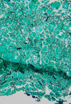
vii
Figures
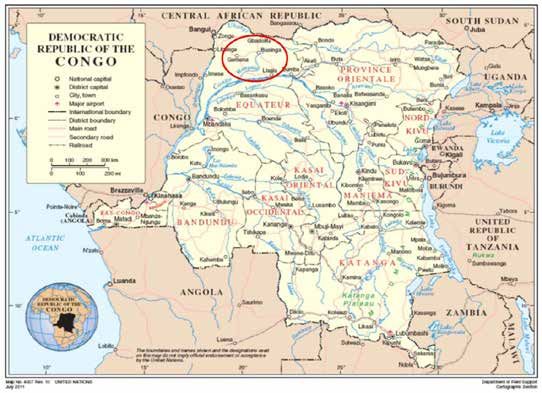
1. Map of the Democratic Republic of the Congo (the Democratic Republic of the Congo
showing the area where the disease investigation was carried out (red circle) 4
2. Inspecting private fish culture ponds near Gemena with no observed sick fish 9
3. Field Investigation Team at the Libala River in Karawa 10
4. Dissecting a munguso fish with lesions at Businga, Mongala River 11
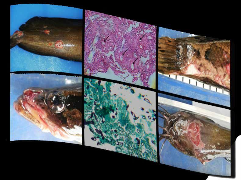
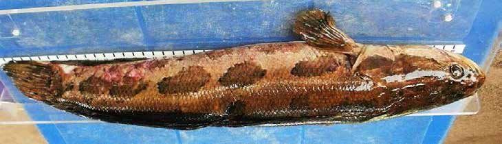
5. Typical position of lesions over the ventral pectoral region in Clarias theodorae
(ngolo or snake catfish) (b) Typical EUS lesions in the tail region of Clarias theodorae (ngolo or
snake catfish) purchased from Gemena live-fish market 16
6. Parachanna obscura (mungusu or snake head) with EUS lesions. (b) Typical position of lesions
on tail of P. obscura. (c) Typical position of EUS lesions on head of P. obscura. Note deep
ulceration into the bony structure of the maxillary portion of the head 17
7. Typical position of lesions on the head of a Protopterus annectens (nzombo or lungfish).
(b) P. annectens for sale at the live-fish market in Gemena 18
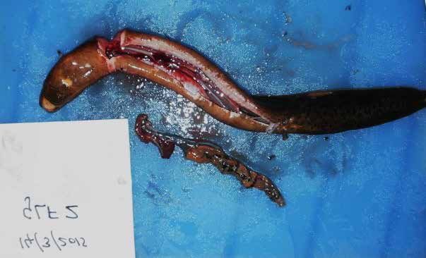
8. Note ulcer ventrally in mandibular area and the severe lesions affecting the intestines
(intestines dissected from body cavity) in a Protopterus annectens (nzombo or lungfish).
Fish purchased from Gemena live-fish market 18
9. Typical position of EUS lesion in a mormyrid fish (Marcusenius macrolepidotus) sampled from
the Libala River at Karawa. 20
10. Examples of fish species collected from various sampling sites. (a) Channallabes apus,
(b) Schilbe mysticus, (c). Hemichromis fasciatus and (d) a member of the family Mormyridae. 20
11. Monthly rainfall patterns in an average year over the Congo River basin (scale represents average
total rainfall in mm/yr). 22
12. PCR results of the direct detection of genomic DNA of Aphanomyces invadans from infected
tissues collected during the International Emergency Fish Disease Investigation Mission on a
suspected Outbreak of Epizootic Ulcerative Syndrome (EUS) to the Democratic Republic of
the Congo (the Democratic Republic of the Congo). The figure represents electrophoresis gel
bands representative of the EUS positive results 1, 2, 4 & 6. Lanes 9 and 10 are negative and
positive controls respectively, while Lanes 3 and 8, have non-specific amplicon. 24
13. Photomicrograph demonstrating typical mycotic granulomas surrounding invasive
oomycete hyphae (black arrows) below the dermis of an EUS-infected lungfish (H&E × 100),
Protopterus annectens. (b) is a Grocott’s stained slide showing black-staining oomycete cell walls
in affected tissue from specimen GLF4, (c) Histopathology of EUS-infected Clarias theodorae
(GCLG1) showing a typical mycotic granuloma surrounding invasive oomycete hyphae (black
arrow) in the skin layer (H&E × 400), while (d) demonstrates black staining oomycete hyphae
(Grocott’s × 400) 24
14. Photomicrograph depicting a granulomatous inflammatory response surrounding necrotic
muscle tissue in association with oomycete hyphae (arrow) in an EUS-infected snakehead,
Parachanna obscura. (MPACH2). (H&E × 100). (b) Section from the same fish stained with
Grocott’s stain demonstrating dark staining oomycete structures in the muscle tissue. 25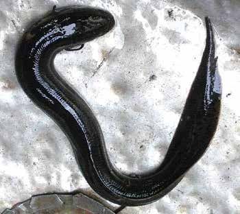
viii

       Acknowledgements
       An International Emergency Fish Disease Investigation Task Force was organized by the
       Aquaculture Branch (FIAA) of the Food and Agriculture Organization of the United Nations (FAO)
       in close coordination with the FAO Representation in the Democratic Republic of the the Congo
       (FAOCD). All members of the Task Force are acknowledged for their important contribution to
       this undertaking.

       The Field Investigation Team was composed of two experts, Dr Bernard Mudenda Hang’ombe
       from the Republic of Zambia and Dr David Huchzermeyer from the Republic of South
       Africa and their outstanding work is very much appreciated. From the Democratic Republic
       of the Congo, Dr Leopold K. Mulumba-Mfumu, Director of the Central Veterinary
       Laboratory (CVL) in Kinshasa is acknowledged for his full participation in the field
       mission. His contributions to the field work, as well as those of his field staff, were invaluable
       to the success of the field mission. Staff at the CVL in Kinshasa are acknowledged for
       assisting with sample preparation. The officers from FAOCD, namely Mr Ndiaga Gueye,
       Mr Marc Bellemans and Mr Chris Pappas are acknowledged for facilitating and covering the
       additional costs of local travel and miscellaneous expenses beyond the Special Fund for Emergency
       and Rehabilitation Activities (SFERA) allocation and for ensuring the safety of the mission.
       The FAO field officer in Gemena, Mr Prudent Landon Osang is thanked for facilitating local
       arrangements in Gemena and for providing access to the fish market in Gemena and to fish culture
       ponds. The District Inspector, North Ubangi District, Mr Vital Selengbe is acknowledged for
       ensuring access to sampling sites and facilitating interaction with local communities and fishers.
       Mr Gerengbo Emmanuel, Chief of Fisheries Cell, North Ubangi District Inspection is thanked for
       facilitating access to the Ubangi River site. The tribal authorities at the various sampling points are
       gratefully acknowledged for their concern and interest in the disease investigation and for taking
       the time to meet with and talk to the mission participants.

       In Cape Town, the Republic of South Africa, Dr Kevin Christison, Specialist Scientist at the
       Research Aquarium of the Department of Agriculture, Forestry and Fisheries (DAFF) of the South
       African Government, and his staff are thanked for the molecular analysis of the samples collected
       from sick fish. Microbiological culture and further molecular analysis as well as histological
       examination of diseased fish tissues were performed by Dr Hang’ombe, and the support of the
       Microbiology Department in the Faculty of Veterinary Science of University of Zambia is gratefully
       acknowledged. Dr Johan Steyl of the University of Pretoria is acknowledged for performing the
       Grocott’s staining of histological sections. Ms Varinee Panyawachira, EUS Expert at the Aquatic
       Animal Health Research Institute, Bangkok and OIE Reference Laboratory for EUS provided
       validation of histopathology slides and suggested to use Grocott’s or Uvitex stain to demonstrate
       the oomycete hyphae.

       From FAO headquarters in Rome, Italy, Dr Rohana Subasinghe, Supervising Officer and
       Dr Melba B. Reantaso, Aquaculture Officer, Aquaculture Branch (FIAA), of the Fisheries and
       Aquaculture Department are acknowledged for initiating the emergency disease investigation.
       Dr Reantaso is thanked for the overall technical oversight and planning of the mission. The
       Assistant Director General of the Technical Cooperation Department (TC) and the Department
       of Fisheries and Aquaculture of the FAO are gratefully acknowledged for securing the financial
       support from SFERA that made this disease investigation possible.
ix Acronyms and abbreviations AAHRI Aquatic Animal Health Research Laboratory CVL Central Veterinary Laboratory DAFF Department of Agriculture, Forestry and Fisheries EUS Epizootic ulcerative syndrome FAO Food and Agriculture Organization of the United Nations FAODC FAO Representation in the Democratic Republic of the Congo FAO-IAEA International Atomic Energy Agency of the United Nations FIAA Aquaculture Branch (FAO) GPA Glucose-peptone agar GPS Global positioning system H&E Haematoxylin and eosin OIE World Organisation for Animal Health PARSSA Programmes for the Support of Water Sector Reform PCR Polymerase chain reaction SARNISSA Sustainable Aquaculture Research Networks in sub-Saharan Africa SFERA Special Fund for Emergency and Rehabilitation Activities TC Technical Cooperation Department (FAO) TCP Technical Cooperation Programme (FAO) TSA Tryptone soya agar UNZA University of Zambia
x

    Executive summary
    In response to a request for an emergency technical assistance from the Government of the
    Democratic Republic of the Congo in connection with a serious disease affecting fish in the
    Lokame River in Loko and in Mbanza Oton, 60 km from Gbadolite, FAO formed an International
    Emergency Fish Disease Investigation Task Force. The overall objective of the Task Force was to:
    (1) confirm that an outbreak was happening, establish a case definition and make a presumptive
    diagnosis of the causative agent; (2) collect and process fish samples for relevant laboratory tests;
    (3) identify risk factors, confirm diagnosis and define further investigation or follow-up work;
    (4) recommend border/cross-border control measures to prevent further spread of the disease;
    (5) identify specific short-term and medium-term biosecurity action plans that the government
    may undertake; and (6) provide further recommendations to FAO on how to prevent the spread
    of the disease.

    Some members of the Task Force, i.e. the Field Investigation Team, travelled to the Democratic
    Republic of the Congo from 13 to 19 March 2015 and conducted field investigations. Laboratory
    tests of field samples followed, including validation of results by the World Organisation for Animal
    Health’s (OIE) Reference Laboratory for epizootic ulcerative syndrome (EUS).

    The disease. EUS is a serious finfish disease that is listed by the OIE. It is caused by an infection
    with Aphanomyces invadans, which has swept across Japan, Australia, many countries in Asia and
    the United States of America since the first outbreaks were reported in the early 1970s, causing
    significant loss of income to fishers and fish farmers. In Africa, EUS was first diagnosed in
    the Republic of Botswana (2007) and has been confirmed in the Republic of Namibia (2007), the
    Republic of Zambia (2007), the Republic of South Africa (2010) and suspected in the Republic
    of the Republic of Zimbabwe (undated). More than 50 finfish species are susceptible to EUS. In
    Africa alone, there are more than 20 finfish species infected (farmed and wild fish populations).
    EUS outbreaks threaten food security for subsistence fishers and fish farmers and subsequently
    people’s physical health, as fish are an important source of animal protein for people in the affected
    countries.

    Diagnosis. Fish showing clinical signs similar to EUS (small to large red spots and open dermal
    ulcerative lesions) were reported in the Equateur Province of the Democratic Republic of the Congo
    in December 2014, with a record of heavy fish mortalities. During the March 2015 investigation1,
    12 families of freshwater fish were inspected for evidence of EUS, of which members of the families
    Clariidae, Channidae, Protopteridae and Mormyridae were most severely affected. Confirmatory
    diagnosis of infection with A. invadans in clinically affected fish was done through OIE standards, i.e.
    (1) histopathology, which demonstrated granulomas in Parachanna obscura (snakehead), Protopterus

    ¹The Field Investigation Team to the Democratic Republic of the Congo conducted from 13−19 March 2015 was
    composed of two international consultants, Dr Bernard Mudenda Hang’ombe from the Republic of Zambia and Dr
    David Huchzermeyer from the Republic of the Republic of South Africa with assistance from a local Task Force led by
    Dr Leopold K. Mulumba-Mfumu, Director of the Central Veterinary Laboratory (CVL) in Kinshasa, the Democratic
    Republic of the Congo and staff and officials of the FAO Representation in the Democratic Republic of the Congo
    (FAODC). A preliminary report based on the field findings was presented to FAODC for the Government of the
    Democratic Republic of the Congo on 19 March 2015. Laboratory tests were conducted between April and September
    2015. Analysis, validation and report finalization was completed in March 2016.
xi
annectens (lungfish) and Clarias theodorae (snake catfish) and (2) polymerase chain reaction (PCR),
which confirmed the presence of genomic DNA of A. invadans in collected specimens.
It was concluded that permissive factors favoured the propagation, infectivity and disease
occurrence of EUS in the rivers (Libala, Loko and Mongala rivers and their tributaries) investigated
in the Equateur Province of the Democratic Republic of the Congo. The habitat preference for
well-vegetated backwaters of forest streams and floodplains that represent ideal conditions for
infection with A. invadans explains the high prevalence of disease among fish in these four families.
The findings showed that environmental, climatic, water quality and human demographic
conditions in the Congo River basin support the possibility of pandemic spread of the disease.
The findings were validated by the EUS Expert at the Aquatic Animal Health Research Institute
(AAHRI) and the OIE Reference Laboratory for EUS.
Control and prevention of EUS. There is no known protective vaccine nor effective
chemotherapeutant for EUS. Treatment of EUS in natural waterbodies is not possible. Practical
control is limited to identifying and reducing pathways of spread.
Public health significance. The agent causing EUS does not pose any direct human health threats.
Except for the fish exhibiting deep ulcerations and tissue decay, which could harbour secondary
pathogens which may have human health consequences, the fish infected with A. invadans do
not pose health hazards for consumers. However, it is recommended not to eat EUS-infected fish
unless properly and thoroughly cooked.
Risk of further spread. There is a high risk of spread to other African waterbodies from one lake
or river system to another, endangering susceptible fish species. Factors include heavy rainfall and
flooding, poor biosecurity, including movement of infected fish, as well as natural spread by fish and
birds. Members of the Clariidae, Channidae and Protopteridae are of greatest concern regarding the
spread of EUS, as fish in these families represent an important food commodity in the Democratic
Republic of the Congo. Additionally, all three families represent air-breathing fish, and marketable fish
are transported to and from markets, live thus providing an effective pathway for pathogen transfer.
Specific recommendations:
• Immediate notification by the veterinary authority in the Democratic Republic of the
Congo (Animal Health Services and Chief Veterinary Officer of the Democratic Republic
of the Congo) to the OIE of an outbreak of EUS (infection with A. invadans) in the Libala,
Loko and Mongala rivers and in their tributaries in Equateur Province of the Democratic
Republic of the Congo.
• Active surveillance of fish markets should be initiated immediately, with tracing of the
actual source (origin) of all infected fish. Affected streams, rivers and villages should be
mapped through initiation of local surveillance programmes and the spread of the disease
outbreaks monitored.
• The movement of live fish between markets and between river systems in areas affected
by EUS should be restricted (e.g. by quarantine and biosecurity measures); as well, a ban
of fishing activities during EUS-outbreak seasons should be considered and alternative
livelihood options for affected fishers should be determined.xii
• An initial national workshop for all personnel involved with fish and aquaculture activities
should be urgently organized to initiate public awareness and extension programmes
to raise understanding of the disease and the measures required to reduce its impacts
(technical assistance).
• Funding should be urgently sourced to mobilize regional EUS expertise for a more
in-depth epidemiological investigation of the extent of EUS in the Democratic Republic
of the Congo and to provide training on EUS recognition, biology, diagnostics, pathology
and surveillance procedures, emergency preparedness and contingency planning for
both veterinary and fisheries officials and especially extension officers in the Democratic
Republic of the Congo.
• The epidemiological investigation of the disease in the Democratic Republic of the Congo
should be aimed at establishing the EUS status in the country (i.e. zones, farms etc.) and
the critical areas near or along the border of affected areas in the Democratic Republic of
the Congo or affected countries (e.g. the Republic of Zambia), the prevalence of EUS, the
extent
• of natural waterbodies affected, the seasonality, the full range of species affected or susceptible
(cultured and wild populations), potential carriers, risk factors and other pathways
(e.g. fish markets, fish vendors etc.) and the risk to inland fisheries and human food security.
The outcome of such an investigation should promote the development of appropriate risk
management responses for the Democratic Republic of the Congo.
• The laboratory facilities at the Central Veterinary Laboratory (CVL) in Kinshasa should be
urgently strengthened to enable the laboratory to initiate and perform effective fish disease
investigations. This will require implementation of the fish pathology laboratory that has
been initiated by the European Union and the International Atomic Energy Agency of the
United Nations (joint division FAO-IAEA).
• Capacity building for local staff, particularly veterinary, fisheries and aquaculture officials,
is urgently needed to enable them to educate the populace (consuming public, capture
fishers, fish farmers, fish vendors, sports fishers/anglers) on the significance of this
disease, the importance of implementing biosecurity measures, and the need for vigilance
in collecting relevant field information; so that they can provide appropriate advice and
technical assistance (e.g. who to contact and what they need to do when some mortalities
are observed).
o for consuming public: that the disease has no public health significance;
o for capture fishing communities: season/time of year that the disease is likely to occur
(heavy rainfall, flood events, low temperature season, e.g. when water temperature
ranges between 18–25 °C), and reporting of any observed mortalities;
o for farming communities: not to culture susceptible species or to avoid farming
susceptible species during the EUS season; implement farm level biosecurity; reporting
of observed mortalities.
General recommendations
• Aphanomyces invadans, the causative agent of EUS is not known to be infectious to humans
and warm-blooded animals, and poses no direct health risk to human populations. Fish
suffering from EUS may have extensive changes to the muscles underlying visible lesions
on the skin. The quality of such flesh is poor. This may be compounded by secondaryxiii
bacterial invasion of the tissues rendering such fish unsuitable for human consumption.
It is therefore recommended not to eat EUS-infected fish unless properly and thoroughly
cooked. Such information needs to be conveyed urgently to human communities in
affected areas.
• A clear risk communication strategy should be formulated so that accurate information
can be provided and to avoid creating panic among relevant stakeholders.
• Dialogue on information-sharing systems about EUS status should be initiated.
Neighbouring countries and an FAO Regional Technical Cooperation (TCP) Programme
for EUS covering Central African countries should be established to initiate a practical
action plan for the region to include subregional disease surveillance, monitoring,
preparedness and response programmes. Dissemination of the findings to countries of the
region should be undertaken as part of an early warning of potential wider spread of the
disease.
• Authorities managing natural waterbodies in the region should be informed and considered
as important stakeholders to address this EUS problem.
• Countries in the region (including the Democratic Republic of the Congo) should be
encouraged to formulate national aquatic biosecurity strategies as part of long-term plans.
• A study should be undertaken to assess: (1) the socio-economic impacts of EUS in
affected countries; (2) the efficacy of mitigation measures and other actions implemented
by countries in Africa and elsewhere; and (3) other lessons learned from other aquatic
disease investigations.
The way forward. The current challenge is to formulate concrete, coordinated and effective
responses and actions by the Government of the Democratic Republic of the Congo, with the
support of FAO, OIE and other relevant organizations and stakeholders to curtail the spread of the
disease.
Strong and timely collaboration among different internal and external stakeholders is needed.
Collaboration may begin with the fish farmers and Democratic Republic of the Congo government
officials having close contact with each other. A strong and open communication may lead to timely
identification and control of the disease at its beginning stages. Ensuring that government officials’
capacity to identify, monitor and manage aquatic animal diseases, particularly in dealing with the
current and future outbreaks, will enhance the success of relevant interventions. It is also essential
that dialogue among the Democratic Republic of the Congo, its neighbouring countries, FAO and
OIE on the status of the current EUS outbreak continues with timely and relevant information
shared. Lastly, it may involve a consultative process of establishing clear short- and medium-term
guidelines and policies aimed at responding to current and future EUS outbreaks.
The work and accomplishments of the International Emergency Fish Disease Investigation Task
Force provide an impetus for further support to improve aquatic biosecurity awareness in the
country.1 1. Background The Democratic Republic of the Congo is endowed with a vast Congo Basin drainage system of rivers, which are shared with the Republic of Angola, the Republic of Burundi, the Central African Republic, the Republic of the Congo, the Republic of Rwanda, the Republic of the Sudan, the United Republic of Tanzania, the Republic of Uganda and the Republic of Zambia. Of these neighbours, the Republic of Zambia has reported cases of epizootic ulcerative syndrome (EUS) in indigenous species of fish since 2007 (Andrew et al., 2008; FAO, 2009; Hang’ombe, Huchzermeyer and Mulumba-Mfumu, 2015), whereas the Democratic Republic of the Congo, sharing its waters with the Republic of Zambia, has until recently had no reports of this disease. EUS is a serious disease of fish that is notifiable (infection with Aphanomyces invadans) to the World Organisation for Animal Health (OIE) (OIE, 2014b). The disease is caused by an infection with A. invadans or (also formerly known as A. piscicida), an oomycete or water mould (OIE, 2014b). OIE member countries are obliged to notify the OIE on the occurrence of new outbreaks of EUS. Many countries have specific-pathogen-free import certification requirements that include disease-status guarantees relevant to A. invadans. In 2014, information and news received from sources including the Sustainable Aquaculture Research Networks in sub-Saharan Africa (SARNISSA), the private sector and OIE delegates from the Democratic Republic of the Congo indicated fish mortalities in the country, particularly in Lokame River in Loko and Mbanza Oton, 60 km from Gbadolite. Based on reported clinical signs and affected species, the disease was suggestive of EUS. This disease has swept across Japan, Australia, many countries in Asia, parts of the United States of America and recently Africa. In Africa, the disease has been recorded in the Republic of Botswana, the Republic of Namibia, the Republic of South Africa, the Republic of Zambia and the Republic of Zimbabwe since 2006, but never before from the Democratic Republic of the Congo. From the southern parts of the Republic of Zambia, the disease has now covered all major river systems in the country, with a serious threat of the disease spreading to other parts of Africa. Since the first outbreaks were reported in the early 1970s, and as the disease affects both farmed and wild fish species, fishers and fish farmers have suffered significant loss of income. Farmed and wild fish worldwide are affected, with natural infection having been reported for some 76 finfish species (e.g. barbs, breams, catfish, gourami, eel, mullet, pike, tigerfish, tilapias and snakehead) (FAO, 2009). First reports of the disease were from Japan in 1971 (Egusa and Masuda, 1971; Hatai and Egusa, 1978, 1979; Hatai et al., 1977), which identified A. piscicida as the cause of the commonly known disease ulcerative mycosis. Subsequent reports from Australia in 1972 (Callinan et al., 1995) implicated the same organism as the cause of red spot disease. In the United States of America, the disease was first described as ulcerative menhaden disease in 1978 (Blazer et al., 2002), and in South and Southeast Asia in 1986 as EUS (Das and Das, 1993; Lilley et al., 1997). First reports of EUS from southern Africa followed an outbreak in 2006 (Andrew et al., 2008; FAO, 2009; Mudenda, 2010; Huchzermeyer and Van der Waal, 2012; Nsonga et al., 2012; Songe et al., 2012). EUS now affects 24 countries on four continents (Africa, Asia, Australia and North America) (FAO, 2009). Most countries, particularly those in Asia (e.g. Bangladesh, Cambodia, India, Nepal, Philippines, Thailand and Viet Nam), but also Australia and Japan, have reported EUS among wild fish populations (OIE, 2014a).
2

    In Africa, the disease has been reported in the Republic of Botswana, the Republic of Namibia,
    the Republic of South Africa, the Republic of Zambia (FAO, 2009; OIE, 2014a). the Republic of
    Zambia is by far the most severely affected country in the southern African region. For example, the
    western province of the Republic of Zambia, with a population of over 850 000, is solely dependent
    on subsistence fishing, and represents one of the poorest regions of the Republic of Zambia, with
    18 percent HIV/AIDs prevalence, and where more than 85 percent of the population is living in
    villages along the Zambezi River. Over 2 000 villages are affected because of EUS (Musumali et al.,
    2009). In most of the countries currently experiencing outbreaks of EUS, the decision by respective
    governments to ban fishing during the EUS season has further negatively impacted the livelihood
    and food-fish source of the communities dependent on subsistence fishing (Bondad-Reantaso,
    Subasinghe and Hang’ombe, unpublished paper).

    The African region is home to a wide variety of indigenous and enzootic species, at least
    3 200 freshwater species having been reported (FishBase, 2004). Many of these have been evaluated
    as suitable aquaculture candidates, the most important being tilapia and catfish (Brummett, 2007).
    With the exception of the Nile tilapia, Oreochromis niloticus (OIE, 2014a), other varieties of tilapia
    and catfish are susceptible to EUS. In Africa alone, there are more than 20 finfish species infected
    (farmed and wild fish populations). Thus, there is a high risk of EUS being spread through the
    movement of live fish for aquaculture purposes. Movement of live fish from one river or lake system
    to another with the same or closely related fish fauna within the African continent may occur
    through several pathways, such as movement of fish species for aquaculture, marketing, angling
    and ornamental trade, as well as by the natural upstream and downstream movement of fish.

    The importance of fisheries in Africa for food security has been documented, and significant effects
    of EUS on fisheries and local communities in the Republic of Zambia, the Republic of Botswana
    and the Republic of Namibia have been reported (WorldFish Center, 2005). The outbreak of fish
    mortality in the Democratic Republic of the Congo is alarming and needs to be attended seriously.
    The disease will potentially affect the livelihood and food security of communities dependent on
    affected river systems. These communities are already under pressure because, apart from fishing
    and hunting, rural populations in the Equateur Province in the Democratic Republic of the Congo
    do not have other major subsistence activities. As hunting is a major risk factor to a broad range of
    zoonotic diseases like ebola, monkey pox, yellow fever, Crimean Congo fever, etc. this is justifying
    the “Projet d’ Appui à la Réhabilitation et la Relance du Secteur Agricole/Agriculture Rehabilitation
    and Recovery Support Project” (PARSSA) funded by the World Bank in the South and North
    Ubangi districts (World Bank, 2009).

    There is high risk of EUS spreading further into other African waterbodies due to heavy rainfall
    and flooding that may interlink drainage systems, the activities of humans not conforming to
    appropriate biosecurity measures, and other poorly studied risk factors such as bird migrations.

    Fish is a major source of protein and income in the Democratic Republic of the Congo; as in other
    countries affected by EUS, there is a threat to food security for subsistence fishers and fish farmers,
    and subsequently people’s physical health. As only a few people are engaged in livestock farming,
    fish plays an important role in food security and rural economies. The Congo Basin is home
    to a wide variety of indigenous and enzootic species, a number of which are good aquaculture
    candidates. The catchment forms a large, well-watered area covering most of the country, with a
    distinct annual flood regime in response to seasonal rainfall cycles. The rivers include young as
    well as mature stretches with extensive floodplain and swamp reaches. This makes the basin an
    important fish habitat and highlights the need to know the causative agent of fish mortalities in the
    Democratic Republic of the Congo.
3
In response to a request for technical assistance to investigate this serious disease outbreak, an
International Emergency Fish Disease Investigation Mission on a Suspected Outbreak of Epizootic
Ulcerative Syndrome (EUS) in the Democratic Republic of the Congo was organized by the FAO,
led by the Aquaculture Branch (FIAA) in close coordination with the Technical Cooperation
Department (TC) and FAO Representation in the Democratic Republic of the Congo (FAODC).
2. International Emergency Fish Disease Investigation
Task Force
The International Emergency Fish Disease Investigation Task Force on EUS (subsequently
referred to as EUS Investigation Task Force) was composed of EUS experts and officers from FAO
(headquarters in Rome, Italy and representation office in Kinshasa, the Democratic Republic of the
Congo), two international experts on EUS and a local task force consisting of officials and staff of
the Central Veterinary Laboratory (CVL). The composition of the EUS Investigation Task Force
can be found in Appendix 1.
The responsibilities of the EUS Investigation Team are indicated below:
• FAO headquarters staff to:
o mobilize resources to support the field investigation mission;
o organize an international team of experts;
o provide technical oversight in the general planning, operational aspects and overall
implementation of the mission, including organizing of Skype and teleconference
calls;
o consult with the OIE Reference Laboratory for EUS and other relevant laboratories;
o finalize the report of the investigation on the suspected EUS outb reak in the
Democratic Republic of the Congo; and
o provide feedback to SFERA and other relevant authorities.
• International experts and local Democratic Republic of the Congo Task Force
(Field Investigation Team) to:
o undertake a field mission from 13 to19 March 2015;
o confirm that an outbreak was happening; establish a case definition and
presumptive diagnosis of the causative agent;
o collect and process fish samples for histopathology, polymerase chain reaction
(PCR), microbiology and mycology and water samples for relevant laboratory
tests;
o identify risk factors, confirm diagnosis and define further investigation or follow-
up work (EUS active surveillance);
o recommend border/cross-border control measures to prevent further spread of
the disease;
o identify specific short-term and medium-term biosecurity action plans that the
government would undertake;
o provide additional recommendations to FAO on how to prevent the further spread
of the disease; and
o participate in video/teleconference or Skype calls to discuss technical and
operations issues.4
• Staff to:
o Mobilize resources to support the field investigation mission;
o coordinate with the Government of the Democratic Republic of the Congo and
other relevant local agencies and institutions to assist in organizing a local task
force;
o provide logistics and operational support to the Field Investigation Team;
o organize a briefing and debriefing session for the Field Investigation Team; and
o participate in video/teleconference or Skype calls to discuss technical and
operations issues.
Funds were secured through the Special Fund for Emergency and Rehabilitation Activities (SFERA)
and from the FAODC to conduct an investigation of the aforementioned fish mortalities.
The Field Investigation Team was composed of two EUS experts and a local task force. Appendices
1 and 2, respectively, show the members of the EUS Investigation Task Force and the itinerary of
the Field Investigation Team, who undertook the mission from 13 to 19 March 2015. The Field
Investigation Team visited several areas in the Democratic Republic of the Congo where the
outbreak was reported (Figure 1).
FIGURE 1 Map of the Democratic Republic of the Congo showing the area where the disease investigation
was carried out (red circle)5 3. Methodology: field observations and laboratory analysis 3.1 General planning for the disease outbreak investigation (with local task force) On 13 March 2015, general planning of the field mission was discussed with the FAO Representation in Kinshasa. The Field Investigation Team would be taken to the sampling sites as indicated in Appendix 2. Methods to collect fish depended on the conditions at each site. If fish were needed to be collected using gill nets, those already made up and set by local fishers would be used in exchange for new nets, if necessary. Alternatively, scoop nets would be used to catch fish in the shallows as far as these might be accessible. Where possible, fish markets would be inspected to determine prevalence of the disease among fish on offer. The planning session was followed by a security briefing. Field sampling materials sourced from Kinshasa (including portable car fridge, folding table and chairs) and sampling materials supplied by the CVL (i.e. microscope, formalin, ethyl alcohol, disinfectant gel, sampling containers and liquid nitrogen) were briefly inspected for completeness, and the field mission was equipped with a global positioning system (GPS) and satellite phone for use during the field work. A GPS reading was to be taken at each sampling site and water temperature and pH were to be recorded from all waterbodies visited. In addition, a 500 ml water sample would be collected for later analysis in Kinshasa. Immediately on arrival at each sampling point, discussions on local logistics were held with resident staff to understand what was required and how the sampling would proceed. The realistic time situation required for fish sampling and specimen collection in the area was discussed. Local staff highlighted the difficulties of accessing fishing sites, as these were located deep in the forests. It was agreed to target live fish markets, fishers encountered coming from the river and if possible, active sampling in river sections that could be accessed along the road. The outbreak investigation was carried out through physical inspection of fish at live fish markets. Suspected fish exhibiting red spots and evidence of ulceration were purchased from fish traders. At river sites, local people were engaged in collecting samples using a scoop net, while the Field Investigation Team was involved in interviewing fishers and other concerned local people. After the collection of fish samples, the specimens were processed through identification of fish species, taking length and weight measurements, clinical observations and collection of samples for further laboratory examination. For this purpose, a make-shift laboratory was set-up at various points. 3.2 Fish sampling As decided during the planning session, the Field Investigation Team made provision for collection of fish samples by gill nets and by scoop nets. Gill nets were purchased in Kinshasa and two scoop nets were purchased in the Republic of South Africa. Decision on which collection method to use was left until the respective sites were reached. Few sites represented active fishing locations, and inspection of local catches on the river banks was not possible. The Gemena market selling live and large numbers of three air-breathing species of fish provided material for a preliminary prevalence determination and collection of suitable specimens for further laboratory evaluation. During the March 2015 investigation, 12 families of freshwater fish were inspected for evidence of EUS.
6

    3.3 Description of field procedures for gross examination of fish and collection of samples
    for laboratory analysis

    Field laboratories were set up at the hotel in Gemena, at Karawa town, at the convent in Businga
    and at the hotel in Gbadolite. Depending on species, live fish were euthanized by decapitation,
    stunning or by an exposure to an overdose of eugenol. The species, sex and length of each fish were
    recorded. For larger fish, the weights were also recorded. The type and distribution of lesions was
    assessed and recorded, and, where systemic pathology was evident, this was also recorded. All fish
    specimens were allocated an identification code and this was photographed together with each
    sampled fish. Where the identification of a species was unclear, a small tissue sample was fixed in
    70 percent ethanol and, in the case of small fishes, the entire fish was fixed in 10 percent formalin
    for later taxonomic identification. The same code was used to identify samples collected from the
    fish for laboratory analysis. Dissection of all the euthanized fish was performed, internal organs
    were examined and samples were collected from diseased tissues. In the case of skin lesions, one
    section of muscle was dissected from the edge of the lesion and placed onto culture medium for
    microbiology. Triplicate sections of muscle were removed extending from the center of a skin lesion
    through the perimeter into healthy tissue. These samples were fixed in 70 percent ethyl alcohol
    for molecular diagnosis. A second, but larger, set of triplicate samples was collected in the same
    manner and fixed in 10 percent formalin for histological examination. A further sample taken
    in the same fashion from a lesion was placed into liquid nitrogen for storage. Tissues preserved
    in liquid nitrogen during the trip were transferred for storage to a freezer at minus 90 °C at the
    CVL immediately after arrival in Kinshasa. From one lungfish, tissue from the edge of a skin ulcer
    and from the intestine was fixed in 2.5 percent glutaraldehyde for storage in the eventuality that
    electron microscopic examination might be necessary. From several lungfish, a full set of organ
    samples was collected in 10 percent formalin for histological examination. Wet-preparation tissue
    squashes were prepared from affected muscle tissue from one snake catfish and two lungfish, and
    from affected intestine from one lungfish. These were examined by light microscopy for presence
    of oomycete hyphae.

    3.4 Water quality, meteorological and other environmental data

    At each sampling point, a GPS reading was recorded, including coordinates and elevation above
    sea level. A number of sites were inspected for water quality, through visual inspection by checking
    the colour, turbidity and pH level. In all cases, the rivers and streams had sand bottom substrate
    with evidence of decaying vegetation, which seemed to influence the colour of the water.

    Water temperature and pH were recorded and 500 ml of river water was collected. River sites were
    photographed. Water samples were transferred to the CVL in Kinshasa for future testing of the
    water for salinity, hardness and alkalinity.

    3.5 Consultation with women fish marketers, fishers, fish farmers

    Women selling fish at the market in Gemena and Businga were interviewed about the origin of
    the fish and presence of sick fish. Fishers in Karawa, Businga, Loko, Mobayi Mbonge and Bobotu
    were shown photos of southern African fish suffering from EUS and were interviewed about the
    incidence and prevalence of sick fish in their respective areas.
7 3.6 Laboratory investigation procedures 3.6.1 Gross clinical signs (on site) The collected fish were examined for gross clinical signs that included red spots on the body surface, head, opercula or caudal peduncle. Large red ulcers with brown necrosis at the periphery were also noted, as were large superficial lesions on the dorsum. Since the main sampling method was through live fish purchases, purchased fish were selected for presence of one or more of the above lesions. All collected fish with gross clinical signs were subjected to further pathogen examination and analysis. 3.6.2 Parasitology (on site) The skin, fins and gills were examined for any evidence of parasites by the naked eye, with particular attention being paid to ulcerated areas of the skin. 3.6.3 Bacteriology (on site) Fish samples showing gross clinical disease signs were subjected to bacteriological examination using standard bacteriological procedures (AAHRI, 1999; Table 8). The bacterial isolates were subcultured before transferring to transport media containing tryptone soya agar (TSA). Some samples from fish showing gross clinical disease lesions were collected in Carry–Blair transport medium and brain heart infusion broth for further bacteriological analysis. 3.6.4 Mycology (on site and at the laboratory facility at UNZA) Fish with ulcerative lesions were observed for external fungal growth or infection. Using aseptic technique, sections of the ulcerative lesions were carefully excised and placed on Petri dishes containing glucose-peptone agar (GPA) for oomycete hyphal growth. The Petri dishes were sealed using Parafilm and were transported to the University of Zambia (UNZA) laboratory for further culture and processing. 3.6.5 Virology (on site and at the laboratory facility of CVL) Tissue specimens from ulcerative lesions were collected and stored at minus 196°C in liquid nitrogen for possible viral analysis. On return to Kinshasa, the specimens were immediately transferred for storage to a freezer at minus 90°C at the CVL. 3.6.6 Histopathology (on site and at the laboratory facility of UNZA and University of Pretoria, the Republic of South Africa) Specimens from fish samples with clinical lesions were collected for histopathology using standard procedures. Sections from selected lesions, kidneys, spleen, liver and heart were collected and immediately placed in 10 percent formalin. In the case of the lungfish, intestines were also collected. After the tissues had been fixed in 10 percent formalin for at least 24 hours, the fixed tissues were then placed into small ziplock bags with formalin-soaked tissue paper for transportation to the laboratory. In the laboratory, tissue samples were processed by standard histological technique and 5 µm thick sections were stained with haematoxylin and eosin (H&E). The sections were examined
8

    using a standard light microscope for presence of mycotic granulomas and other pathology in the
    tissue. Some sections showing mycotic granulomas were stained with Grocott’s stain to demonstrate
    oomycete (mycotic) structures.

    3.6.7 Polymerase chain reaction (PCR) (on site and at the laboratory of UNZA and DAFF)

    Samples that included infected tissue, spleen and kidney were collected for the direct detection
    of DNA belonging to A. invadans. A small section of affected tissue was excised and placed in
    70 percent ethanol. In the laboratory, genomic DNA was extracted using the DNA extraction
    commercial kit (ZR genomic DNATM-Tissue Mini Prep Catalog No. D3051) following the
    manufacturer’s instructions (Zymo Research Irvine, California, United States of America). The
    extracted DNA was subjected to PCR using species-specific primer sites located in the ITS1
    and ITS2 regions. The forward primer ITS11 (5’-GCC-GAA-GTT-TCG-CAA-GAA-AC-3’)
    and the reverse ITS23 (5’-CGT-ATA-GAC-ACA-AGC-ACA-CCA-3’) were used. The PCR
    product was analysed by agarose gel electrophoresis to observe the targeted product of 550 bp
    (Phadee et al., 2004).

    3.7 Analysis of available reports and other correspondences/information based on initial
    information received (i.e. clinical signs and affected species)

    Reports received from several sources raised suspicion that a serious fish disease outbreak
    was affecting wild fish at several locations in the Democratic Republic of the Congo. From
    descriptions of sick fish received, it appeared that the disease of concern may be what is known
    as EUS. Information from the following sources was taken into account: SARNISSA network,
    private-sector observations and information from delegates of the Democratic Republic
    of the Congo participating in the OIE Aquatic Animal Health Conference in Viet Nam on
    20 January 2015. A report viewed at the CVL in Kinshasa indicated that Clarias spp. with ulcerative
    lesions had already been observed at Kasangulu, near Kinshasa in 2013, and it appeared to be
    common knowledge that fish were imported from the Republic of Zambia for sale in the markets
    of Kinshasa.

    4. Results
    4.1 General planning for the disease outbreak investigation (with local task force)

    4.1.1 Case definition

    For the purposes of this disease investigation, the case definition used was “fish with cutaneous
    lesions including red spots, erosions, ulcers and wounds”. This definition conforms to that observed
    in previous outbreaks in other countries, and as demonstrated by the relevant photographs that
    were brought for comparison during this field investigation. Fish with scale loss resulting from
    netting injuries were precluded.

    4.2 Sampling location and field activities

    The Field Investigation Team (Drs Hang’ombe, Huchzermeyer and Mulumba) travelled to Gemena,
    met with the FAO field officer, Mr Prudent Landon and other officials (Appendix 3) and further
9
discussed the field activities and received local security briefing. A live fish market in Gemena
was visited where a substantial number of fish were on offer for sale, including many fish showing
lesions suggestive of EUS. Figure 1 shows the location where the disease investigation was carried
out. Fish samples were examined and specimens for laboratory analysis were collected. Several
fish culture ponds on the outskirts of the city were also visited to ascertain the type of aquaculture
practices in use (Figure 2).
FIGURE 2 Inspecting private fish culture ponds near Gemena
with no observed sick fish
In another location, Karawa, where the Libala River (Site 1) is located, the Field Investigation Team
first met with the local chief and veterinary field officers (Appendix 3) and was informed that sick
fish had been collected and had been placed in a freezer at a nearby house, but the owner was
unavailable. The Field Investigation Team showed two cardboards with photographic illustrations
of species of southern African fish suffering from EUS. These proved very useful, as the lesions
were immediately recognized as similar to those noticed by fishers in local fish since December
2014. The fishers reported very high morbidity and high mortality among fish at the outset of
the outbreak, and described having seen large numbers of dead fish floating in the streams. At
the Libala River (Figure 3), two fishers were asked to use the scoop nets along the vegetated river
banks, which yielded three fish, two of which had lesions typical of EUS. There was no sign of gill
net use in the river and there was no landing site where fishers brought in catches.You can also read