STIM-Orai Channels and Reactive Oxygen Species in the Tumor Microenvironment - MDPI
←
→
Page content transcription
If your browser does not render page correctly, please read the page content below
cancers
Review
STIM-Orai Channels and Reactive Oxygen Species in
the Tumor Microenvironment
Janina Frisch 1,2,† , Adrian Angenendt 1,† , Markus Hoth 1 , Leticia Prates Roma 1,2, *          and
Annette Lis 1, *
 1   Department of Biophysics, Center for Integrative Physiology and Molecular Medicine, Medical Faculty,
     Saarland University, 66421 Homburg, Germany; Janina.Frisch@uks.eu (J.F.);
     Adrian.Angenendt@uks.eu (A.A.); markus.hoth@uks.eu (M.H.)
 2   Center for Human and Molecular Biology, Saarland University, 66421 Homburg, Germany
 *   Correspondence: leticia.prates-roma@uks.eu (L.P.R.); annette.lis@uks.eu (A.L.)
 †   These authors contributed equally to this work.

 Received: 22 February 2019; Accepted: 27 March 2019; Published: 30 March 2019

 Abstract: The tumor microenvironment (TME) is shaped by cancer and noncancerous cells,
 the extracellular matrix, soluble factors, and blood vessels. Interactions between the cells, matrix,
 soluble factors, and blood vessels generate this complex heterogeneous microenvironment. The TME
 may be metabolically beneficial or unbeneficial for tumor growth, it may favor or not favor
 a productive immune response against tumor cells, or it may even favor conditions suited to hijacking
 the immune system for benefitting tumor growth. Soluble factors relevant for TME include oxygen,
 reactive oxygen species (ROS), ATP, Ca2+ , H+ , growth factors, or cytokines. Ca2+ plays a prominent
 role in the TME because its concentration is directly linked to cancer cell proliferation, apoptosis,
 or migration but also to immune cell function. Stromal-interaction molecules (STIM)-activated Orai
 channels are major Ca2+ entry channels in cancer cells and immune cells, they are upregulated in
 many tumors, and they are strongly regulated by ROS. Thus, STIM and Orai are interesting candidates
 to regulate cancer cell fate in the TME. In this review, we summarize the current knowledge about
 the function of ROS and STIM/Orai in cancer cells; discuss their interdependencies; and propose
 new hypotheses how TME, ROS, and Orai channels influence each other.

 Keywords: Orai; STIM; calcium; reactive oxygen species; H2 O2 ; tumor microenvironment

1. Introduction
      The tumor microenvironment (TME) (Figure 1) has a significant influence on carcinogenesis
(tumor development). The TME is generated by cancer and noncancerous cells, including immune
cells, cell–cell interactions, the extracellular matrix, and soluble factors. Soluble factors include
oxygen; nutrients; reactive oxygen species (ROS); reactive nitrogen species (RNS); ATP; Ca2+ , H+ ,
and other ions; growth factors; chemokines; cytokines; or waste products [1–4]. The intracellular
Ca2+ concentration ((Ca2+ )int ) is a key regulator of (cancer) cell proliferation and apoptosis and, thus,
should play an important role in tumor growth and development. Ca2+ influx across the plasma
membrane is a major mechanism to shaping (Ca2+ )int in all cells, including cancer and immune
cells [5–9]. Stromal-interaction molecules (STIM)-activated Orai channels represent the main Ca2+
channel type in most electrically unexcitable cells including immune cells [6,7,9] but also many cancer
cells [5,10,11]. Their expression in cancer cells is found to be correlated with metastatic progression,
a poor prognosis, and a shorter survival. Since malignant cells exhibit a strong dependence on Ca2+
flux for proliferation, Orai channels could be considered a potential therapeutic target to inhibit
cancer growth.

Cancers 2019, 11, 457; doi:10.3390/cancers11040457                               www.mdpi.com/journal/cancers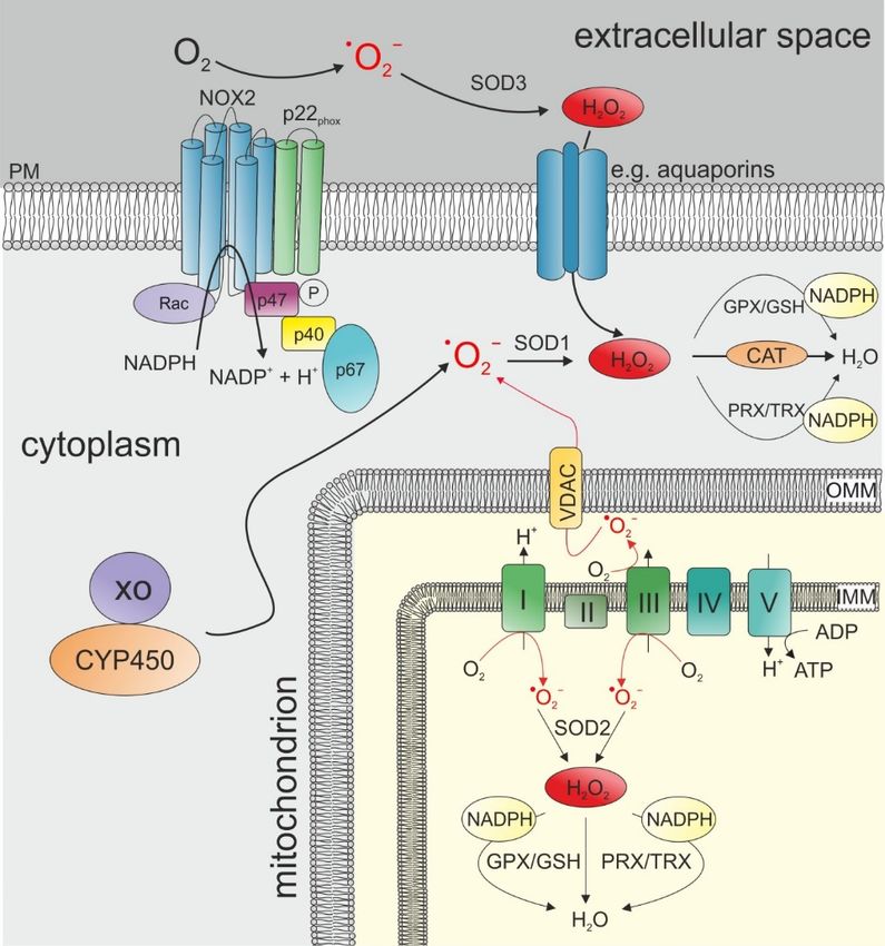
Cancers 2019, 11, 457 2 of 28
Cancers 2019, 11, x 2 of 27
1. An overview of the tumor microenvironment (TME): The TME is composed by a diverse
Figure 1.
range of
of cell
celltypes,
types,including
includingtumor
tumorcells, immune
cells, immune cells, epithelial
cells, cells,
epithelial andand
cells, stromal cells.cells.
stromal Areas of low
Areas of
nutrients and Oand
low nutrients 2 result
O2 in necrotic
result in regions.
necrotic The TME
regions. controls
The TME tumor growth
controls by
tumor diverse
growth mechanisms
by diverse
that are further
mechanisms discussed
that in the
are further text. in the text.
discussed
ROS
ROS have
haverecently beenbeen
recently in theinfocus
the offocus
TME research
of TMEbecause,
research depending
because,ondepending
their concentrations,
on their
ROS may be decisive
concentrations, ROS formaythebelife and death
decisive of cancer
for the cells
life and [12,13].
death Since Orai1
of cancer and Orai2
cells [12,13]. butOrai1
Since not Orai3
and
channels are strongly regulated by ROS [14–16], Orai channels are interesting targets to integrate Ca 2+
Orai2 but not Orai3 channels are strongly regulated by ROS [14–16], Orai channels are interesting
influx
targetsand
to ROS signaling
integrate Ca2+ in the TME.
influx and In ROSthissignaling
review, we infocus on theIn
the TME. interactions of Orai
this review, we channels
focus on andthe
ROS in the TME
interactions and on
of Orai their potential
channels and ROS relevance
in thefor TMEand
TME development. We propose
on their potential a scenario
relevance forwhere
TME
redox changes We
development. alterpropose
Orai function
a scenario Ca2+ influx
andwhere redox in both malignant
changes alter Oraiand nonmalignant
function cells, such
and Ca2+ influx as
in both
immune cells, resulting in changes in (Ca 2+ ) with a direct impact on tumor fate.
malignant and nonmalignant cells, such as immune int cells, resulting in changes in (Ca )int with a direct
2+
impact on tumor fate.
2. The Tumor Microenvironment (TME)
2. The Tumor Microenvironment
According to the World Health (TME)
Organization (WHO), cancer is “the second leading cause of
deathAccording
globally and is estimated to account
to the World Health Organization for 9.6 million deaths
(WHO), in 2018”
cancer (World
is “the Health
second Organization).
leading cause of
The
deathprocess of cancer
globally and development
is estimated and to progression
account for is9.6 called carcinogenesis
million deaths inand is divided
2018” (World intoHealth
3 to 4
distinct steps called initiation, promotion, progression, and metastasis [17].
Organization). The process of cancer development and progression is called carcinogenesis and is
In solid
divided into tumors, the tumor
3 to 4 distinct stepsmass
calledisinitiation,
formed bypromotion,
a diverse milieu which and
progression, is composed
metastasis of [17].
malignant
and nonmalignant cells such as endothelial cells, cancer-associated fibroblasts,
In solid tumors, the tumor mass is formed by a diverse milieu which is composed of malignant immune cells, adipose
cells, and neuroendocrine cells in addition to vascular and lymphatic networks
and nonmalignant cells such as endothelial cells, cancer-associated fibroblasts, immune cells, adiposeand the extracellular
matrix (ECM)
cells, and [1]. This dynamic
neuroendocrine and complex
cells in addition multicellular
to vascular environment
and lymphatic networks is known
and theas the tumor
extracellular
microenvironment (TME) (Figure 1). The TME has long been considered
matrix (ECM) [1]. This dynamic and complex multicellular environment is known as the an important factor for tumor
tumor
growth: The first publications
microenvironment (TME) (Figure are 1).
from theTME
The 19thhascentury
long [18]!
been In the past few
considered years, the TME
an important factorand
for
noncancerous
tumor growth: The first publications are from the 19th century [18]! In the past few years,therefore,
cells have been recognized as major players for tumor growth [19,20] and, the TME
as
andpotential targets for
noncancerous cellsdrug
haveactions. However, due
been recognized to its complexity,
as major players for the TMEgrowth
tumor and its [19,20]
cancer-type
and,
specific features still remain an obstacle for efficient cancer therapy [3].
therefore, as potential targets for drug actions. However, due to its complexity, the TME and its Many of the molecular
mechanisms of signaling
cancer-type specific featurespathways within
still remain the TMEfor
an obstacle areefficient
not well-understood
cancer therapyand [3]. the
Many complex
of the
interactions between cellular and non-cellular TME components are
molecular mechanisms of signaling pathways within the TME are not well-understood and the not well-defined. A detailed
complex interactions between cellular and non-cellular TME components are not well-defined. A
detailed understanding of the TME and its interactions would allow for better pharmacological
treatment and a better tumor prognosis.
Cancers 2019, 11, 457                                                                                3 of 28

understanding of the TME and its interactions would allow for better pharmacological treatment and
a better tumor prognosis.
      Depending on the cellular and non-cellular composition, the local milieu can be highly variable
between different tumors or even within the same tumor (Figure 1). The metabolism of different
cell types, cell–cell interactions, the architecture of TME formed by remodeling of ECM proteins
(which create a stiff fibrotic matrix), and the blood supply together create an environment composed
of oxygen; nutrients; reactive oxygen species (ROS); reactive nitrogen species (RNS); ATP; Ca2+ , H+ ,
and other ions; growth factors; chemokines; cytokines; or waste products (Figure 1). This environment,
together with locally secreted molecules from different cells, leads to pH gradients, differences in
oxygen tension, and interstitial pressure across the tumor [2,4,21]. These parameters have an impact
on the metabolism of surrounding cells and consequently cellular function, where the environment is
constantly changing and adapting according to new challenges and interactions with the host.
      It has been shown that the TME can promote cancer growth and metastasis as a result of
bidirectional interactions between cancer cells, noncancerous cells, and the surrounding environment.
In the worst scenario, the TME forms a niche favorable to tumor growth and less favorable to other
cells that could eliminate or limit tumor cell proliferation. In fact, it has been shown that most of
the nonmalignant cells within the TME often adopt a tumor-promoting phenotype after the local
milieu modifies their cellular functions [22]. Such alterations include changes in gene expression
and cellular activity. For example, hypoxia, which develops at the beginning of tumor growth and
induces cell necrosis (Figure 1), leads to the activation of hypoxia-responsive genes in malignant
and nonmalignant cells. It also promotes the recruitment and survival of immune cells that are
mainly glycolytic, such as macrophages, which produce large amounts of ROS [3]. This has drastic
consequences for the cells present in this niche, as they need to adapt to be able to survive in this highly
oxidative environment. Increased ROS also activates pathways in leukocytes to secrete more cytokines
that favor tumor growth, leads to new cellular mutations, and may, therefore, transform other cells
and induce apoptosis. Regions that undergo cycling hypoxia are also present in the TME. Cycling
here refers to situations of fluctuating oxygen levels, going from deep hypoxia to moderate hypoxia
states, similar to the reoxygenation of blood vessels in the human heart after ischemia, which bares the
risk of ischemia-reperfusion injury [23]. Cycling hypoxia can affect cells near blood vessels, which are
not efficiently perfused, contrary to hypoxic regions which are normally far from blood vessels [24].
Cells exposed to cycling hypoxia need to deal with and to adapt to two conditions: a lack of oxygen and
sudden reoxygenation, which leads to very high amounts of ROS. All these changes can create different
sub-microenvironments within the TME, each of them having distinct characteristics, populated by
different cells, and exposed to different molecules.

3. STIM/Orai Channels in Cancer
      Like all cells, cancer cells of the TME express a whole range of different ion channels. Considering
the importance of Ca2+ for cell signaling including proliferation, migration, and apoptosis in
combination with the strong dependence of cancer growth on these mechanisms, Ca2+ channels
should play a very important role in the TME. Among Ca2+ channels, Orai channels have a prominent
role in many electrically unexcitable cells [6,7,9]. They represent the main Ca2+ channel in most
immune cells in which they were initially discovered [25], but they are also highly expressed in cancer
cells and correlate with metastatic progression, a poor prognosis, and a shorter survival [5–8,10,11].
Since malignant cell functions depend on Ca2+ flux, considerable interest has emerged in the
therapeutic potential of inhibiting Orai for many cancer types.

3.1. Short Introduction to STIM and Orai
      Store-operated Ca2+ entry (SOCE) is the major Ca2+ entry pathway including cancer and immune
cells [5–8,10,11]. Its well-known activation mechanism is depicted in Figure 2. The two known SOCE
activators, STIM1 and STIM2, sense the ER Ca2+ store content and activate Orai channels in the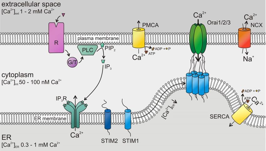
Cancers 2019, 11, 457 4 of 28
plasma membrane. STIMs are known to form homomultimers (as depicted in Figure 2) but may also
form heteromultimers [26]. STIM1 is less sensitive to ER luminal Ca2+ compared to STIM2 [27] but
activates Orai channels significantly stronger [28]. There are three known isoforms of the tetraspanning
Cancers 2019, 11, x 4 of 27
hexameric Orai channels, Orai1, Orai2, and Orai3 (Figure 2), with characteristic properties [29,30].
All of them 2+ from the ER, possess a high selectivity for Ca2+ ,
of them are are activated
activated after
after the the depletion
depletion of 2+Cafrom
of Ca the ER, possess a high selectivity for Ca2+, are
are inwardly rectifying, and show a Ca2+ -dependent inactivation (CDI)
inwardly rectifying, and show a Ca -dependent inactivation (CDI)
2+ [30].[30].
Figure 2.
Figure 2. Store-operated
Store-operated Ca Ca2+2+ entry (SOCE) by stromal-interaction molecule (STIM)/Orai channels in
entry stromal-interaction molecule (STIM)/Orai
cancer cells:
cells: Following
Followingthe thestimulation
stimulationofofG G protein-
protein- or or
tyrosine
tyrosine kinase-coupled
kinase-coupled (G/T) receptors
(G/T) (R),
receptors
phospholipase
(R), phospholipase C (PLC) C (PLC) hydrolyses
hydrolysesphosphatidylinositol
phosphatidylinositol4,5-bisphosphate
4,5-bisphosphate (PIP (PIP2)2 ) to
to inositol
trisphosphate (IP (IP33).).The latter
The binds
latter to itstoreceptor,
binds its receptor, Ca2+ channel
a Ca2+ arelease in the endoplasmic
release channel reticulum
in the endoplasmic
and opens it, which induces the Ca 2+
reticulum (ER) membrane and opens it, which induces the Ca depletion of ER Ca stores. A (Ca
(ER) membrane 2+ depletion of ER Ca 2+ stores.2+
A drop of drop2+)ER
of
(Ca 2+
activates luminal Ca
)ER activates 2+
luminalsensor 2+
Ca proteins, the STIMs.
sensor proteins, the Activated STIMs oligomerize
STIMs. Activated and move
STIMs oligomerize and to the
move
plasma
to membrane
the plasma where they
membrane where bind and
they open
bind andOrai channels,
open Orai channels, a Ca2+ influx
leading toleading to a Ca 2+ influx
across the plasma
across
membrane.
the Plasma membrane
plasma membrane. Plasma Ca 2+ ATPases
membrane 2+
Ca (PMCAs),
ATPases Na +
(PMCAs), 2+ +
–Ca exchangers 2+ (NCX), and (NCX),
Na –Ca exchangers sarco-
/endoplasmic
and reticulum Ca
sarco-/endoplasmic 2+ ATPases
reticulum Ca2+(SERCAs)
ATPasesexport
(SERCAs)Ca2+export
from the 2+ from the cytosol.
Cacytosol.
3.2. STIM/Orai in
3.2. STIM/Orai in Tumor
Tumor Initiation
Initiation and
and Promotion
Promotion
Initiation
Initiation isisthethefirst step
first in cancer
step development
in cancer developmentwherewhere
normalnormal
cells undergo irreversible
cells undergo changes,
irreversible
transform, escape the immune surveillance, undergo continuous unregulated
changes, transform, escape the immune surveillance, undergo continuous unregulated proliferation, proliferation, and are
able to form tumors at the end. The loss of growth control involves a whole range
and are able to form tumors at the end. The loss of growth control involves a whole range of critical of critical mutations
and is the sum
mutations and of the sum
is the accumulated abnormalities
of the accumulated in a cell’s regulatory
abnormalities in a cell’s systems.
regulatory Whether
systems. changes
Whetherin
(Ca 2+ ) are relevant for cancer initiation is currently not clear [5].
int
changes in (Ca )int are relevant for cancer initiation is currently not clear [5].
2+
A
A mechanistic
mechanisticlink between
link betweenCa2+Ca homeostasis
2+ homeostasisand chromosome
and chromosome instability was recently
instability was proposed
recently
in hepatocytes with Hepatitis B viral (HBV) infection, as the driver of hepatocellular
proposed in hepatocytes with Hepatitis B viral (HBV) infection, as the driver of hepatocellular carcinoma [31].
The hepatocytes carry a gain-of-function mutation in the preS2 region of a large
carcinoma [31]. The hepatocytes carry a gain-of-function mutation in the preS2 region of a large surface antigen (LBHS),
one of two
surface HBV-encoded
antigen (LBHS), one oncoproteins, that is linked
of two HBV-encoded to the early that
oncoproteins, onsetisof hepatocellular
linked to the early carcinoma.
onset of
The preS2 mutation promotes ER-plasma membrane (PM) connections
hepatocellular carcinoma. The preS2 mutation promotes ER-plasma membrane (PM) connections through ER stress and causes
the permanent activation of SOCE in these cells, inducing chromosome
through ER stress and causes the permanent activation of SOCE in these cells, inducing chromosome instability, aneuploidy,
and anchorage-independent
instability, growth [31]. However,growth
aneuploidy, and anchorage-independent more in vivo
[31]. studies more
However, and direct
in vivoevidence are
studies and
necessary to prove or disprove a role of SOCE and, thus, Ca 2+ as cancer initiators or drivers.
direct evidence are necessary to prove or disprove a role of SOCE and, thus, Ca as cancer initiators 2+
or drivers.
3.3. STIM/Orai in Tumor Proliferation/Growth
Orais and STIMs are expressed in the vast majority of tumors [5,10]. Their expression levels seem
to correlate with metastatic progression, a poor prognosis, and a shorter survival in studies using
patient specimens. Colorectal cancer patients with a positive expression of STIM1 [32] or/and with a
high Orai1 expression had poorer prognoses and shorter overall survival rates [33]. Comparable
results for Orai1 were also shown for non-small cell lung cancer [34], esophageal squamous cell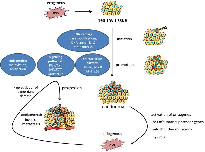
Cancers 2019, 11, 457                                                                                 5 of 28

3.3. STIM/Orai in Tumor Proliferation/Growth
     Orais and STIMs are expressed in the vast majority of tumors [5,10]. Their expression levels seem
to correlate with metastatic progression, a poor prognosis, and a shorter survival in studies using
patient specimens. Colorectal cancer patients with a positive expression of STIM1 [32] or/and with
a high Orai1 expression had poorer prognoses and shorter overall survival rates [33]. Comparable
results for Orai1 were also shown for non-small cell lung cancer [34], esophageal squamous cell
carcinoma [35], and gastric cancer [36]. Additionally, Orai3 expression was increased in tumor
tissues of lung adenocarcinoma [37], prostate cancer [38], and breast cancer [39] and correlated with
overall survival and metastasis-free survival [37]. Interestingly, an analysis of a microarray from
McAndrew and colleagues revealed a significantly poorer prognosis [40] for breast cancer patients
with a STIM1-high and STIM2-low phenotype. Considering that not only the ratio between STIM1,
STIM2, Orai1, Orai2, and Orai3 but also the discovery of STIM2 splice variants [41,42] are highly
relevant for Ca2+ channel activity [43–47], the relative composition of STIMs and Orais needs to be
carefully addressed not only in tumors.
     Cell proliferation is dependent on the cell cycle and transitions between different phases (G0/G1,
S phase, and G2/M phase) which are tightly controlled through Ca2+ -dependent checkpoints (reviewed
in Reference [48]). SOCE alters cancer cell proliferation in vitro [49–51] and also in vivo [33,35,36,52–54].
However, how Ca2+ controls distinct checkpoints is not well-understood. Increases in the basal or
transient fluctuation of Ca2+ are involved, but also (Ca2+ )ext needs to be considered. Cell cycle arrest in
the G0/G1 phase in U251 cells [53], in neck squamous cell carcinoma cell lines [54], and at the S and
G2/M phases in cervical cancer cells [52] by STIM1-silencing has been reported. A pro-proliferative
role of STIM1 in vivo using U251 human glioma xenograft model in mice revealed that knocking
down STIM1 in xenografts demonstrated a diminished growth [53]. In contrast, an elevated Orai1
and/or STIM1 expression can promote cell proliferation [36,54]. In non-small lung cancer cells,
nicotine promotes cell proliferation by upregulating Orai1 expression and therefore by enhancing
SOCE and increasing basal Ca2+ concentration [55]. In esophageal squamous cell carcinoma zinc
is able to inhibit Orai1-mediated SOCE, Ca2+ oscillations, and subsequent cell proliferation [56].
The pharmacological inhibition or knocking down of Orai channel could block human esophageal
squamous cell carcinoma proliferation in vitro and tumor growth in vivo [35]. Another study shows
that high Ca2+ diet in a mouse model of slowly evolving prostate cancer accelerated its progression
by promoting proliferation [57] indicating the importance of (Ca2+ )ext . Growth factors as fibroblast
growth factor 4 (FGF4) may also have an impact on Orai1 expression, resulting in increased SOCE,
and may promote epithelial-mesenchymal transition and enhanced cell proliferation [58]. One of the
most important and interesting cascades in this context is the mTOR (mechanistic target of rapamycin)
pathway (reviewed in Reference [59]). As the catalytic subunit of two distinct protein complexes
(mTORC1 and C2), this serine/threonine protein kinase plays a central role for the cell perception of
the environment in the regulation of metabolism, cell cycle, and growth. It is, thus, essential for the
adaptability of cells to specific changes and needs as in the TME. A recent study showed an interesting
relationship between mTORC1 and STIM1 expression as a novel potential therapeutic approach for
patients with tuberous sclerosis complex (TSC) tumors [60].
     Not only the main players of SOCE, STIM1 and Orai1, but also the slightly “neglected” STIM2 and
Orai3 are involved in proliferation and growth of tumor cells [61]. The overexpression of STIM2
inhibits cell proliferation and tumor growth in colorectal cancers in vivo [62] but promotes cell
migration in primary melanoma in vivo [63], implicating the contribution of STIM2 signaling at
different stages of tumor progression. Furthermore, the high Orai1 and STIM2 expression found in
melanoma biopsies at the rim of invading tumors are linking their possible role in tumor invasion
and/or metastasis in vivo [63]. In human carcinoma cells versus normal mucosa cells, STIM2 protein
was nearly depleted in contrast to an upregulation of STIM1 and all three Orai proteins [64].
The involvement of Orai3 in the machinery of tumorigenesis has been reported, including breast,
prostate, and lung cancer [38,65–67]. By using mice xenograft models, it was shown that Orai3 plays
Cancers 2019, 11, 457 6 of 28
a crucial role in prostate cancer development in vivo [38]. The knockdown of Orai3 significantly
reduced SOCE and inhibited proliferation by arresting non-small cell lung cancer cell lines in the
G0/G1 phase [67]. Orai3 transcripts are differentially expressed in the different subtypes of breast
cancer and regulated by estrogen receptor alpha (ERα). The silencing of ERα causes a decreased
expression of Orai3 and cell proliferation in vitro [65]. The same study places Orai3 as an important
player in tumorigenesis in vivo, since the growth of breast tumors was significantly reduced by
Orai3 knockdown before the transfer to the recipient mice with severe combined immunodeficiencies
(SCID) [65]. Orai3 expression seems to be regulated positively and negatively by miRNA and to
act directly on Orai3 30 UTR [68]. Another interesting finding places Orai1–Orai3 channel complexes
in the center of attention in a variety of prostate cancer cells [38]. The study reports an oncogenic
switch in which cells, especially those exposed to tumor microenvironmental factors (here, arachidonic
acid), change from homologous Orai1 complexes to more heterogeneous Orai1–Orai3 complexes by
upregulating Orai3 expression. This switch could be an excellent adaptation, where a shift from
a pro-apoptotic to more pro-proliferative phenotype is beneficial for cell proliferation and growth [38].
3.4. STIM/Orai in Tumor Survival/Apoptosis
A well-controlled balance between cell proliferation and cell death is necessary to avoid excessive
proliferation leading to cancer development. In cancer, a scenario with too little apoptosis dominates,
causing the expansion of malignant cells that are resistant to apoptosis. Despite being part of the
problem, apoptosis is a popular target in cancer treatment. Therefore, a better understanding of the
complex underlying mechanism of apoptosis is the key to developing more specific targets to execute
the lethal hit against cancer cells. Ca2+ plays a pivotal role in the mechanistic induction of apoptosis.
During apoptosis, (Ca2+ )int is dramatically increased, and as a consequence, mitochondria take up
large amounts of it and induce apoptosis. Interrupting the prolonged Ca2+ influx through SOCE by
knocking down Orai/Stim or blocking it with specific inhibitors can counteract cell apoptosis.
Human colon carcinoma cells show increased Orai and STIM1 expression, but STIM2 is
almost depleted [64]. In noncancerous cells, the knockdown of STIM2 decreases SOCE while it
promotes apoptosis resistance. This finding suggests that the loss of STIM2 contributes to apoptosis
resistance in tumor cells. In addition, the blockage of STIM1-mediated SOCE can significantly
enhance chemotherapy-induced apoptosis in lung and pancreatic cancer cells [69,70]. In a pancreatic
adenocarcinoma cell line, a siRNA-mediated knockdown of Orai1 and/or STIM1 increases apoptosis
induced by chemotherapy drugs 5-fluorouracil or gemcitabine [70]. In addition, Orai1 downregulation
has been shown to contribute to the formation of an apoptosis-resistant phenotype in prostate cancer
cells [71]. Furthermore, the presence of an increased Orai3 expression, leading to the assembly of
more Orai1/Orai3 channel complexes can increase the resistance to apoptosis, as has been suggested
in the context of pancreas carcinoma [38]. Similar results are found in breast cancer cells where the
downregulation of Orai3 arrests cell-cycle progression and induces apoptosis but not in normal breast
epithelial cells [39].
A recent study placed Bcl-2 as a SOCE regulator to modify ER stress-induced apoptosis [72].
The Bcl-2 family plays a major role in the regulation of apoptosis. Its pro- or anti-apoptotic members
act mainly at the mitochondria level. Bcl-2 is the first identified anti-apoptotic protein capable of
preventing apoptosis in a Ca2+ -dependent manner [73]. A mutant of Bcl-2 used in the study increased
the expression of SOCE components and depleted Ca2+ in the ER lumen, causing a massive Ca2+ influx
leading to caspase activation and apoptosis [72]. Additionally, several anticancer drugs that are used
to induce cancer cell apoptosis function through the dysregulation of Ca2+ signaling, for example,
in colon cancer cells or triple-negative breast cancer [74,75].
3.5. STIM/Orai in Epithelial-to-Mesenchymal Transition (EMT)/Cancer Progression
Changes in cell phenotypes, defined as epithelial-to-mesenchymal transition (EMT), are important
for tumor metastasis [76,77]. This transition is associated with an improvement in migratory andCancers 2019, 11, 457 7 of 28
invasive properties. The list of EMT inducers is long, including growth factors secreted by the tumor
environment, cytokines, hypoxia, and metabolic changes. Additionally, the indispensable change
in gene expression is activated by complex regulatory networks, involving transcriptional control,
transcriptional factors, miRNAs, alternative splicing, posttranslational regulation, protein stability,
and subcellular localization [78]. Several studies already reported that an altered SOCE is linked to
EMT in prostate cancer [79], colon cancer [32], and gastric cancer [36]. Transforming growth factor
β1 (TGF-β1) in MCF7 breast cancer cells enhanced SOCE. Silencing the transcription factor Oct4
or significant inhibition via TGF-β1 upregulated the expression of STIM1 and Orai1 and promoted
invasion and metastasis by inducing EMT [80]. TGF-β-induced EMT seems to be differently regulated
by the expression of STIM2 (regulating non-store-operated Ca2+ entry) and by the expression of
STIM1 (regulating store-dependent Ca2+ entry) [81]. Another study describes the Orai3 and STIM1
requirement for TGF-β-dependent Snai1 transcription, a transcription factor upregulated during
EMT [82]. A similar study in lung adenocarcinoma cell lines shows that FGF4 was also able to induce
EMT by elevating Ca2+ entry via the expression of Orai1 channels [58].
3.6. STIM/Orai in Tumor Metastasis/Angiogenesis
Cell motility is partly mediated by a (Ca2+ )int gradient [83,84], and several components of
migration mechanisms, such as cytoskeleton remodeling, leading edge guidance, and matrix
degradation, are Ca2+ sensitive [85]. Over the last ten years, in vitro and in vivo evidence
has accumulated that SOCE components are involved in cell motility, invasion, and tumor
metastasis [86]. The inhibition of SOCE or their components inhibit metastasis of breast
cancer [57,87,88], melanoma [63,89], colorectal cancer [32], prostate cancer [51], and gastric cancer [36].
Consistently, the overexpression of STIM1 enhances cell migration in cervical cancer [52] and colorectal
cancer [49]. Cell motility and, thus, metastasis are regulated by dynamic interactions between
cytoskeleton, myosin II and focal adhesions [90], which assemble and disassemble to mediate
cell migration [91]. STIM1-dependent signaling regulates focal adhesion turnover [52,87] required
for leading edge protrusion and trailing tail retraction and regulates actomyosin contractility [92].
Furthermore, silencing STIM1 significantly alters podosome dynamics, reduces cell invasiveness [93],
and regulates the dephosphorylation of focal adhesion kinase (FAK) by modulating focal adhesion
turnover [94,95] and the recruitment and association of active pTyr397-FAK and talin at focal
adhesions [92]. Similar results were found in breast cancer cells in a murine tumor metastasis
model [87], in colorectal cancer following a destabilization of STIM1 [96], and in glioma cell lines [95].
One important pathway to modulate cell migration, invasiveness, and metastasis is the
PI3K/Akt/mTOR signaling pathway. Akt plays a central role by phosphorylating many proteins
involved in the stabilization of actin cytoskeleton and by promoting migration via remodeling. SOCE is
positively regulated by the PI3K/Akt pathway, and this effect might be suppressed by targeting
receptor tyrosine kinases (RTK) [97]. The RTK protein family includes epidermal growth factor
receptors (EGFRs), FGF receptors, and vascular endothelial growth factor receptors (VEGFRs) [98]
which are heavily involved in cell migration [98] and angiogenesis. Upregulated VEGF production
by a high STIM1 expression in human cervical cancer cells regulates the focal-adhesion dynamics
of migratory cells [52]. Accordingly, the inactivation of PI3K/Akt signaling pathway by STIM1
knockdown reduced the migration and invasion of prostate cancer cells [51]. Furthermore, a lipid raft
SK3/TRPC1/Orai1 complex promotes cell migration in metastatic colorectal cancer. The formation
of this complex is favored by the phosphorylation of STIM by epidermal growth factor (EGF) and
the activation of Akt [99]. A SK3-Orai1 complex also plays a critical role in cell migration and bone
metastasis [88,99].
The proliferation and motility of cells are critical steps in angiogenesis. Due to continuous and
fast growth, tumors rely on the formation of new blood vessels to ensure an adequate supply of
oxygen and nutrients [100]. Low oxygen tension (hypoxia) promotes metastasis and is considered
as the key driver in angiogenesis [101,102]. Hypoxia is sensed by the tissue and triggers theCancers 2019, 11, 457 8 of 28
cellular production of hypoxia-inducible factor 1 (HIF-1), a transcription factor that activates many
downstream pathways [103] including the expression of VEGF, TGF-β, and platelet-derived growth
factor (PDGF-β) [104]. Hypoxia is a common feature of the TME of most solid tumors, and hypoxic
cancer cells secret VEGFs to initiate tumor angiogenesis [100,105]. Interestingly, VEGF and FGF (and
others) increase (Ca2+ )int . However, molecular mechanisms underlying SOCE-mediated angiogenesis
remain poorly understood.
In colon cancer [106] and in triple-negative breast cancer [107], hypoxia leads to the upregulation
of Orai1 by the Notch1 pathway. These data are in line with the findings that Notch1 signaling
pathways activate NFκB [108] and that NFκB regulates the expression of Orai1 and STIM1 [109].
Orai1 upregulation potentiates SOCE and activates the nuclear factor of activated T cells, NFAT4,
contributing to hypoxia-induced invasion and angiogenesis [106,107]. Hypoxia leads to the
accumulation of hypoxia-inducible factor 1-alpha (HIF-1α), a subunit of the transcription factor HIF,
which responds to alterations of available oxygen [110]. The hypoxia-induced accumulation of HIF-1α
was found to correlate with the overexpression of STIM1 in human and murine hepatocarcinoma
cells (HCCs) [111]. The increase in STIM1 expression is a consequence of a direct HIF-1α binding to
the promoter of STIM1 and leads to an increase in SOCE in HCCs promoting tumor growth [111].
During hypoxia, HIF-1α is stabilized and induces many genes like VEGF for a better adaptation
to this condition [112]. Since the production of ROS is augmented under hypoxic conditions [113]
and ROS inhibits the SOCE mediated by STIM1 and Orai1 or Orai2 [14–16], the overexpression of
STIM1 might be a countermeasure of the HCCs to provide the necessary Ca2+ signals for vital cell
functions. Furthermore, the expression of STIM2 was also reported [114] under hypoxic conditions.
In rat pulmonary arterial smooth muscle cells, hypoxia-related STIM2 overexpression is accompanied
by an increased SOCE and proliferation [114]. Apart from the hypoxia-induced induction of Orai1 and
STIMs, it was recently also reported that Orai3 expression was induced by HIF1α in MDA-MB-468 ERα
negative cells [115]. Unlike in ERα positive MCF-7 breast cancer cells [65,66] where Orai3 silencing
reduces SOCE, the upregulation of Orai3 did not contribute to SOCE in ERα negative cells [115].
However, since the Orai3 expression in breast cancer is highly dependent on the ERα expression itself,
one might speculate that the Orai1 complexes contribute significantly to Ca2+ signals in ERα negative
cells. These results place the channels and sensors as new targets in regulating hypoxia.
4. ROS Production and Elimination
As mentioned before, reactive oxygen species (ROS) are one of the key factors to influence the
TME, and they also modulate Orai channels (see below). In the following section, we summarize the
most common pathways and mechanisms in ROS metabolism with specific regard to the TME inspired
by two reviews [116,117].
ROS comprise a group of molecules that are generated via the partial reduction of O2 and establish
high chemical reactivity [116–118]. The one-electron reduction of oxygen leads first to the formation of
superoxide anion radical (• O2 ¯), which in turn is dismutated and further reduced to hydrogen peroxide
(H2 O2 ) which is finally either fully reduced to water or partially reduced and split to hydroxyl radical
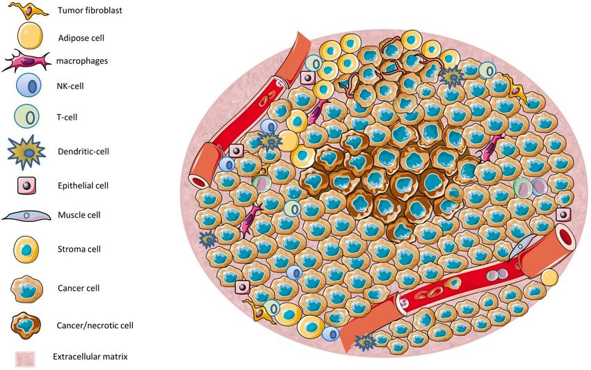
(OH• ) and hydroxyl anion (OH¯) [119]. ROS is mainly produced by mitochondria, NADPH oxidases
(NOX), and other enzymes like xanthine oxidase and cytochrome P450 [116,117,120,121] (Figure 3).
Very high ROS levels can be produced in mitochondria when • O2 ¯ molecules are released during
ATP generation in the electron transport chain (ETC) [116,122]. Several endogenous and exogenous
factors influence mitochondrial ROS production, including mitochondrial membrane potential [123],
hypoxia, and nutrient metabolites but also cancer- and immune-related factors like TNF-α and Toll-like
receptors [117,124–127].
NOX complexes also produce very high levels of ROS (Figure 3). The production of ROS via the
NOX family occurs during the catalysis of electron transfer from NADPH to O2 and thereby produces
• O ¯ [117]. NOX-dependent ROS production requires the functional assembly of the NOX complex,
2
which can be mediated by different signaling molecules such as growth factors and, again, TNF-α andCancers 2019, 11, 457 9 of 28
Toll-like receptors [117,128,129]. Similar to the mitochondrial ROS production, the release of • O2 ¯ is
dependent on the location of the NOX molecules. NOX comprises NOX1-5 and Duox1/2 [116] which
are present in the plasma membrane and intracellular membranes of the nucleus, mitochondria and
the endoplasmatic reticulum (ER) [117]. The specific isoforms at particular sites can lead to ROS release
either to the intracellular or extracellular spaces [117,121].
Cancers 2019, 11, x 9 of 27
Figure 3.
Figure 3. The
The main
main sources of ROS production and elimination: ROS ROS can
can be generated either by
NADPHoxidases
activated NADPH oxidases(here
(here represented
represented by NOX2)
by NOX2) located
located in theinplasma
the plasma membrane
membrane or by
or by several
several complexes
complexes of the transport
of the electron electron transport chain
chain in the in the
inner inner membrane
membrane of mitochondria.
of mitochondria. Other
Other metabolic
metabolicinclude
enzymes enzymes includeoxidase
xanthine xanthine(XO)oxidase (XO) and cytochrome
and cytochrome P450 (CYP450) P450that
(CYP450)
directlythat
form directly form
superoxide
superoxide
in in the cytoplasm.
the cytoplasm. SuperoxideSuperoxide
dismutases dismutases
(SODs) can (SODs)
convertcan• O
convert
2 ¯ to H •O2¯ to H2O2. SOD1 is located
O
2 2 . SOD1 is located in the
in the cytoplasm,
cytoplasm, SOD2 SOD2 is located
is located in the in the mitochondrial
mitochondrial matrix,matrix,
and SOD3and SOD3 is located
is located in the in the extracellular
extracellular space.
space.
The The further
further elimination
elimination of H2 O 2O2catalase,
of2Hvia via catalase, GPX/GSH,
GPX/GSH, and/or
and/or PRX/TRXcan
PRX/TRX caneither
either bebe initiated
cytoplasm upon the transport
directly in the mitochondrial matrix or in the cytoplasm transport of of H
H22O22 via
via aquaporins.
aquaporins.
CYP450: cytochrome
cytochrome P450;
P450; IMM:
IMM: inner
inner mitochondrial
mitochondrial membrane; OMM: OMM: outer mitochrondrial
membrane; PM: plasma membrane; VDAC: voltage-dependent anion channel; XO: xanthine oxidase. oxidase.
ROS have
have long
long been
been considered
consideredtotobe besolely
solelydeleterious
deleteriousfor
forcells
cells causing
causing oxidative
oxidative damage
damage in
in different
different molecules
molecules like like
DNA, DNA,
lipids,lipids, or proteins.
or proteins. However, However,
moderatemoderate
ROS levelsROS
are levels are also
also important
important
for variousfor physiological
various physiological
cellularcellular functions,
functions, including
including intracellular
intracellular signaling,cell
signaling, cell survival,
survival,
proliferation and immune responses [117,122,130,131]. Hence,
immune responses [118,123,131,132]. Hence, a unique redox a unique redox homeostasis is
required to control
control the
the balance
balance between
between production
productionand andelimination
elimination[118].
[117].ROSROS elimination,
elimination, or
or “antioxidant defense”, is mainly performed by four enzymatic
“antioxidant defense”, is mainly performed by four enzymatic systems: systems: superoxide dismutases
(SODs), peroxiredoxin
peroxiredoxin (PRX)/thioredoxin
(PRX)/thioredoxin (TRX) system, system, glutathione
glutathione peroxidase
peroxidase(GPX)/glutathione
(GPX)/glutathione
(GSH) system, and catalase (Figure
(Figure 3).
3).
The quantification of ROS levels is a challenging task. Table 1 summarizes the most commonly
used approaches.
For more comprehensive overviews over certain technologies, we acknowledge the following
reviews which are also relevant for the conceptual design of Table 1: references [133–151].Cancers 2019, 11, 457 10 of 28
The quantification of ROS levels is a challenging task. Table 1 summarizes the most commonly
used approaches.
For more comprehensive overviews over certain technologies, we acknowledge the following
reviews which are also relevant for the conceptual design of Table 1: references [132–150].
Table 1. A summary of the most commonly used tools and probes to measure various ROS.
Technique or Method Tools and Examples Specificity (Potential) Applications
Dihydroethidium (DHE) • O ¯, if used with HPLC
2
Dihydrorhodamine (DHR) not specific
Fluorescence- 20 ,70 -dichlorodihydrofluorescein in vitro, extra- and
not specific intracellular,
based assays (DCFH2 )
cell suspensions
Amplex Red, Amplex UltraRed H2 O2
Hydroxyphenyl Fluorescein (HPF) not specific
Aminophenyl Fluorescein (APF) not specific
roGFP2 EGSH
Genetically roGFP2 coupled to glutaredoxins EGSH in vivo and in vitro,
encoded fluorescent roGFP2 coupled to peroxidases or intracellular, single cells,
probes H2 O2 tissues, subcellular
peroxiredoxins
compartments
HyPer (different variants
H2 O2
including HyPer-Red)
Lucigenin
Chemiluminescence
Luminol not specific in vitro, cell suspensions
assays
Isoluminol
Cytochrome C, Superoxide
Enzymatic assays dismutase (SOD), Horseradish •O ¯ in vitro, cell suspensions
2
Peroxidase (HRP)
Prussian Blue, Paraquat
(1,10 -Dimethyl-4,40 bipyridium H2 O2 , peroxides and
Chemical assays in vitro, cell suspensions
dichloride), FOX (containing others
xylenol orange)
Electrodes of various types and
sizes (macro-, mini-, micro- • O ¯, in vitro; single cells,
2 H2 O2 and other
Electrochemical assays ultramicro- and nanoelectrodes), cell suspensions, extra-
ROS
arrays, chips; additional redox and intracellular
mediators
DMPO (5, • O ¯,
2 OH•
5-dimethyl-1-pyrroline-N-oxide)
DEPMPO
Electron paramagnetic • O ¯, in vitro and in vivo,
[5-(diethoxyphosphoryl)-5-methyl- 2 OH•
resonance (EPR) cell suspensions, extra-
1-pyrroline-N-oxide]
spectroscopy using spin and intracellular
probes Horseradish Peroxidase assay
(enzymatic using cyclic H2 O2
hydroxylamines)
in vivo EPR with different
Various ROS
functional spin traps and probes
5. Impact of Reactive Oxygen Species (ROS) in the Tumor Microenvironment (TME)
ROS likely plays a dual role in cancer, both as initiating factors as well as downstream signaling
molecules as a result of malignant transformations. In the literature, numerous reviews are available
that deal with cancer and ROS, but it is often hard to distinguish between cancer development and
progression and between “good” and “bad” ROS, although these differences play important roles in
the whole topic [116].Cancers 2019, 11, 457 11 of 28
5.1. How Can ROS Support Carcinogenesis?
The first step of carcinogenesis is called initiation and refers to the alteration, change, or mutation
of genes that develop either spontaneously or due to an exogenous source [17]. ROS can induce
detrimental DNA damage via base modifications; inter-strand, intra-strand and DNA-protein
crosslinks; and the induction of double strand breaks [151] (Figure 4). The promotion of cancer
(cells)
Cancers is considered
2019, 11, x a clonal expansion and accumulation of the pre-neoplastic cells that result from the
11 of 27
initiation process, while progression already refers to a malignant conversion of the cells into invasive
carcinoma [17,152].
carcinoma [17,153]. Once tumors are initiated, there are
are several
several ways
ways that
that ROS
ROS can
can drive
drive promotion
promotion
and expansion.
and expansion. These can be roughly divideddivided into
into 33 subgroups:
subgroups: the
the impairment
impairment ofof transcription
transcription
factors, signaling
factors, signaling pathways,
pathways, and epigenetics (Figure 4). We We discuss
discuss the
the mechanisms most frequently
involved in
involved in the
the majority
majority of
of cancer
cancer subtypes
subtypes but
but cannot
cannot acknowledge
acknowledgeevery
everymechanism
mechanismin-depth.
in-depth.
Figure
Figure 4.4. ROS
ROS inin the
the TME:
TME: Non
Non cancer-related
cancer-related ROS
ROS can
can be
be deleterious
deleterious for healthy
healthy tissue and thereby
lead
lead to
to tumor
tumor initiation
initiation and promotion via DNA damage and the impairment of signaling pathways,
transcription
transcription factor
factor expression,
expression, andand epigenetic
epigenetic changes.
changes. After the tumor
tumor initiation
initiation upregulation
upregulation ofof
oncogenes, the loss of tumor suppressor genes, mitochondria mutations, and hypoxic
oncogenes, the loss of tumor suppressor genes, mitochondria mutations, and hypoxic conditions can conditions can
lead
lead to
to further
further elevated
elevated tumor-related
tumor-related ROSROS levels.
levels. In a positive
positive feedback
feedback loop
loop system,
system, rising
rising ROS
ROS
levels
levels in
in the
the TME
TME can can in
in turn
turn support
support tumor
tumor progression,
progression, angiogenesis,
angiogenesis, invasion,
invasion, and
and metastasis
metastasis via
via
an
an amplification
amplification of of the
the pathways
pathways involved
involved in
in initiation
initiation and
and promotion.
promotion. Finally,
Finally, to
to protect
protect against
against
threshold-crossing
threshold-crossing toxic toxic ROS
ROS levels,
levels, tumor
tumor cells
cells initiate
initiate the
the upregulation
upregulation of of antioxidant
antioxidant defense
defense
mechanisms
mechanisms in order to prevent from ROS-related deleterious events like apoptosisor
in order to prevent from ROS-related deleterious events like apoptosis ornecroptosis.
necroptosis.
The invasion of newly synthesized blood vessels to the network of tumor cells (angiogenesis)
The invasion of newly synthesized blood vessels to the network of tumor cells (angiogenesis) is
is an important event in carcinogenesis which is responsible not only for the supply with nutrients,
an important event in carcinogenesis which is responsible not only for the supply with nutrients,
immune cells, and oxygen but also for the disposal of waste products as well as tumor spreading
immune cells, and oxygen but also for the disposal of waste products as well as tumor spreading
(metastasis) [153] (Figure 4). One of the most prominent pro-angiogenic factors is the vascular
(metastasis) [154] (Figure 4). One of the most prominent pro-angiogenic factors is the vascular
endothelial growth factor (VEGF), which has been shown to be a key regulator in cancer angiogenesis
endothelial growth factor (VEGF), which has been shown to be a key regulator in cancer angiogenesis
upon stimulation via several pathways, factors, and conditions [154]. One of the transcription factors
upon stimulation via several pathways, factors, and conditions [155]. One of the transcription factors
that leads to increased VEGF expression is HIF-1α [155]. Elevated ROS levels can suppress HIF-1α
that leads to increased VEGF expression is HIF-1α [156]. Elevated ROS levels can suppress HIF-1α
degradation, finally leading to an increased VEGF expression and subsequent angiogenesis in distinct
cancer types such as prostate and ovarian cancer and fibrosarcoma [117,157]. Another important
transcription factor in cancer is the nuclear factor kappa-light-chain-enhancer of activated B cells (NF-
κB) [158]. Besides its well-known role in immune and inflammatory responses, cell proliferation, andCancers 2019, 11, 457 12 of 28
degradation, finally leading to an increased VEGF expression and subsequent angiogenesis in distinct
cancer types such as prostate and ovarian cancer and fibrosarcoma [116,156]. Another important
transcription factor in cancer is the nuclear factor kappa-light-chain-enhancer of activated B cells
(NF-κB) [157]. Besides its well-known role in immune and inflammatory responses, cell proliferation,
and apoptosis [158], NF-κB can also promote tumor proliferation [116,117]. Interestingly, mitochondrial
ROS have been shown to activate NF-κB with the subsequent upregulation of the EGF receptor in
pancreatic cancer, inducing the formation of pre-neoplastic lesions [117].
Another ROS-sensitive transcription factor involved in cell transformation, proliferation,
and apoptosis is activator protein 1 (AP-1) [116,159]. In human colon cancer cells, the upregulation
of AP-1 due to H2 O2 has been documented, leading to increased MMP7 levels that are involved in
tumor metastasis [160]. Furthermore, activated AP-1 enhances the expression of genes involved in
growth stimulation like cyclin D1 but suppresses genes involved in the growth inhibition of cell cycle
inhibitor p21, finally leading to an increased cell proliferation [116,161,162]. The tumor suppressor
gene p53 is a key regulator of anti-proliferative cellular responses and is mutated in many tumor cells,
resulting in a deleterious loss of function [163]. The ROS-dependent impairment of p53 expression
and its activation/inactivation are controversially discussed in the literature. It is evident that ROS can
directly inactivate p53 via the oxidation of cysteine residues in its DNA-binding domain [164], but the
downstream effects of this inactivation remain ambivalent. In general, the inactivation of p53 will
most likely leads to the loss of anti-proliferative effects, which are part of its tumor suppressor gene
function. On the other hand, elevated ROS levels can promote apoptosis, senescence, and DNA-repair
in a p53-dependent manner, finally leading to a reduced malignant transformation [116,165–168].
Of note, an impaired p53 expression can, in turn, have effects on ROS production itself, and functional
p53 can enhance the expression of antioxidants like GPX, catalase, and SOD2 [116]. This downstream
effect can be lost upon the mutation and absence of p53, finally leading to ROS accumulation and
a pro-tumorigenic phenotype [116,169]. Again contradictorily, p53 has been shown to maintain
mitochondrial health and to subsequently limit ROS generation and tumor development [116,170].
In summary, ROS-dependent p53 impairment and downstream effects are variable and seem to react
in feedback loop systems that can be both pro- and anti-tumorigenic.
ROS can further influence other signaling pathways that are important for cancer progression
such as the phosphatidylinositol-4,5-bisphosphate 3-kinase (PI3K) protein kinase B (Akt) pathway
(PI3K/Akt) which plays an important role in cell metabolism, growth, proliferation, and survival [171].
Enhanced ROS levels have also been shown to inactivate negative regulators of this pathway (e.g.,
PTEN: phosphatase and tensin homolog and PTP1B: protein tyrosine phosphatase 1B) via cysteine
oxidation [116,117,172,173].
Another possibility of how ROS can affect tumor development and progression is through
epigenetic regulation. Epigenetic alterations in the TME are usually linked to DNA methylation
or acetylation in the promoter regions of genes that are crucial for cancer cell proliferation or
migration. In this regard, ROS can, for example, increase histone H3 acetylation of the promoter
region of the Snai2 gene, which leads to an increased slug transcription factor expression and the
subsequent enhancement of cell proliferation and migration [116,174]. Another study demonstrated
a H2 O2 -dependent downregulation of E-cadherin in hepatocellular carcinoma cells that was described
as a result of a hypermethylation in the promoter region upon the recruitment and ROS-dependent
upregulation of histone deacetylase 1 (HDAC1) and DNA methyltransferase 1 (DNMT1). In turn,
the loss of E-cadherin has been associated with epithelial-mesenchymal transition (EMT) that is favored
by Slug and Snail expression, resulting in metastatic cancer [116,175,176]. Furthermore, DNA oxidation
itself may lead to both hypermethylation and hypomethylation and subsequently inactivate tumor
suppressor genes or activate oncogenes, respectively [116,177–179].Cancers 2019, 11, 457 13 of 28
5.2. Sources of ROS in The Tumor Microenvironment
The importance of ROS for carcinogenesis immediately leads to the question of if there are ROS
sources in the TME (Figure 4). Already 30 years ago, it was shown that cancer cells can induce
pathologically increased ROS release [180]. However, analyzing ROS in the TME remains to be difficult,
despite the fact that more and more techniques to measure different ROS are being developed (Table 1).
A big challenge is that the unique TME is disrupted during preparation, leading to errors in analyses,
in particular with regard to tumor cell metabolism. Furthermore, finding proper controls for in vitro
studies is likewise challenging, as fast proliferating tumor cells can establish different ROS levels
already due to their diverse metabolic state and not necessarily due to malignant transformation [181].
Nevertheless, several reliable studies have been performed to analyze ROS formation in the TME as
reviewed in Reference [182]. The activation of oncogenes, the loss of tumor suppressor genes as well
as mitochondrial DNA mutations and hypoxia may lead to an enhancement of ROS levels in tumor
cells that further support carcinogenesis and malignancy [182]. Other publications further describe the
same mechanisms to be responsible for the NOX-dependent ROS release by tumor cells [183,184].
As mentioned earlier, the cellular environment of a tumor includes several noncancerous cell
types that are recruited upon tumor formation, such as cytokine-secreting T cells, macrophages,
neutrophils, and fibroblasts. Cytokines are very important drivers of ROS production in the TME and,
thus, further contribute to the mechanisms described in the section above. The exposure of tumor cells
to certain important cytokines like IFNγ, TNFα, and IL-1 was shown to increase ROS production by
tumor cells themselves in various cancer types [185], while the elevated ROS levels were attributed to
NOX elevation or mitochondria activity [186]. In addition, neutrophils and macrophages are known
to induce a rapid burst of superoxide formation during their killing activity, finally leading to the
enhanced production of hydrogen peroxide in the TME [185,187,188].
In summary, several studies revealed the existence of both tumor cell-related and noncancerous
sources of ROS that are available in a solid TME (Figure 4). Together, they build up a complex network,
partially influencing each other and thereby further amplifying ROS formation in the TME. However,
since each tumor has a distinct metabolic state, it is nearly impossible to make predictions on ROS
levels in a given TME. The prediction of ROS levels is further complicated by the fact that the tumor is
able to initiate an antioxidant defense upon a continuous exposure to high ROS concentrations.
5.3. What Are The Downstream Effects of Increased ROS in The Tumor Microenvironment?
Tumor-induced ROS release can further activate the signaling pathways discussed before, i.e.,
the modulation of transcription factors or epigenetic changes, which are involved in tumor initiation,
promotion, and progression. This might induce a positive feedback loop system beneficial for tumor
growth. In contrast, very high ROS levels may be toxic and may induce tumor cell death. To avoid this,
tumor cells activate a defense mechanism to escape cell death [117]. This is of particular importance in
metastatic tumors where cancer cells, detached from the extracellular matrix, are exposed to elevated
ROS levels, for example, in oxidizing environments like blood and viscera [117,189,190]. Antioxidant
pathways upregulated upon elevated ROS levels include nuclear factor erythroid 2 (Nrf2) upregulation,
the activation of the JNK/p38 pathway, and GSH and NADPH elevation.
It is of great importance to distinguish between “good” and “bad” ROS with regard to cancer
initiation and progression. In general, low ROS levels seem to be beneficial for tumor cells in
order to support the proliferative and invasive properties, but upon crossing a distinct threshold,
ROS can be toxic for tumor cells. Of note, some studies revealed that even low ROS levels
can induce anti-tumorigenic signaling in cancer, leading, for example, to cell cycle arrest and
senescence [116]. As an example, gene deletions in NOX4 have been documented in association
with the hepatocellular carcinoma of a high tumor grade, and NOX4 knockdown studies showed
an elevated proliferative capacity of liver tumor cells in vitro [191]. Several pathways are involved in
anti-tumorigenic ROS-related signaling, finally leading to downstream tumor cell apoptosis, autophagy,
or necroptosis [12].You can also read