Epigenetic Regulation of Neuroinflammation in Parkinson's Disease - MDPI
←
→
Page content transcription
If your browser does not render page correctly, please read the page content below
International Journal of
               Molecular Sciences

Review
Epigenetic Regulation of Neuroinflammation in
Parkinson’s Disease
Madiha Rasheed † , Junhan Liang † , Chaolei Wang, Yulin Deng                                   and Zixuan Chen *

                                             School of Life Science, Beijing Institute of Technology, Beijing 100081, China; madiharasheed@bit.edu.cn (M.R.);
                                             3220191185@bit.edu.cn (J.L.); 3220201326@bit.edu.cn (C.W.); deng@bit.edu.cn (Y.D.)
                                             * Correspondence: zx-chen@bit.edu.cn; Tel.: +86-185-1902-0651
                                             † These authors contributed equally to this work as first authors.

                                             Abstract: Neuroinflammation is one of the most significant factors involved in the initiation and
                                             progression of Parkinson’s disease. PD is a neurodegenerative disorder with a motor disability linked
                                             with various complex and diversified risk factors. These factors trigger myriads of cellular and molec-
                                             ular processes, such as misfolding defective proteins, oxidative stress, mitochondrial dysfunction, and
                                             neurotoxic substances that induce selective neurodegeneration of dopamine neurons. This neuronal
                                             damage activates the neuronal immune system, including glial cells and inflammatory cytokines,
                                             to trigger neuroinflammation. The transition of acute to chronic neuroinflammation enhances the
                                             susceptibility of inflammation-induced dopaminergic neuron damage, forming a vicious cycle and
                                             prompting an individual to PD development. Epigenetic mechanisms recently have been at the
                                             forefront of the regulation of neuroinflammatory factors in PD, proposing a new dawn for breaking
                                   this vicious cycle. This review examined the core epigenetic mechanisms involved in the activation
                                      and phenotypic transformation of glial cells mediated neuroinflammation in PD. We found that
Citation: Rasheed, M.; Liang, J.;            epigenetic mechanisms do not work independently, despite being coordinated with each other to
Wang, C.; Deng, Y.; Chen, Z.                 activate neuroinflammatory pathways. In this regard, we attempted to find the synergic correlation
Epigenetic Regulation of                     and contribution of these epigenetic modifications with various neuroinflammatory pathways to
Neuroinflammation in Parkinson’s             broaden the canvas of underlying pathological mechanisms involved in PD development. More-
Disease. Int. J. Mol. Sci. 2021, 22, 4956.   over, this study highlighted the dual characteristics (neuroprotective/neurotoxic) of these epigenetic
https://doi.org/10.3390/
                                             marks, which may counteract PD pathogenesis and make them potential candidates for devising
ijms22094956
                                             future PD diagnosis and treatment.

Academic Editor: Mercè
                                             Keywords: Parkinson’s disease; neurodegeneration; neuroinflammation; epigenetics; astrocytes;
Pallas Lliberia
                                             microglia
Received: 19 March 2021
Accepted: 29 April 2021
Published: 7 May 2021
                                             1. Introduction
Publisher’s Note: MDPI stays neutral              Parkinson’s disease (PD) is the second most common neurodegenerative disorder,
with regard to jurisdictional claims in      after Alzheimer’s disease (AD), affecting the old age population [1]. It is characterized by
published maps and institutional affil-      the subclinical feature of cytoplasmic proteinaceous inclusions of Lewy bodies (LBs) in the
iations.                                     substantia nigra (SN) of the PD brain and degeneration of dopaminergic neurons (DA).
                                             Extensive degeneration of dopaminergic neurons lowers dopamine levels in the brain,
                                             resulting in clinical manifestations such as postural instability, static tremor, bradykinesia,
                                             and ankylosis arthritis [2–4]. Following the dopaminergic loss, various non-dopaminergic
Copyright: © 2021 by the authors.            neurons also degenerate and lead to several dopamine-resistant symptoms, including
Licensee MDPI, Basel, Switzerland.           insomnia, olfactory dysfunction, autonomic dysfunction, pain, and sensory manifesta-
This article is an open access article       tions [5]. Though these symptoms are treated at primary stages with dopaminergic drugs
distributed under the terms and              (Levodopa), their effectiveness reduces with symptoms severity due to successive un-
conditions of the Creative Commons           derlying neurodegeneration [2,6]. Hence, it makes PD treatment quite challenging and
Attribution (CC BY) license (https://        complicated.
creativecommons.org/licenses/by/
4.0/).

Int. J. Mol. Sci. 2021, 22, 4956. https://doi.org/10.3390/ijms22094956                                               https://www.mdpi.com/journal/ijms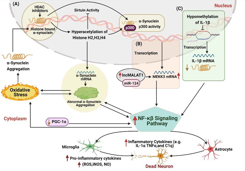
Int. J. Mol. Sci. 2021, 22, 4956 2 of 35
For decades, the underlying pathophysiology of the PD has remained unclear. It
is believed to be caused by both genetic mutations (SNCA, LRRK2, PARKIN, and DJ-1),
termed as familial PD, and environmental factors (exogenous neurotoxins, age, diet, and
lifestyle) in sporadic PD [7]. It is observed that these factors induce various molecular
and cellular events, such as oxidative stress, α-synuclein oligomerization, mitochondrial
dysfunction, higher iron concentration to trigger neurodegeneration and neuroinflam-
mation. Nevertheless, the core molecular and cellular processes involved in the PD are
divergent. However, all of these events participate over time in neuronal apoptosis by
activating the microglia and astrocytes population in particular regions of the brain [5].
Any initial stimulus, either in the form of cellular stresses (genotoxic, osmotic, hypoxic, or
oxidative stress) or environmental stresses (exogenous neurotoxins or stressful lifestyle),
activates the neuronal immune responses, including microglia, astrocytes, inflammatory
cytokines, etc. [8]. However, the continual activation of the neuronal immune system leads
to a vicious cycle of neuroinflammation that causes chronic inflammatory environments
that are likely to ensue neuronal dysfunction and ultimately the death of the endangered
neuronal population. A significant number of PD studies, including animal and human
models, have strongly linked neuroinflammation as environmental stressors that trigger
progressive degeneration of DA neurons. Therefore, on-time delivery of anti-inflammatory
therapy is required to provide neuroprotective effects in PD patients at risk of genetic and
epigenetic dysfunction.
Epigenetic factors have been observed to play a dynamic role in regulating neuroin-
flammation in both forms of PD. These factors, including DNA/RNA methylation, histone
modification, chromatin remodeling, non-coding RNAs (ncRNAs), etc., are sought to bridge
environmental factors and genes by altering gene expression and chromosome function
without changing the DNA sequence. Moreover, these epigenetic mechanisms regulate
the immune cells and inflammatory factors and are believed to be part of the underlying
mechanism countering neuroinflammation-induced DA damage in PD patients. It is worth
mentioning that these epigenetic mechanisms act synergistically and do not regulate the
neuroinflammation in PD independently. For instance, circRNAs and lncRNA sponge with
miRNAs and regulate various target genes that activate microglia and astrocytes, thus
trigger neuroinflammation in PD. Similarly, methylases cooperate with ncRNAs to acti-
vate various inflammatory pathways to decide the fate of glial cells in PD. Unfortunately,
detailed studies of epigenetic mechanisms and their synergic interaction with neuroinflam-
matory pathways in PD have not been discussed until now. Therefore, in this review, we
have discussed the impact of epigenetic mechanisms in regulating neuroinflammation in
PD and identified their synergic interactions with various neuroinflammatory pathways.
In addition, we have also highlighted various epigenetic modifications based on their
neuroprotective and neurotoxic properties to identify their potential use in future PD
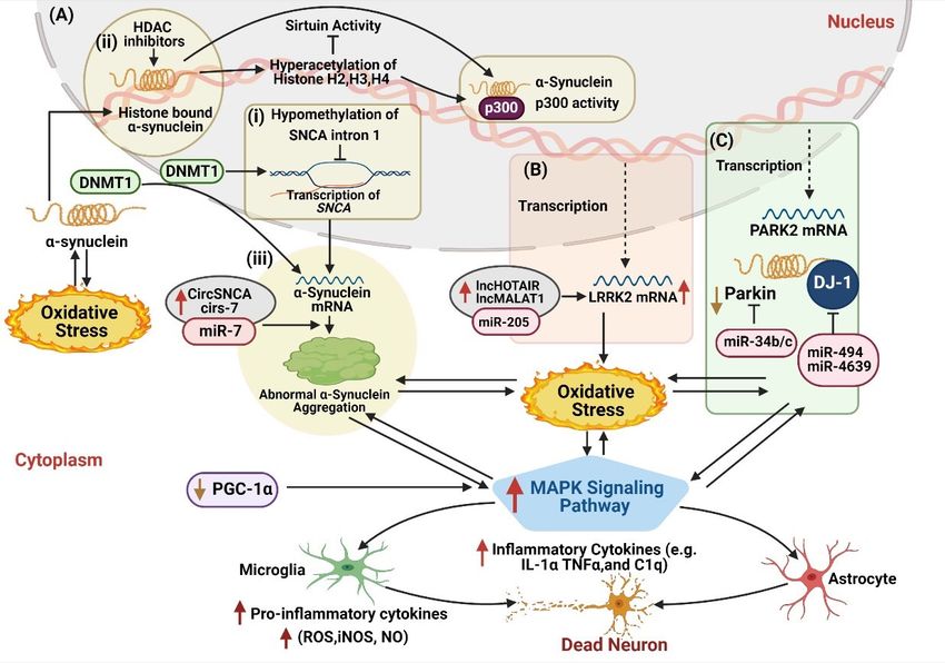
management (Figure 1).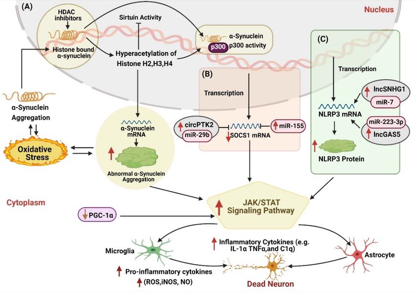
Int. J. Mol. Sci. 2021, 22, 4956 3 of 35
Int. J. Mol. Sci. 2021, 22, x FOR PEER REVIEW 3 of 34
Figure
Figure 1. Impact
1. Impact of of epigeneticmechanisms
epigenetic mechanismsregulating
regulating neuroinflammation
neuroinflammation ininParkinson’s
Parkinson’sdisease. TheThe
disease. neuroinflammation
neuroinflammation
cycle
cycle is activated
is activated onon DADA neurondamage,
neuron damage,which
which triggers
triggers epigenetic
epigenetic modifications
modifications and disturbs
and thethe
disturbs normal function
normal of of
function
inflammatory responses. Epigenetic regulators are divided into two categories: neurotoxic and neuroprotective. Neurotoxic
inflammatory responses. Epigenetic regulators are divided into two categories: neurotoxic and neuroprotective. Neuro-
epigenetic regulators enhance inflammatory factors and ROS production, transform glial cells to an inflammatory phenotype,
toxic
andepigenetic
promote regulators
dopaminergicenhance inflammatory
neuron factors and
death. In contrast, ROS production,
neuroprotective transform
epigenetic glial cells
regulators to antherapeutic
display inflammatory
phenotype, and promote
characteristics dopaminergic
and inhibit neuronby
neuroinflammation death. In contrast,
alleviating neuroprotective
DA neuronal damage inepigenetic
Parkinson’sregulators
disease. display therapeutic
characteristics and inhibit neuroinflammation by alleviating DA neuronal damage in Parkinson’s disease.
2. Crosstalk of Neuroinflammation in PD Progression
2. Crosstalk of Neuroinflammation
Neuroinflammation in PD Progression
acts as a double‐edged sword in the nervous system. Under
normal circumstances, microglia
Neuroinflammation and astrocytes sword
acts as a double-edged protectinagainst pathogenic
the nervous system. attack
Under or nor-
cellular
mal stress. However,
circumstances, during
microglia andPD progression,
astrocytes inflammatory
protect cytokines attack
against pathogenic activateorglial
cellular
cells and
stress. change their
However, duringphenotype. This process
PD progression, triggers inflammatory
inflammatory mediators,
cytokines activate reactive
glial cells and
change species
oxygentheir (ROS), and
phenotype. nitric
This oxidetriggers
process synthaseinflammatory
(NOS) production, leading
mediators, to massive
reactive oxygen
dopaminergic
species neuronal
(ROS), and apoptosis
nitric oxide and α‐synuclein
synthase aggregation,
(NOS) production, thus
leading intensifying
to massive PD
dopaminer-
development. Although the initial damage of DA neuron
gic neuronal apoptosis and α-synuclein aggregation, thus intensifying PD development. does not trigger
neuroinflammation,
Although the deadof
the initial damage remains
DA neuronof DAdoes
neurons
not usually induce chemokines release
trigger neuroinflammation, the dead
that promotes the penetration of activated microglia
remains of DA neurons usually induce chemokines release that to clear the dead remains
promotes the of nerve
penetration
offragments
activatedand thus results in neuroinflammation [9]. In 2016, Gerhard et al., performed
microglia to clear the dead remains of nerve fragments and thus results in
PET scans on the brains of PD patients. They observed an increased level of
neuroinflammation [9]. In 2016, Gerhard et al., performed PET scans on the brains of PD
neuroinflammation in the pons, basal ganglia, striatum, frontal, and temporal cortex areas
patients. They observed an increased level of neuroinflammation in the pons, basal ganglia,
along with the massive apoptosis of DA neurons [10]. Thus, it proposed that the apoptotic
striatum, frontal, and temporal cortex areas along with the massive apoptosis of DA neu-
process of DA neurons and the development of neuroinflammation occurs
rons [10]. Thus, it proposed that the apoptotic process of DA neurons and the development
simultaneously. Moreover, it is observed that the midbrain region enriched with DA
ofneurons
neuroinflammation occurs simultaneously. Moreover, it is observed that the midbrain
possesses the highest number of microglia [11]. Unlike neurons in the
region enriched with DA
hippocampus or cortex, midbrainneurons DA possesses
neuronstheexhibited
highest number of microglia
more sensitivity [11]. Unlike
for cytokines
neurons
such as tumor necrosis factor (TNF) [12,13] and were directly associatedsensitivity
in the hippocampus or cortex, midbrain DA neurons exhibited more with
for cytokines such
inflammation. as tumor necrosis
In conclusion, factor degeneration
the consistent (TNF) [12,13]ofand were directly
dopaminergic associated
neurons in PDwith
inflammation.
patients’ brainsInareconclusion,
the outcomesthe ofconsistent degeneration of dopaminergic
chronic neuroinflammatory reactions. neurons in PD
patients’ brains are the outcomes of chronic neuroinflammatory reactions.
Molecular Insight of Neuroinflammation in Parkinson’s Disease
Molecular Insight of Neuroinflammation in Parkinson’s Disease
Neuroinflammation occurs on the activation and proliferation of microglia and
Neuroinflammation
astrocytes. Microglia are occurs
immuneoncellsthe in
activation
the brainand
thatproliferation
participate inofthe
microglia and astro-
inflammatory
cytes. Microglia are immune cells in the brain that participate in the inflammatory
response in the central nervous system (CNS). Astrocytes are the most abundant type response
of
inglial
the cells
central
in the CNS that support brain structure. When inflammatory factors suchcells
nervous system (CNS). Astrocytes are the most abundant type of glial as in
the CNSand
TNFα thatinterleukins
support brain structure.
(IL‐6, When
IL‐1β, and inflammatory
IFN‐γ) factors
are recognized andsuch as TNFα
received, and inter-
microglia
leukins (IL-6, IL-1β, and IFN-γ) are recognized and received, microglia differentiate into
the pro-inflammatory M1 phenotype. M1 phenotypic microglia secrete TNF-α, IL-6, IL-1β,
ROS, and nitric oxide (NO) and reduce the secretion of neurotrophic factors to aggravate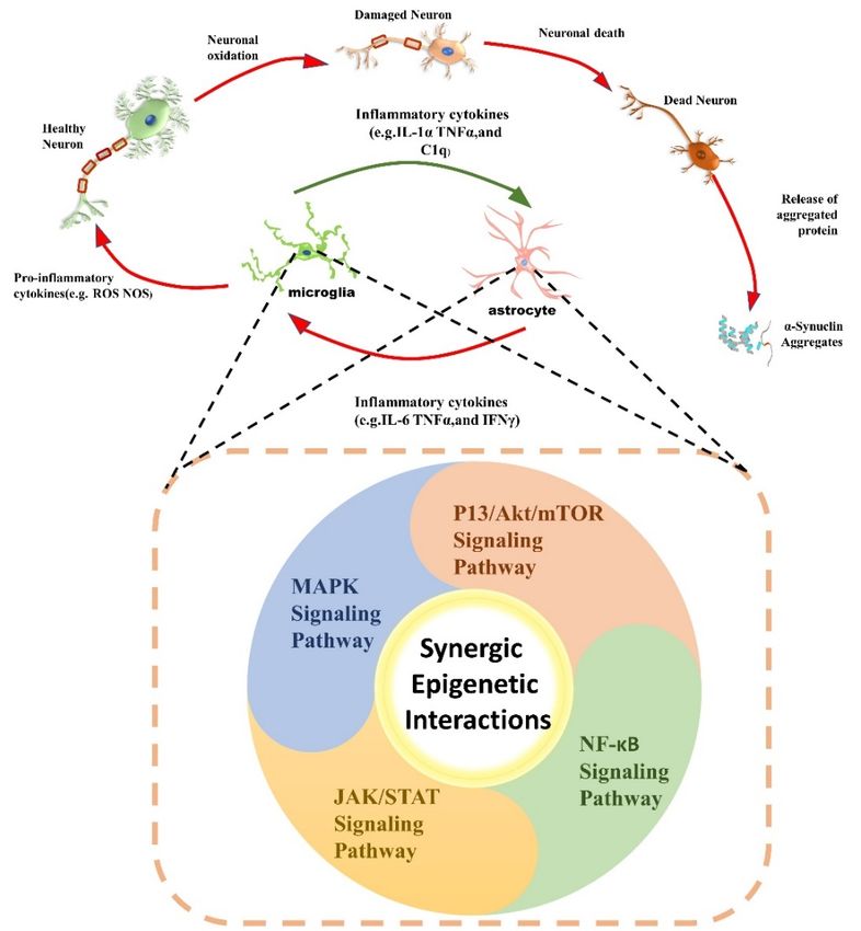
Int. J. Mol. Sci. 2021, 22, 4956                                                                                          4 of 35

                                   the damage of nerve cells [14,15]. In contrast, when stimulated by IL-4, IL-10, and other
                                   cytokines, microglia differentiate into the anti-inflammatory M2 phenotype. Microglia
                                   with M2 phenotype produces various anti-inflammatory compounds and has an immuno-
                                   suppressive effect by antagonizing M1 microglia [15]. Microglia with M2 phenotype can
                                   also secrete IL-4, IL-13, TGF-β, and neurotrophic factors such as IGF-1 (insulin-like growth
                                   factor-1), displaying a neuroprotective role [14,15]. Similar to the microglia, astrocytes A1
                                   phenotype is also programmed to secrete neurotoxic pro-inflammatory mediators after
                                   receiving inflammatory factors. However, while receiving anti-inflammatory factors, astro-
                                   cytes with A2 phenotype produces neurotrophic factors and show neuroprotective role.
                                   Thus, the abnormal or over-activation of microglia and astrocytes M1/A1 phenotype and
                                   lower M2/A2 phenotype can be a possible mechanism that induces neuroinflammation
                                   and neurodegeneration in PD.
                                        The underlying mechanism involved in chronic neuroinflammation is currently elu-
                                   sive and is thought to be affected by many factors. According to D Cherry’s review, the
                                   M1/M2 phenotype of microglia is mostly related to the transformation of acute neuroin-
                                   flammation to a chronic state. It is observed that failure of the M1 to M2 phenotype of
                                   microglia reduces neurotropic factors (such as IGF1 or brain-derived neurotrophic factor)
                                   and leads to the production of inflammatory factors and ROS, which causes chronic neu-
                                   roinflammation and results in neuronal apoptosis [16]. The M1/M2 phenotype transition
                                   of microglia is associated with cytokines and growth factors. For instance, when IL-6s
                                   are released, the JAK/STAT signaling pathway activates both STAT1 and STAT3 genes
                                   that triggers other neuroinflammation-related genes such as Jmjd3 and pro-inflammatory
                                   cytokines, thus promotes microglia-mediated neuroinflammation [17]. Contrarily, anti-
                                   inflammatory signal factors such as IL-10 and STAT3 would induce the expression of the
                                   SOCS1 gene and other anti-inflammatory genes to inhibit the pro-inflammatory response,
                                   mediated by TLR4 and IL-6 [18,19]. In addition, some regulatory drugs also change the
                                   phenotype of microglia. Such as LPS (lipopolysaccharide), TNF-α inducer stimulates the
                                   polarization of microglia into the M1 phenotype. Adding fasudil (Rho-kinase inhibitor)
                                   can reduce the activities of inflammatory cytokines IL-1β, IL-6, TNF-α, and NF-κB and
                                   trigger microglia towards the M2 phenotype [20]. Moreover, the NADPH inhibitor enzyme
                                   convert LPS induced M1 microglia into M2 [21]. Moreover, epigenetic modifications may
                                   affect the M1/M2 phenotype transition of microglia. For example, miR-155 drive microglia
                                   to M1 phenotype [22].
                                        In PD, astrocytes induce the death of dopaminergic neurons by secreting the pro-
                                   inflammatory cytokines and other toxic molecules (NO, etc.) [23]. In contrast, there are
                                   other reports that astrocytes protect neurons by inhibiting neuroinflammation [24]. The
                                   paradoxical performance of astrocytes in PD neuroinflammation is due to its different
                                   polarization phenotypes. Excessive activation of the A1 phenotype and lack of A2 pheno-
                                   type are important factors in developing neuroinflammation in PD. Cytokines and growth
                                   factors regulate the A1/A2 phenotype transition of astrocytes. For instance, the release
                                   of glycoprotein (gp130) leads to the A2 phenotype by astrocytes. Moreover, gp130 acti-
                                   vated STAT1 and STAT3 and mediated the SHP2/Ras/ERK signal cascade to limit the
                                   neuroinflammation development. [25]. However, when IL17 was released, it bound to the
                                   heterologous transmembrane receptors, which led to the recruitment of NF-kB activator
                                   1 (Act1) and the formation of signal complexes, thereby exhibiting the A1 phenotype
                                   and promoting the production of pro-inflammatory cytokines [26]. Except for cytokines,
                                   PD-related genes could also regulate the phenotype of astrocytes. For example, researchers
                                   found altered gene expression in astrocytes when exposed to α- synuclein and accelerated
                                   the inflammation process. They speculated that the SNCA gene might activate astrocytes in
                                   PD through IFN-c and TNF-α and regulate inflammation progression [27–29]. Similarly, in
                                   mouse models of PD, mutations in Parkin cause astrocyte dysfunction and aggravate neu-
                                   ronal death [30]. However, DJ-1 induces the A2 phenotype of astrocytes through the STAT1
                                   pathway and protected neurons by inhibiting brain inflammation in PD patients [28,31,32].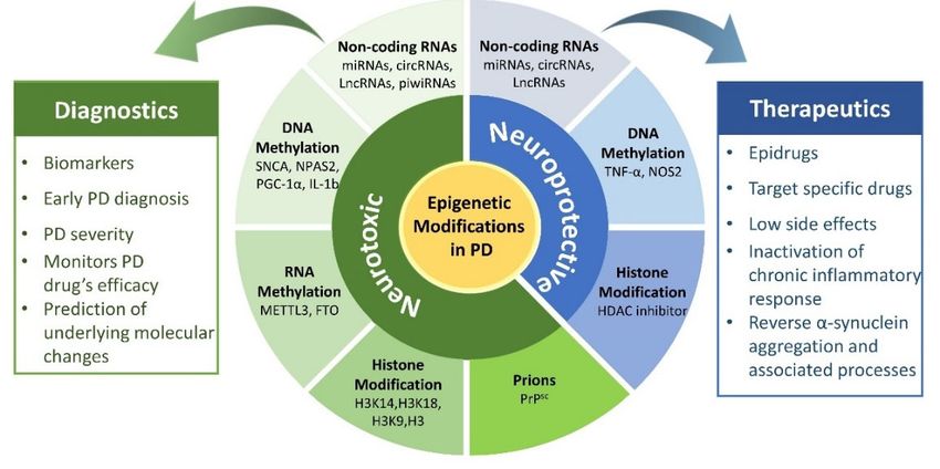
Int. J. Mol. Sci. 2021, 22, 4956                                                                                          5 of 35

                                         Although the source and structure of microglia and astrocytes are quite different, they
                                   work mutually in any stimulus (brain injury). The synergy between them in neurodegen-
                                   erative diseases has become a hot point in recent years. According to Ilia D. Vainchtein’s
                                   review, both microglia and astrocytes have specific receptors for norepinephrine, purines,
                                   and bacterial metabolites in the blood, which seem to induce synchronous reactions be-
                                   tween them. Transforming growth factors (TGF-β) released by astrocytes could let neurons
                                   release certain components which act on microglia while astrocytes and microglia interact
                                   directly with molecules as IL-1α, TNF-α, and IL-33. For example, IL-1α released by astro-
                                   cytes changed the permeability of the blood–brain barrier, thereby causing the activation
                                   of microglia [33]. The activated microglia secreted Il-1α, TNF-α, and C1q both in vivo
                                   and in vitro, which induced A1 astrocytes to function [34]. In PD, the two coordinated
                                   regulation mechanisms jointly regulate the progression of neuroinflammation. Research
                                   by Kaoru Saijo et al., showed that Nurr1 (a monocyte receptor) inhibited the expression of
                                   pro-inflammatory neurotoxic mediators in microglia and astrocytes and reduced the death
                                   of DA neurons. Decreased expression of Nurr1 made astrocytes more susceptible to the
                                   influence of microglia and promoted inflammation [35]. Hayate Javed et al., found that
                                   glial maturation factor (GMF) was a pro-inflammatory factor, which may be a co-receptor of
                                   microglia and astrocytes. However, inhibiting GMF reduced the activation of NLRP3 (NLR
                                   family pyrin domain containing 3) inflammasomes and regulated the cytotoxic function of
                                   microglia and astrocytes, thus prevented PD development [36]. Studies by Bortolanza et al.,
                                   also showed that 7-nitroindazole (a NOS inhibitor) could reduce the iNOS expression in PD
                                   rat models to downregulate neuroinflammation by inhibiting the activation of astrocytes
                                   and microglia [37]. Thus, more research in this domain may provide valuable insight
                                   into the deep understanding of the underlying mechanism of glial cell coordination and
                                   communication that may help researchers identify the novel targets for PD therapy.

                                   3. Epigenetics and Neuroinflammation in PD
                                        Epigenetic modifications possess a dual function in the regulation of the nervous
                                   system. Their neurotoxic and neuroprotective properties make them ideal candidates
                                   for disease management. Ample studies have shown that neuroinflammation in PD
                                   is regulated by various epigenetic modifications such as non-coding RNA, DNA/RNA
                                   methylation, and histone acetylation. They regulate neuroinflammation by governing
                                   immune cells, including macrophages, T lymphocytes, microglia, and astrocytes. For
                                   instance, histone 3 lysine 27 (H3K27) methylation enhances the inflammatory phenotype
                                   of macrophages and microglia, which is known as the M1 response. In contrast, H3K27
                                   histone demethylase jumonji-domain Protein 3 (Jmjd3) upregulates the anti-inflammatory
                                   M2 phenotype of microglia [38]. Thus, a detailed study of these epigenetic mechanisms
                                   may unravel the hidden mystery of neuroinflammation involved in PD progression.

                                   3.1. Non-Coding RNA and Neuroinflammation
                                   3.1.1. MicroRNAs
                                        MicroRNAs belongs to the class of small non-coding RNA (sncRNA) with a length of
                                   17–22 nt. They inhibit gene expression level by binding to the 3’untranslated region (3’UTR)
                                   of mRNA, resulting in mRNA degradation, adenylation, or translational inhibition [39].
                                   Over the past few years, various miRNAs target PD-related genes that induce neuroin-
                                   flammation and accelerate the neurodegeneration of dopaminergic neurons (Table 1). For
                                   instance, downregulation of miR-133b in the mid-brain tissues of Parkinson’s patients has
                                   resulted in the malfunctioned dopaminergic neurons and induces neuroinflammation in
                                   PD [40]. Another study reported that dysregulated Let-7 had promoted neuroinflammation
                                   through the activation of microglia and macrophages by acting as DAMP of TLR7 (Toll-
                                   like receptors 7) that further induced microglia to secrete inflammatory factors [41–43].
                                   Contrarily, Let-7 also promoted the M2 phenotype in microglia by targeting the C/EBP-δ
                                   transcription factor and inhibiting neuroinflammation-induced apoptosis [44,45]. Similarly,
                                   SOCS1 and SOCS3 genes involved in regulating the inflammatory signal transduction of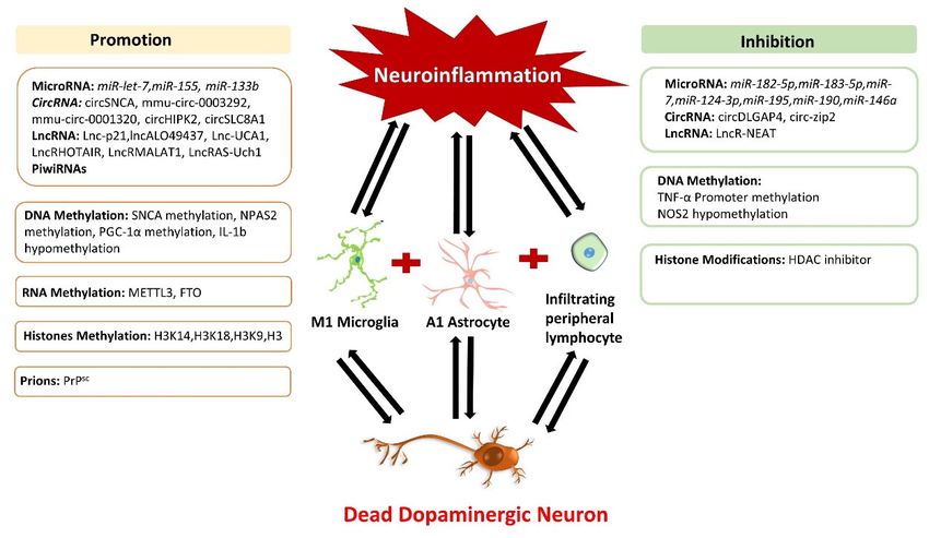
Int. J. Mol. Sci. 2021, 22, 4956                                                                                                   6 of 35

                                       microglia were found downregulated by miR-155 and enhanced neuroinflammation in
                                       PD [46].

      Table 1. MicroRNAs involved in the neuroinflammation in Parkinson’s disease. ↑ shows up-regulated, ↓ shows down-
      regulated.

                                                                      miRNA
     microRNAs                     Model                 Target                                Function                  References
                                                                     Expression
                                                       miRNA with Neuroprotective Effect
                                                                                    Downregulate α-synuclein and
                             PD patients, MPTP
                                                                                    acted as a neuroprotective agent
                                mice model,
        miR-7                                            SNCA            ↑               against oxidative stress,          [47]
                             MPP+ -SH-SY5Y cell
                                                                                    mitochondrial impairment, and
                                   model
                                                                                           neuroinflammation
                                                                                   Downregulate NLRP3 expression
        miR-7               MPTP mouse model             NLRP3           ↑        and reduced neuronal damage, and          [48]
                                                                                      improved microglia function
                                                                                         Mimic glial cell-derived
     miR-182-5p             Primary neuronal cell                                   neurotrophic factor (GDNF) and
                                                         GDNF            ↓                                                  [49]
     miR-183-5p              model of midbrain                                        protected the survival of DA
                                                                                                  neurons
                                                                                    Downregulate the production of
                             MPP+ -SH-SY5Y cell                                     caspase-3, inflammatory factors
     miR-124-3p                                          STAT3           ↓                                                  [50]
                                   model                                          TNF-α, IL-1β, and reactive oxygen
                                                                                                  species
                            LPS-BV-2 cell models,                                    Downregulate the activation of
                             MPTP mice model,                                          microglia, the production of
       miR-124                                          MEKK3            ↓                                                  [51]
                             MPP+ -SH-SY5Y cell                                    pro-inflammatory factors, and cell
                                   model                                                         apoptosis
                                                                                       Downregulate the release of
                                                                                  pro-inflammatory cytokines (iNOS,
                                                                                         IL-6, and TNF-α) while
       miR-195              LPS-BV-2 cell model         ROCK1            ↓                                                  [52]
                                                                                       upregulating the release of
                                                                                   anti-inflammatory cytokines (IL-4
                                                                                                and IL-10)
                            LPS-BV-2 cell model,                                    Downregulate pro-inflammatory
       miR-190                                           Nlrp3           ↓                                                  [53]
                             MPTP mice model                                      cytokines and microglia activation
                                                                                     Inhibit inflammatory response
                                                        IL-6 and
      miR-146a             Human glial cell lines                        ↑        mediated by glial cells and provide       [54]
                                                         COX-2
                                                                                             neuroprotection.
                                                         miRNA with Neurotoxic Effect
                                Microglia and
                                                                                     Upregulate the expression of
                             astrocytes cultured
       miR-155                                          SOCS-1           ↑          pro-inflammatory mediators in           [46]
                            from DJ-1-knockout
                                                                                       microglia and astrocytes
                                 mouse brain
                           Pitx3 mutant Aphakia                                    Downregulate Pitx3 and induced
      miR-133b             mice, 6OHDA-treated           Pitx3           ↓         DA neuronal damage and trigger           [40]
                                     mice                                                 neuroinflammation
                              Animal, human                                         Let-7 act as a signal activator of
         Let-7             biopsies, cell lines, and     TLRs            ↑         TLR7 in microglia and induced an       [41–43]
                            primary cell cultures                                    inflammatory response in PD

                                            Interestingly, various microRNAs possess a therapeutic potential and provide neu-
                                       roprotection against neuroinflammation by targeting PD-associated genes (Table 1). In
                                       2018, Roser et al., reported the neuroprotective role of miR-182-5p and miR-183-5p, which
                                       downregulated GDNF (glial cell-derived neurotrophic factor) and protected DA neurons
                                       against neuronal damage in the PD model [49]. A cytoplasmic protein NLRP3 belongs to an
                                       inflammatory signaling complex, inflammasome, plays a significant role in inflammation-
                                       mediated pyrophosphorylation neurodegeneration of DA in substantia nigra par compacta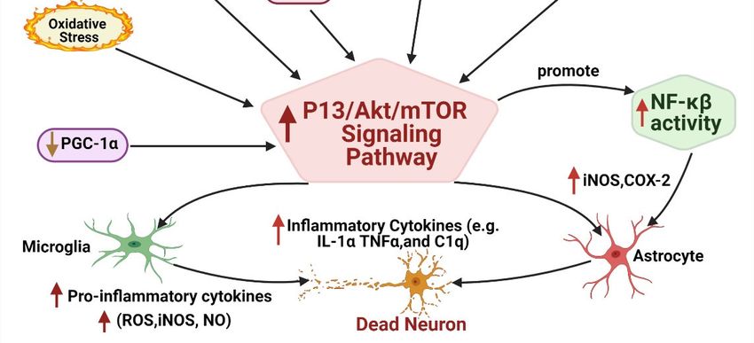
Int. J. Mol. Sci. 2021, 22, 4956                                                                                         7 of 35

                                   (SNpc) in PD [55–57]. A study performed on the striatum of the PD mouse model showed
                                   that miR-7 lowers the expression of the NLRP3 gene and SNCA, which resulted in reduced
                                   neuronal damage and improved microglia function in PD [47,48]. Another study on the
                                   PD model showed that inhibition of STAT3 (transcription factor regulating monoamine
                                   oxidase A) and MAPK (mitogen-activated protein kinase) pathway reduced neuroinflam-
                                   mation and behavioral changes caused by microglia [58]. miR-124-3p was observed to
                                   exert its neuroprotective role in DA neurons by downregulating STAT3 gene expression in
                                   the PD cell model induced by MPP [50]. In another study, miR-124 targeted the MEKK3
                                   in the NF-Kb pathway and the nuclear factor kappa light chain enhancer that reduced
                                   inflammatory cytokine levels in the PD mouse model [51].
                                         Other than targeting inflammation-related genes in PD, miRNAs are also involved
                                   in regulating microglia and astrocytes. Recently, PD pathogenesis was investigated on
                                   LPS induced in vitro model for microglia activation. This study showed that upregulated
                                   miR-195 inhibited the release of pro-inflammatory cytokines such as inducible nitric oxide
                                   synthase, IL-6, and TNF-a and increased the release of anti-inflammatory cytokines IL-4 and
                                   IL-10. Moreover, miR-195 was observed to downregulate ROCK1 gene expression in the PD
                                   model, which resulted in lowered microglia activation and reduced neuroinflammation [52].
                                   Higher expression of miR-190 in the PD model inhibited pro-inflammatory mediators such
                                   as iNOS, IL-6, TNF-α, and TGF-β1 and enhanced the release of anti-inflammatory media-
                                   tors, such as IL-10. Thus, it postulates that miR-190 negatively regulates the expression
                                   of the Nlrp3 gene to inhibit the activation and inflammatory response of microglia, which
                                   abridged neuronal damage in SNpc [53].
                                         Another study on PD mouse models treated with LPS and IFN-γ showed differential
                                   expression of miRNAs in cortical astrocytes. For instance, miR-146a, miR-146b, and miR-
                                   155 were found upregulated, whereas miR-351, miR-455, and miR-149 were downregulated.
                                   These subsets of differential miRNAs regulated TNF-α signaling pathway genes and
                                   activated astrocyte immune response in the PD [54,59]. Further investigation by Iyer et al.,
                                   in 2012, demonstrated that upregulated miR-146a displayed neuroprotection by reducing
                                   the expression of IL-6 and COX-2 and inhibited the inflammatory response mediated
                                   by astrocytes [54]. Therefore, through the previous literature, it was observed that most
                                   of the miRNAs involved in neuroinflammation are interlinked and induce subsequent
                                   molecular changes along with neuroinflammation in the PD. Thus, it is difficult to predict
                                   the exact mechanism of miRNAs involved in PD progression at this stage and requires
                                   more researcher’s attention to gain insight into the underlying PD-associated molecular
                                   mechanism. Nevertheless, neurotoxic and neuroprotective properties of these miRNAs
                                   propose their potential as candidate biomarkers for specific diagnostics and therapeutics of
                                   future PD patients.

                                   3.1.2. CircRNA
                                        CircRNAs are naturally occurring endogenous ncRNAs, widely distributed through-
                                   out the body. They are single-stranded RNA molecules up to 100 nucleotides in length
                                   and are covalently linked with 30 to 50 ends to form a loop-like structure [60]. They are
                                   highly expressed in the brain and regulate various gene expressions by acting as a sponge
                                   with miRNAs. Due to the higher percentage in the brain, they regulate various neuronal
                                   processes, where their deregulated expression footmarks several neurological disorders.
                                   During PD progression, various circRNAs sponge with miRNA to induce neuroinflamma-
                                   tion along with other sub-molecular manifestations (Table 2).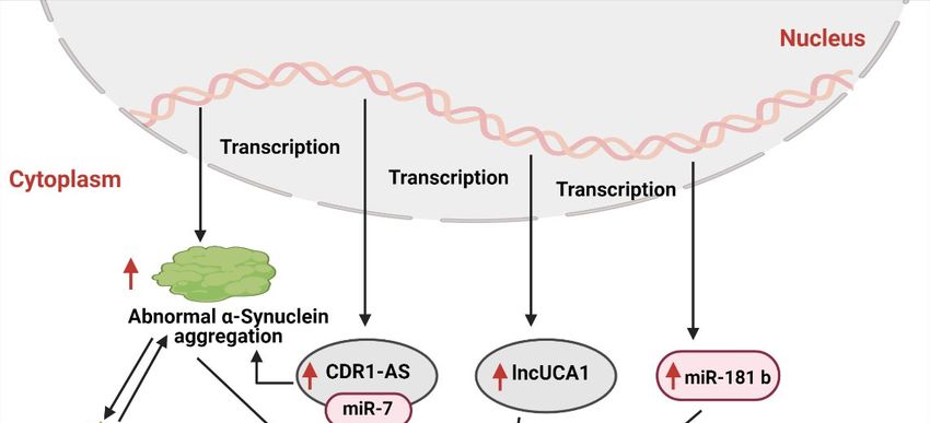
Int. J. Mol. Sci. 2021, 22, 4956                                                                                                   8 of 35

             Table 2. CircRNAs involved in the neuroinflammation in PD. ↑ shows up-regulated, ↓ shows down-regulated.

     Circular              miRNA
                                        Expression     Target         Study Model                   Function                References
      RNA                  Sponge
                                                                                              Upregulate miR-7 that
                                                                      MPTP-mouse
                                                                                             downregulate SNCA and
    circSNCA                miR-7          ↓           SNCA             model                                                  [61]
                                                                                            reduce cell apoptosis, and
                                                                     MPP+ -SH-SY5Y
                                                                                               improves autophagy
                                                                                             Downregulate NR4A2 to
      mmu-
                                                                      MPTP- mouse            impair DA neurons and
    circRNA-            miRNA-132          ↑           NR4A2                                                                   [62]
                                                                        model              promote neurodegeneration
     0003292
                                                                                             and neuroinflammation
                                                                                           Regulate the activation of the
                                                                     MPP+ -SH-SY5Y           CREB signal to affect the
  circDLGAP4             miR-134-5p        ↓           CREB         cells, MPTP mouse     expression of BDNF, Bcl-2, and       [63]
                                                                           models           PGC-1α in cells, and exert
                                                                                              neuroprotective effects.
                                                                     Sigmar1KO mice,
                                                                                          Regulates astrocyte activation,
                                                                           human
    circHIPK2           miR124-2HG         ↑         SIGMAR1                              autophagy, and endoplasmic           [64]
                                                                     astrocytoma cell
                                                                                                reticulum stress
                                                                         line A172
                                                                       Transgenic C.
                                                                                           Target miR-60 to protect DA
     circzip-2             miR-60          ↑            Zip-2        elegans model of                                          [65]
                                                                                                    neurons
                                                                             PD
                                                                    Substantia nigra of
   circSLC8A1              miR-128         ↑            Ago2          PD patients and       Promote oxidative stress         [66–68]
                                                                      healthy donors
                                                                     LPS-treated BV2         Sponge with miR-124 to
                                                     MEKK3/NF-
 mmu_circRNA                                                              cells and        induce neuroinflammation
                        miRNA-124          ↑         κB signaling                                                             [62,69]
  _0001320                                                             MPTP-mouse           through MEKK3/NF-κβ
                                                       pathway
                                                                           model               signaling pathway

                                            The neuroprotective microRNA miR-7 was found downregulated due to the spong-
                                      ing effect of ciRS-7 and circSNCA in PD models. Downregulated miR-7 upregulated the
                                      SNCA gene expression that resulted in α-synuclein aggregation accompanied by higher
                                      oxidative levels, mitochondrial dysfunction, neuroinflammation, and neuronal death [70].
                                      Contrarily, the downregulation of circSNCA under pramipexole (PPX) treatment proposed
                                      its therapeutic effect, reduced PD cell apoptosis, and improved autophagy [61]. Recently,
                                      transcriptomic profiling of PD brains of a mouse model showed that mmu-circRNA-
                                      0003292 sponges with miRNA-132 to downregulate the expression of the NR4A2 gene [62].
                                      Lower expression of NR4A2 caused impaired development and differentiation of midbrain
                                      neurons that promoted DA neurodegeneration and neuroinflammation, hallmark parkin-
                                      sonism [62,71]. Another investigation on the PD model showed that circSLC8A1 sponged
                                      with miR-128 and downregulated SIRT1 and BMI1 transcripts. This circSLC8A1/miR-128
                                      sponged influences protein homeostasis and chronic neuroinflammation mitochondrial
                                      dysfunction and promotes neurodegeneration to induce PD progression [66–68].
                                            Since the past few years, circRNAs have gained great attention due to their neu-
                                      roprotective effects against PD pathogenesis. Recently, Zhong Feng et al., showed that
                                      circDLGAP4 exerts its neuroprotective effect through regulating the miR-134-5p/CREB
                                      pathway in human and mouse PD models [63]. Moreover, circDLGAP4 sponged with
                                      miR-134-5p to regulate the activation of CREB pathway and CREB-associated genes, such
                                      as BDNF, Bcl-2, and PGC-1α, which promoted neuron viability by reducing mitochon-
                                      drial dysfunction, neuroinflammation, and enhanced autophagy [72–74]. In another study,
                                      mmu_circRNA_0001320 sponged with miRNA-124 and inhibited neuroinflammation in
                                      PD by regulating the MEKK3/NF-κB signaling pathway [62,69]. Thus, it speculates that
                                      the mmu_circRNA_0001320/miRNA-124/MEKK3 axis plays an integral part in regulating
                                      the levels of the inflammatory factors in PD neuroinflammation. Knockdown of circHIPK2
                                      in the PD model presented therapeutic properties against neuroinflammation. It caused
Int. J. Mol. Sci. 2021, 22, 4956 9 of 35
lower expression of MIR125-2HG and SIGMAR1 gene expression and inhibited astrocyte
activation through the modulation of autophagy and reduced endoplasmic reticulum stress
levels [64]. Circzip-2 has been reported to protect the dopaminergic neurons by targeting
miR-60 that downregulated the zip-2 expression. This circzip-2/miR-60 axis provided
resistance against oxidative stress and reduced neuroinflammation in the PD model [65].
Therefore, due to their precise involvement and stability in PD development, it is proposed
that circRNAs can serve as a potential tool for PD management strategies.
3.1.3. LncRNA
Long non-coding RNA (lncRNAs) is a RNA transcript with a length greater than 200
nucleotides [75]. LncRNAs are highly expressed in the central nervous system and regulate
several neurobiological processes such as neural plasticity, neurogenesis, brain develop-
ment, etc., where any dysregulation results in various neurodegenerative diseases [75].
Converging evidence has reported that various lncRNAs are involved in PD progression, as
summarized in Table 3. In 2017, Ni Y et al., reported differential expression of 87 lncRNAs
in the substantia nigra of PD patients [76]. Whereas 13 lncRNAs were found differentially
expressed in the peripheral blood leukocytes of PD patients [77], postulating that lncRNAs
actively participate in PD pathogenesis.
Table 3. Long non-coding RNAs involved in neuroinflammation in Parkinson’s disease. ↑ shows up-regulated, ↓ shows
down-regulated.
LncRNA
LncRNAs Study Model Target Function References
Expression
Upregulated oxidative stress and
LncRNA UCA1 6-OHDA rat model ↑ PI3K/AKT inflammation through PI3K/Akt [78]
signaling pathway
MPP+ -SH-SY5Y
Regulate miR-126-5p and RAB3IP
LncRNA HOTAIR cells, MPTP-mouse ↑ miR-126-5p [79]
to promote the progression of PD.
models
Upregulated LRRK2 expression by
MPP+ -SH-SY5Y
LncRNA- miR-205-5p- miR-205 inhibition and induce
cell model, ↑ [80]
HOTAIR LRRK2 neuronal apoptosis and
MPTP-mice model
neuroinflammation
Upregulated LRRK2 expression by
MPP+ -SH-SY5Y
LncRNA- miR-205-5p- miR-205 inhibition and induce
cell model, ↑ [81]
MALAT1 LRRK2 neuronal apoptosis and
MPTP-mice model
neuroinflammation
Downregulated ASUch1 expression
iMN9D cells, is regulated by downregulated
LncRNA-AS Uch1 ↓ Nurr1 [82]
MPTP-mice model Nurr1 activity, results in
dopaminergic dysfunction
Target miR-625 to regulate TRPM2
MPP+ -SH-SY5Y expression that increase the levels
Lnc-p21 ↑ miR-625 [83]
cells of ROS, TNF-α, IL-1β and IL-6, and
trigger cell apoptosis
MPP+ -SH-SY5Y Regulated the miR-205-5p/MAPK1
LncRNA
cells, MPTP-mouse ↑ miR-205-5p axis to increase the levels of [84]
AL049437
model inflammatory factors and ROS
Inhibited the degradation of PINK1
MPP+ -SH-SY5Y
to increase autophagy and
LncRNA NEAT1 cells, MPTP-mouse ↑ PINK1 [85]
displayed a neuroprotective role in
model
PD
LncRNAs display an integral role in neuroinflammation development in PD. For in-
stance, lncRNA UCA-1 (urothelial carcinoma-associated-1) is reported to induce apoptosis
and neuroinflammation in the PD model through targeting P13K/Akt signaling pathways.
However, its knockdown has reduced oxidative stress and inflammatory responses, whichInt. J. Mol. Sci. 2021, 22, 4956 10 of 35
lowered the degeneration of dopaminergic neurons [78]. Another investigation showed
that LncRNA-p21 (long non-coding RNA-p21) binds with miR-625 to induce cytotoxicity
and neuronal apoptosis in the PD cell model. In contrast, silencing of LncRNA-p21 has
reduced cytotoxicity along with TNF-α, IL-1β, and IL-6 levels, which increased the SOD
activity and reversed neuronal damage and neuroinflammation [83]. lncRNA HOTAIR
has been found to target the miR-126-5p and RAB3IP in a ceRNA- dependent manner
and enhanced PD progression [79]. Moreover, lncRNA HOTAIR and lncRNA-MALAT1
bind with miR-205 and downregulated LRRK2 gene expression to induced oxidative stress,
neuronal apoptosis, and neuroinflammation [80,86] Contrarily, downregulation of these
lncRNAs nullified the α-synuclein aggregation and the apoptosis of dopaminergic neurons.
Further studies showed that lower expression of lncRNA ASUchl1 (antisense to the mouse
Ubiquitin carboxy-terminal hydrolase 1) in the PD model had downregulated Nurr1 gene
expression, which also contributed to neuronal damage and associated neuroinflamma-
tion [82]. Though a wide range of lncRNAs is found dysregulated in PD, but still not
enough to understand the exact mechanism of lncRNAs in PD development. Therefore,
detailed investigations are required to figure out the exact mechanism involved in PD
progression.
In addition to the neurotoxic role of lncRNAs in PD, several LncRNAs displayed
therapeutic significance by protecting neurons against neuroinflammation and oxidative
stress in PD models. lncRNA NEAT (nuclear-enriched assembly transcript-1) has been
reported to exert a neuroprotective role by upregulating the PINK1 gene expression, which
inhibited the PINK1 protein deterioration in PD models and reduced neuronal injury and
neuroinflammation [85]. Recent research by Zhang et al., showed that silencing of lncRNA
AL049437 had reduced TNF-α, iL-6, and ROS production in MPP+ induced PD model,
which significantly reduced the neuroinflammation and oxidative stress [84]. Though a
limited number of lncRNAs are reported with neuroprotective properties, it still broadens
the vision for their useful application in PD therapeutics.
3.1.4. Piwi Interacting RNAs
Piwi-interacting RNAs (piRNAs) are small non-coding RNAs (26 to 31 nt), referred
to as genomic guardians. They are involved in protecting the genome by facilitating the
transcriptional and post-transcriptional silencing of transposable elements through DNA
methylation and RNA interference. It is recently reported that piRNAs regulate various
brain functions and are involved in various neurological diseases, including PD [87,88].
An investigation on PD patients reported lowered expression of SINEs (short inter-
spersed nuclear element) and LINEs (Long interspersed nuclear element), which caused
significant alterations in PGC-1α and CREB-pathways, resulted in the mitochondrial dys-
function and associated neuroinflammation [89]. Further study on long-term memory in
the Aplysia CNS showed that piRNA plays a key role in regulating the methylation of
CREB2 gene promoter region [90]. Thus proposes that piRNAs might lower CREB gene
expression that progresses neuroinflammation in PD patients [91]. So far, limited studies
have been reported in this domain; therefore, we require extensive research to elucidate
their role in the pathogenesis of PD.
3.2. Methylation Regulation and Neuroinflammation
3.2.1. DNA Methylation
DNA methylation is an epigenetic modification that involves the covalent transfer
of a methyl group to the C-5 position of cytosine by DNA methyltransferase (DNMT) to
induce chromatin conformation and inhibits the transcription mechanism, which results in
abnormal gene expression [92]. DNA methylation is important for normal body develop-
ment. It plays a vital role in various processes such as genomic imprinting, inactivation of
X-chromosome, and downregulation of repetitive element transcription and transposition,
where any deregulation results in various diseases, including neurodegenerative disease. A
significant number of disturbed methylation patterns have been observed in various genesInt. J. Mol. Sci. 2021, 22, 4956 11 of 35
that trigger neuroinflammation and PD development, summarized in Table 4. Accumulat-
ing evidence showed that lower methylation levels in SNCA mRNA of PD patients resulted
in the higher translation of SNCA mRNA and α-synuclein aggregation [93–95]. Similarly,
another investigation by Masliah et al., showed that nuclear DNMT1 levels were reduced
in PD brain samples and SNCA transgenic mouse models, leading to insufficient DNA
methylation in the CpG island upstream of the SNCA, SEPW1, and PRKAR2A genes [96].
Interestingly, the methylation pattern of the SNCA gene in the brain was found similar to
the blood of PD patients [97]. Hence DNA methylation patterns from peripheral blood of
PD patients can serve as substitute biomarkers for PD progression [98].
Table 4. DNA methylation involved in the neuroinflammation in Parkinson’s disease. ↑ shows up-regulated and ↓ shows
down-regulated.
DNA Methylation Model Target Expression Function References
Induces α-syn aggregation and
PD patients, induces DA neuronal damage and
SNCA promoter SNCA ↓ [93–95]
healthy controls activates glial cells to trigger
neuroinflammation
PD patients, Disturb the circadian rhythm of PD
NPAS2 promoter NPAS2 ↓ [99]
healthy controls patients
Transform microglia to the M1
IL-1β promoter Mouse model IL-1β ↓ phenotype, that triggered neuronal [79,100]
damage and neuroinflammation
Human brains of
PD patients and Increase inflammatory gene
PGC-1α promoter PGC-1α ↑ [101]
healthy controls, expression and ROS production
DM SYN mice
PD patients, Regulates the inflammatory
TNF-α promoter TNF-α ↓ [102]
healthy controls phenotype of microglia
PD patients, Reduce NO production to avoid
NOS2 promoter NOS2 ↓ [103]
healthy controls activation of microglia
TET enzyme encoded by the TET gene family is reported to play a prominent role
in DNA demethylation. A study performed by Li Shu et al., on 1657 PD patients and
1394 control subjects showed that the TET1 gene might influence PD risk by regulating
the 5hmC levels and subsequent gene expression [104]. Likewise, the Clock gene that
controls circadian rhythm presented significant variations in its expression in PD patients
and animal models [99,105,106]. Investigation of the seven clock genes (PER1, PER2,
CRY1, CRY2, Clock, NPAS2, and BMAL1) revealed that PD patients’ leucocytes showed
reduced NPAS2 promoter methylation, whereas the rest of the clock genes’ promoters
found unmethylated [99]. Isil Ezgi Eryilmaza et al., in 2017, observed that variations in the
methylation patterns increased the transcription regulation of PARK2, which resulted in
mitochondrial dysfunction, dopaminergic neuron apoptosis, and neuroinflammation in
PD [107]. Overall, these studies indicate that DNA methylation and demethylation play a
significant role in the pathological process of PD.
PGC-1α is a transcriptional co-regulator, which is found downregulated during PD
pathogenesis [107]. Analysis of human PD brain samples showed increased methylation
at the PGC-1α promoter region. In evidence of this study, ICV administration of palmitic
acid (induces epigenetic modification in neurons, microglia, and astrocytes) to α-synuclein
transgenic mice resulted in hypermethylation of the PGC-1α promoter in the substantial
nigra (SN), leading to the lower expression of PGC-1α gene and mitochondrial content along
with higher expression of inflammation-related genes [101]. However, increased expression
of the PGC-1α gene in PD models has presented the neuroprotective effects in PD models
by increasing the expression of mitochondrial respiratory chain nuclear coding subunits
and hence, prevented the loss of dopaminergic neurons [108]. In another study of active
multiple sclerosis MS, Philip G Nijland et al., 2014 found that overexpression of PGC-1α inInt. J. Mol. Sci. 2021, 22, 4956 12 of 35
astrocytes could significantly reduce pro-inflammatory IL-6 and production and secretion
of chemokine (CC motif) ligand 2. This led to reduced ROS production and inhibited
oxidative damage and inflammation, thereby lowering the neurodegeneration [109]. Thus,
we speculate that increased methylation of PGC-1α in PD may inhibit the neuroprotective
effect of astrocytes and transform them to the A2 phenotype to promote neuroinflammation
development.
During PD pathogenesis, the inflammatory factors induce neuroinflammation by
changing the phenotype of microglia and astrocytes. Excessive activation of microglia
led to the upregulation of TNF-α, il-1, il-6, il-12, and other pro-inflammatory molecules,
along with mitochondrial ROS production and loss of mitochondrial membrane potential,
which leads to neuronal damage [110]. Thus, TNF-α activates microglia to promote the
transformation of their inflammatory phenotype, while the inflammatory response of
microglia induces more production of TNF-α to form a vicious circle. Increasing evidence
has proposed that higher TNF expressions malfunctioned the glutamate receptors that
disturb the transportation of specific ions across nerve cells and lead to neurodegenerative
diseases, such as AD and PD [111]. Various studies show that PD patients exhibited higher
levels of TNF-α in their cerebrospinal fluid and TNF-α receptor 1 (TNFR 1, p55TNFR) in
SNpc [112–114]. However, TNF-α knockdown inhibited microglia activation and reduced
the neurotoxicity of MPTP in DA [114]. DNA methylation is significantly involved in
regulating the expression of inflammatory factors. In 2008, Heike C. Pieper et al., reported
that PD patient’s SNpc cells possess a lesser degree of DNA methylation in the TNF-α
promoter than the DNA of other brain parts. Additionally, they observed that specific
methylation in the CpGs dinucleotides of the TNF-α promoter activity lowered the binding
of specific transcription factors (AP-2 and Sp1) that downregulated the TNF-α promoter
activity which increased the sensitivity of dopaminergic neurons to TNF-α-mediated in-
flammation [102]. In summary, DNA methylation may regulate the phenotype of microglia
and inflammatory response mediated by inhibition of TNF-α expression.
iNOS is a pro-inflammatory factor encoded by the NOS2 gene and is highly expressed
in the SNpc of PD patients. NOS2 gene is mostly regulated at the transcriptional level or
partly by the methylation of CpG dinucleotides [115]. Hypermethylation of the CpG site in
the 5’promoter region of the NOS2 gene reduces the iNOS activity [115,116]. In contrast,
hypomethylation of NOS2 may increase the iNOS activity. A study on PD patients exposed
to welding fumes (mainly containing Mn) reported insufficient NOS2 methylation [103],
which resulted in higher iNOS activity. In contrast, inhibiting iNOS reduced the neuronal
stress and downregulated the activation of microglia caused by signal molecules (such as
MMPs), thus prevented neuronal damage [117,118]. Collectively, it can be deduced that
the methylation level of NOS2 affects the iNOS activity and regulates the activation of
microglia and the neuroinflammation caused by it.
Interleukin 1 (IL-1) has been implicated in various neurological conditions due to
its significant role in the central nervous system and neurodegeneration. Increased ex-
pression of IL-1β has been observed in the cerebrospinal fluid and striatal regions of
patients with various neurological diseases, including PD, inducing neuronal death and
damage [119–121]. Various PD studies reported that IL-1β induced neuroinflammation by
disabling microglia. Hypomethylation of the IL-1β promoter region has been observed to
convert microglia to the M1 phenotype, triggering neuronal damage and neuroinflamma-
tion [79,100]. Thus, methylation of IL-1β may be one of the influencing factors of PD and
other diseases closely related to age.
3.2.2. RNA Methylation
RNA methylation (m6A) is the most prevalent post-transcriptional modification of
RNA, which involves binding the methyl group to the N6 site of adenine (RNA base).
It is widely distributed in mRNA and long coding RNA (lncRNA). Most of the m6A
modifications occur in exons, and it remains in the mature mRNA after splicing and in-
fluences the translation of m6A-containing mRNA. Methyltransferases and demethylasesInt. J. Mol. Sci. 2021, 22, 4956 13 of 35
catalyze m6A modifications. Common methyltransferases include METTL3, METTL14,
RBM15/B, etc., whereas demethylases include FTO (obesity-related protein) and ALKBH5
(alkylated DNA repair protein alkB homolog 5) [122–125]. Recently it has been observed
that protein families containing the YTH domain may interact with m6A demethylases and
contribute to the translation, degradation, and splicing of RNA [126]. Substantial evidence
suggests that m6A modifications play a crucial role in numerous mechanisms, such as
splicing, transportation, location, and stability of mRNAs, impacting various biological pro-
cesses, including stem-cell differentiation, somatic-cell reprogramming, biological rhythms,
etc. [127]. However, the actual mechanism of m6A modifications remains elusive and
requires more investigations.
In the adult brain, m6A modifications are highly expressed. Substantial evidence has
shown that dysregulation of m6A modifications is associated with many neuronal pro-
cesses, such as dopaminergic signaling, neurogenesis [128], learning, and memory [129–131],
proposing the close association of m6A modifications and brain activity. Recently, a study
on the mouse model showed that dysregulated m6A modifications induced by circSTAG1
affected the stability of FAAH (an indispensable membrane enzyme that can degrade fatty
acid amide family) mRNA, which caused astrocyte dysfunction and depressive behav-
iors [132]. Transcriptome-wide profiling of m6A modifications of mice with depressive
behavior demonstrated a high degree of m6A modifications in the prefrontal cortex with
lowered levels of FTO (nucleic acid demethylase), which resulted in the degradation of
subsets of proteins involved in neuronal plasticity and led to the impaired memory-related
processes [129–131].
More recently, m6A modifications have gained wide attention due to the significance of
epitranscriptomic regulation in PD progression. A study performed on PD mouse and cell
models showed that a high level of FTO reduced the m6A modifications in dopaminergic
cells, resulted in the overexpression of NMDA receptor 1 (N-methyl-D-aspartate), oxidative
stress, and Ca2+ influx, leading to neuronal damage and neuroinflammation [133]. Another
investigation on m6A modifications in Chinese Han people with sporadic PD showed
common and rare variations in m6A regulating genes (METTL3, METTL14, WTAP, FTO,
ALKBH5, YTHDF1, YTHDF2, YTHDF3, HNRNPC, and ELAVL1). Gene-wise association
analysis shows that no significant association has been found between these genes, and it
is proposed that ethnic factors might restrict this association [134].
However, Methyltransferase-like 3 (METTL3), an RNA methyltransferase, is also
involved in regulating various immune and inflammation processes. For instance, in an
investigation on osteoarthritis, increased levels of IL-1β have been observed to enhance the
expression of METTL3 that induced the significant m6A modifications. In contrast, METTL3
knockdown has reduced IL-1β-induced inflammatory cytokine levels and activation of
NF-κB signaling, thus reduced neuronal damage [135,136]. Due to very limited research
on m6A modifications in PD so far, the impact of METTL3 on neuroinflammation has not
been studied for PD-associated m6A modifications. Though m6A modifications are widely
dysregulated in PD, still this domain is barely touched and requires more investigation to
understand the underlying mechanism in PD development.
3.3. Histone Modification
Histones are the basic structural proteins of chromosomes divided into H1, H3, H2A,
H2B, and H4. They are involved in post-transcriptional modifications, including acetyla-
tion, phosphorylation, methylation, ubiquitination, sumoylation, ADP-ribosylation, etc.,
where variations in histones may lead to the dysfunction of the DNA transcription ma-
chinery through abnormal condensation of chromatin [137] and lead to various disorders.
Various PD studies have shown a strong association between histone modification and
neuroinflammation, implicating its role in PD progression.
Accumulating studies have shown a significantly higher level of histone modifications
in PD models [138]. Increased histone acetylation is attributed to the imbalance between
histone deacetylase (HDAC) and histone acetyltransferase (HAT). During PD development,Int. J. Mol. Sci. 2021, 22, 4956 14 of 35
downregulation of histone deacetylase 1 (HDAC1) induces an abnormal neuronal cell
cycle, leading to DNA damage and neuronal apoptosis [139]. In 2016, Sugeno reported
that α-synuclein directly bound to histones, which upregulated histone acetyltransferase
and reduced the level of acetylated histone H3 in the PD cell model [140]. This interaction
resulted in reduced sirtuin activity, abnormal α-synuclein aggregation, mitochondrial
dysfunction, and oxidative stress, resulting in DA damage and neuroinflammation [141].
Further studies have shown that PD patients possessed increased acetylation of histone
H3 in the brain samples due to increased acetylation of histone H3 lysine 14 (H3K14) and
histone H3 lysine 18 (H3K18) and decreased deacetylation of histone H3 lysine 9 (H3K9).
This higher level of H3 acetylation followed by the downregulation of histone deacetylase
led to an over-acetylation of core histones, which inhibited the gene transcription in the
primary motor cortex and accelerated the degeneration of dopaminergic neurons [142,143].
In an eleven-year follow-up of four million Norwegians, Shuchi Mittal et al., found
that the β2AR agonist, salbutamol (a brain-permeable asthma drug), can effectively reduce
the incidence of PD. Further studies have shown that β2AR ligand regulates the SNCA’s
transcription by affecting the acetylation of histone 3 lysine 27 of SNCA promoter and
enhancer [144]. Moreover, it is observed that the activation of microglia in PD is significantly
related to the overexpressed histone acetylation. AGK2 is an inhibitor that targets the
SIRT-2 site on HDAC and is reported to protect dopaminergic neurons and reduced
microglial activation [145]. Similarly, nicotinamide (an amide converted from vitamin
B3) also presented a neuroprotective effect in PD animal models [146]. Recently a study
on the PD rat model showed that nicotinamide induced hyperacetylation of histones
and increased the expression levels of various neurotrophic and anti-apoptotic factors
in the brain, thereby presenting therapeutic effects against PD progression. However,
nicotinamide overdose caused neurotoxicity and led to dopaminergic neuronal damage,
suggesting an appropriate dose range is needed for effective therapeutic results [147].
Studies by Xuefei Wu et al., showed that HDAC inhibitors, including sodium butyrate (SB)
and trichostatin A (TSA), upregulated the expression of GDNF and BDNF in astrocytes
and inhibited the HDAC to protect DA neurons [148]. In rodent models of central nervous
system inflammatory diseases, BDNF can prevent NF-κB-mediated neuroinflammation
and apoptosis [149]. In addition, HDAC inhibitor TSA can also alleviate the MPP+ -induced
astroglial glutamate uptake disorder, which may be a new mechanism for HDAC inhibitors
to promote neuroprotection [150].
3.4. Prions
Prions are lethal and pathogenic proteinaceous agents involved in various neurological
disorders, including PD [151,152]. They possess epigenetic inheritance and can transmit
genetic information to both mitotic and meiotic cell divisions without altering the genomic
sequence [153]. Recently, the “prion transmission” of α-syn has been extensively studied
in PD. It is observed that α-syn can spread throughout the body during the pathogenic
process and promote microglial activation and neuroinflammation in PD. These activated
microglia further promote the aggregation of α-syn and may provoke neuroinflammation,
forming a vicious cycle of neurodegeneration of DA neurons in PD. Therefore, it affirms that
neuroinflammation induces α-syn prion-like behavior to feed-forward neuronal damage.
Moreover, it is also proposed that the gastrointestinal tract and olfactory epithelium are
directly associated with neuroinflammation. Disturbed gastrointestinal tracts are the initial
sites of α-syn accumulation presumed to be transmitted to the nervous system through
“prion-like behavior” and tune progressive neuroinflammation [153]. Unfortunately, the
underlying mechanism of α-syn accumulation in the intestinal track has not been elucidated
until now, hence makes a loophole in the hidden mystery of PD origin. However, based
on these studies, it can be hypothesized that dietary patterns and gut microbiota might
play an active part in α-syn accumulation in the intestine and triggers PD onset through
prion-like behavior. Further studies are required to unravel the exact pathogenic role of
α-syn prions and their association with intestinal health and PD progression.You can also read