Structural Insights into RNA Dimerization: Motifs, Interfaces and Functions - MDPI
←
→
Page content transcription
If your browser does not render page correctly, please read the page content below
molecules
Review
Structural Insights into RNA Dimerization: Motifs,
Interfaces and Functions
Charles Bou-Nader *          and Jinwei Zhang *
 Laboratory of Molecular Biology, National Institute of Diabetes and Digestive and Kidney Diseases,
 50 South Drive, Bethesda, MD 20892, USA
 * Correspondence: charles.bou-nader@nih.gov (C.B.-N.); jinwei.zhang@nih.gov (J.Z.)

 Academic Editor: Quentin Vicens

 Received: 10 May 2020; Accepted: 19 June 2020; Published: 23 June 2020

 Abstract: In comparison with the pervasive use of protein dimers and multimers in all domains
 of life, functional RNA oligomers have so far rarely been observed in nature. Their diminished
 occurrence contrasts starkly with the robust intrinsic potential of RNA to multimerize through
 long-range base-pairing (“kissing”) interactions, self-annealing of palindromic or complementary
 sequences, and stable tertiary contact motifs, such as the GNRA tetraloop-receptors. To explore the
 general mechanics of RNA dimerization, we performed a meta-analysis of a collection of exemplary
 RNA homodimer structures consisting of viral genomic elements, ribozymes, riboswitches, etc.,
 encompassing both functional and fortuitous dimers. Globally, we found that domain-swapped
 dimers and antiparallel, head-to-tail arrangements are predominant architectural themes. Locally,
 we observed that the same structural motifs, interfaces and forces that enable tertiary RNA folding also
 drive their higher-order assemblies. These feature prominently long-range kissing loops, pseudoknots,
 reciprocal base intercalations and A-minor interactions. We postulate that the scarcity of functional
 RNA multimers and limited diversity in multimerization motifs may reflect evolutionary constraints
 imposed by host antiviral immune surveillance and stress sensing. A deepening mechanistic
 understanding of RNA multimerization is expected to facilitate investigations into RNA and RNP
 assemblies, condensates, and granules and enable their potential therapeutical targeting.

 Keywords: RNA; intermolecular interaction; dimerization; structure; domain swapping; folding;
 ribozymes; riboswitches

1. Introduction
     Quaternary structures of biological macromolecules are frequently at the core of functional
assemblies that enable life.        Such higher-order structures form through a network of
intermolecular interactions between individual modules employing recurring interfacial motifs
to drive multimerization. Association of identical or different building blocks produce homo or
hetero-multimers, respectively. These oligomeric assemblies often lead to overall structural symmetry
or pseudosymmetry that can be important for biological function and evolution [1–4]. More than 65%
of proteins are believed to rely on homo-oligomeric states for their activities [1,5]. By comparison,
only a handful of naturally occurring RNAs are known to function in homodimeric states, despite
the frequent occurrences of palindromic or complementary, dimer-forming sequences and a plethora
of RNA structural motifs that can engage specific and robust long-range contacts [6]. One of the
most striking examples of a functional RNA dimer lies in the peptidyl-transferase center (PTC) of
the ribosome. In the PTC, a cleft formed between a pair of near-symmetrical RNA helical motifs is
proposed to catalyze ancient chemical reactions, such as peptide bond formation, constituting a dimeric
proto-ribosome ribozyme [7].

Molecules 2020, 25, 2881; doi:10.3390/molecules25122881                        www.mdpi.com/journal/molecules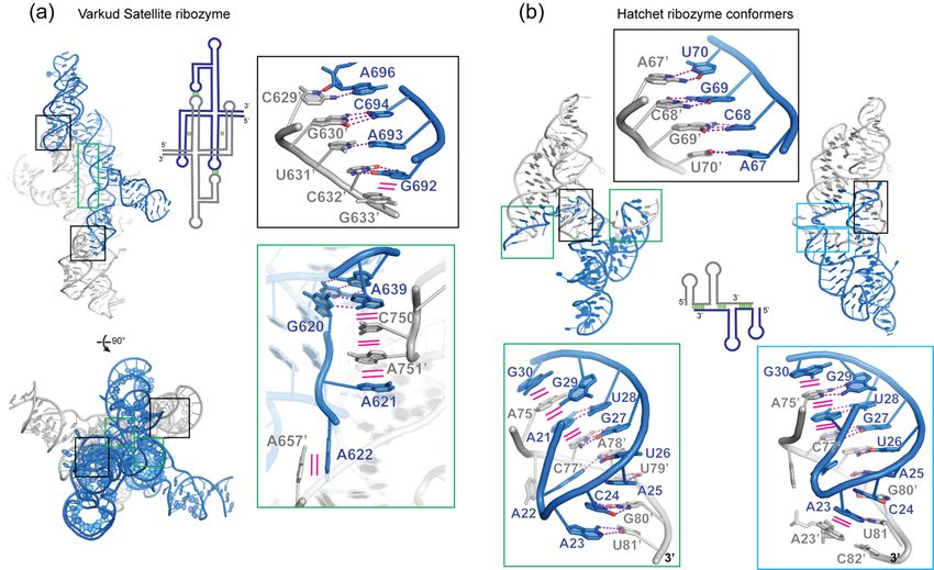
Molecules 2020, 25, 2881                                                                            2 of 28

      The simplest, most common form of RNA-RNA interactions occur through base pairing of
complementary single-stranded sequences, forming heterodimeric double-stranded RNA (dsRNA).
These specific interactions are widespread in diverse forms of post-transcriptional gene regulation and
RNA modification and processing, such as the pairing of small interfering RNAs (siRNA), microRNAs
(miRNA), or small nucleolar RNAs (snoRNAs), etc., to their complementary RNA targets. [8–12].
These hybridization events produce heterodimeric dsRNAs that are abundant in cells. However,
for the purpose of this review, we generally exclude simple dsRNAs that form solely through
sense-antisense hybridization and treat dsRNAs as monomers to focus on dimerization that involve
tertiary structural contacts.
      Consecutive base pairing positions adjacent base planes to stack with each other, either within the
same strand or crossing into the opposing strand—termed cross-strand stacking. Notably, stacking can
also occur without base pairing as the aromatic stacking itself provides significant favorable enthalpy.
These non-covalent, attractive interactions between polarized aromatic nucleobases daisy-chain the
base pairs into helices and frequently further concatenate adjacent helices to form long, coaxial
helical stacks. In so doing, aromatic-aromatic (or π-π) interactions to a large extent dictate the overall
shape of most RNA tertiary and higher-order structures. In addition to base pairing and stacking,
other types of tertiary interactions also contribute to RNA-RNA interactions, including dimerization,
such as purine-minor groove interactions, ribose zippers, tetraloop-tetraloop receptor interactions,
etc. These tertiary and quaternary interactions between discrete RNA elements can be dramatically
stabilized by the presence of Mg2+ ions and, to a lesser extent, by monovalent cations, such as K+ .
Mg2+ ions play the dual roles of serving as diffuse counter ions that ameliorate the electrostatic stress
from juxtaposing densely charged phosphate backbones, as well as bridging specific tertiary contacts
in the form of chelated ions [13].
      In contrast to their apparent biological rarity in cells, RNA multimers frequently emerge in vitro
and often present a nuisance in the laboratory. Most of such in vitro multimers are believed to occur via
fortuitous pairing of short segments of complementary sequences, when the RNA was heat-denatured
and cooled slowly—a process that encourages strand annealing. As such these in vitro multimers
are similar in origin to those dsRNAs produced by sense-antisense hybridization events in cells.
Various RNA refolding procedures have been developed with an explicit or implicit objective to enrich
monomers, while dimers and higher oligomeric states are reduced and usually discarded as artifacts
or physiologically irrelevant entities. For instance, many unmodified tRNAs exhibit strong tendencies
to dimerize and oligomerize and require a “snap-cool” procedure to refold into monomers [14].
However, the view that RNA oligomers are largely refolding artifacts is rapidly changing due to the
recent recognition that multivalent, intermolecular RNA-RNA contacts can induce functional RNA
oligomerization, condensation, phase separation or formation of ribonucleoprotein (RNP) granules
that critically regulate cellular metabolism and cell fate [8,15–19]. These recent findings accentuate the
importance and previously underappreciated role of RNA multimerization in biology. Although our
molecular understanding of RNA quaternary structures is still in its infancy, RNA nanotechnologies
have utilized artificially engineered RNA assemblies for more than two decades to create programmable
supramolecular architectures [20–25].
      In order to understand how RNA dimerizes and multimerizes in general, we survey a
representative collection of RNAs that rely on their homodimeric states to exert their biological
functions and also those that dimerize fortuitously, such as in crystallo. Interestingly, in several
riboswitches, we are able to compare the monomeric and dimeric forms of essentially the same
RNA. We maintain a focus on RNA dimerization interfaces and structural motifs used to drive
self-assembly with an emphasis on tertiary contacts in addition to base pairing. This meta-analysis
revealed that kissing-loop interactions and complementary strand swapping are the predominant
dimerization motifs. Since only a handful of recurring RNA structural motifs are responsible for
mediating dimerization in most instances, we organize the discussions based on RNA functional
classes instead of the dimerization motifs that they employ. In the following sections, we examine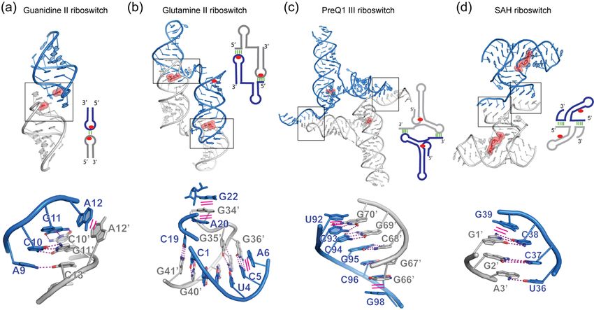
Molecules 2020, 25, 2881                                                                                                3 of 28
Molecules 2020, 25, x FOR PEER REVIEW                                                                                   3 of 27

instances
 instances of
           of RNA
              RNA dimerization
                   dimerization that
                                that occur
                                     occur in
                                            in retroviral
                                               retroviral genomes,
                                                          genomes, mRNA
                                                                   mRNA localization,
                                                                         localization, ribozymes
                                                                                       ribozymes and
                                                                                                   and
riboswitches,
 riboswitches, and fluorogenic RNAs, as well as consequences of RNA multimerization in
               and fluorogenic RNAs,   as well  as  consequences of RNA multimerization    in diseases
                                                                                              diseases
and
 and immunity.
      immunity.
2. Dimerization of Retroviral Genomic RNAs
 2. Dimerization of Retroviral Genomic RNAs
      Perhaps the most studied physiologically relevant RNA homodimerization phenomenon is the
       Perhaps the most studied physiologically relevant RNA homodimerization phenomenon is the
non-covalent linkage of two viral genomic RNA (gRNA) copies in retroviruses [26–31]. This conserved
 non-covalent linkage of two viral genomic RNA (gRNA) copies in retroviruses [26–31]. This
quaternary architecture of several kilobases of gRNA enable multiple biological functions in the
 conserved quaternary architecture of several kilobases of gRNA enable multiple biological functions
viral lifecycle. First, templated genomic repair during reverse transcription (RT) is facilitated by
 in the viral lifecycle. First, templated genomic repair during reverse transcription (RT) is facilitated
switching of the reverse transcriptase between the two spatially close strands [32]. Second, genetic
 by switching of the reverse transcriptase between the two spatially close strands [32]. Second, genetic
diversity is enhanced by homologous recombination between the two gRNA copies [33]. Lastly,
 diversity is enhanced by homologous recombination between the two gRNA copies [33]. Lastly,
interconversion between the monomeric and dimeric gRNAs modulate their translation and packaging
 interconversion between the monomeric and dimeric gRNAs modulate their translation and
into viral particles [34,35]. The latter is controlled by modulating the exposure of binding sites for
 packaging into viral particles [34,35]. The latter is controlled by modulating the exposure of binding
viral nucleocapsid (NC) domains [30,36,37]. Retroviral gRNA dimerization frequently occurs through
 sites for viral nucleocapsid (NC) domains [30,36,37]. Retroviral gRNA dimerization frequently occurs
a segment of their 50 leader region termed the dimer linkage site (DLS), as detailed below [38–40]
 through a segment of their 5′ leader region termed the dimer linkage site (DLS), as detailed below
(Figure 1).
 [38-40] (Figure 1).

      Figure 1.
      Figure  1. Homodimerization
                  Homodimerization of    of retroviral
                                            retroviral genomic
                                                       genomic RNAs
                                                                RNAs (gRNA).
                                                                       (gRNA).(a) (a)Schematic
                                                                                      Schematicview
                                                                                                  viewof   the55′0 leader
                                                                                                        ofthe      leader
      dimerlinkage
      dimer    linkagesitesite  (DLS)
                            (DLS)       of human
                                   of human           immunodeficiency
                                               immunodeficiency            virus
                                                                  virus type       type driving
                                                                              1 (HIV-1)   1 (HIV-1)
                                                                                                  gRNA driving     gRNA
                                                                                                          dimerization.
      dimerization.
      Left: monomeric Left:state
                              monomeric
                                  with thestate  with the dimerization
                                             dimerization               initiation
                                                           initiation site          site (DIS) sequestrated
                                                                           (DIS) sequestrated     by the U5 by     the U5
                                                                                                               segment.
      segment.
      Right:      Right:state
             dimeric      dimeric  state
                               driven  bydriven   by intermolecular
                                          intermolecular             DIS-DIS’ interaction.
                                                           DIS-DIS’ interaction.              The Gag
                                                                                    The Gag start       start site
                                                                                                   site AUG’        AUG’
                                                                                                                element
      element
      pairs      pairswith
            in trans   in trans  with
                             the U5    the U5 segment.
                                    segment.              These conformational
                                                These conformational               changes
                                                                       changes allow     the allow
                                                                                             HIV-1 the  HIV-1togenome
                                                                                                    genome        switch
      to switch between translation (monomeric) and packaging (dimeric) functions. Structural elements
      followed by a prime symbol indicate that they belong to the other protomer. (b) and (c) Crystal and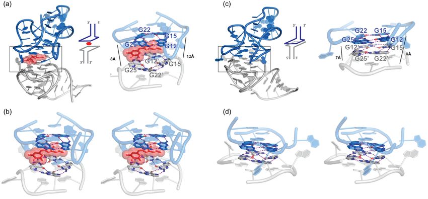
Molecules 2020, 25, 2881                                                                               4 of 28

      between translation (monomeric) and packaging (dimeric) functions. Structural elements followed
      by a prime symbol indicate that they belong to the other protomer. (b,c) Crystal and NMR structure
      of the DIS kissing loop interaction, respectively (PDB 2B8S and 1BAU). (d,e,f) Crystal, NMR, and
      Cryo-EM structures of the DIS homodimer in its extended duplex state (PDB 1Y99, 2GM0 and 6BG9).
      (g) Schematic view of the 50 leader structure of the Moloney murine leukemia virus (MoMuLV) and
      Moloney murine sarcoma virus (MoMuSV) regulating gRNA dimerization. Four elements in this
      RNA drive its dimerization: palindromic sequences in PAL1 and PAL2 and kissing loop interactions
      between Stem-loop 1 (SL1) and Stem-loop 2 (SL2). (h) Structure of MoMuLV homodimer SL2 (PDB
      1F5U). (i) Structure of MoMuSV homodimer SL1-SL2 (PDB 2L1F). Interface regions are boxed and
      detailed below each structure. Hydrogen bonding interactions are shown as magenta dashed lines,
      while stacking interactions are shown as solid magenta lines.

2.1. HIV-1 gRNA
      In the case of human immunodeficiency virus type-1 (HIV-1), a dimerization initiation site (DIS)
was identified within the ~ 350 nucleotide DLS [41,42]. The DIS contains a six-nucleotide GC-rich
palindrome of the sequence GUGCAC in an apical loop. Mutation of this palindromic sequence blocks
gRNA dimerization, while compensatory mutations restore it. Importantly, the DIS can form two
types of dimers in vitro that have been extensively characterized structurally. A kissing loop dimer
is formed at low temperatures in the presence of Mg2+ [43–47] (Figure 1b,c), while a more stable
extended duplex dimer forms through refolding at higher temperatures or in the presence of NC acting
as a chaperone [48–52] (Figure 1d,e,f). The kissing loop dimer is considered a transient intermediate
leading to the extended dimer. Curiously, dimeric gRNAs extracted from mature virions are more
stable than those from immature viral particles [53]. Thus, it is possible that a transition from a loose
kissing loop dimer to a more stable extended DIS occurs during viral maturation.
      The crystal and nuclear magnetic resonance (NMR) structures (Figure 1b,c) of the DIS kissing
loop show an identical self-pairing of the palindromic GUGCAC loop [43,44]. The flanking 30 A
(A16 in Figure 1b,c) stacks on one of the closing Watson-Crick pairs. The DIS palindromic sequence
is flanked by strictly conserved purines whose substitution or deletion block dimerization [54,55].
Thus, this cross-strand stacking is required to stabilize the dimeric interface. Nonetheless, the crystal
structure shows a bulged-out conformation of the flanking 50 purine, while the NMR structure reveals
a non-bulged conformation. This hints at the flexibility of the DIS kissing loop which could be required
to transition towards a more stable extended duplex, a notion supported by single-molecule FRET
analysis [56]. Structures of the extended dimeric DIS reveal an A-form dsRNA structure generated by
self-pairing of the palindromic loop and strand swapping between 50 and 30 strands of both protomers
(Figure 1d,e,f) [48,49,52]. This more than doubles the surface of the dimerization interface (Table 1,
compare PDB 1BAU & 2B8S versus 1Y99 which have similar sequence lengths) rationalizing the
increased stability. In the extended DIS crystal structure, the RNA is colinear with a single bulged
out nucleotide. In contrast, the NMR and cryo-electron microscopy (cryo-EM) structures reveal bent
conformers of the extended DIS. In the latter, an S-turn at the 50 region was observed but it remains
unknown if this RNA motif only forms in the context of the extended DIS duplex.
      Conformational changes in the HIV-1 50 leader regulate the oligomeric state of the gRNA [52,57,58].
The palindromic loop of the DIS is sequestrated intramolecularly by pairing to the U5 element in
the monomeric DLS [59–61] (Figure 1a). Exposure of the DIS is achieved by pairing of the Gag start
site (AUG in Figure 1a) to the U5 thus inducing the dimeric DLS required for genome packaging.
AUG was proposed to pair either in cis or in trans with the U5 element of the same or the other
gRNA copy. It is tempting to speculate that the differences in stability of dimeric gRNAs between
immature and mature viral particles could originate from the nature of AUG pairing to U5 (cis vs.
trans). The structure of a minimal HIV-1 RNA packaging signal in its monomeric state consisting
of the U5, AUG, DIS mutant and splice donor (SD) was recently determined [62,63]. Further work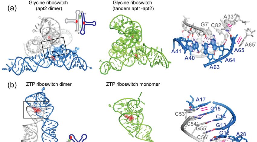
Molecules 2020, 25, 2881 5 of 28
is needed to fully describe the dimerization interface of the HIV-1 DLS and decrypt the monomer
to dimer structural switch [35]. Indeed, the adjacent trans-activation response element (TAR) has
also been proposed to directly or indirectly contribute to gRNA dimerization, as TAR mutations and
deletions exhibited strong influences on gRNA dimerization [64–67]. Furthermore, tRNALys3 annealing
to the primer binding site (PBS) in the DLS was suggested to enhance gRNA dimerization [61,68].
Taken together, it is likely that multiple structural elements (DIS, AUG, U5, TAR, PBS, and even poly-A)
within the dynamic 50 leader coordinately modulate the exposure of the DIS palindrome, nucleation of
the gRNA monomers and subsequent structural re-organizations enabling propagation of the dimer
interface. Conceivably, other RNA viral genomes may utilize similar strategies to sequentially engage
multivalent contacts, as well as morph global and local structures, to ultimately achieve prescribed
dimer configurations.
Table 1. RNA homodimers discussed in this review. N.D.: not determined. The dimerization interface
area (in Å2 ) was computed using PISA [69].
Number of Method of
Name PDB Code Dimerization Motif Interface (Å2 ) Nucleotides Structural
Per protomer Determination
Palindromic kissing loop (six X-ray
2B8S 365.2 23
nucleotides) diffraction
Human immunodeficiency
virus type-1 DIS kissing loop Palindromic kissing loop (six
1BAU 593.6 23 NMR
nucleotides)
6BG9 Strand swapping 2251.6 47 Cryo-EM
Human immunodeficiency
X-ray
virus type-1 DIS extended 1Y99 Strand swapping 1083.1 23
diffraction
duplex
2GM0 Strand swapping 1771.4 35 NMR
Moloney murine sarcoma Palindromic kissing loop (two
1F5U 246.7 18 NMR
virus SL2 nucleotides)
Moloney murine leukemia Palindromic kissing loop (two
2L1F 1146.3 66 NMR
virus SL1-SL2 nucleotides)
Palindromic kissing loop (six
Oskar 30 -UTR N.D. N.D. N.D. N.D.
nucleotides)
Kissing loop
Bicoid 30 -UTR N.D. N.D. N.D. N.D.
(six nucleotides)
Kissing loop X-ray
4R4P
(four nucleotides) 2612.6 186 diffraction
Varkud satellite ribozyme
Reciprocal base intercalation X-ray
4R4V
diffraction
Palindromic kissing loop (four X-ray
6JQ5 2157.3 82
nucleotides) diffraction
Hatchet ribozyme
Strand swapping X-ray
6JQ6 2254.1 81
diffraction
X-ray
3OWI A-minor interactions Reciprocal 1219 88
diffraction
Glycine riboswitch base intercalation
X-ray
3OX0 1243 88
diffraction
X-ray
5NDI Palindromic kissing loop (two 339.1 20
diffraction
nucleotides)
Guanidine II riboswitch X-ray
Reciprocal base intercalation
5VJ9 342.5 16
diffraction
Pseudoknot
X-ray
Glutamine II riboswitch 6QN3 Reciprocal base intercalation 1615.7 50
diffraction
Base triple
X-ray
preQ1 III riboswitch 4RZD Pseudoknot 1212.6 101
diffraction
X-ray
SAH riboswitch 3NPQ Strand swapping 513 54
diffraction
X-ray
ZTP riboswitch 4XWF Pseudoknot 1945 64
diffraction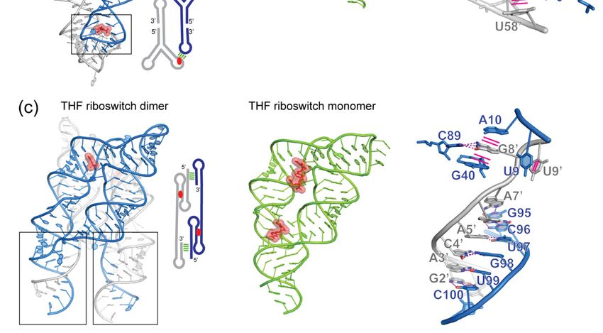
Molecules 2020, 25, 2881                                                                                                6 of 28

                                                    Table 1. Cont.

                                                                                                 Number of      Method of
             Name                 PDB Code       Dimerization Motif           Interface (Å2 )   Nucleotides      Structural
                                                                                                Per protomer   Determination
                                                                                                                    X-ray
                                   3SUX                                           1860.9            101
                                                                                                                 diffraction
        THF riboswitch                             Strand swapping
                                                                                                                    X-ray
                                   3SUY                                           1909.6            101
                                                                                                                 diffraction
                                                                                                                    X-ray
                                    5BJO                                          569.7             36
                                                                                                                 diffraction
           Corn RNA                             G-quadruplex stacking
                                                                                                                    X-ray
                                    6E80                                          670.2             36
                                                                                                                 diffraction
  Internal ribosomal entry site                                                                                     X-ray
                                    2IL9          Domain swapping                 2087.5            142
    (IRES) of Dicistroviridae                                                                                    diffraction
   Designed GAAA tetraloop
                                    2I7Z     Tetraloop – tetraloop receptor       766.7             43             NMR
       receptor complex
      Abbreviations in the table:       S-adenosylhomocysteine (SAH), 5-aminoimidazole-4-carboxamideriboside
      50 -triphosphate (ZTP) and tetrahydrofolate (THF).

2.2. MoMuLV and MoMuSV gRNAs
      Another well-characterized retroviral dimeric gRNA is the ~380 nucleotide DLS of Moloney murine
leukemia virus (MoMuLV) and the closely related Moloney murine sarcoma virus (MoMuSV) [70–74].
Switching between a monomeric and dimeric gRNA is achieved by four dimerization motifs.
Intermolecular pairing of two distinct palindromic sequences PAL1 and PAL2 induce a register
shift in the DLS which exposes self-complementary GACG loops in SL1 and SL2 (also named SL-C and
SL-D) (Figure 1g). Remarkably, the NMR structure of the isolated SL2 from MoMuSV reveals a stable
homodimer mediated by a kissing loop [73] (Figure 1h, PDB 1F5U). This is among the smallest RNA
dimeric interface reported to date spanning 247 Å2 (Table 1). Only two base pairs are formed between
both palindromic loops (C10:G110 and G11:C100 , Figure 1h) and an adenosine stacks on each side of
the kissing loop helix (A9 and A90 in Figure 1h). The NMR structure of the MoMuLV domain SL1-SL2
shows an identical dimeric interface as the isolated MoMuSV SL2, but formed between the loops of
SL1 and SL2 with SL20 and SL10 of the second monomer, respectively [74] (Figure 1g,i). It is likely that
the strength and bend of the stems act as topological tools to restrict the intermolecular self-pairing to
SL1-SL20 /SL2-SL10 instead of SL1-SL10 /SL2-SL20 .

2.3. Other Viruses
     Several other classes of viruses also rely on RNA dimerization for specific functions. A palindromic
kissing loop in the SARS coronavirus genome was shown to regulate ribosomal frameshifting, thus
affecting viral growth [75]. The 30 untranslated region (UTR) of the hepatitis C virus can form two
alternative dimeric interfaces [76–79]. One relies on the complementarity between the DLS in the
30 -UTR and the distal stem-loop 5BSL3.2 of the 50 -UTR which is proposed to enhance translation.
The other dimer is mediated by a palindromic kissing loop in the 30 -UTR and proposed to regulate
genome packaging.
     In summary, a number of RNA viruses, including retroviruses, rely on kissing loop-loop
interactions through palindromic or other complementary sequences to induce genome
homodimerization for various essential functions. To buttress these generally short duplexes,
single-stranded purines frequently cross-strand stack with both flanks of the intermolecular helices
stabilizing them. The stacking of such bases contributes significantly to the stability of the dimeric
interface by minimizing exposure of hydrophobic bases to the solvent and by favorable enthalpy
changes through the aromatic interactions [80]. This strategy is reminiscent of the stabilization of
codon-anticodon interactions in the ribosome [81,82] and T-box riboswitches [83–86], as well as a 3-bp
pseudoknot in the adenovirus VA-I RNA [87].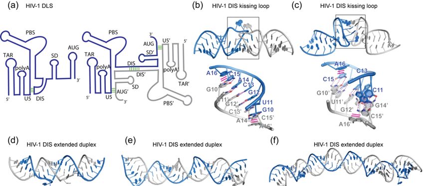
Molecules 2020, 25, 2881                                                                           7 of 28

3. Dimerization-Mediated mRNA Localization
     The 30 -UTRs of eukaryotic mRNAs are regulatory hubs for their translation, decay and cellular
localization, etc. [88]. Non-uniform distributions of certain mRNAs in the cell create spatio-temporal
patterns of gene expression and localized translation which are crucial for cell polarity during cell
differentiation and embryo development, etc. 30 -UTRs contain sequence and structural elements that
can recruit distinct protein partners to exert localized functions [89]. Some of these 30 -UTR signatures
act as “zip-codes” and dictate the cellular localization of the mRNA. Diverse mechanisms orchestrate
mRNA localization and have been extensively reviewed [90–95]. A peculiar case is the targeting of
the oskar (osk) and bicoid (bcd) mRNAs to opposite poles of Drosophila embryos [96–98]. In both cases,
the Staufen protein [99,100] mediates the transport of both mRNAs through movement on microtubules,
while other factors repress translation until osk or bcd reach their respective destinations [101–103].
Importantly, homodimerization of these mRNAs is required for efficient subcellular localization.

3.1. Oskar mRNA
     Spliced osk is localization-competent, while unspliced osk is not [104]. Nonetheless, the latter can
be localized at the pole of the cell by hitchhiking onto spliced osk through intermolecular interactions.
A conserved six-nucleotide, GC-rich palindromic loop sequence in the region of 714-827 of the osk
30 -UTR was shown to promote homodimerization in vitro [105] (Figure 2a). Such a kissing loop-loop
interaction is reminiscent of the DIS of HIV-1 (see above). However, the conversion into an extended
duplex has not yet been observed for osk and may not form due to a larger loop flanked by pyrimidines.
Double nucleotide substitutions in the palindromic sequence blocked dimerization by preventing
kissing interactions. Importantly, dimerization is not required for localization of spliced osk but
necessary for hitchhiking by unspliced osk [105]. Although the functional role of osk localization
or translation activation through dimerization remains unclear, it exemplifies how mRNAs lacking
“zip-codes” could still be targeted to specific cellular regions by piggybacking with other RNAs through
RNA multimerization.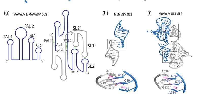
Molecules 2020, 25, 2881 8 of 28
Molecules 2020, 25, x FOR PEER REVIEW 8 of 27
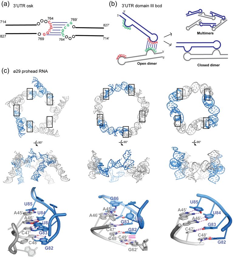
Figure2.2.Homodimerization
Figure Homodimerizationin inmRNA
mRNAtransport.
transport. (a)
(a) oskar
oskar mRNA
mRNA dimerize
dimerizethrough
throughaaconserved
conservedGC GC
richkissing
rich kissinglooploopininthe 0 -untranslated region
the33′-untranslated region (UTR).
(UTR). (b)
(b)bicoid
bicoidmRNA
mRNAdimerization
dimerization motif motif relies
reliesonon
the complementarity
the complementarity of the theapical
apicalloop
loopand
andananinternal bulge
internal bulgein the domain
in the domainIII of its 30 -UTR.
IIIitsof3′-UTR. This leads
This
to formation
leads to formation of closed dimers
of closed dimersor open
or opendimers
dimersthat
thatcan
canfurther
furtheroligomerize.
oligomerize. (c) (c) Structures of of the
the
proheadRNA
prohead RNA of of bacteriophage ø29 ø29 driven
drivenbybykissing
kissinginteractions
interactionsbetween
between two
two stem-loops.
stem-loops. From
Fromleft
to right,
left twotwo
to right, cryo-EM
cryo-EMstructures of the
structures of pentameric
the pentamericassembly of pRNA
assembly (PDB(PDB
of pRNA 1FOQ1FOQ and 6QYZ) and a
and 6QYZ)
and a crystal
crystal structurestructure of a pRNA
of a pRNA tetramer tetramer (PDBThe
(PDB 3R4F). 3R4F). The conserved
conserved feature the
feature amongst amongst the three
three structures
structures is the Watson-Crick
is the Watson-Crick pairing between
pairing between the 45AACC48
the 45AACC48 and 82GGUU85
and 82GGUU85 segments, segments, which higher-
which drives drives
higher-order assembly. Interface regions are boxed and detailed below each
order assembly. Interface regions are boxed and detailed below each structure. Hydrogen bonding structure. Hydrogen
bonding interactions
interactions are shownare asshown as magenta
magenta dasheddashed lines, while
lines, while stacking
stacking interactions
interactions are shownare shown as
as solid
solid magenta
magenta lines.lines.
3.2. Bicoid mRNA
3.2. Bicoid mRNA
The localization signal of bcd mRNA, found in the domain III of its 30 -UTR, was shown to form
The localization signal of bcd mRNA, found in the domain III of its 3′-UTR, was shown to form
dimers in vitro [106–108]. Pairing of the apical loop of domain III with a complementary internal
dimers in vitro [106–108]. Pairing of the apical loop of domain III with a complementary internal
bulge sequence form the dimerization motif via six Watson-Crick pairs (Figure 2b). At least two
bulge sequence form the dimerization motif via six Watson-Crick pairs (Figure 2b). At least two
sequential steps are involved during self-assembly and are kinetically controlled [107]. Open dimers
sequential steps are involved during self-assembly and are kinetically controlled [107]. Open dimers
are formed initially through pairing of a single motif between two protomers. This initial nucleation
are formed initially through pairing of a single motif between two protomers. This initial nucleation
event then leads to a closed and stable (nearly irreversible) dimer with both motifs paired (Figure 2b).
event then leads to a closed and stable (nearly irreversible) dimer with both motifs paired (Figure 2b).
Mutations that disrupt the complementarity between the loop and the bulge block dimerization in
vitro and inhibit cellular targeting of bcd in vivo [106,107]. This suggests that bcd multimerizationMolecules 2020, 25, 2881 9 of 28
Mutations that disrupt the complementarity between the loop and the bulge block dimerization in vitro
and inhibit cellular targeting of bcd in vivo [106,107]. This suggests that bcd multimerization could
bring into close proximity different duplex regions of the 30 -UTR, which in turn recruit one or multiple
double-stranded RNA-binding domains (dsRBDs) [109] of Staufen to form large ribonucleoprotein
particles prior to loading on microtubules.
Interestingly, bcd forms exclusively dimers in vitro, while the isolated domain III assembles into
dimers, trimers, and tetramers [107]. It is likely that neighboring regions of domain III sterically and
topologically affect its oligomerization. Furthermore, protein factors may also impact bcd quaternary
structure. This multimerization mechanism and the ability to form more than one type of multimer is
reminiscent of the bacteriophage ø29 prohead RNA (pRNA), discussed below [110–113] (Figure 2c).
3.3. ø29 Prohead RNA
The bacteriophage ø29 pRNA is a component of the packaging motor required for viral
genomic DNA encapsulation. pRNA forms dimers in vitro through a kissing loop interaction
mediated by four complementary base pairs between two distinct stem-loops [114,115]. However,
pentamers or hexamers of pRNA are thought to be the functional assemblies and form through
interactions with other protein factors of the packaging motor (protein connector, protein capsid,
and ATPase). In this model, pRNA “open homodimerization” is proposed to be the nucleation
point that precedes the formation of higher-order oligomers. Structures of the pentameric and
tetrameric pRNAs [111–113] (Figure 2c) revealed that a range of oligomeric assembly configurations
can be produced by mix-and-matching two complementary loop sequences on either end of the
RNA protomer in head-to-tail configurations. The intrinsic structural flexibility of the RNA protomer
may permit formation of a linear or circular chain of protomers of variable lengths. This use of two
complementary but non-palindromic sequences to build multimers contrasts with the tendency of
RNAs bearing single palindromic sequences to form dimers. This architectural modularity has been
extensively used in RNA nanotechnology [21,23,116–118].
4. Homodimeric Ribozymes
Ribozymes are RNA catalysts that accelerate various chemical transformations, including
self-cleavage, splicing, tRNA aminoacylation and peptidyl transfer, etc. Many naturally occurring
ribozymes catalyze post-transcriptional RNA processing, such as backbone cleavage or ligation, and
usually proceed through a general acid-base mechanism and produce 20 ,30 -cyclic phosphate and 50
hydroxyl termini [119,120]. Several ribozymes are known to function as homodimers.
4.1. VS Ribozyme
The Varkud Satellite (VS) ribozyme, the largest known nucleolytic ribozyme, was discovered
30 years ago in the mitochondria of Neurospora fungi. It has both self-cleavage and ligation properties
and functions in the processing of RNA intermediates of rolling-circle replication [121]. It is organized
into seven helical regions that can be trans-cleaved following self-assembly into a homodimer [122,123].
Its structure and mechanism have been the subject of extensive studies [124]. The crystal structure of
the entire VS ribozyme revealed an intertwined symmetric dimer spanning an interface of 2612 Å2 ,
the largest RNA homodimerization interface described to date (Table 1). Stem-loop 1 (P1) forms a
kissing loop interaction with stem-loop 50 (P50 ) of the second protomer through three Watson-Crick
base pairs and one non-canonical C629-A6960 pair (Figure 3a) [123,125]. Interestingly, cross-strand
stacking by G633 bolsters the 4-bp intermolecular duplex, analogous to the aforementioned retroviral
RNAs. This leads to a domain-swapped configuration where helix P1 docks with helices P20 , P50
and P60 of the other subunit, thus forming a composite active site. Here, the general base G638 of
helix P1 and the general acid A7560 of helix P60 flank the scissile phosphate between G620 and A621,
driving its in-line nucleophilic attack, stabilization of the transition state and ultimately departure
of the leaving group. The intermolecular kissing interaction between the P1 and P50 loops probablyMolecules 2020, 25, 2881 10 of 28
initiates VS ribozyme dimerization. Subsequently, another interaction between the middle section of P1
and P20 and P60 brings into proximity the reactants forming the catalytic site (Figure 3a). Intriguingly,
Molecules 2020, 25, x FOR PEER REVIEW 10 of 27
this second interaction is primarily stacking in nature with the bases of A7510 and C7500 inserted
between between
inserted the aromatic rings of A621
the aromatic ringsand
of A639. Despite
A621 and A639.notDespite
employing
not any base-pairing
employing interactions,
any base-pairing
this secondary contact likely contributes significantly to dimerization by assembling a coaxial
interactions, this secondary contact likely contributes significantly to dimerization by assembling array ofa
5 nucleobases
coaxial array ofstabilized by consecutive
5 nucleobases stabilized π-π stacking interactions.
by consecutive π-π stacking interactions.
Figure
Figure 3.
3. Structures
Structures ofofdimeric
dimericribozymes.
ribozymes.(a) (a)Crystal
Crystal structure
structure of Varkud
of Varkud Satellite
Satellite ribozyme
ribozyme (PDB(PDB
code
code 4R4P & 4R4V) with the insets showing the intermolecular kissing loop interaction
4R4P & 4R4V) with the insets showing the intermolecular kissing loop interaction between stem-loop between stem-1
loop 1 (P1)
(P1) and and stem-loop
stem-loop 50 (P505′), (P5′), as as
as well well
theasintercalation
the intercalation of stacked
of stacked nucleobases
nucleobases between
between P1 and
P1 and P60 .
P6′. (b) Crystal
(b) Crystal structures
structures of the
of the Hatchet
Hatchet ribozyme
ribozyme (PDB6QJ6
(PDB 6QJ6&&6QJ5)
6QJ5)showing
showing twotwo types
types of
of dimeric
dimeric
interfaces.
interfaces. The
Thetoptopinset
insethighlights
highlightsthe thepalindromic
palindromicsequence
sequencepaired
pairedwith
with the
the same
same sequence
sequence ofof the
the
second protomer. Hydrogen bonding interactions are shown as magenta dashed
second protomer. Hydrogen bonding interactions are shown as magenta dashed lines, while stacking lines, while stacking
interactions
interactions are
are shown
shown asas solid
solid magenta
magenta lines.
4.2. Hatchet
4.2. HatchetRibozyme
Ribozyme
The Hatchet
The Hatchet ribozyme
ribozyme is is aa small
small ribozyme
ribozyme that that was
was recently
recently discovered
discovered in in several
several genes
genes of
of
Veillonella sp.
Veillonella sp. [126] and shown to form stable homodimers [127]. The dimerization is driven by aa
[126] and shown to form stable homodimers [127]. The dimerization is driven by
tetranucleotidepalindromic
tetranucleotide palindromicinternal
internallooploopsequence
sequenceACGU ACGU(nts (nts67-70)
67-70) that
that forms
forms aa symmetric
symmetric helixhelix
(Figure 3b). This leads to the exchange of the 3 0 strand between protomers forming a hybrid helix P2
(Figure 3b). This leads to the exchange of the 3′ strand between protomers forming a hybrid helix P2
(pairing between
between A21 A21 toto G29
G29 with
with A75 0 to U810 ) and extending the dimeric interface. Although the
(pairing A75′ to U81′) and extending the dimeric interface. Although the
palindromic sequence is essential for efficient
palindromic sequence is essential for efficient dimerization, dimerization, itit is is not
not required
required forfor catalysis
catalysis and
and aa
monomeric hatchet ribozyme maintained efficient cleavage [127]. Interestingly,
monomeric hatchet ribozyme maintained efficient cleavage [127]. Interestingly, two conformers of two conformers of
the homodimeric
the homodimeric hatchethatchet ribozyme
ribozyme were were trapped
trapped in in different
different crystal
crystal lattices.
lattices. Their
Their cores
cores are
are nearly
nearly
identical with an RMSD of 1.8 Å over 64 residues but the orientation of the swapped 3 0 strands are
identical with an RMSD of 1.8 Å over 64 residues but the orientation of the swapped 3′ strands are
rotated by
rotated ~80◦(Figure
by ~80° (Figure3b).3b).This
Thisled ledtototwo
twodistinct
distinctconfigurations
configurationswith withthe
theprotomers
protomers either
either forming
forming
an antiparallel
an trans-likesymmetric
antiparalleltrans-like symmetricdimer dimeroror a more
a more intertwined
intertwined cis-like
cis-like pseudosymmetric
pseudosymmetric dimer.
dimer. In
In solution, both conformers and potentially others are likely sampled
solution, both conformers and potentially others are likely sampled as a result of the apparent as a result of the apparent
flexibility of
flexibility of the
the 3′30 extended
extendedtail tailwith
withthetheembedded
embeddedpalindromic
palindromic sequence.
sequence. An Anunanswered
unanswered question
question
is whether
is whether this
this dimerization
dimerization occursoccurs in in vivo
vivo andand for
for what
what purpose,
purpose, since
since the
the palindromic
palindromic sequence
sequence is
is
found in the Hatchet ribozymes of multiple organisms but not
found in the Hatchet ribozymes of multiple organisms but not strictly conserved [126].strictly conserved [126].
5. Homodimerization of Riboswitches
Riboswitches are cis-acting noncoding elements found in the 5′-UTR of some mRNAs (mostly in
prokaryotes) [128–130]. They regulate the transcription, translation or splicing of downstream genes
through direct binding of small-molecule metabolites [131] or tRNAs [132,133] to an aptamer domain,Molecules 2020, 25, 2881 11 of 28
5. Homodimerization of Riboswitches
Riboswitches are cis-acting noncoding elements found in the 50 -UTR of some mRNAs (mostly in
prokaryotes) [128–130]. They regulate the transcription, translation or splicing of downstream genes
through direct binding of small-molecule metabolites [131] or tRNAs [132,133] to an aptamer domain,
which in turn controls a conformational switch in the expression platform. Most riboswitches function
with a single aptamer but some are organized into a tandem arrangement of two to three aptamers that
bind the same or even different metabolites, thus linking two or more metabolic pathways [134]. In the
following section, we discuss 7 classes of riboswitches that were crystallized as dimers. Among them,
3 classes (Glycine, ZTP, and THF) also crystallized as monomers, allowing direct comparisons with
their dimeric counterparts. The biological relevance and potential functions of these dimers will also
be discussed. In cases where the tandem aptamers are highly homologous, such as the glycine and
guanidine-II riboswitches, dimeric structures of single aptamers in crystallo likely represented the way
the natural tandem aptamers interact with each other and act in concert.
5.1. Glycine Riboswitches
One prominent example of tandem riboswitches is the tandem glycine riboswitch formed by two
covalently linked glycine aptamers (apt1 and apt 2) followed by a single expression platform [135].
Although it was proposed early on that cooperative binding of individual glycine molecules to each
aptamer produced a sharper digital response [135–137], later work suggested that each aptamer
largely binds glycine independently and the double-aptamer quaternary structure is stabilized by
an intervening P0 helix and a K-turn connecting the two aptamers [138–141]. The crystal structure
of an isolated Vibrio cholerae glycine apt2 revealed a crystallographic homodimer formed between
helix P3 of one protomer and helix P10 of the other [142] (Figure 4a). Four A-minor interactions [143]
stabilize this interface. A40 and A64 from P3 form Type-I A-minor interactions with G840 -C50 and
G70 -C820 from P10 , respectively, while both A41 and A63 engage Type-II A-minor interactions with the
same U60 -A830 pair (Figure 4a). In addition, extrusion and intermolecular stacking between A65 and
A650 stabilize the crystallographic homodimer and facilitates ligand binding by flipping A65 outside
of the glycine-binding pocket. Remarkably, the manner by which this apt2-apt20 homodimer forms
in crystallo, initially thought to be physiologically non-functional, is nearly the same as the way a
natural tandem Fusobacterium nucleatum glycine riboswitch forms an apt1-apt2 intramolecular interface
(RMSD of 2.66 Å over 104 residues; Figure 4a). This is driven by structural similarity between the two
aptamers and inherent symmetry of the dimer interface [144].
Further studies showed that singlet glycine riboswitches bind glycine with comparable affinities
as the more common tandem version, but require a neighboring “ghost aptamer”, which is likely
a degenerate, reduced version of the original partner aptamer that maintained the inter-aptamer
contacts for structural stabilization. There are multiple proposals on why most glycine riboswitches
have retained a tandem arrangement, as such an arrangement does not apparently enhance ligand
binding (at least in vitro) [145–150]. Here, we describe one additional hypothesis. The ancestral
glycine binding aptamer, presumably “A”-shaped like their descendent aptamers, may have by chance
folded into a structure that is in a sense “palindromic”. That means the donors (A-rich bulge or
loop) and receptors (minor groove of G-C/C-G pairs) of the A-minor interactions were spaced and
angled in such a way that two oppositely oriented copies of the aptamer can simultaneously engage
two sets of A-minor interactions with each other, thereby seeding the dimer configuration. This
notion is analogous to the design of a dimer tectoRNA, which took advantage of appropriately spaced
tetraloop and tetraloop receptor motifs, achieving a dimerization Kd of ~4 nM [151–153]. The resulting
mutual reinforcement of both glycine-binding aptamers, clearly advantageous for folding, overall
stability, and possibly also tighter binding of small ligands, such as glycine, led to retention of the
dimeric form. Through evolution, depending on gene-specific regulatory needs, certain tandem glycine
riboswitches had degraded into singletons that function with a ghost aptamer [146]. For tandem
adenosines embedded in a helix to effectively engage the minor groove, the donor helix needs to beMolecules 2020, 25, 2881 12 of 28
inclined or angled relative to the recipient helix [143]. This incline presumably produced the ~60◦
angle between the two connecting helices (P1 and P10 ) at the inter-aptamer junction, which then drove
the evolution of a K-turn. Once adopted, the stable K-turn could have taken charge of the overall
dimer architecture, playing a key role in placing donor adenosines in close proximity to their receptor
minor grooves. The A-minor interactions, compared to kissing loops and other base-pairing contacts,
lack the specificity to find each other and engage unassisted. Thus, this theory rationalizes the critical
requirement of the K-turn and the appearance of ligand-binding cooperativity in its absence. A similar
scenario was recently observed in the T-box riboswitches, where an essential K-turn brings the Stem
II S-turn motif to dock, via an extended ribose zipper, with Stem I, forming a composite binding
groove for the incoming tRNA anticodon [84,85]. Taken together, recent studies of tandem and singlet
glycine riboswitches have produced a wealth of important insights not just into ligand recognition by
RNA, but into RNA-RNA interactions and the evolution of quaternary structure and geometric motifs.
Undoubtedly, the saga of the glycine riboswitches will continue.
Molecules 2020, 25, x FOR PEER REVIEW 12 of 27
Comparison of
Figure 4. Comparison of monomeric
monomeric and dimeric structures of the same same riboswitches.
riboswitches. (a) Glycine
aptamer 22crystallized
aptamer crystallizedin aindimer
a dimer
(PDB(PDB
3OWI)3OWI) configuration
configuration similar
similar to to theglycine
the tandem tandem glycine
aptamer1-
aptamer2 (PDB 3P49). (b) ZTP riboswitch dimer structure (PDB 4XWF) juxtaposed with its
aptamer1-aptamer2 (PDB 3P49). (b) ZTP riboswitch dimer structure (PDB 4XWF) juxtaposed with
monomeric version
version (PDB
(PDB5BTP).
5BTP).(c)(c)THF
THFriboswitch
riboswitchcrystallized
crystallizedin dimeric
in dimeric(PDB 3SUY
(PDB andand
3SUY 3SUX) and
3SUX)
monomeric
and monomericversion (PDB
version 4LVV).
(PDB Ligands
4LVV). are shown
Ligands in stick
are shown representation
in stick representation andand
semi-transparent red
semi-transparent
spheres.
red Interface
spheres. regions
Interface regionsareare
boxed
boxed and detailed
and onon
detailed the
theright
rightofofeach
eachdimeric
dimericstructure. Hydrogen
structure. Hydrogen
bonding interactions
bonding interactions are
are shown
shown asas magenta
magenta dashed
dashed lines,
lines, and
and stacking
stacking interactions
interactions are
are shown
shown as
as solid
solid
magenta lines.
magenta lines.
Further studies showed that singlet glycine riboswitches bind glycine with comparable affinities
as the more common tandem version, but require a neighboring “ghost aptamer”, which is likely a
degenerate, reduced version of the original partner aptamer that maintained the inter-aptamer
contacts for structural stabilization. There are multiple proposals on why most glycine riboswitchesMolecules 2020, 25, 2881 13 of 28
5.2. ZTP Riboswitches
Two crystal structures of the ZTP (5-aminoimidazole-4-carboxamideriboside 50 -triphosphate)
riboswitch were solved (Figure 4b) [154–156]. In the Fusobacterium ulcerans ZTP riboswitch, two
subdomains formed by P1-P2 and P3, respectively, bind each other forming a P4 pseudoknot. This cis
tertiary interface forms the binding pocket for ZTP. Remarkably, in the Schaalia odontolytica ZTP
riboswitch structure, this pseudoknot is formed in trans by domain swapping in a crystallographic
dimer leading to G12-G15 pairing with C540 -C570 (Figure 4b). The two structures are similar with an
overall RMSD of 2.85 Å over 40 residues. Importantly, nearly identical ZTP-binding pockets were
observed in the two structures, suggesting that the crystallographic dimer structure from S. odontolytica
actually captured the biological relevant RNA-ligand interface.
5.3. THF Riboswitches
The structure of the Eubacterium siraeum tetrahydrofolate (THF) riboswitch exhibited an unraveled
P1 helix that strand-exchanged with the P10 of another monomer forming a scissors-like structure
where the intertwined P1-P10 forms the hinge (Figure 4c) [157]. This homodimerization occurred
through seven Watson-Crick pairs between the 50 G10 -A70 of one monomer and the 30 G95-C101 of
another (Figure 4c). In addition, C89 forms a Watson-Crick pair with G80 that is stacked between A10
and G40, while U9 stacks with U90 further stabilizing this dimeric interface. By contrast, the P1 helix
in the Streptococcus mutans THF riboswitch remained paired forming a monomer. Both dimeric and
monomeric assemblies are structurally similar (Figure 4c), but the latter captured two THF molecules
rather than one in the former structure [158]. Both structures brought valuable insights into the function
of THF riboswitch and led to the design of artificial homodimeric RNAs responsive to THF through
a loop-receptor intermolecular interface [159]. Similar artificial constructs were previously formed
through a GAAA tetraloop-receptor mediated homodimerization interface [151,152,160].
5.4. Guanidine-II Riboswitches
The guanidine-II riboswitch, originally termed the mini-ykkC motif, is the simplest of the three
known classes of guanidine riboswitches [161]. It is comprised of two small stem loops (P1 and
P2) linked by a variable size linker (7 to 40 nucleotides) [162]. Both stem loops were shown to
bind a single guanidine each in a cooperative manner. The ACGR sequence of both loops are
highly conserved, shared between both P1 and P2 and were protected by guanidine in in-line
probing experiments, while the linker was not. The crystal structures of the individual Escherichia
coli and Pseudomonas aeruginosa P1 and P2 aptamers bound to guanidine revealed homodimeric
interfaces through a kissing loop interaction [163,164] (Figure 5a). In both instances, complementary
Watson-Crick pairs are formed between C10-G110 and G11-C100 from the loops of both protomers,
which are further coaxially stacked. This is reminiscent of the kissing loop between SL1 and SL2 of the
MoMuSV and MoMuLV discussed previously (Figure 1g–i). Two guanidine molecules bind inside
both ACGR loops at the dimerization interface but interact only with one of the protomer. Although
this homodimerization occurred in crystallo, it is likely that in the natural full-length guanidine-II
riboswitch, a similar kissing loop interaction occurs intramolecularly between P1 and P2. In this
context, the dynamics and strength of coaxial stacking might be tuned by the linker for conditional
switching. From a thermodynamic perspective, the loop-loop interaction likely pre-organizes the
binding site for guanidine, reducing the entropic cost of binding [163,164]. Remarkably, only two base
pairs form at the interface. Their engagement is likely facilitated by the covalent P1-P2 tether which
brings the pairs into proximity. Further, coaxial stacking across the dimer interface is expected to
significantly stabilize the loop-loop interaction and ligand binding. Dimerization presumably requires
extension of the variable single-stranded linker, as evidenced by its altered pattern of in-line cleavage
upon guanidine binding [162]. It would be interesting to consider how the length and sequence of the
P1-P2 linker affect this heterodimerization, ligand binding, and, in turn, gene expression.which brings the pairs into proximity. Further, coaxial stacking across the dimer interface is expected
to significantly stabilize the loop-loop interaction and ligand binding. Dimerization presumably
requires extension of the variable single-stranded linker, as evidenced by its altered pattern of in-line
cleavage upon guanidine binding [162]. It would be interesting to consider how the length and
sequence
Molecules of25,the
2020, 2881P1-P2 linker affect this heterodimerization, ligand binding, and, in turn, gene
14 of 28
expression.
Figure5.5. Additional
Figure Additional in
in crystallo
crystallo dimeric
dimeric riboswitches.
riboswitches.(a) (a)Guanidine
GuanidineIIIIriboswitch
riboswitch(PDB
(PDB 5NDI
5NDI and
and
5VJ9). (b)
5VJ9). (b) Glutamine
Glutamine riboswitch
riboswitch IIII (PDB
(PDB 6QN3).
6QN3).(c)(c)Class
ClassIII
IIIpreQ1
preQ1riboswitch
riboswitch(PDB
(PDB 4RZD).
4RZD). (d)(d)
SAH
SAH
riboswitch(PDB
riboswitch (PDB 3NPQ).
3NPQ). Ligands
Ligands are
areshown
shownin instick
stickrepresentation
representationandand semi-transparent
semi-transparent redred
spheres.
spheres.
Interface regions are boxed and detailed below each structure. Hydrogen bonding
Interface regions are boxed and detailed below each structure. Hydrogen bonding interactions interactions areare
shown as magenta dashed lines, and stacking interactions are shown as solid magenta
shown as magenta dashed lines, and stacking interactions are shown as solid magenta lines. lines.
5.5.
5.5.Glutamine-II
Glutamine-II Riboswitches
riboswitches
Therecent
The recentcrystal
crystalstructure
structureofofthe
theProchlorococcus
Prochlorococcussp.
sp.glutamine-II
glutamine-IIriboswitch
riboswitchhas hasshed
shedlight
lightononits
its ligand-binding
ligand-binding properties
properties [165].
[165]. TheThe functional
functional formform of this
of this genegene regulator
regulator is likely
is likely monomeric
monomeric but it
but it crystallized
crystallized as a homodimer
as a homodimer (Figure
(Figure 5b). 5b). This
This occurs occurs
through thethrough
formation theofformation of a 5-bp
a 5-bp intermolecular
intermolecular pseudoknot-like
pseudoknot-like helix formed
helix formed between between
C1-C5 of the C1-C5 of the firstand
first monomer monomer 0
G36 -G41 0
and G36′-G41′ of
of the second
the second Furthermore,
protomer. protomer. Furthermore, a triplex-like
a triplex-like interaction
interaction involvinginvolving a base(G18•G2-C39
a base triple 0 ) and the
triple (G18•G2-C39′)
and the sandwiching
sandwiching of G340 inofbetween
G34′ in between
A20 and A20 G22 and G22 complete
complete the homodimer
the homodimer interface.interface.
Overall, Overall,
this leads
this leads to a domain-swapped dimer configuration with a P1-pseudoknot-P2′
0
to a domain-swapped dimer configuration with a P1-pseudoknot-P2 helical stack representing helical stackthe
representing
functional the functional
monomeric unit. monomeric unit. Notably,
Notably, a similar domain aswapping
similar domain swapping
configuration wasconfiguration was
seen in the crystal
seen in the crystal structure of a Dicistroviridae internal ribosomal entry site (IRES) [166].
structure of a Dicistroviridae internal ribosomal entry site (IRES) [166]. Although these domain-swapped Although
dimer configurations presumably do not occur in vivo, the dimeric structures nonetheless were able to
be interpreted to understand the functional monomers.
5.6. PreQ1 Class III Riboswitches
Like the glutamine-II riboswitch and Dicistroviridae IRES, the Faecalibacterium prausnitzii preQ1 class
III riboswitch bound to its ligand also crystallized in a homodimeric and partially domain-swapped
configuration. In its previously elucidated class I and II counterparts, preQ1 binding at the helical
junctions between P1 and P2 stabilizes coaxial stacking and directs helical sequestration of the
ribosome-binding site (RBS, or Shine-Dalgarno sequence) [167–171]. Intriguingly, the class III variant
has its RBS located more downstream and embedded in a single-stranded 30 tail region [167].
The homodimer structure revealed that upon preQ1 binding, the 30 RBS-containing tail formed
a robust 6-bp helix with a complementary sequence from the loop of P4 of the other monomer, forming
a second pseudoknot [172] (Figure 5c). In this open configuration, U92-C97 of the 30 tail is paired
with G660 -U710 of the P40 loop. The resulting helix is further reinforced by cross-strand stacking by
G98. Molecular dynamics simulations and single-molecule FRET analysis suggested that the 30 -tail
can rapidly dock and undock from the P4 loop within the same monomer (closed configuration), as
modulated by preQ1 binding. Although this homodimerization is not known to occur in solution, theMolecules 2020, 25, 2881 15 of 28
same dimerization interface is likely used within a single riboswitch monomer for gene regulation by
this novel mechanism.
5.7. SAH Riboswitches
The crystal structure of the Ralstonia solanacearum S-adenosylhomocysteine (SAH) riboswitch
revealed an intermolecular P1 helix formed by strand swapping between two monomers [173]
(Figure 5d). Four Watson-Crick pairs formed intermolecularly between U36-G38 of a junctional region
(J1/4) and G10 -A30 of frayed P1 helix of the other monomer. Intriguingly, this created an unusual
X-shaped structure that resembles a stacked Holliday junction [174]. Selective 2’-hydroxyl acylation
analyzed by primer extension (SHAPE) analysis revealed that the terminal two base pairs of P1 tend
to unravel in solution, which likely facilitated strand exchange and the observed dimerization in
crystallo [173]. Similarly, this dimerization interface is not thought to function in vivo.
In summary, most known riboswitches carry out their regulatory functions in monomeric states.
However, their homodimerization is frequently observed in crystallo through strand or domain
swapping. This is likely the result of their conformational flexibility, especially in their gene-regulatory
P1 regions that are prone to strand fraying and subsequent selection by the crystallization process.
Despite not being completely natural, these dimeric structures have brought essential functional
insights into specific recognition of various small molecules by RNA. Intriguingly, most of these
crystallographic dimers are structurally similar to their monomeric counterparts, not only in their
ligand-binding pockets but also in their overall architectures. This is exemplified by the glycine, ZTP,
and THF riboswitches, of which both monomeric and dimeric structures are available for comparison
(Figure 4). For riboswitches that are only known to function as singlet monomers but crystallized as
dimers, it remains possible that future discovery of their tandem versions in other species would bring
biological relevance to these dimers. Indeed, with ongoing gene duplication, horizonal transfer, and
phage-mediated genome recombination events, it is conceivable that singlet riboswitches can and have
evolved into tandem versions to leverage cooperativity or other traits to effect a modified regulatory
behavior, mirroring how certain tandem glycine riboswitches have devolved into singlets.
6. Dimeric Fluorogenic RNA Aptamers
Another notable class of RNAs that bind small molecules are the in vitro selected fluorescence-
enhancing RNA aptamers [175]. When a weakly fluorescent small molecule binds to the aptamer,
its intrinsic fluorescence can be dramatically enhanced making it a versatile tool to visualize RNA
in cells. Different flavors of fluorescent RNAs have been designed or selected and now span
the entire visible spectrum region [176]. Recently, the Corn RNA was selected to bind DFHO
(3,5-difluoro-4-hydroxybenzylidene- imidazolinone-2-oxime), a compound that mimics the fluorescent
properties of the red fluorescent protein (RFP). The Corn aptamer was shown to dimerize in solution
through direct stacking between the two G-quadruplex base planes from each protomer in the absence
of DFHO [177,178] (Figure 6). Although stacking of G-quadruplexes in solution is well-known [179],
this structure represents the first example of a homodimeric RNA relying solely on stacking by
G-quadruplexes and unpaired purines with no inter-protomer base pairing in the dimerization
interface. The fluorophore binding pocket lies between both G-quadruplexes at the dimerization
interface and a single DFHO molecule intercalates between the quartets through extensive stacking.
Both G-quadruplexes and an auxiliary enclosure of 3 single-stranded adenosines collaborate to
encapsulate DFHO, restrict its conformational freedom and augment its fluorescence. G-quadruplexes
are a prominent motif that is frequently used to bind fluorescence dyes by these aptamers [175].
Interestingly, several new aptamers have been reported not to require this motif, such as the
dimethylindole red (DIR) aptamers [180] and the dimeric fluorogenic RNA aptamer o-Coral, which is
a RNA dimer selected to bind a dimeric, self-quenched fluorophore (sulforhodamine B) [181].You can also read