Redox Signaling and Sarcopenia: Searching for the Primary Suspect - MDPI
←
→
Page content transcription
If your browser does not render page correctly, please read the page content below
International Journal of
               Molecular Sciences

Review
Redox Signaling and Sarcopenia: Searching for the
Primary Suspect
Nicholas A. Foreman † , Anton S. Hesse †                        and Li Li Ji *

                                             Laboratory of Physiological Hygiene and Exercise Science, School of Kinesiology,
                                             College of Education and Human Development, University of Minnesota, 1900 University Ave, Minneapolis,
                                             MN 55455, USA; forem056@umn.edu (N.A.F.); hesse151@umn.edu (A.S.H.)
                                             * Correspondence: llji@umn.edu; Tel.: +1-612-624-9989
                                             † These authors contributed equally to this work.

                                             Abstract: Sarcopenia, the age-related decline in muscle mass and function, derives from multiple eti-
                                             ological mechanisms. Accumulative research suggests that reactive oxygen species (ROS) generation
                                             plays a critical role in the development of this pathophysiological disorder. In this communication,
                                             we review the various signaling pathways that control muscle metabolic and functional integrity such
                                             as protein turnover, cell death and regeneration, inflammation, organismic damage, and metabolic
                                             functions. Although no single pathway can be identified as the most crucial factor that causes sar-
                                             copenia, age-associated dysregulation of redox signaling appears to underlie many deteriorations at
                                             physiological, subcellular, and molecular levels. Furthermore, discord of mitochondrial homeostasis
                                             with aging affects most observed problems and requires our attention. The search for the primary
                                             suspect of the fundamental mechanism for sarcopenia will likely take more intense research for the
                                   secret of this health hazard to the elderly to be unlocked.


Citation: Foreman, N.A.; Hesse, A.S.;        Keywords: aging; mitochondria; redox signaling; sarcopenia; skeletal muscle; peroxiredoxin
Ji, L.L. Redox Signaling and
Sarcopenia: Searching for the Primary
Suspect. Int. J. Mol. Sci. 2021, 22, 9045.
https://doi.org/10.3390/                     1. Introduction
ijms22169045
                                                  Sarcopenia is defined as the loss of mass and strength of skeletal muscle, largely as
                                             a result of aging [1]. Sarcopenia negatively affects quality of life and is associated with
Academic Editors: Jaime M. Ross and
                                             failure of independent living and early mortality. In addition, the prevalence of sarcopenia
Giuseppe Coppotelli
                                             is expected to increase as the world’s population ages [1]. This health problem brings
                                             an enormous financial burden to society: those with sarcopenia incur an additional cost
Received: 26 July 2021
Accepted: 19 August 2021
                                             of $2316 per person per year compared to those without sarcopenia [2]. In addition, the
Published: 22 August 2021
                                             estimated cost of hospitalization for those with sarcopenia is $40.4 billion annually [2].
                                             Therefore, understanding the mechanisms that underlie sarcopenia may lead to therapies
Publisher’s Note: MDPI stays neutral
                                             and drugs that mitigate these negative impacts. Unfortunately, despite decades of research,
with regard to jurisdictional claims in
                                             the exact mechanisms that underlie sarcopenia have yet to be delineated.
published maps and institutional affil-           Among all the potential etiological foundations of sarcopenia, the generation of reac-
iations.                                     tive oxygen species (ROS), with the associated oxidative damage and/or defective redox
                                             signaling, has stood out as a viable explanation [3–5]. With age, cells produce more ROS,
                                             even at the resting state, primarily from the mitochondria (mtROS) and from NADPH
                                             oxidase (NOX) [3,4]. Although antioxidant enzyme activities in muscle increase with
Copyright: © 2021 by the authors.
                                             age [5], this compensatory adaptation does not completely counteract the rising oxidative
Licensee MDPI, Basel, Switzerland.
                                             stress. Deleterious oxidative modification of macromolecules causes not only multiple cel-
This article is an open access article
                                             lular dysfunctions but also distortion of signal transduction pathways that control protein
distributed under the terms and              turnover, mitochondrial homeostasis, energy metabolism, antioxidant gene expression, and
conditions of the Creative Commons           redox balance (Figure 1). In this short communication, we provide an update and critical
Attribution (CC BY) license (https://        review on how increasing ROS with age may modulate the various contributing factors in
creativecommons.org/licenses/by/             the development of sarcopenia, such as impaired protein turnover, mitochondrial dysfunc-
4.0/).                                       tion, improper 50 adenosine monophosphate-activated protein kinase (AMPK) signaling,

Int. J. Mol. Sci. 2021, 22, 9045. https://doi.org/10.3390/ijms22169045                                          https://www.mdpi.com/journal/ijms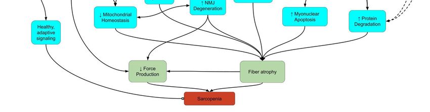
Int. J. Mol. Sci. 2021, 22, x FOR PEER REVIEW 2 of 23
Int. J. Mol. Sci. 2021, 22, 9045 2 of 22
factors in the development of sarcopenia, such as impaired protein turnover, mitochon-
drial dysfunction, improper 5′ adenosine monophosphate-activated protein kinase
(AMPK)
apoptosissignaling, apoptosisinflammation,
and regeneration, and regeneration, inflammation,
neuromuscular neuromuscular
dysfunction, dysfunc-
and nicotinamide
+
tion, and dinucleotide
adenine nicotinamide (NADadenine dinucleotide
) depletion. (NADwe
Finally, +) depletion.
focus on Finally, wetofocus
the effort onfor
search thea
potential
effort key link
to search for between impaired
a potential key linkredox signaling
between impairedandredox
functional deterioration
signaling in sar-
and functional
copenic muscle
deterioration in and highlight
sarcopenic the recent
muscle discoveries
and highlight about
the thediscoveries
recent role of peroxiredoxin (Prx),ofa
about the role
family of antioxidant
peroxiredoxin (Prx), a enzymes
family ofthat respond enzymes
antioxidant to peroxide
thatlevels and to
respond thereby mediate
peroxide levelssignal
and
transduction
thereby in mammalian
mediate cells [6].in mammalian cells [6].
signal transduction
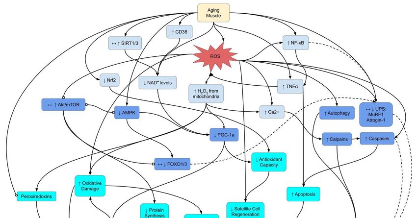
Selectredox
Figure 1.1.Select
Figure redox signaling
signaling pathways
pathways underlying
underlying sarcopenia.
sarcopenia. Lines
Lines between
between boxesboxes indicate
indicate signaling
signaling pathwaypathway
con-
connections.
nections. Arrowheads
Arrowheads represent
represent an increase
an increase or or promotion,
promotion, while
while block
block headsrepresent
heads representinhibition.
inhibition.Dashed
Dashedlines
linesindicate
indicate
pathways
pathways with a minor
minoror ornegligible
negligiblerole
roleinin sarcopenia.
sarcopenia. Arrows
Arrows within
within boxes
boxes showshow changes
changes in sarcopenia
in sarcopenia basedbased on up-
on upstream
stream signals. Abbreviations: AMPK:
0 5′ adenosine monophosphate-activated protein kinase;
2+ Ca 2+: calcium ions; CD38:
signals. Abbreviations: AMPK: 5 adenosine monophosphate-activated protein kinase; Ca : calcium ions; CD38: cluster
cluster of differentiation
of differentiation 38;: H
38; H2 O 2O2: hydrogen peroxide; mTOR: mechanistic target of rapamyacin; NAD + +: nicotinamide ad-
2 hydrogen peroxide; mTOR: mechanistic target of rapamyacin; NAD : nicotinamide adenine
enine dinucleotide; NF-κB: nuclear factor kappa-light-chain-enhancer of activated B cells; NMJ: neuromuscular junction;
dinucleotide; NF-κB: nuclear factor kappa-light-chain-enhancer of activated B cells; NMJ: neuromuscular junction; Nrf2:
Nrf2: nuclear factor erythroid 2-related factor 2; PGC-1α: peroxisome proliferator-activated receptor gamma coactivator
nuclear factor erythroid 2-related factor 2; PGC-1α: peroxisome proliferator-activated receptor gamma coactivator 1-alpha;
1-alpha; ROS: reactive oxygen species; SIRT: sirtuin; TNFα: tumor necrosis factor alpha; UPS: ubiquitin-proteosome sys-
ROS: reactive oxygen species; SIRT: sirtuin; TNFα: tumor necrosis factor alpha; UPS: ubiquitin-proteosome system.
tem.
2. Protein Turnover
2. Protein Turnover
The quantity of muscle mass is determined by the rates of protein synthesis and
The quantity of muscle mass is determined by the rates of protein synthesis and deg-
degradation. With age, protein synthesis decreases while degradation increases, tipping
radation. With age, protein synthesis decreases while degradation increases, tipping the
the balance towards atrophy [7].
balance towards atrophy [7].
2.1. Protein Degradation
2.1. Protein Degradation
Muscle protein degradation is governed by four major proteolytic pathways: the
Muscle protein degradation
ubiquitin–proteasome is the
system (UPS), governed
calpains,by
thefour majorand
caspases, proteolytic pathways: the
the autophagy-lysosomal
ubiquitin–proteasome system (UPS), the calpains, the caspases, and the autophagy-lyso-
pathway [7]. Two of the most important E3 ubiquitin ligases with respect to UPS are Mus-
somal pathway [7].
cle RING-finger Two of(MuRF-1)
protein-1 the most important
and atrophy E3gene-1/muscle
ubiquitin ligases with respect
atrophy to UPS
f-box (Atrogin-
1/MAFbx). These proteins are responsible for the degradation of several myofibrillar, sar-
comeric, and related regulatory proteins [8,9]. The UPS is commonly upregulated in manyInt. J. Mol. Sci. 2021, 22, 9045 3 of 22
other wasting conditions and is heavily influenced by the forkhead box class O (FOXO)
transcription factor family [10]. In sarcopenia, however, upregulation of the UPS and the
influence of FOXO likely play a minor role [7,11]. With some exceptions [12], the FOXO
pathway, including MuRF-1 and Atrogin-1, does not change or is even downregulated with
age [1]. Instead, the calpains and autophagy pathways are thought to exert a larger influence
during sarcopenia [7].
The calpain (calcium-dependent, non-lysosomal cysteine protease) system is a calcium-
dependent ATP-independent pathway that cleaves myofibril proteins [13]. Calpain content
and activity are known to increase with age [14], and H2 O2 has been shown to increase
calpain 1 and 2 expression and activity in C2C12 myotubes and in human myoblasts [15,16].
Thus, age-related increases in H2 O2 may upregulate calpain-mediated degradation. The
mechanism of age-related activation of the calpain system by ROS may be in part due to a
higher intracellular Ca2+ concentration. Previous research suggests that oxidative damage
induces a “leaky” ryanodine receptor [17], which impairs Ca2+ reuptake via sarcoplasmic
reticulum Ca2+ -ATPase (SERCA) pumps [18–20], thus leading to a higher intracellular Ca2+
concentration [21]. Additionally, ROS can increase the susceptibility of protein degradation
by calpains [22], because oxidative modification alters protein secondary and tertiary
structure [23]. In addition, oxidized proteins are more readily degraded by the UPS
because they are more easily ubiquitinated [21,24]. Furthermore, some proteosomes are
known to degrade oxidized proteins without ubiquitination [25]. This evidence suggests
that elevated ROS in sarcopenic muscles triggers excessive calpain proteolysis.
Caspases are cysteine-dependent, aspartate-targeting proteases that play an important
role mainly in programmed cell death and have been investigated within the context
of sarcopenia [13]. Caspases, particularly caspase-3, increases with H2 O2 in C2C12 my-
otubes [26]. Inhibition of caspase-3 has been shown to reduce cleavage of MyHC-2 and
limit a calpain-2–caspase-3 interaction that degrades both α-actinin and MyHC-2 [14].
Despite this, the proteolytic role of caspases in sarcopenia is likely minor. With age, nNOS
activity is reduced; this limits S-nitrosylation, which impairs calpain activity. However,
transgenic nNOS expression to limit the decline in S-nitrosylation with age did not change
caspase-3-dependent proteolysis [14]. It seems the role of caspases in sarcopenia is more
important with respect to apoptosis, as is discussed later.
2.2. Protein Synthesis
The most well-known pathway for muscle protein synthesis is the PI3K/Atk(PKB)/
mechanistic target of rapamycin (mTOR) pathway. Details of this pathway can be found in
many previous reviews [10,13,27]. This pathway is stimulated by insulin, growth factors,
mechanical loading, and feeding [27]. In addition, Akt inhibits FOXO activity, thereby
reducing activation of proteolysis and autophagy [13]. Research on the activation of this
pathway has revealed inconsistent results regarding the changes in mTOR pathway with
age and its impact on muscle protein levels [28]. Basal levels of protein synthesis are not
marred with age, but the response of protein synthesis machinery to anabolic stimuli is
blunted in a phenomenon known as anabolic resistance [29]. For example, aged subjects
show impaired Akt/mTOR/p70S6K signaling after muscle contraction [28], by nutritional
interventions [30], and after insulin receptor binding [31].
ROS and inflammation may contribute to anabolic resistance at several levels in
sarcopenia. Elevated levels of ROS such as H2 O2 can inhibit phosphorylation of Akt,
mTOR, and the downstream mTOR targets 4E-BP1 and p70S6K [13,21]. Excess ROS
produced in aged muscles may inhibit key components of the Akt/mTOR pathway, thereby
limiting their capacity to respond to exercise stimuli. Indeed, reducing oxidative stress in
aging can improve exercise adaptation. For example, antioxidant supplementation was
reported to restore age-related defective leucine stimulation on protein synthesis in rats [32].
Unfortunately, antioxidant supplementation alone may be insufficient to maintain muscle
mass, as this approach was not shown to be successful in two human studies [33,34].Int. J. Mol. Sci. 2021, 22, 9045 4 of 22
The above evidence demonstrates that oxidative stress from excessive ROS is capable
of blunting protein synthesis during sarcopenia. However, ROS levels alone may not be
sufficient to determine the effect on protein synthesis. Experiments in C2C12 cells by Tan
et al. [35] showed that the effect of ROS on Akt phosphorylation depended on the balance
between thiol oxidation of Akt, phosphatase and tensin homolog (PTEN), and protein
phosphatase 2A (PP2A), as both PTEN and PP2A can dephosphorylate and suppress Akt
signaling. The authors reported greater Akt phosphorylation when Akt, PTEN, and PP2A
were all oxidized, whereas Akt phosphorylation was diminished when Akt alone was
oxidized. It was concluded that Akt was the enzyme most sensitive to ROS-induced thiol
oxidation, and low but chronic oxidative stress may lead to impaired protein synthesis.
Ironically, although anabolic resistance presents an issue in sarcopenia, constitutive
activation of the Akt/mTOR pathway promotes sarcopenia [11,36,37]. Sandri et al. [11]
reported that Akt overexpression accelerated a sarcopenic phenotype in mice, demonstrat-
ing the importance of protein degradation to preserve muscle quality. Newer research
bolsters this idea by using rapamycin to delay sarcopenia in mice with constitutively active
mTORC1 [36]. The same study reported that atrophy induced by adding inflammatory
cytokines to C2C12 cells was blunted with rapamycin treatment. It seems the elevated
mTORC1 signaling in sarcopenia could be due to excessive oxidation of the phosphatases
that normally provide a check on this pathway [35].
Although muscular adaptation to anabolic stimuli decreases in the elderly as previ-
ously mentioned, exercise is proven to improve muscle mass and function [38]. Both animal
and human research indicates that aged skeletal muscle maintains sensitivity to exercise-
induced redox signaling. Endurance training promotes mitochondrial biogenesis mainly
through the upregulation of the PGC-1α signaling pathway [39]. Resistance training in the
elderly can improve muscle strength and mass by favorably altering autophagy, apoptosis,
and potentially muscle protein synthesis. These changes are likely mediated by changes
to the IGF-1/Akt/mTOR and Akt/FOXO pathways, but more research is necessary to
elucidate these mechanisms [40].
Another powerful intervention of sarcopenia is by nutritional supplementation, in-
cluding proteins, amino acids, Omega-3 fatty acids, and phytochemicals. Limited by the
scope and focus, we are unable to cover this topic in the current review. Interested readers
are referred to several excellent review articles below [41–43].
In summary, protein turnover during sarcopenia is characterized by higher proteolysis
via calpains and autophagy. These increases are driven in part by higher levels of oxida-
tive stress. In contrast to other degradation pathways, UPS activity is stagnant or even
decreased during aging, whereas proteolysis by caspases is insignificant. Compared to
younger individuals, elderly individuals cannot effectively synthesize protein in response
to anabolic stimuli. This may be caused in part by elevated ROS that promotes excessive
Akt/mTOR oxidation and inactivation.
3. Mitochondrial Homeostasis
3.1. Mitochondrial Oxidant Production
Mitochondria play an important role not only in cellular energy production but also
in regulating ROS generation and antioxidant defense against oxidative stress, a major
contributor to sarcopenia. Here, we focus on the importance of H2 O2 , a reactive species
linked to both oxidative stress and adaptive redox signaling [5]. Both in vitro and in vivo
studies indicate that mitochondria are the major source of basal H2 O2 production [44].
Mitochondrial production of H2 O2 increases with age in mice [45,46], rats [47,48], and
humans [49]. Increases in H2 O2 concentration induce atrophy in cell culture models [50],
and time-course analysis shows that this increase takes place prior to skeletal muscle atro-
phy [51]. Exogenous H2 O2 also decreases mitochondrial membrane potential that precedes
mitochondrial fragmentation, despite no changes in transcripts related to mitochondrial
fusion or fission protein expression [52]. Acute and chronic progression of H2 O2 concentra-
tion similar to the chronic, low-grade inflammation seen in aging induces muscle atrophyInt. J. Mol. Sci. 2021, 22, 9045 5 of 22
in C2C12 myocytes, probably through increased protein degradation [26]. According to
a variety of experimental models, increases in H2 O2 precede and in some cases induce
alterations in skeletal muscle volume with aging, suggesting that this plays a role in the
development of sarcopenia.
A common compensatory response to increased oxidative stress is the upregulation
of cellular antioxidant defenses [5]. Changes in muscle antioxidant enzyme activity with
aging have been observed for decades [53], with the majority of research showing increases
in activity of SOD1 [45], SOD2 [54], catalase [45,54,55], and GPx [55]. Moreover, mRNA
levels of SOD1 and SOD2 have been shown to increase in 24-month-old mice with muscle
atrophy, suggesting a transcriptional activation [45]. However, the same study showed
decreases in SOD2 and GPx protein contents in aging muscles [45]. It seems possible
that aging influences gene expression of antioxidant enzymes at both transcriptional and
post-transcriptional levels [56], which warrants further investigation.
Two questions may arise as to the role of mitochondrial source of ROS in developing
sarcopenia: (1) Are increased mtROS the cause of muscle atrophy, and (2) if so, can increased
antioxidant defense protect against muscle loss? A recent study by Eshima et al. [56] has
provided some insight. Using a mitochondrial catalase-overexpression mouse model,
the authors showed that eliminating mtROS did not prevent muscle atrophy induced by
hindlimb unloading. Additionally, acute antioxidant treatments did not rescue the tempo-
rary loss of force following fatiguing contractions in young or aged skeletal muscle [57,58].
On the contrary, chronic manipulation of antioxidant enzyme content or activity has yielded
mixed results. For example, Umanskaya et al. [59] showed that mitochondrial catalase over-
expression reduced the age-dependent loss of muscle mass and function through decreased
mitochondrial ROS and improved calcium handling. Because the substrate for catalase is
H2 O2 and aging can alter H2 O2 concentration and distribution in different compartments
of muscle cells [56], these results provide strong support for the notion that H2 O2 might
be a mechanistic driver of sarcopenia. Interestingly, in the senescence-accelerated mice
(SAM), catalase, SOD, and GPx activities severely decreased by the age of 24 weeks, along
with increases in oxidative stress, but a loss of grip strength did not occur until 32 weeks
of age [12]. These studies reveal that mitochondrial production of ROS and the resultant
redox status change are intimately involved in the development of sarcopenia.
3.2. Mitochondrial Biogenesis and PGC-1α Signaling
Mitochondrial homeostasis is controlled by mitochondrial biogenesis and degradation
partially governed by autophagy/mitophagy and fusion/fission dynamics [60–62]. Due to
the critical role it plays in mitochondrial biogenesis, PGC-1α has long been suspected as
an important factor in the development of sarcopenia [63,64]. Mitochondrial biogenesis
decreases in aging muscle cells, driven by an age-related decline in PGC-1α expression and
possibly by decreased SIRT1 and SIRT3 levels as well, causing acetylation and inactivation
of PGC-1α [65,66]. PGC-1α not only regulates mitochondrial biogenesis but also modulates
the crosstalk of signaling pathways of mitochondrial quality control in old age [67]. For
example, PGC-1α controls the expression of Mfn2, a main player in fusion dynamics and
mitophagy, and aging decreases Mfn2 levels [68]. PGC-1α also promotes the expression of
SIRT3, thus deacetylating the key mitochondrial metabolic and antioxidant enzymes [69,70].
Most studies demonstrate that aged muscle has significantly lower levels of PGC-1α
content, signaling activity, and associated functions [64,71]. However, whether aging per
se or other age-related deteriorative factors cause the downregulation of PGC-1α gene
expression is still difficult to conclude. Recently, the SAM model has shed some light
on muscular aging research including sarcopenia [12]. SAM mice demonstrated clear
downregulation of genes involved in mitochondrial biogenesis including PGC-1α, nuclear
respiratory factor-1 (NRF-1), Tfam, and cytochrome c oxidase, as well as mitochondrial
fusion proteins Mfn2 and Opa1. This suggests that declined PGC-1α signaling capacity
may be programmed in mammalian life. Supporting evidence also comes from PGC-Int. J. Mol. Sci. 2021, 22, 9045 6 of 22
1α knockout studies, wherein deletion of PGC-1α resulted in sarcopenia and shorter
lifespan [72].
On the other hand, PGC-1α overexpression results in amelioration of not only muscle
atrophy and deterioration caused by immobilization [72–74] but also attenuation of age-
related decline in mitochondrial protein and Tfam expression, mitochondrial metabolic
function, and muscle atrophy. Yet, whether PGC-1α overexpression can attenuate loss
of muscle mass in aging is still controversial. Although previous research consistently
shows positive effects of PGC-1α in boosting mitochondrial biogenesis and improving
age-associated muscle function, some authors showed that PGC-1α overexpression via
electroporation did not affect muscle fiber size or muscle/body weight ratio in old mice [75].
Additionally, PGC-1α had no effect on atrogen-1 or MuRF1 levels or protein ubiquitination,
suggesting that there is no direct connection between these two signaling pathways.
Other than PGC-1α, nuclear factor erythroid 2-related factor 2 (Nrf2) deficiency has
recently been implicated in frailty and sarcopenia by impairing mitochondrial biogenesis
and dynamics in aged muscle [76,77]. In Nrf2 KO mice, muscle oxidative function and
performance were reduced compared to those in the mid-age and old wild-type animals,
along with decreased PGC-1α, Tfam, Mfn1/2, and Opa1 levels. However, whether Nrf2
prevented muscle fiber atrophy was not conclusive. The authors attributed the protective
role of Nrf2 to ameliorating antioxidant gene expression and decreasing oxidative stress,
thereby improving mitochondrial biogenesis and homeostasis at old age.
3.3. Autophagy and Mitophagy
Autophagy is one of four major proteolytic pathways that take place in skeletal
muscle cells. There are three types of autophagy: macroautophagy, microautophagy, and
chaperone-mediated autophagy [78]. Targeted degradation of mitochondrion by autophagy
is termed mitophagy. Autophagy is a normal and necessary process to maintain healthy
cells. With insufficient autophagy, the cell accumulates dysfunctional and malformed
proteins, whereas excessive autophagy promotes muscle wasting [79]. Until the last few
years, autophagy was generally thought to decrease with age [80]. This was supported
by evidence that knockout of atrogenes and other autophagy proteins could induce early
sarcopenia, while overexpressing atrogenes prevented losses in muscle function and ex-
tended lifespan [81]. However, more recent experiments have challenged this proposition
by measuring autophagic flux in aged muscle [82–84]. Measuring only protein content
depicts a static snapshot of autophagy, while flux measurements clarify discrepancies in
results and interpretation. The findings of the above three studies revealed that basal
autophagy and mitophagy flux both increase with age, but that aging blunted increases
in mitophagy flux in response to exercise [84]. Other reviewers have noted that elevated
basal mitophagic flux may be insufficient to maintain a pool of healthy mitochondria,
given that dysfunctional mitochondria accumulate with age [85]. It was further noted that
the inability to upregulate mitophagic response to exercise stimulus may exacerbate the
accumulation of dysfunctional organelles [86].
Although autophagy is regulated by multiple pathways, several lines of evidence
indicate that ROS stimulates autophagy in skeletal muscle [87]. For example, oxidative
stress caused by SOD1 mutation [88] or by the addition of H2 O2 to C2C12 cells induces
autophagy [89–91]. Removing oxidative stress with antioxidant treatments also attenuates
autophagy [89,92]. However, a study in mdx mice showed that excessive NOX-2-dependent
ROS production suppressed autophagy [93].
The mechanism by which increases in autophagy following elevated ROS appears to
involve mTORC1 and AMPK. An active mTORC1 generally suppresses autophagy by in-
hibiting ULK1 activity, whereas AMPK accelerates autophagy by phosphorylating ULK1
and by inhibiting mTORC1 [87]. It has been shown that oxidative stress induced by muscle
immobilization activates proteolytic pathways and inhibits mTOR [94]. Studies in C2C12
cells also demonstrated ROS-mediated autophagy due to AMPK activation [89]. Further-
more, recent studies showed that Nrf2 deficiency, which increases oxidative stress, increasedInt. J. Mol. Sci. 2021, 22, 9045 7 of 22
autophagic flux and AMPK activation in a manner similar to aging [82]. Detailed research
has revealed that the concentration of ROS may dictate the outcome of autophagic response
in muscles. For example, Meijles et al. [95] found that low levels of ROS promoted mTOR
phosphorylation in cardiomyocytes, while high ROS levels could deplete ATP and activate
AMPK. This study did not assess autophagy, but the concentration-dependent, divergent
responses to ROS highlighted the complication of this topic. The characteristic increase in
ROS at old age may in part explain the increased autophagic flux in aged muscle.
ROS also stimulates mitophagy by interacting with mitochondrial inner-membrane
anion channels and mitochondrial permeability transition pores. An increase in ROS release
by the mitochondria decreases mitochondrial inner membrane potential, thus labeling
the dysfunctional mitochondria for degradation by mitophagy [96]. In support of this
scenario, reducing oxidative stress from H2 O2 with adiponectin treatment has been shown
to reduce mitophagy in C2C12 cells [97]. A recent study demonstrated that suppressing
oxidative stress with phytochemical antioxidant apigenin alleviated muscle atrophy due
to inhibition of hyperactive mitophagy and apoptosis in aged mice [98]. Furthermore,
PGC-1α overexpression by local transfection was shown to effectively suppress the age-
related increase in mitophagy protein expression in mice, suggesting crosstalk between
mitochondrial biogenic and mitophagic signaling pathways [75].
Molecular mechanisms explaining the redox control of autophagy/mitophagy are hot
topics in muscle biology. One potential target of altered redox signaling in aging is PTEN, a
phosphatase that normally dephosphorylates mTORC1 and suppresses Akt/PKB signaling
pathway [99]. Posttranslational modifications of PTEN includes oxidation of its Cys 124 to
form a disulfide bond, thus inhibiting its activity, leading to enhanced PI3K/Akt/mTOR
signaling [99]. Moreover, Kim et al. reported that during muscle differentiation, mitochon-
drial ROS can oxidize PTEN, thereby promoting phosphorylation of Ser 317 on ULK1, a
change that induces autophagy [100].
In sum, autophagy and mitophagy are redox-sensitive pathways whose basal levels in
skeletal muscle rise with age due to increased mitochondrial oxidative damage. However,
aging may impair the ability of skeletal muscle to activate mitophagy in response to
elevated oxidative stress, such as during muscle contraction.
4. AMPK Signaling
AMPK is a pivotal enzyme in skeletal muscle that responds to cellular energy status
and regulates a variety of cellular functions, and its activity is known to change with
aging [101]. While AMPK itself does not control muscle mass, it coordinates downstream
effectors, such as SIRTs and FOXO, which are key regulators of muscle protein turnover.
AMPK is a heterotrimeric kinase with a catalytic α subunit, and β and γ regulatory subunits,
which can combine to make up to 12 unique AMPK isoforms [102]. AMPK is a redox-
sensitive kinase, though some controversy exists as to whether it can be directly activated
by ROS or if the activation is a result of an indirect, ROS-induced decrease in cellular
ATP levels. There is strong support for activation of AMPK independent of changes in
ATP concentration across a variety of stimuli, such as addition of exogenous H2 O2 [103],
endogenous increases in H2 O2 [104], and hypoxia [105]. Modification of cysteine residues
on AMPKα appears to play an important part in these manipulations [104,106]. While
exposure of AMPK to H2 O2 resulted in increased kinase activity, mutation of Cys 299 to
alanine diminished the ability of H2 O2 to activate the enzyme, and mutation of Cys 304 to
alanine totally abolished the stimulating effect of H2 O2 [107]. On the other hand, oxidation
of Cys 130 and Cys 174 on AMPKα inhibits its activity and interferes with its activation by
AMPK kinases (AMPKK) [106]. Reduction of Cys 130/Cys 174 was shown to be essential
for activation of AMPK during energy starvation [106]. Rabinovitch et al. [108] showed that
mitochondrial ROS activated AMPK, which triggered a PGC-1α-dependent antioxidant
response that decreased mitochondrial ROS. Cells lacking AMPK activity displayed higher
mitochondrial ROS and premature senescence. These findings clearly demonstrate that inInt. J. Mol. Sci. 2021, 22, 9045 8 of 22
addition to being sensitive to energy status, AMPK is redox-sensitive and mitochondrial
ROS regulates AMPK activity via its α-subunit cysteine redox regulation.
Based on the fact that AMPK is redox-sensitive, one would expect AMPK activity to
increase in the presence of increased ROS with aging. This would be important for further
research on sarcopenia, as increased basal activation of AMPK has been shown to diminish
muscle overload-induced hypertrophy in both young and old muscle fibers [109]. However,
AMPK activity has been shown to decrease with age in rat and human skeletal muscle,
despite maintenance of its protein content [110,111]. Interestingly enough, combined
treadmill running and resistance training or aerobic training alone in rodents were able to
attenuate sarcopenia in part by upregulating AMPK and PGC-1α [112,113].
Understanding the mechanisms of the interaction between AMPK and aging is crucial
for elucidating the prevention and treatment of sarcopenia. Several potential pathways
have recently been explored. During the past two decades, the double-negative feedback
loop between mTORC1 and AMPK was postulated to explain the enhanced autophagy
under oxidative stress [114,115]. This theory claims that under physiological conditions,
mTORC1 inhibits AMPK and thus keeps ULK1, the initiator of autophagy, in check. An
increase in AMPK activity under oxidative stress and nutritional deficiency not only down-
regulates mTORC1, the master regulator of protein synthesis, but also removes the negative
feedback inhibition on AMPK. Hyperactivation of AMPK can phosphorylate ULK1, leading
to increased autophagic flux. In our analysis, this double-negative theory may account in
part for the age-associated decline in protein synthesis as well as increased proteolysis via
autophagy. Another potential explanation is attributed to redox resistance within AMPK,
supported by a recent study by Caldeira et al. [116]. In this study, AMPK from peripheral
blood mononuclear cells of middle-aged individuals was able to be activated in response to
an oxidative challenge and reduce oxidative stress. In contrast, the same stimulus did not
induce an antioxidant response in cells from elderly individuals. Recently, Nrf2-mediated
downregulation of AMPK was studied in an attempt to elucidate the crosstalk between
these two signaling pathways in autophagy under oxidative stress [117]. Nrf2 has a funda-
mental role in cell homeostasis by regulating antioxidant and electrophilic stress responses.
While prolonged oxidative stress, as is seen in sarcopenia, increases AMPK activity and
content, it also activates Nrf2 and subsequently modulates AMPK activity and autophagy.
It was suspected that this reduction in AMPK prevents hyper-autophagic responses. While
this work was performed in HEK293T cells and C. elegans, it may shed some light on the
role of AMPK and autophagy in aging and sarcopenia.
The decrease in AMPK detailed above is distinct from the increase in AMPK activity
seen in other muscle-wasting conditions such as immobilization. In those conditions,
myocytes enter a low energy state, upregulating AMPK activity [62]. This inhibits the
PI3K/Akt/mTOR pathway and upregulates FOXO-mediated transcription. The net result
is accelerated autophagy and increased degradation, primarily by MuRF1, atrogin-1, and
several atrogenes [62]. However, as stated above, AMPK and FOXO activities do not
increase with age. Instead, sarcopenia is attenuated through increases in AMPK activity.
As stated in the Mitochondrial Biogenesis section, this is likely mediated by AMPK phos-
phorylation of PGC-1α. We and others have demonstrated exercise to be one of the most
successful means to increase AMPK activity and mitigate sarcopenia [65,112,118]. The
divergent responses of AMPK to these conditions highlight the multi-functional nature of
this kinase. Excessive AMPK activity may be relevant to sarcopenic individuals requiring
immobilization or bedrest, and how these stressors together affect AMPK, FOXO, and
atrophy requires further research.
5. Apoptosis and Regeneration
One of the molecular mechanisms of sarcopenia is postulated to be an imbalance when
apoptosis outpaces regeneration [16]. Indeed, the imbalance comes from both directions:
myonuclear apoptosis increases [119] while satellite cell function decreases [29]. There
are two main apoptotic pathways. The first is the intrinsic, mitochondria-dependentInt. J. Mol. Sci. 2021, 22, 9045 9 of 22
pathway in which mitochondria release cytochrome c and promote cell death [120]. This
pathway is accelerated by elevated concentrations of intracellular calcium ions [121] and
ROS [122]. The extrinsic pathway relies on death receptor binding, such as TNFα, and
directly stimulates apoptosis or induces the intrinsic pathway. TNFα binds to membrane-
bound receptors such as tumor necrosis factor receptor 1 (TNFR1), which recruits adapter
proteins such as tumor necrosis factor receptor type 1-associated DEATH domain (TRADD)
that eventually activate caspase-3. For details on the role of TNFα and apoptosis, readers
are referred to other reviews [119,123,124]. Currently, it is unclear whether the intrinsic or
the extrinsic pathway is the major player in sarcopenic apoptosis [29].
5.1. Apoptosis
In the intrinsic pathway of apoptosis, elevated ROS leads to higher intracellular Ca2+
concentrations mediated in part by redox modifications to the ryanodine receptor that pro-
mote Ca2+ influx into the cell [17]. This phenomenon is also observed in hypobaric hypoxia,
another condition that elevates ROS and promotes muscle atrophy. Briefly, oxidative stress
hypernitrosylates the ryanodine receptor and impairs binding with FKBP12/calstabin-1
and other channel complexes [17]. Furthermore, elevated ROS due to SOD2 knockout [19],
chronic stimulation [20], and aging [18] can inhibit the activity of SERCA pumps and
lead to higher intracellular Ca2+ levels, which increases the permeability of mitochondria,
release of cytochrome c, and activation of caspase-3 [119,121]. Furthermore, calpain ac-
tivity stimulated by higher intracellular Ca2+ induces apoptosis by activating caspase-12
and prompting pro-apoptotic factor endonuclease G (EndoG) release from the mitochon-
dria [13,125].
As mentioned above, TNFα is a prominent pro-apoptotic ligand that can initiate the
extrinsic apoptotic pathway by binding with its receptors. Moreover, TNFα binding stim-
ulates ROS production, inducing mitochondria-dependent apoptosis [126]. Importantly,
TNFα levels increase with age in both rats and humans, and TNFα levels are inversely
proportional to muscle force production [127]. These findings strongly suggest that TNFα
is one of the mechanistic drivers of sarcopenia, and it has been postulated as a key player
in the inflammatory theory of aging [1,38].
5.2. Satellite Cells and Regeneration
The ability of satellite cells (SC) to participate in muscle regeneration declines with
age, and this process is partially mediated by elevated oxidative stress. Specifically, SC
differentiation and proliferation decrease with muscle aging [29], even in response to
anabolic stimuli [128]. Additionally, in aged muscles, more SCs enter the permanent
senescence stage from reversible quiescence [129]. Furthermore, antioxidant capacity in SC
diminishes with age [130], whereas ROS production increases with age [131]. In light of
the role of redox balance within the SC, antioxidant supplementation was found to renew
the regeneration capability and improve antioxidant defense in human senescent SCs [132].
In addition to myocytes, there are ROS-dependent alterations in autophagy in SCs [132].
Basal autophagy flux within SCs declines with age, and this contributes to their senescence
and dysfunction. While geriatric SCs produced more ROS, inhibiting ROS restores the
proliferation and regeneration capacity of SCs in geriatric cells [132]. Finally, a reduced
as opposed to a more oxidized cellular environment promotes myoblast differentiation
in C2C12 cells [133]. These findings demonstrate the importance of redox environment
within SCs to preserve regeneration and delay senescence.
There is clear evidence that elevated ROS from aging negatively affects the regener-
ation ability of SC. However, precise mechanisms underlying the observations are still
unknown. Changes in several redox-sensitive signaling pathways in myogenesis such as
Notch, Wnt, p38/MAPK, and JAK/STAT3 have been observed in satellite cells [134]. It
remains unclear why the responses of these redox-sensitive pathways change with age and
why they are no longer adaptive as the organism ages.Int. J. Mol. Sci. 2021, 22, 9045 10 of 22
6. Inflammation
The impact of inflammation on almost all aging-related degenerative diseases including
sarcopenia has been increasingly recognized by the scientific community in the last decade,
and this association clearly hinges on cell signaling [135]. Several cytokines play an impor-
tant role in the pathogenesis of muscle wasting, most notably TNF-α, but also IL-1β, IL-6,
interferon (IFN)-γ, and transforming growth factor (TFG)-β [136]. Inflammation represents
a pathophysiological state that is intimately related to cellular redox stress and in turn sub-
stantially alters cellular redox homeostasis. Inflammation has long been considered a major
threat during muscle disuse for various reasons [137,138]. The main trigger of inflammation
in immobilized muscle is activation of the NF-κB pathway. Transgenic mice overexpressing
IKKβ, the primary activator of NF-κB, suffer from muscle wasting via the upregulation of
MuRF1 [136]. In contrast, deletion of p105/p50 subunits of NF-κB was shown to be resistant
to IM-induced muscle atrophy [139]. NF-κB activation is mostly associated with an overpro-
duction of pro-inflammatory cytokines, as mentioned above. TNFα, the most potent activator
of NF-κB pathway, promotes a positive feedback loop by activating NF-κB, which in turn
induces TNFα and drives NF-κB mediated muscle wasting. TNFα, IL-1, and IL-6 directly in-
hibit PI3K/Akt activity and thus release their inhibition on FOXO [140]. NF-κB also promotes
the expression of MuRF1, which degrades muscle contractile proteins [136]. Furthermore,
inflammation is associated with an overexpression of myostatin due to FOXO activation and
can induce muscle atrophy independently of NF-κB [141]. Thus, muscle inflammation can be
a sustained disturbance to muscle redox homeostasis due to the crosstalk of multiple redox
signaling pathways that control protein turnover.
Recent research indicates that PGC-1α has an anti-inflammatory effect in skeletal
muscle atrophy. PGC-1α modulates local or systemic inflammation by suppressing the
expression of pro-inflammatory cytokines and myokines such as TNF-α and IL-6 [142]. The
inhibitory effects of PGC-1α on inflammation may result from several cellular mechanisms.
First, intact PGC-1α signaling is mandatory for antioxidant enzyme gene expression [143].
Second, PGC-1α inhibits NF-κB by interaction with the p65 subunit in the nucleus, thus
attenuating its binding to DNA [144]. Third, PGC-1α suppresses nuclear retention of
FOXO3 caused by AMPK activation [145]. Finally, SIRT expression is at least partially
controlled by PGC-1α, and SIRT1 negatively regulates FOXO and p65 via their deacetyla-
tion [10]. However, although overexpression of PGC-1α via in vivo transfection was found
to ameliorate mitochondrial function and reduce upregulated mitophagy and ubiquitin
proteolysis in aged muscle, it failed to prevent age-associated muscle atrophy [75].
Convincing evidence exists linking inflammation with muscle metabolic dysfunctions
marked by disturbed mitochondrial homeostasis, insulin resistance, and protein imbalance.
Especially, mitochondrial dysfunction is recognized as a major factor of age-related muscle
degeneration [146]. However, the molecular mechanism connecting sarcopenia to inflam-
mation is poorly understood. Studies based on large databases have generated a more
complicated picture and controversial conclusions. For example, one recent study using
meta-analysis reported that, independently of disease state, higher levels of C-reactive
protein (CRP), IL-6, and TNFα were associated with lower arm and leg strength and muscle
mass [147]. Yet another meta-analysis study, while finding that sarcopenic subjects had
significantly higher levels of CRP than control subjects, showed that serum IL-6 and TNFα
levels were not significantly different when people with sarcopenia were compared with
controls [148].
However, NF-κB alone does not appear to be a driver of sarcopenia. p65 protein ex-
pression and NF-κB binding did not increase in aged rats despite elevations in TNFα [149],
suggesting a disconnect between NF-κB and its typical stimuli with age. Rather, the neg-
ative impact of NF-κB on sarcopenia is due to impaired adaptation to anabolic stimuli.
Specifically, elderly individuals showed increased nuclear Ser468 phosphorylation of p65, a
DNA binding region of NF-κB, in response to a bout of resistance exercise. In comparison,
young participants saw no change in p65 phosphorylation, suggesting an impaired NF-κB
response to exercise with age [150]. This study also demonstrated reduced phosphorylationInt. J. Mol. Sci. 2021, 22, 9045 11 of 22
of S6K1 and FOXO with exercise, again highlighting the role of anabolic resistance in the
elderly. In aged rats, impairing inflammatory cytokines with ibuprofen improves acute Akt
phosphorylation of FOXO3 after feeding [151]. Although NF-κB proteins were not mea-
sured, minimizing low-grade inflammation may mitigate anabolic resistance and therefore
improve outcomes in sarcopenia. Despite theoretical similarities to immobilization, the
role of inflammation in sarcopenia may be related more to anabolic resistance than to direct
protein degradation.
7. Neuromuscular Junction
Degradation of the nervous system with age is well established [1]. Defects of neu-
romuscular function not only have negative impacts on muscle contractile function but
also result in oxidative stress, as denervation increases mitochondrial peroxide production
in neighboring, innervated muscle fibers [152]. Much of the mechanistic understanding
of the changes in neuromuscular function with age centers on the use of SOD1 KO mice.
Complete SOD1 KO induces a sarcopenic phenotype, characterized by a loss of strength,
muscle mass, and reduced physical activity [153]. Importantly, muscle-specific SOD1 KO
can lead to clear reductions in contractile force but little to no loss in muscle mass [154,155].
Normal expression of SOD1 in nervous tissue is critical, as neuron-specific SOD1 KO
resulted in contractile force loss in muscle [156], while restoration of strength and mass
was observed when this line of SOD1 was restored [157]. The disconnect between muscle
mass and function is due to the decrease in excitation–contraction coupling, specifically
mediated by impaired SERCA pump function and damage to the ryanodine receptor [158].
Recently, a SERCA activator was shown to reverse the alterations in muscle mass, function,
oxidative damage, and autophagy markers back to wild-type levels in SOD1 KO mice [159].
The above-mentioned tissue-specific SOD1 KO model reveals that the signaling mech-
anism of the neuromuscular junction (NMJ) is probably distinct from the skeletal muscle
fiber atrophy. Neuron-specific SOD1 KO induced progressive NMJ degeneration and
gradual loss of strength and muscle mass, which was most pronounced at 24 months of age.
This occurred despite no alterations in mitochondrial oxidative damage or function [160].
However, muscle-specific SOD1 KO does not lead to degradation at the NMJ or alterations
in redox homeostasis, which are both found in a whole-body SOD1 KO model [154]. Thus,
the results of SOD1 KO mice can be synthesized into a two-hit model, where both loss of
redox homeostasis in motor neurons and increased skeletal muscle ROS are necessary to
induce the sarcopenic phenotype seen in whole-body SOD1 KO [161]. These findings indi-
cate that dysregulation in redox homeostasis within motor neurons is a major contributor
to sarcopenia [154–157].
Recent evidence suggests that signaling can also occur in a retrograde fashion, i.e.,
from the muscle cells back to the innervating nerves. This scenario is best supported by
a series of SOD1 mutant studies, initially designed for the study of amyotrophic lateral
sclerosis (ALS) [162]. Mice with a muscle-specific SOD1 mutation (SOD1G93A ) develop fiber
atrophy, loss of strength, and mitochondrial dysfunction [88], making it a possible model
for sarcopenia. Expression of SOD1G93A specifically in skeletal muscle lead to disruptions
in NMJ structure, improper mitochondrial localization, and impaired transmembrane
potential in pre- and post-synaptic mitochondria [163]. It has been shown that transgenic
expression of PGC-1α caused morphological and functional remodeling, including im-
proved NMJ, providing further evidence for retrograde signaling from skeletal muscle back
to the NMJ [164].
Several studies have attempted to further understand the mechanisms behind muscle-
driven or nerve-driven disorders of signaling in sarcopenia. Comparison of full SOD1
KO vs. muscle-specific SOD1 KO showed divergent oxidation statuses of the catalytic
site of Prx5 [165]. Despite these changes in Prx, the peripheral nerves of the whole-body
SOD1 KO mice did not show any evidence of oxidative damage. This is consistent with
previous research showing that the catalytic cysteine residue of Prx6 in peripheral nerves
was oxidized in sarcopenic mice, alongside increases in superoxide and peroxynitriteInt. J. Mol. Sci. 2021, 22, 9045 12 of 22
production [165]. Oxidation of Prx leads to their inactivation [166], impairing their ability
to form so-called redox relays with downstream effectors [167]. This implies that Prxs of
the peripheral nerves in sarcopenic mice are unable to effectively transmit the increase in
H2 O2 into adaptive signaling, leading to increased oxidative stress and cellular dysfunction.
Overall, these data suggest that manipulation of signaling due to changes in the redox
status of cysteine residues, particularly those on enzymes responsible for redox signal
transduction, instead of the oxidative damage itself, could be a potential mechanistic driver
of sarcopenia.
Nevertheless, oxidative damage should not be totally discounted in the development
of sarcopenia, particularly within the mitochondria. SOD1 mutation in mice was shown
to induce loss of mitochondrial inner membrane (MIM) potential near the NMJ, which
interfered with propagating Ca2+ waves and signaling in skeletal muscle [168]. This defect
of neuromuscular coupling was proposed to underlie the pathogenesis of ALS-induced
muscle atrophy. On the other hand, transgenic expression of SOD1 in the mitochondrial
intermembrane space was able to reduce mitochondrial oxidative damage, allow motor
neurons to propagate, prevent NMJ damage, and eliminate the loss of muscle mass and
function in SOD1 KO mice [169]. Furthermore, in SOD1 mutant mice developing sar-
copenia, treatment with Trolox was able to rescue mitochondrial function and subcellular
organization, restore NMJ morphology, and return acetylcholine receptor turnover to
age-matched WT levels [163]. The same study also examined the role of PKCθ , which is
a redox-sensitive kinase expressed in the post-synaptic region of the NMJ. PKCθ serves
to eliminate acetylcholine receptors in skeletal muscle and mediate nerve-muscle inter-
actions [170,171]. Treatment of SOD1 mutant mice with a PKCθ inhibitor rescued loss
of mass and force [163]. This implies that PKCθ -mediated oxidative stress may activate
downstream pathways related to NMJ degradation. Because treatment with Trolox pro-
duced similar results to the PKCθ inhibition, available evidence suggests a role for redox
signaling in the degradation of the NMJ in sarcopenia. Taken together, research up to this
date highlights the importance of maintaining redox signaling in muscle neurons and the
NMJ in the prevention of sarcopenia.
8. NAD+ Homeostasis
Increased protein acetylation plays an important role in the mitochondrial and func-
tional decline in aged muscle and can be a potential mechanism for sarcopenia [172]. Over
100 lysine sites in mitochondrial proteins may be acetylated, which may alter mitochon-
drial function in an adverse manner. This can include the inactivation of PGC-1α and key
enzymes in the TCA cycle and ETC, inactivation of SOD2 and other antioxidant enzymes,
and activation of NF-κB and inflammatory pathways [173–175]. Thus, maintenance of
cellular protein acetylation/deacetylation balance is critical for maintaining functional
integrity in aging muscle.
Protein deacetylation is carried out mainly by the NAD+ -dependent deacetylase
SIRTs [176]. Previous studies have shown that aging downregulates SIRT1 activity, the
multi-functional deacetylase in the cell; however, recent research indicates that SIRT1
protein levels are not altered with age and some studies even reported positive corre-
lations between SIRT content and age in muscle despite elevated acetylation of protein
content [177]. Thus, cellular SIRT levels per se do not appear to determine protein acety-
lation/deacetylation status in skeletal muscle. Instead, it is suggested that the cellular
content of NAD+ , the substrate and mandatory acceptor of lysine residues from acety-
lated protein in SIRT-catalyzed reactions, is significantly downregulated during aging
in general [172,176,177] and in skeletal muscle in particular [178]. Thus, the insufficient
supply of NAD+ may be a limiting factor in intracellular deacetylation capacity in aged
muscle, because deteriorated mitochondrial function and inflammation are both important
etiological mechanisms for sarcopenia [179].
Cellular mechanisms by which aging diminishes the cellular NAD+ pool are com-
plicated and under intense research. While the metabolic pathways that use NAD+ andInt. J. Mol. Sci. 2021, 22, 9045 13 of 22
NADH as coenzymes to transfer electrons do not result in net NAD+ decline in the cell,
several signaling pathways are known to consume NAD+ , such as SIRTs, poly(ADP-ribose)
polymerase (PARP)-1, and a cluster of differentiation (CD) 38 [176,177]. During aging,
each of these pathways is activated and competes for NAD+ . For example, aging induces
mitochondrial protein hyperacetylation in muscle, requiring increasing amounts of NAD+
in the TCA cycle, ETC, and SOD2 [172]. Activation of inflammatory pathways during
aging demands NAD+ for deacetylating NF-κB-p65 and FOXO in the nucleus [176]. Fur-
thermore, aging is known to increase DNA oxidative damage [180]. Repairing oxidized
DNA base pairs uses NAD+ as a substrate to transfer poly ADP-ribose backbone, cat-
alyzed by PARP-1. Research shows that up to 80% of NAD+ decline during aging may
be explained by increased DNA damage [176]. Indeed, PARP-1 was upregulated in aged
mouse muscle [178]. Interestingly, PARP-1 inhibition was shown to improve mitochondrial
function due to SIRT1 activation, presumably due to increased NAD+ availability [181].
Finally, aging increases protein expression of CD38, also known as cyclic ADP ribose
hydrolase, a multi-functional enzyme that produces ADP ribose and cyclic ADP-ribose
(minor function) from NAD+ [182]. Inhibition of CD38 or CD38 gene knockout has been
found to preserve intracellular NAD+ levels and SIRTs activity [183,184]. CD38 protein
content was reported to increase dramatically in aged mouse skeletal muscle, along with
decreased mitochondrial oxidative enzyme expression, increased protein and transcription
factor acetylation, and greater oxidative damage [178].
Under physiological conditions, decreased cellular NAD+ concentration may be re-
plenished by elevated NAD+ synthesis by either the Preiss-Handler pathway that converts
dietary nicotinic acid (NA) to NAD+ or the de novo NAD+ biosynthesis from dietary
tryptophan [176]. Whether the enzyme activities in the Preiss–Handler pathway and de
novo biosynthesis pathway are downregulated or upregulated in aging muscle is un-
clear. More importantly, NAD+ levels may be restored by the so-called salvage pathway
controlled by nicotinamide phosphoribosyltransferase (NAMPT), the rate-limiting enzyme-
converting nicotinamide (NAM) generated by SIRTs, CD38, and PARP-1 to nicotinamide
mononucleotide (NMN) and eventually NAD+ . This pathway recycles NAM to main-
tain intracellular NAD+ levels and relieves NAM inhibition on SIRTs [176]. Aging may
decrease NAMPT activity, thus reducing the conversion of NAM to NAD+ , resulting in
muscle degeneration [185,186]. However, this area is still controversial as Yeo et al. [178]
showed an upregulation of NAMPT in quadriceps and gastrocnemius muscles in aged
mice, yet with decreased NAD+ levels. Dietary intake of NAD+ precursors could have a
strong influence on NAD+ homeostasis in aged animals, as some recent research indicates
that supplementation of NAM, or nicotinamide ribose (NR) can boost intracellular and
mitochondrial NAD+ levels, improve metabolic function, and even increase longevity in
aged mice [177].
In summary, aged muscles suffer from NAD+ deficit due to increased competition
from SIRTs, CD38, and PARP-1 pathways, whereas neither de novo synthesis nor NAD+
salvage is sufficient to replenish the intracellular NAD+ pool. The shortage of NAD+ may
retard the ability of SIRT to deacetylate key enzymes and transcription factors. Whether
or not NAD+ deficiency constitutes a major mechanism for sarcopenia requires more
investigation.
9. Peroxiredoxins and Redox Signaling
Since the free radical theory of aging was postulated more than half a century ago,
research on the mechanisms of aging has been one of the most vibrant branches of modern
biology. Sarcopenia is proven to underlie numerous health hazards beyond muscle itself.
The connection of increased ROS with age and sarcopenia does not seem to be coincidental.
Most age-related alterations reviewed in this article are linked with an imbalance between
ROS production and antioxidant defense, favoring an oxidized cellular milieu. While some
of the enzymes and transcription factors are directly modified by H2 O2 and other reactive
species, others show sensitivity to intracellular redox status that either change their geneYou can also read