The Consequences of Our Changing Environment on Life Threatening and Debilitating Fungal Diseases in Humans - MDPI
←
→
Page content transcription
If your browser does not render page correctly, please read the page content below
Journal of
              Fungi
Review
The Consequences of Our Changing Environment on Life
Threatening and Debilitating Fungal Diseases in Humans
Norman van Rhijn                  and Michael Bromley *

                                          Manchester Fungal Infection Group, University of Manchester, Manchester M13 9PL, UK;
                                          norman.vanrhijn@manchester.ac.uk
                                          * Correspondence: mike.bromley@manchester.ac.uk

                                          Abstract: Human activities have significantly impacted the environment and are changing our
                                          climate in ways that will have major consequences for ourselves, and endanger animal, plant and
                                          microbial life on Earth. Rising global temperatures and pollution have been highlighted as potential
                                          drivers for increases in infectious diseases. Although infrequently highlighted, fungi are amongst the
                                          leading causes of infectious disease mortality, resulting in more than 1.5 million deaths every year. In
                                          this review we evaluate the evidence linking anthropomorphic impacts with changing epidemiology
                                          of fungal disease. We highlight how the geographic footprint of endemic mycosis has expanded, how
                                          populations susceptible to fungal infection and fungal allergy may increase and how climate change
                                          may select for pathogenic traits and indirectly contribute to the emergence of drug resistance.

                                          Keywords: fungal disease; climate change; global warming; fungal pathogens; Aspergillus; Candida;
                                          Cryptococcus; endemic mycoses


Citation: van Rhijn, N.; Bromley, M.      1. Orientation
The Consequences of Our Changing               There is no doubt that our climate is changing. Mean global temperatures have risen
Environment on Life Threatening and       by 0.8 degrees Celsius since 1975 and are expected to rise by a further 2 to 5 degrees by
Debilitating Fungal Diseases in           2070 [1–3] with local fluctuations predicted to be even more dramatic [4,5]. Average sea
Humans. J. Fungi 2021, 7, 367.
                                          temperatures are expected to rise in line with global temperature shifts leading to a rise
https://doi.org/10.3390/jof7050367
                                          in water levels due to thermal expansion and melting of the ice caps [6]. Overall rainfall
                                          levels will decrease however, as the atmosphere can hold more water, rainfall will be more
Academic Editor: Johanna Rhodes
                                          extreme, leading to more flooding [7]. Generally, land areas will become more arid and
                                          saline, winds will become more extreme and there are expected to be changes in the levels of
Received: 13 April 2021
Accepted: 4 May 2021
                                          ultraviolet radiation reaching the Earth’s surface [8–10]. These changes to our environment
Published: 7 May 2021
                                          alongside geopolitical and geoeconomic pressures will require significant adaptation of
                                          human behaviour. Although the effects of anthropomorphic activities, especially climate
Publisher’s Note: MDPI stays neutral
                                          change, have been extensively discussed in the context of displacement and loss of plant
with regard to jurisdictional claims in
                                          and animal species, the impact on microorganisms is rarely discussed [11]. There is a
published maps and institutional affil-   theoretical concern that climate change will affect the frequency, type, morbidity and
iations.                                  mortality rates of infectious diseases and these theories are beginning to be backed up by
                                          a growing body of evidence. In this review we will explore the potential impact climate
                                          change will have on the fungal infections of man, either in their role as primary pathogens
                                          of disease or adjuncts to other infections.
Copyright: © 2021 by the authors.
                                          2. The Ability to Infect Requires Pathogens to Adapt to a Hostile Host Environment
Licensee MDPI, Basel, Switzerland.
This article is an open access article
                                               The fungal kingdom emerged 1.5 billion years ago and can be found all across the
distributed under the terms and           globe [12–14]. Around 120,000 different fungal species have been catalogued; however,
conditions of the Creative Commons        there are estimated to be a further 2.1 to 3.7 million different species yet to be identified [15].
Attribution (CC BY) license (https://     Many of these are thought to be located in little explored habitats, particularly in tropical
creativecommons.org/licenses/by/          regions and biodiversity hotspots such as The Guiana Shield and neighbouring Amazon
4.0/).                                    basin regions [16–18]. Only a few hundred of the catalogued species are known to infect

J. Fungi 2021, 7, 367. https://doi.org/10.3390/jof7050367                                                      https://www.mdpi.com/journal/jof
J. Fungi 2021, 7, 367                                                                                           2 of 18

                        humans [19] and fewer still are considered primary pathogens. The ability of most fungi
                        to infect humans is thought to be as a consequence of the way they have adapted to their
                        ecological niche and hence they are often considered to be accidental pathogens [20]. The
                        key factor that provides a barrier to infection of warm-blooded mammals is their core
                        temperature, as most fungi are unable to adapt to grow at body temperature [21]. This
                        “thermal restriction” has been hypothesised to be one of the major drivers of the evolution
                        of fungal pathogenic potential [22]. The mammalian host is a hostile environment for
                        pathogens for a number of other reasons, which further restrict fungal growth. Hosts
                        can deploy a number of sequestration strategies to limit micronutrient levels, generate
                        highly localised acidic environments, induce oxidative and aerobic stress and release
                        bioactive antimicrobial agents [23,24]. Primary pathogens evade these host responses
                        by avoiding detection, hijacking the host immune systems, countering with their own
                        bioactive agents, generating competing high affinity nutrient sequestration molecules, and
                        rapidly modulating their metabolism to survive and prosper in nutrient- and oxygen-poor
                        environments [25–28]. Most fungal pathogens however require components of the host’s
                        immune system to fail or become dampened before they can establish an infection. These
                        failures in the immune system can arise from genetic defects or impacts of other primary
                        infections [23].
                              As climates change, complex and geographically distinct factors will directly affect the
                        health and stability of local ecosystems [29,30]. Temperatures are predicted to rise, so are
                        levels of CO2 , aridity, salinity, and nutrient availability [31–34]. This will have an impact
                        directly on the environment of microbiota, changing competitive landscapes in a way that
                        will favour species that have the flexibility to adapt quickly [35]. In the sections below, we
                        highlight where evidence supports the suggestions that human fungal pathogens will have
                        competitive fitness advantages as their local niches expand bringing them into contact
                        with more people. We will also highlight evidence showing that the number of individuals
                        predisposed to fungal infection will increase, due to increases in primary infections, and
                        how climate change may impact the interactions between pathogens and the host immune
                        system [36].

                        3. How Climate Change Could Affect Species Commonly Associated with Infections
                        of Humans
                        3.1. Cryptococcus
                              Cryptococcosis, is a fungal disease caused by the encapsulated yeast species Cryptococ-
                        cus, and has a broad host range including humans, dogs and marine mammals. Infections
                        in humans are mainly attributed to two species, Cryptococcus neoformans and Cryptococcus
                        gattii [37]. Cryptococcus neoformans has a worldwide distribution and is a relatively common
                        complication of human immunodeficiency virus (HIV) infection. Estimates suggest that
                        there are over 220,000 cases of cryptococcal meningitis in HIV patients annually. Mortality
                        rates in this patient group are high, primarily due to poor diagnosis and availability of
                        antifungal drugs in the developing world, resulting in 180,000 deaths per year [38,39]. It
                        is predicted by 2050, between 11.6 and 16 million additional HIV cases will be attributed
                        to climate change, driven by economic and behavioural changes [40,41]. If a direct linear
                        relationship between HIV cases and Cryptococcus cases is maintained this would result
                        in more than 100,000 additional cases of cryptococcosis and if treatment and diagnostic
                        practices in affected areas remain poor, more than 82,000 deaths would follow.
                              Until recently the geographic distribution of C. gattii was thought to be limited to
                        tropical and subtropical areas in South America, Africa, Asia, and Australia. However,
                        it has recently been identified in Mediterranean regions of Europe and an outbreak of
                        cryptococcosis in the Pacific Northwest revealed that C. gattii was endemic in areas of the
                        USA [42–45]. How it arrived in these geographies is unclear, but anthropomorphic factors
                        such as trade in agricultural products (including trees and livestock), human transmission
                        and natural transmission of spores on air currents by migratory birds and even distribution
                        via tsunamis have been suggested [46]. The expansion and successful survival of C. gattii
                        in these territories has been attributed directly to changes in ecological niches caused
J. Fungi 2021, 7, 367 3 of 18
by global warming [46,47]. The northward expansion of the C. gattii species complex
through the United States into Canada is a real cause for concern, especially as certain
molecular types (VGI—C. gattii sensu stricto and VGII—C. deuterogattii) are able to infect
ostensibly immunocompetent individuals [48,49] and it is noteworthy that, compared to
other Cryptococcus strains, the strains responsible for the Vancouver Island and Pacific
Northwest outbreak; VGIIa and VGIIc, respectively, are more virulent in mouse models of
cryptococcosis [50–52].
Whether increased temperatures or other changes in some environmental niches may
select for strains of Cryptococcus with enhanced virulence characteristics is a matter for
debate and is difficult to prove experimentally. A body of circumstantial evidence has been
used to support this concept; however, the link is not that clear cut. Thermal adaptation
is directly linked to the biophysical properties of the cell envelope in several organisms
(cell membrane, cell wall and capsule) [53,54]. Increased cell and capsule size have also
been correlated to increased thermotolerance in C. gattii sensu stricto and C. deuterogatii
species, when compared to other members of the species group; these species are also
more pathogenic [55]. The presence of a capsule has clearly been shown to be a key
virulence determinant for Cryptococcus species and provides protection from phagocytosis
and detection by the host’s immune system [56,57] however the link between capsule size,
composition and virulence remains unclear. A number of factors make this determination
difficult. The capsule is a dynamic structure that can change due to environmental cues.
In vitro, the capsule size of a strain does not always reflect the capsule size in a patient [58]
meaning that simple correlations between in vitro capsule size and pathogenicity are not
necessarily appropriate. In addition, it has been proposed that strains with larger capsules
may have a fitness advantage in the lung with smaller capsules linked to dissemination
to the brain. Lastly, it has been shown that hyper- and hypo-capsulating strains have
reduced virulence in a murine infection model [59], highlighting that the relationship is
not straightforward.
There is a stronger case to link increases in virulence, thermal adaptation and protec-
tion from UV light via cell wall melanin content. Melanin protects the cell from phago-
cytosis by macrophages and subsequent reactive oxygen species-mediated cell damage.
Melanisation also protects Cryptococcus against extreme temperatures and UV radiation [60],
hence, it is possible that UV exposure from increased solar radiation as well as increased
temperatures could be selecting for strains with increased melanin content in their cell
wall and hence increased pathogenicity [61–65]. This hypothesis has been exemplified
in Aspergillus niger where strains exposed to higher levels of UV contain more melanin,
despite originating from geographically similar locations [66]. Following this rationale,
other fungi that have melanin in their spores, such as members of the Aspergillus genus
(see below) or dematiaceous fungi, such as Exophiala, Cladiphialophora and Alternaria may
have a fitness advantage as these can withstand high levels of UV radiation, potentially
leading to more cases of aspergillosis or phaeohyphomycosis, respectively [61].
3.2. Aspergillus
Aspergillosis is a spectrum of disease that consists of allergic, chronic and invasive
forms [67]. The global burden of disease is large with over 11 million people thought to be
affected by allergic forms, 3 million people with chronic disease, and over 300,000 cases of
invasive aspergillosis annually [68].
The impact of air pollution and climate change on allergic disease has been comprehen-
sively reviewed recently [69]. There are a number of factors that are proposed to contribute
to increases in allergy, these include an increase in the bioavailability of fungal allergens
in the air due to increased growth of fungi as temperatures rise, increases in dispersal
caused by storms and intriguingly but rather speculatively, an increase in expression of
allergens. It is also fascinating that certain air pollutants can interfere with and enhance
the immunogenic potency of Aspergillus fumigatus allergens [70]. Several pollutants are
also known to cause direct damage to the lung epithelia and the local microbiome thatJ. Fungi 2021, 7, 367 4 of 18
provides an innate barrier to infection. When this barrier is damaged, spores which would
ordinarily be cleared before they shed their immunoprotective outer coat [71], have the
opportunity to swell and germinate, releasing a payload of allergenic proteins.
Chronic pulmonary aspergillosis is a complication of chronic obstructive pulmonary
disease (COPD) and is often seen as a sequel to tuberculosis (TB) [72]. There is conflicting
data about the impact of increasing temperature on the frequency of TB. Notifications have
been observed to rise with increasing temperatures in some studies, but this response is
not uniform [73] with some studies highlighting increased incidence in infection during
the winter [74]. Irrespective of this, COPD and TB have been modelled by the WHO to
cause over 6 million deaths globally in 2060, up from 2.5 million currently. This increase is
primarily driven by reduced air quality, and increased CO2 levels [75,76] and will result in
an increased population that will be susceptible to chronic aspergillosis.
Resistance to the azole class of antifungals, which are the first line therapeutics for
treatment of aspergillosis is of significant concern. Mortality increases from an already high
30–50% in patients infected with a drug sensitive isolate, to near 90% for those infected with
a resistant isolate [77]. Azole resistance in some centres in the Netherlands ranges from 15
to 20% [78]. Evolutionarily, drug resistance can be linked to multiple factors, exposure to
the drug in question creating selective pressure for resistance, population size, a pool of pre-
existing resistance strains and the fitness cost associated with resistance [79]. Resistance can
also result as a by-stander effect from other selective pressures. There are theoretical ways
in which climate change has the potential to accelerate the emergence of drug resistance in
A. fumigatus for a number of unique reasons. Azoles target the biosynthesis of ergosterol, a
core component of the fungal membrane. Resistance to the azoles is typically driven by
increases in ergosterol content, either by mutations in the gene encoding the target of the
azoles (lanosterol demethylase) or regulators of the ergosterol biosynthetic pathway [80–82].
Changes in the composition of the cell membrane can lead to increased thermotolerance
in Saccharomyces cerevisiae; however, it is strains with lower ergosterol content that are
more thermotolerant [83,84]. Recently it has been shown that a number of azole-resistant
strains isolated from patients had mutations in hmg1, a gene that encodes HMG-CoA
reductase, an enzyme that appears upstream of lanosterol demethylase. Resistance in these
strains is linked to a modification of the sterol composition in the cell membrane, including
a reduction in ergosterol content [85]. No direct evidence linking thermotolerance and
reduced ergosterol biosynthesis has been described in A. fumigatus and the azole-resistant
Hmg1 mutants were not assessed for fitness at body temperature, although it should be
noted that most strains of A. fumigatus are already able to withstand high temperatures in
excess of 50 ◦ C [86]. Whether strains with reduced ergosterol content would survive well
in the environment, or be more or less pathogenic to humans, is also unclear.
Climate change per se will have a drastic impact on the way we farm. It is estimated
40% of land is used for agriculture, and is still increasing. More intense agricultural
activities can alter microbial diversity [87]. Environmental factors are known to influence
plant pathogens and impact the effectiveness of chemical treatments [88]. This will lead to
different pesticide strategies; either using increased concentrations or multiple applications
of pesticides [89]. Fungicide usage will change due to climate change as well [90]. As
azole resistance is associated with fungicide usage, we may expect increased trends of
resistant isolates within the clinic if the use of azoles is expanded [91,92]. Resistant isolates
could subsequently be spread by the aforementioned factors that are likely to be affected
by climate change, including air currents, human transmission, transport of agricultural
commodities (including flower bulbs) and dispersal by migratory birds [93].
Several species of Aspergillus are able to cause disease in humans, but A. fumigatus is
the most common cause of disease. As A. fumigatus is able to adapt to high temperature
stress better than any other member of the species group [94–96] one would predict that
in hotter environments A. fumigatus would be isolated in patients more than in temperate
climates. Indeed, models suggest that continued elevated temperatures lead to replacement
of species such as Aspergillus flavus by A. fumigatus [97–100]. Paradoxically this doesJ. Fungi 2021, 7, 367 5 of 18
not seem to be the case outside the laboratory. Despite being less thermotolerant than
A. fumigatus, A. flavus is a predominant etiological agent for aspergillosis in Asia, the Middle
East and Africa [101]. This highlights that simple lab-based models for thermotolerance
do not capture the complex conditions found in the environment. The reasons for the
dominance of A. flavus in these regions remains elusive, but it is worrying that A. flavus is
considered to be intrinsically resistant to the salvage therapeutic amphotericin B. As there
are only three classes of antifungal used to treat aspergillosis, this could severely restrict
treatment options.
Much is left unknown about how A. fumigatus will adapt to increases in global tem-
perature and consequently we know little about how these environmental changes will
affect the pathogenic potential of this mould.
3.3. Mucorales
Mucormycosis is caused by fungi in the order Mucorales, most commonly by Mucor,
Rhizopus and Lichtheimia spp. [102,103] and has an estimated annual incidence of around
10,000 patients per year with rates of around 0.1 to 0.3 per 100,000 people, excluding India
and Pakistan [68,104]. The rates of infection in India and Pakistan are 100 times higher
than this at c. 14 cases per 100,000 individuals, pushing the annual global incidence up to
300,000. One cited reason for the massive disparity in Mucorales infections in this part of
the world are the high numbers of individuals with uncontrolled diabetes; however, rates
in India (76% of sufferers with uncontrolled diabetes [105]; 77 million sufferers) are not even
five times that in the USA (44% uncontrolled; [106]; 31 million sufferers) and are similar
to neighboring countries [107]. Therefore it is difficult to discount that other factors are
contributing to disease prevalence [108]. Could the local climate be contributing to the high
level of infections in this region? There is little evidence to support a link between climate
and Mucorales infections other than the fact that these organisms are highly thermotolerant
with some species able to grow in temperatures in excess of 45 ◦ C [109,110]. The problem
is, there are few ecological studies that have been carried out to compare relative levels of
Mucorales and other fungi in soils and the air in different countries and climates, so it is
unclear if the abundance of Mucorales in local environments could be contributing to high
infection rates. A single centre study has described an association between mucormycosis
infection rates and high temperatures/low precipitation however the numbers evaluated
are very low [111].
If local environmental conditions are contributing to Mucorales infection rates, it is
unclear why countries with climates not dissimilar to India and Pakistan have relatively
low levels of infection [104]. The significant efforts of several healthcare workers in these
regions have highlighted the problem with Mucorales infections and have driven more
effective monitoring and diagnostic programs [104] so incidence in other areas may be
under diagnosed. An increase in Mucorales infections would be of serious concern as
most mucoraceous fungi are resistant to key antifungals and mortality rates are high (c.
38%) [112].
Less serious but debilitating cutaneous mucormycosis can occur secondary to pene-
trating trauma [113] and cases have been associated with extreme weather events, such
as tornadoes and tsunamis [114,115]. Climate change is affecting the distribution and
variability of tornadoes [116,117]. In addition, an increased frequency of tsunamis, earth-
quakes and volcanic eruptions has been linked to climate change [118]. As sea levels rise,
ocean tides and storms become stronger and the effect of El Niño will likely contribute to a
further increase of tsunamis [119]. Taken together, there is a potential for an increase of
mucormycosis due to climate change and efforts should be undertaken to establish how
significant this risk is.
3.4. Candida
Candida species are a major cause of nosocomial bloodstream infections, being the
fourth cause of mortality and morbidity in the intensive care unit (ICU). Annually,J. Fungi 2021, 7, 367 6 of 18
3000–11,000 deaths are associated with candidemia [120]. Technological advancement
in medicine has increased patients at risk for Candida infections. These infections are
increasing as more solid organ transplants per year are performed, immunosuppression
becomes prolonged and more patients enter ICU [121]. The main cause of Candida infec-
tions is Candida albicans. However, the epidemiology of Candida infections has changed
over recent years [122]. In the USA, Candida glabrata now accounts for over one third of all
Candida infections [123]. This is of concern, as some non-albicans species are intrinsically
resistant to current antifungal therapies [124]. These changes are not directly attributed to
climate change, but have been hypothesised to be due to antifungal usage in the clinic [125].
However, Candida auris, which has been placed on the CDC antibiotic resistance threats
report register, has emerged as a highly drug resistant pathogen and its appearance and
spread have been linked to climate change.
C. auris was first characterised in 2009, causing otomycosis in a patient in Japan [126].
However, it has been recently shown that imported cases from India were present in
Europe as early as 2007 [127]. Soon after, multiple outbreaks of C. auris infections were
reported [128,129]. This caused the WHO to include C. auris as an emerging problem for
antimicrobial resistance surveillance programmes [130]. C. auris is intrinsically resistant
to most of the antifungals currently in clinical use and has a reported mortality rate of
33–72% [131]. Tracing of C. auris revealed the potential origin as the Indian subconti-
nent [132,133]. From here, it appears it spread rapidly, appearing simultaneously in Africa,
South America and Asia [132,134,135]. The sudden and swift emergence of C. auris has
sparked an interest in the evolution of this fungal pathogen. Potentially, a combination
of abiotic stress adaptation and biotic predation, in particular by amoeba, has driven the
evolution of thermotolerance and halotolerance in this species, resulting in adaptation
to different environmental niches and allowing the breaking of the thermal infection bar-
rier of animals with higher core temperatures [136]. These animals, likely to be avian,
subsequently distribute the fungus to urban areas where it could infect humans [137,138].
4. The Spectre of Newly Arising Fungal Pathogens
Another example of a newly emerging fungal pathogen whose emergence has been
traced to the changing environment is Emergomyces. The earliest detection of this species
was in 1992 and has now been recognised across four continents [139,140]. Similarly,
Sporothrix species outbreaks have been increasingly reported in Brazil. While from 1987 to
1998, only 13 cases were reported, from 2010 to 2014, 129 cases were reported in humans
and between 2012 and 2017, 101 human infections were reported [141,142]. Interestingly,
Sporothrix clinical isolates are tolerant to high temperatures [143]. Whilst associations have
been made with our changing climate, over the same period, our awareness and ability to
diagnose atypical fungal infections has increased. Without further directed research, it will
remain unclear what contribution climate change is making to the rise in these infections.
Fungal keratitis has been increasing drastically over the last decade. While tradition-
ally considered to be more prevalent in dry environments, the increased use of contact
lenses in environments with high abundance of Fusarium species has been linked to a spike
in keratitis in the USA, with similar trends seen in Egypt and India [144–146]. Organisms
responsible for keratitis vary worldwide and even minor damage can leave the eye suscepti-
ble to fungal infection as the immune response in this area of the body is somewhat limited
and the average temperature on the eye surface is around three degrees Celsius lower than
the core body temperature in humans. If, as expected, the abundance of spore forming
fungi in the environment increases with climate change [147], even organisms not typically
associated with invasive infection may become more common in infections of the eye. An
increased number of fungal species more typically associated with plant pathogenesis are
now seen as causative agents of keratitis [148,149]. Fungal outbreaks caused by emergent
and rare species are still being recognised in previously unknown patient cohorts [150,151].
Again further work needs to be undertaken to differentiate improvements in diagnosis
from increases in infection rates linked directly to climate change.J. Fungi 2021, 7, 367 7 of 18
5. Geographic Spread of Endemic Mycoses
Endemic mycoses are typically caused by a group of dimorphic fungi within the
family Onygenaceae. These organisms have a restricted geographical distribution due to
their inability to proliferate outside their specialised ecological niches. They are mainly
located in North and South America, Africa and parts of Southeast Asia, and include
Coccidioides, Paracoccidioides, Histoplasma, Blastomyces, Talaromyes and the recently identified
Emergomyces. Infections caused by these fungi are becoming more common and are increas-
ingly reported in “non-endemic” regions [152,153]. Their emergence has been linked to
changes in geoclimatic factors and anthropogenic behaviour [154,155].
Coccidioidomycoses (Valley Fever) is primarily a respiratory infection and results from
inhalation of spores that have been liberated from the soil. Spore formation is hypothesised
to be promoted by spells of precipitation followed by extreme draught, in combination
with high temperature [156]. Spores are then aerosolised during dust storms or human
activities that disturb the ground [157]. Valley Fever is endemic in the Midwest of the USA,
Mexico and parts of Central and South America [152]. In the Midwest, the prevalence of
Valley Fever is 6.1/100,000 people, while in more northern states this is 1.1–3.5 [158]. In
these patients, dissemination, meningeal infection or severe pneumonia result in 5–10%
mortality and require the long-term use of antifungals [159]. Over the last two decades,
a 50 and a 213% increase in Valley Fever has been observed in California and Arizona,
respectively [160–163] with the weather, including a particularly warm and dry period in
2016/17, and unusual precipitation rates cited as potential contributory factors in increases
in infection rates [157,164–166].
The two causative agents of disease are Coccidioides immitis and Coccidioides posadasii.
C. posadasii is thought to have emerged from southern Arizona and expanded northwards
into central Arizona around 800,000 years ago and southward into Texas and South America
around 500,000 years ago, probably facilitated by the movement of infected mammals [167].
C. immitis is believed to have diverged c. 370,000 years ago in the San Joaquin Valley region
of California [168]. Until recently their geographical distribution was settled; however,
there is clear evidence of modern dispersal of Coccidioides.
In keeping with the rest of the United States, rates of coccidiomycosis in the central
state of Missouri increased significantly from 2004 through to 2013. Although several of
these cases were linked to travel to endemic regions, some were thought to be contracted
locally, as patients had not travelled outside their local district. Sampling of environments
near the patient’s homes, however, were not able to identify evidence of Coccidiodes in the
environment [169]. In Washington State, however, C. immitis has been repeatedly isolated
from soil near to a crash site where a patient is likely to have contracted coccidiomycosis.
The origins of the strain have been tracked to San Joaquin by genome sequencing [169]. Sub-
sequent cases of Valley Fever have been identified in the area confirming that Coccidioides
is now endemic in this region [167,170].
The spread of arid environments, with more dust and moisture may cause a more
northern spread of coccidioidomycosis, doubling the endemic area [171,172]. The further
expansion of arid areas in the Midwest of the USA and a predicted 240% increase in dust
storms may spread Valley Fever to previously nonendemic regions [173–175]. Furthermore,
increased human activity in arid areas and expansion of urban regions has been linked to
the incidence of Valley Fever [157]. Epidemiological models predict that by 2030 cases will
increase by 12%, and in 2100, cases will increase by 50% [172].
Paracoccidioidomycosis has been recognised by the WHO as a rare tropical disease and
is endemic in South America [152,176,177]. This systemic disease is caused by Paracoccid-
ioides brasiliensis and to a lesser extent by the recently discovered Paracoccidioides lutzii [178].
These saphrophytic fungi grow within the soil and have been associated with humid
regions with moderate temperatures in the vicinity of water [179]. When conidia are
aerosolised and inhaled, they can cause an infection in immunocompetent people with mor-
tality rates of around 4% [177]. Over time, changes in the prevalence and distribution of this
mycosis have been observed, hypothesised to be linked to human migration and climateJ. Fungi 2021, 7, 367 8 of 18
change [180]. This increase has been seen around the Amazon regions where construction
and internal migration have led to changes in epidemiology [181]. Deforestation and soil
disruption as well as increased population density have been linked to paracoccidioidomy-
cosis in other regions [182,183]. The incidence of acute/subacute paracoccidioidomycosis
is also associated with higher air temperature and humidity in the year prior to diagno-
sis [184,185]. Additionally, soil moisture seems to affect prevalence [186]. Modelling of
environmental variables with infection rates found a strong link to El Niño events [187].
More frequent extremes as well as global spread El Niño events have been linked to climate
change and may contribute to the expansion of paracoccidioidomycosis [1,188,189].
The Blastomyces dermatitidis complex is endemic to parts of North America and has
been reported in Africa and India [152] and there is evidence that infections are becoming
more frequent in more northerly areas of the globe. For example, in Canada, blastomycosis
has been reported with increasing frequency, from 0.08 cases per 100,000 people 20 years
ago to 0.5 cases now [153]. Inhalation of spores, aerosolised by disruption of soil, can cause
infection in humans and animals. B. dermatitidis can withstand a wide range of temperatures
and its abundance has been associated with drought [190]. Higher temperatures and levels
of precipitation in prior seasons are indicative of the expected infection rate during May
and October, in which the majority of infections occur [191]. Clusters of infections have
been associated with periods of changes in precipitation, followed by drought, similar to
Coccidioides [192–195]. Extremes in precipitation and drought are predicted to increase in
North America, which may lead to drastic changes in prevalence of B. dermatitidis [196–199].
Histoplasma capsulatum is the causative agent of histoplasmosis, and causes infections
in South and North America and Sub-Saharan Africa [140]. The fungus proliferates within
soil areas with bird or bat droppings [200]. Some 90% of people in the river valleys
of the United States are exposed to H. capsulatum within their lifetime, but it causes
infection in less than 1% of people [201]. However, outbreaks have been observed [202,203].
As of 2019, histoplasmosis has been recognised by the CDC as an endemic mycosis in
more northern states of the United States of America [204,205]. While no seasonal or
temporal trends in incidence were observed, data from 2011–2014 showed a geographical
spread to non-endemic regions [153,206]. In Ontario, Canada, no change in cases has been
observed between 1990–2015, while in Alberta, Canada a continuous rise (0.05 to 0.25 per
100,000 people) in cases has been observed from 2015, showing a northward expansion
of histoplasmosis [153]. Modelling of the suitable environmental conditions (distance to
open water, soil pH and land cover type) revealed that climate change has expanded the
geographical niche for H. capsulatum [207]. Furthermore, as H. capsulatum is also found in
bird and bats droppings, the climate change-linked behavioural changes of birds and bats
will have an impact on the spread of histoplasmosis [208–210]. This has been supported
by a shift in outbreaks in rural areas to more urban areas, affecting more people [199].
Interestingly, strains of H. capsulatum that grow at elevated temperature or those exposed
to more light show increased virulence, suggesting that if temperatures and exposure to
UV increases as predicted [197,199,211], selective pressures may drive strains to become
more pathogenic [212,213].
6. Concluding Remarks
Climate change will have an impact on the way we live, farm and interact with our
environment. This will change the epidemiological landscape of pathogens, as fungi
can readily adapt to changing environments. There is yet much work to do on fungi in
relation to climate change, especially since only a small proportion of 1.5 million fungal
species have been identified. Our changing environment will likely expose us to fungi with
which humans have never interacted. Furthermore, more humans will become at risk for
fungal infections. The prevalence and diversity of soil microorganisms will undoubtedly
change, due to climate change. This change has already been observed with endemic fungi
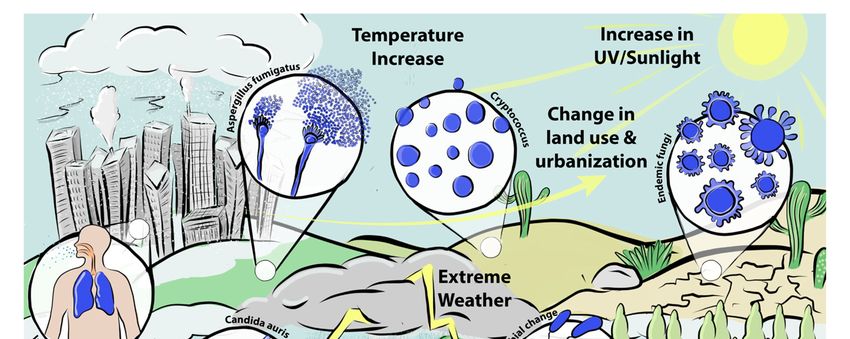
and with the emergence of novel fungal pathogens (Figure 1). New emerging diseases,
such as COVID-19 and severe influenza, are being recognised as risk factors for fungalto climate change, especially since only a small proportion of 1.5 million fungal species
have been identified. Our changing environment will likely expose us to fungi with which
humans have never interacted. Furthermore, more humans will become at risk for fungal
infections. The prevalence and diversity of soil microorganisms will undoubtedly change,
J. Fungi 2021, 7, 367 due to climate change. This change has already been observed with endemic fungi9 of 18
and
with the emergence of novel fungal pathogens (Figure 1). New emerging diseases, such
as COVID-19 and severe influenza, are being recognised as risk factors for fungal infec-
tions [214–216]. The emergence and spread of viral infections is facilitated by increased
infections [214–216]. The emergence and spread of viral infections is facilitated by increased
population
population density. As climate
density. As climate change
change drives
drives the
the increase
increase inin urban
urban populations,
populations, diseases
diseases
that predispose to secondary fungal infections are likely to increase [217,218]. However,
that predispose to secondary fungal infections are likely to increase [217,218]. However,
many
many questions
questions remain
remain unanswered and more
unanswered and more study
study isis required.
required.
Figure 1. Schematic overview of changes in the epidemiological landscape of fungal pathogens and associated changes in
Figure 1. Schematic overview of changes in the epidemiological landscape of fungal pathogens and associated changes in
environmental parameters.
environmental parameters.
Author Contributions: Conceptualization, N.v.R. and M.B.; data curation, N.v.R. and M.B.; writing—
Author
original Contributions: Conceptualization,
draft preparation, N.v.R. and M.B.; N.v.R. and M.B.; and
writing—review dataediting,
curation, N.v.R.
N.v.R. andand M.B.;
M.B.; writ-
funding
ing—original draft preparation, N.v.R. and M.B.; writing—review and editing, N.v.R. and
acquisition, M.B. All authors have read and agreed to the published version of the manuscript. M.B.;
funding acquisition, M.B. All authors have read and agreed to the published version of the manu-
Funding: This research was funded by the Wellcome Trust, grant number 219551/Z/19/Z and
script.
208396/Z/17/Z.
Funding: This research was funded by the Wellcome Trust, grant number 219551/Z/19/Z and
Institutional Review Board Statement: Not applicable.
208396/Z/17/Z.
Informed Consent
Institutional Statement:
Review Not applicable.
Board Statement: Not applicable.
Acknowledgments:
Informed We would Not
Consent Statement: like applicable.
to thank Designs that Cell for designing and realizing Figure 1.
Conflicts of Interest:We
Acknowledgments: M.B. is thelike
would director and Designs
to thank shareholder
that of Syngenics
Cell Limited
for designing and
and PiQ laboratories
realizing Figure 1.
Limited. N.v.R. declares no competing interest. The funders had no role in the design of the study; in
the collection, analyses, or interpretation of data; in the writing of the manuscript, or in the decision
to publish the results.
References
1. Hansen, J.; Sato, M.; Ruedy, R.; Lo, K.; Lea, D.W.; Medina-Elizade, M. Global temperature change. Proc. Natl. Acad. Sci. USA 2006,
103, 14288–14293. [CrossRef]
2. Pachauri, R.K.; Allen, M.R.; Barros, V.R.; Broome, J.; Cramer, W.; Christ, R.; Church, J.A.; Clarke, L.; Dahe, Q.; Dasgupta, P.; et al.
Climate Change 2014: Synthesis Report. Contribution of Working Groups I, II and III to the Fifth Assessment Report of the Intergovernmental
Panel on Climate Change; Pachauri, R., Meyer, L., Eds.; IPCC: Geneva, Switzerland, 2014; 151p.
3. Hansen, J.; Sato, M.; Kharecha, P.; Russell, G.; Lea, D.W.; Siddall, M. Climate change and trace gases. Philos. Trans. R. Soc. A Math.
Phys. Eng. Sci. 2007, 365, 1925–1954. [CrossRef] [PubMed]J. Fungi 2021, 7, 367 10 of 18
4. Seneviratne, S.I.; Donat, M.G.; Mueller, B.; Alexander, L.V. No pause in the increase of hot temperature extremes. Nat. Clim.
Chang. 2014, 4, 161–163. [CrossRef]
5. Hansen, J.; Sato, M. Regional climate change and national responsibilities. Environ. Res. Lett. 2016, 11, 034009. [CrossRef]
6. Karl, T.R.; Trenberth, K.E. Modern global climate change. Science 2003, 302, 1719–1723. [CrossRef]
7. Tabari, H. Climate change impact on flood and extreme precipitation increases with water availability. Sci. Rep. 2020, 10, 13768.
[CrossRef]
8. Bonfils, C.J.W.; Santer, B.D.; Fyfe, J.C.; Marvel, K.; Phillips, T.J.; Zimmerman, S.R.H. Human influence on joint changes in
temperature, rainfall and continental aridity. Nat. Clim. Chang. 2020, 10, 726–731. [CrossRef]
9. Zeng, Z.; Ziegler, A.D.; Searchinger, T.; Yang, L.; Chen, A.; Ju, K.; Piao, S.; Li, L.Z.X.; Ciais, P.; Chen, D.; et al. A reversal in global
terrestrial stilling and its implications for wind energy production. Nat. Clim. Chang. 2019, 9, 979–985. [CrossRef]
10. McKenzie, R.L.; Aucamp, P.J.; Bais, A.F.; Björn, L.O.; Ilyas, M.; Madronich, S. Ozone depletion and climate change: Impacts on
UV radiation. Photochem. Photobiol. Sci. 2011, 10, 182–198. [CrossRef]
11. Weiskerger, C.J.; Brandão, J.; Ahmed, W.; Aslan, A.; Avolio, L.; Badgley, B.D.; Boehm, A.B.; Edge, T.A.; Fleisher, J.M.; Heaney,
C.D.; et al. Impacts of a changing earth on microbial dynamics and human health risks in the continuum between beach water
and sand. Water Res. 2019, 162, 456–470. [CrossRef]
12. Wang, D.Y.C.; Kumar, S.; Hedges, S.B. Divergence time estimates for the early history of animal phyla and the origin of plants,
animals and fungi. Proc. R. Soc. B Boil. Sci. 1999, 266, 163–171. [CrossRef]
13. Tedersoo, L.; Bahram, M.; Põlme, S.; Kõljalg, U.; Yorou, N.S.; Wijesundera, R.L.C.; Ruiz, L.V.; Vasco-Palacios, A.M.; Thu, P.Q.;
Suija, A.; et al. Global diversity and geography of soil fungi. Science 2014, 346, 1256688. [CrossRef]
14. Kubicek, C.P.; Druzhinina, I.S. (Eds.) Fungi in Extreme Environments. In Environmental and Microbial Relationships; Springer:
Berlin/Heidelberg, Germany, 2007; pp. 85–103.
15. Hawksworth, D.L.; Lücking, R. Fungal diversity revisited: 2.2 to 3.8 million species. In The Fungal Kingdom; Heitman, J., Howlett,
B.J., Crous, P.W., Stukenbrock, E.H., James, T.Y., Gow, N.A.R., Eds.; Wiley Online Library: Hoboken, NJ, USA, 2017; pp. 79–95.
[CrossRef]
16. Henkel, T.W.; Aime, M.C.; Chin, M.M.L.; Miller, S.L.; Vilgalys, R.; Smith, M.E. Ectomycorrhizal fungal sporocarp diversity and
discovery of new taxa in Dicymbe monodominant forests of the Guiana Shield. Biodivers. Conserv. 2011, 21, 2195–2220. [CrossRef]
17. López-Quintero, C.A.; Straatsma, G.; Franco-Molano, A.E.; Boekhout, T. Macrofungal diversity in Colombian Amazon forests
varies with regions and regimes of disturbance. Biodivers. Conserv. 2012, 21, 2221–2243. [CrossRef]
18. Truong, C.; Mujic, A.B.; Healy, R.; Kuhar, F.; Furci, G.; Torres, D.; Niskanen, T.; Sandoval-Leiva, P.A.; Fernández, N.; Escobar, J.M.;
et al. How to know the fungi: Combining field inventories and DNA-barcoding to document fungal diversity. New Phytol. 2017,
214, 913–919. [CrossRef] [PubMed]
19. Kwon-Chung, K.; Bennett, J. Medical mycology. Revista do Instituto de Medicina Tropical de São Paulo 1992, 34, 504. [CrossRef]
20. Naranjo-Ortiz, M.A.; Gabaldón, T. Fungal evolution: Major ecological adaptations and evolutionary transitions. Biol. Rev. 2019,
94, 1443–1476. [CrossRef] [PubMed]
21. Robert, V.A.; Casadevall, A. Vertebrate Endothermy Restricts Most Fungi as Potential Pathogens. J. Infect. Dis. 2009, 200,
1623–1626. [CrossRef] [PubMed]
22. Casadevall, A. Thermal restriction as an antimicrobial function of fever. PLoS Pathog. 2016, 12, e1005577. [CrossRef] [PubMed]
23. Romani, L. Immunity to fungal infections. Nat. Rev. Immunol. 2011, 11, 275–288. [CrossRef]
24. Malavia, D.; Crawford, A.; Wilson, D. Nutritional immunity and fungal pathogenesis: The struggle for micronutrients at the
host–pathogen interface. Adv. Microb. Physiol. 2017, 70, 85–103. [PubMed]
25. Beattie, S.R.; Mark, K.M.; Thammahong, A.; Ries, L.N.A.; Dhingra, S.; Caffrey-Carr, A.K.; Cheng, C.; Black, C.C.; Bowyer, P.;
Bromley, M.J.; et al. Filamentous fungal carbon catabolite repression supports metabolic plasticity and stress responses es-sential
for disease progression. PLoS Pathog. 2017, 13, e1006340. [CrossRef] [PubMed]
26. Schrettl, M.; Bignell, E.; Kragl, C.; Joechl, C.; Rogers, T.; Arst, H.N., Jr.; Haynes, K.; Haas, H. Siderophore biosynthesis but not
reductive iron assimilation is essential for Aspergillus fumigatus virulence. J. Exp. Med. 2004, 200, 1213–1219. [CrossRef]
27. König, A.; Müller, R.; Mogavero, S.; Hube, B. Fungal factors involved in host immune evasion, modulation and exploitation
during infection. Cell. Microbiol. 2021, 23, 13272. [CrossRef] [PubMed]
28. Moyes, D.L.; Wilson, D.; Richardson, J.P.; Mogavero, S.; Tang, S.X.; Wernecke, J.; Höfs, S.; Gratacap, R.L.; Robbins, J.; Runglall, M.;
et al. Candidalysin is a fungal peptide toxin critical for mucosal infection. Nat. Cell Biol. 2016, 532, 64–68. [CrossRef] [PubMed]
29. Gaston, K.J.; Fuller, R.A. Commonness, population depletion and conservation biology. Trends Ecol. Evol. 2008, 23, 14–19.
[CrossRef]
30. Sala, O.E. Global Biodiversity Scenarios for the Year 2100. Science 2000, 287, 1770–1774. [CrossRef] [PubMed]
31. Crowley, T.J.; Berner, R.A. CO2 and climate change. Science 2001, 292, 870–872. [CrossRef] [PubMed]
32. Berdugo, M.; Delgado-Baquerizo, M.; Soliveres, S.; Hernández-Clemente, R.; Zhao, Y.; Gaitán, J.J.; Gross, N.; Saiz, H.; Maire, V.;
Lehmann, A.; et al. Global ecosystem thresholds driven by aridity. Science 2020, 367, 787–790. [CrossRef]
33. Lynch, J.P.; Clair, S.B.S. Mineral stress: The missing link in understanding how global climate change will affect plants in real
world soils. Field Crop. Res. 2004, 90, 101–115. [CrossRef]
34. Corwin, D.L. Climate change impacts on soil salinity in agricultural areas. Eur. J. Soil Sci. 2021, 72, 842–862. [CrossRef]J. Fungi 2021, 7, 367 11 of 18
35. Cavicchioli, R.; Ripple, W.J.; Timmis, K.N.; Azam, F.; Bakken, L.R.; Baylis, M.; Behrenfeld, M.J.; Boetius, A.; Boyd, P.W.; Classen,
A.T.; et al. Scientists’ warning to humanity: Microorganisms and climate change. Nat. Rev. Genet. 2019, 17, 569–586. [CrossRef]
[PubMed]
36. O’Connor, E.A.; Hasselquist, D.; Nilsson, J.-Å.; Westerdahl, H.; Cornwallis, C.K. Wetter climates select for higher immune gene
diversity in resident, but not migratory, songbirds. Proc. R. Soc. B Boil. Sci. 2020, 287, 20192675. [CrossRef] [PubMed]
37. Khawcharoenporn, T.; Apisarnthanarak, A.; Mundy, L.M. Non-neoformans Cryptococcal Infections: A Systematic Review. Infection
2007, 35, 51–58. [CrossRef] [PubMed]
38. Rajasingham, R.; Smith, R.M.; Park, B.J.; Jarvis, J.N.; Govender, N.P.; Chiller, T.M.; Denning, D.W.; Loyse, A.; Boulware, D.R.
Global burden of disease of HIV-associated cryptococcal meningitis: An updated analysis. Lancet Infect. Dis. 2017, 17, 873–881.
[CrossRef]
39. Kneale, M.; Bartholomew, J.S.; Davies, E.; Denning, D.W. Global access to antifungal therapy and its variable cost. J. Antimicrob.
Chemother. 2016, 71, 3599–3606. [CrossRef] [PubMed]
40. Lieber, M.; Chin-Hong, P.; Whittle, H.J.; Hogg, R.; Weiser, S.D. The Synergistic Relationship Between Climate Change and the
HIV/AIDS Epidemic: A Conceptual Framework. AIDS Behav. 2021, 1–12. [CrossRef]
41. Baker, R.E. Climate change drives increase in modeled HIV prevalence. Clim. Chang. 2020, 163, 237–252. [CrossRef]
42. Kidd, S.E.; Chow, Y.; Mak, S.; Bach, P.J.; Chen, H.; Hingston, A.O.; Kronstad, J.W.; Bartlett, K.H. Characterization of environmental
sources of the human and animal pathogen Cryptococcus gattii in British Columbia, Canada, and the Pacific Northwest of the
United States. Appl. Environ. Microbiol. 2007, 73, 1433–1443. [CrossRef]
43. Ellis, D.H.; Pfeiffer, T.J. Natural habitat of Cryptococcus neoformans var. gattii. J. Clin. Microbiol. 1990, 28, 1642–1644. [CrossRef]
44. Acheson, E.S.; Galanis, E.; Bartlett, K.; Klinkenberg, B. Climate Classification System-Based Determination of Temperate Climate
Detection of Cryptococcus gattii sensu lato. Emerg. Infect. Dis. 2019, 25, 1723–1726. [CrossRef] [PubMed]
45. Engelthaler, D.M.; Casadevall, A. On the emergence of Cryptococcus gattii in the Pacific Northwest: Ballast tanks, tsunamis, and
black swans. mBio 2019, 10. [CrossRef] [PubMed]
46. Granados, D.P.; Castañeda, E. Influence of climatic conditions on the isolation of members of the Cryptococcus neoformans species
complex from trees in Colombia from 1992–2004. FEMS. Yeast. Res. 2006, 6, 636–644. [CrossRef]
47. Kidd, S.E.; Bach, P.J.; Hingston, A.O.; Mak, S.; Chow, Y.; MacDougall, L.; Kronstad, J.W.; Bartlett, K.H. Cryptococcus gattii Dispersal
Mechanisms, British Columbia, Canada. Emerg. Infect. Dis. 2007, 13, 51–57. [CrossRef] [PubMed]
48. Mitchell, D.H.; Sorrell, T.C.; Allworth, A.M.; Heath, C.H.; McGregor, A.R.; Papanaoum, K.; Richards, M.J.; Gottlieb, T. Cryptococcal
disease of the CNS in immunocompetent hosts: Influence of cryptococcal variety on clinical manifestations and outcome. Clin.
Infect. Dis. 1995, 20, 611–616. [CrossRef] [PubMed]
49. Mak, S.; Klinkenberg, B.; Bartlett, K.; Fyfe, M. Ecological niche modeling of Cryptococcus gattii in British Columbia, Canada.
Environ. Health Perspect. 2010, 118, 653–658. [CrossRef]
50. D’Souza, C.A.; Kronstad, J.W.; Taylor, G.; Warren, R.; Yuen, M.; Hu, G.; Jung, W.H.; Sham, A.; Kidd, S.E.; Tangen, K.; et al. Genome
Variation in Cryptococcus gattii, an Emerging Pathogen of Immunocompetent Hosts. mBio 2011, 2, e00342-10. [CrossRef]
51. Byrnes, E.J.; Li, W.; Lewit, Y.; Ma, H.; Voelz, K.; Ren, P.; Carter, D.A.; Chaturvedi, V.; Bildfell, R.J.; May, R.C.; et al. Emergence and
Pathogenicity of Highly Virulent Cryptococcus gattii Genotypes in the Northwest United States. PLoS Pathog. 2010, 6, e1000850.
[CrossRef]
52. Fraser, J.A.; Giles, S.S.; Wenink, E.C.; Geunes-Boyer, S.G.; Wright, J.R.; Diezmann, S.; Allen, A.; Stajich, J.E.; Dietrich, F.S.; Perfect,
J.R.; et al. Same-sex mating and the origin of the Vancouver Island Cryptococcus gattii outbreak. Nat. Cell Biol. 2005, 437, 1360–1364.
[CrossRef]
53. DaCal, M.; Bradford, M.A.; Plaza, C.; Maestre, F.T.; García-Palacios, P. Soil microbial respiration adapts to ambient temperature in
global drylands. Nat. Ecol. Evol. 2019, 3, 232–238. [CrossRef]
54. Bradford, M.A.; McCulley, R.L.; Crowther, T.W.; Oldfield, E.E.; Wood, S.A.; Fierer, N. Cross-biome patterns in soil microbial
respiration predictable from evolutionary theory on thermal adaptation. Nat. Ecol. Evol. 2019, 3, 223–231. [CrossRef]
55. Fernandes, K.E.; Dwyer, C.; Campbell, L.T.; Carter, D.A. Species in the Cryptococcus gattii complex differ in capsule and cell size
following growth under capsule-inducing conditions. Msphere 2016, 1, e00350-16. [CrossRef]
56. Kozel, T.R.; Pfrommer, G.S.T.; Guerlain, A.S.; Highison, B.A.; Highison, G.J. Role of the Capsule in Phagocytosis of Cryptococcus
neoformans. Clin. Infect. Dis. 1988, 10, S436–S439. [CrossRef]
57. Bojarczuk, A.; Miller, K.A.; Hotham, R.; Lewis, A.; Ogryzko, N.V.; Kamuyango, A.A.; Frost, H.; Gibson, R.H.; Stillman, E.; May,
R.C.; et al. Cryptococcus neoformans Intracellular Proliferation and Capsule Size Determines Early Macrophage Control of Infection.
Sci. Rep. 2016, 6, 21489. [CrossRef] [PubMed]
58. Robertson, E.J.; Najjuka, G.; Rolfes, M.A.; Akampurira, A.; Jain, N.; Anantharanjit, J.; Hohenberg, M.v.; Tassieri, M.; Carlsson, A.;
Meya, D.B.; et al. Cryptococcus neoformans ex vivo capsule size is associated with intracranial pressure and host immune response
in HIV-associated cryptococcal meningitis. J. Infect. Dis. 2014, 209, 74–82. [CrossRef]
59. Pool, A.; Lowder, L.; Wu, Y.; Forrester, K.; Rumbaugh, J. Neurovirulence of Cryptococcus neoformans determined by time course of
capsule accumulation and total volume of capsule in the brain. J. NeuroVirol. 2013, 19, 228–238. [CrossRef]
60. Rosas, Á.L.; Casadevall, A. Melanization affects susceptibility of Cryptococcus neoformans to heat and cold. FEMS Microbiol. Lett.
1997, 153, 265–272. [CrossRef]J. Fungi 2021, 7, 367 12 of 18
61. Grishkan, I. Ecological Stress: Melanization as a Response in Fungi to Radiation. In Extremophiles Handbook; Horiko-shi, K., Ed.;
Springer: Tokyo, Japan, 2011; pp. 1135–1145.
62. Dadachova, E.; Bryan, R.A.; Huang, X.; Moadel, T.; Schweitzer, A.D.; Aisen, P.; Nosanchuk, J.D.; Casadevall, A. Ionizing Radiation
Changes the Electronic Properties of Melanin and Enhances the Growth of Melanized Fungi. PLoS ONE 2007, 2, e457. [CrossRef]
63. Firacative, C.; Duan, S.; Meyer, W. Galleria mellonella model identifies highly virulent strains among all major molec-ular types of
Cryptococcus gattii. PLoS ONE 2014, 9, e105076. [CrossRef] [PubMed]
64. Fernandes, K.E.; Brockway, A.; Haverkamp, M.; Cuomo, C.A.; van Ogtrop, F.; Perfect, J.R.; Carter, D.A. Phenotypic variability
correlates with clinical outcome in Cryptococcus isolates obtained from Botswanan HIV/AIDS patients. mBio 2018, 9, e02016-18.
[CrossRef] [PubMed]
65. De Sousa, H.R.; de Oliveira, G.P.; de Oliveira Frazão, S.; de Melo Gorgonha, K.C.; Rosa, C.P.; Garcez, E.M.; Junior, J.L.; Correia, A.F.;
de Freitas, W.F.; Paes, H.C.; et al. Faster Cryptococcus melanization increases virulence in experimental and human cryptococcosis.
bioRxiv 2020. [CrossRef]
66. Singaravelan, N.; Grishkan, I.; Beharav, A.; Wakamatsu, K.; Ito, S.; Nevo, E. Adaptive melanin response of the soil fungus
Aspergillus niger to UV radiation stress at “Evolution Canyon”, Mount Carmel, Israel. PLoS ONE 2008, 3, e2993. [CrossRef]
67. Latge, J.P.; Chamilos, G. Aspergillus fumigatus and Aspergillosis in 2019. Clin. Microbiol. Rev. 2019, 33. [CrossRef]
68. Bongomin, F.; Gago, S.; Oladele, R.O.; Denning, D.W. Global and Multi-National Prevalence of Fungal Diseases—Estimate
Precision. J. Fungi 2017, 3, 57. [CrossRef] [PubMed]
69. Reinmuth-Selzle, K.; Kampf, C.J.; Lucas, K.; Lang-Yona, N.; Fröhlich-Nowoisky, J.; Shiraiwa, M.; Lakey, P.S.J.; Lai, S.; Liu, F.;
Kunert, A.T.; et al. Air Pollution and Climate Change Effects on Allergies in the Anthropocene: Abundance, Interaction, and
Modification of Allergens and Adjuvants. Environ. Sci. Technol. 2017, 51, 4119–4141. [CrossRef]
70. Lang-Yona, N.; Shuster-Meiseles, T.; Mazar, Y.; Yarden, O.; Rudich, Y. Impact of urban air pollution on the allergenicity of
Aspergillus fumigatus conidia: Outdoor exposure study supported by laboratory experiments. Sci. Total. Environ. 2016, 541,
365–371. [CrossRef] [PubMed]
71. Bertuzzi, M.; Hayes, G.E.; Icheoku, U.J.; Van Rhijn, N.; Denning, D.W.; Osherov, N.; Bignell, E.M. Anti-Aspergillus Activities of
the Respiratory Epithelium in Health and Disease. J. Fungi 2018, 4, 8. [CrossRef]
72. Denning, D.W.; Pleuvry, A.; Cole, D.C. Global burden of chronic pulmonary aspergillosis as a sequel to pulmonary tu-berculosis.
Bull. World Health Organ. 2011, 89, 864–872. [CrossRef]
73. Gelaw, Y.A.; Yu, W.; Magalhães, R.J.S.; Assefa, Y.; Williams, G. Effect of Temperature and Altitude Difference on Tuberculosis
Notification: A Systematic Review. J. Glob. Infect. Dis. 2019, 11, 63–68. [CrossRef] [PubMed]
74. Fernandes, F.M.D.C.; Martins, E.D.S.; Pedrosa, D.M.A.S.; Evangelista, M.D.S.N. Relationship between climatic factors and air
quality with tuberculosis in the Federal District, Brazil, 2003–2012. Braz. Infec. Dis. 2017, 21, 369–375. [CrossRef] [PubMed]
75. Sleeman, K.E.; de Brito, M.; Etkind, S.; Nkhoma, K.; Guo, P.; Higginson, I.J.; Gomes, B.; Harding, R. The escalating global burden
of serious health-related suffering: Projections to 2060 by world regions, age groups, and health conditions. Lancet Glob. Health
2019, 7, e883–e892. [CrossRef]
76. Ayres, J.G.; Forsberg, B.; Annesi-Maesano, I.; Dey, R.; Ebi, K.L.; Helms, P.J.; Medina-Ramon, M.; Windt, M.; Forastiere, F.; on
behalf of the Environment and Health Committee of the European Respiratory Society. Climate change and respiratory disease:
European Respiratory Society position statement. Eur. Respir. J. 2009, 34, 295–302. [CrossRef]
77. Lin, S.-J.; Schranz, J.; Teutsch, S.M. Aspergillosis Case-Fatality Rate: Systematic Review of the Literature. Clin. Infect. Dis. 2001, 32,
358–366. [CrossRef] [PubMed]
78. Lestrade, P.P.; Buil, J.B.; van der Beek, M.T.; Kuijper, J.; van Dijk, K.; Kampinga, G.A.; Rijnders, B.J.A.; Vonk, A.G.; Greeff, S.C.d.;
Schoffelen, A.F.; et al. Paradoxal trends in azole-resistant Aspergillus fumigatus in a national multicenter surveillance program, the
Netherlands, 2013–2018. Emerg. Infect. Dis. 2020, 26, 1447. [CrossRef]
79. Brockhurst, M.A.; Harrison, F.; Veening, J.-W.; Harrison, E.; Blackwell, G.; Iqbal, Z.; MacLean, C. Assessing evolutionary risks of
resistance for new antimicrobial therapies. Nat. Ecol. Evol. 2019, 3, 515–517. [CrossRef] [PubMed]
80. Furukawa, T.; Van Rhijn, N.; Fraczek, M.; Gsaller, F.; Davies, E.; Carr, P.; Gago, S.; Fortune-Grant, R.; Rahman, S.; Gilsenan, J.M.;
et al. The negative cofactor 2 complex is a key regulator of drug resistance in Aspergillus fumigatus. Nat. Commun. 2020, 11, 1–16.
[CrossRef]
81. Gsaller, F.; Hortschansky, P.; Furukawa, T.; Carr, P.D.; Rash, B.; Capilla, J.; Müller, C.; Bracher, F.; Bowyer, P.; Haas, H.; et al. Sterol
Biosynthesis and Azole Tolerance Is Governed by the Opposing Actions of SrbA and the CCAAT Binding Complex. PLoS Pathog.
2016, 12, e1005775.
82. Camps, S.M.T.; Rijs, A.J.M.M.; Klaassen, C.H.W.; Meis, J.F.; O’Gorman, C.M.; Dyer, P.S.; Melchers, W.J.G.; Verweij, P.E. Molecular
Epidemiology of Aspergillus fumigatus Isolates Harboring the TR34/L98H Azole Resistance Mechanism. J. Clin. Microbiol. 2012,
50, 2674–2680. [CrossRef]
83. Caspeta, L.; Chen, Y.; Ghiaci, P.; Feizi, A.; Buskov, S.; Hallström, B.M.; Petranovic, D.; Nielsen, J. Altered sterol composition
renders yeast thermotolerant. Science 2014, 346, 75–78. [CrossRef]
84. Swan, T.M.; Watson, K. Stress tolerance in a yeast sterol auxotroph: Role of ergosterol, heat shock proteins and trehalose. FEMS
Microbiol. Lett. 1998, 169, 191–197. [CrossRef]
85. Rybak, J.M.; Ge, W.; Wiederhold, N.P.; Parker, J.E.; Kelly, S.L.; Rogers, P.D.; Fortwendel, J.R. Mutations in hmg1, Challenging the
Paradigm of Clinical Triazole Resistance in Aspergillus fumigatus. mBio 2019, 10, e00437-19. [CrossRef] [PubMed]You can also read