Extracellular Metalloproteinases in the Plasticity of Excitatory and Inhibitory Synapses
←
→
Page content transcription
If your browser does not render page correctly, please read the page content below
cells
Review
Extracellular Metalloproteinases in the Plasticity of Excitatory
and Inhibitory Synapses
Grzegorz Wiera *              and Jerzy W. Mozrzymas *

                                          Department of Biophysics and Neuroscience, Wroclaw Medical University, 50-368 Wroclaw, Poland
                                          * Correspondence: grzegorz.wiera@umed.wroc.pl (G.W.); jerzy.mozrzymas@umed.wroc.pl (J.W.M.);
                                            Tel.: +48-71-784-15-50 (J.W.M.)

                                          Abstract: Long-term synaptic plasticity is shaped by the controlled reorganization of the synaptic pro-
                                          teome. A key component of this process is local proteolysis performed by the family of extracellular
                                          matrix metalloproteinases (MMPs). In recent years, considerable progress was achieved in identi-
                                          fying extracellular proteases involved in neuroplasticity phenomena and their protein substrates.
                                          Perisynaptic metalloproteinases regulate plastic changes at synapses through the processing of extra-
                                          cellular and membrane proteins. MMP9 was found to play a crucial role in excitatory synapses by
                                          controlling the NMDA-dependent LTP component. In addition, MMP3 regulates the L-type calcium
                                          channel-dependent form of LTP as well as the plasticity of neuronal excitability. Both MMP9 and
                                          MMP3 were implicated in memory and learning. Moreover, altered expression or mutations of
                                          different MMPs are associated with learning deficits and psychiatric disorders, including schizophre-
                                nia, addiction, or stress response. Contrary to excitatory drive, the investigation into the role of

                                          extracellular proteolysis in inhibitory synapses is only just beginning. Herein, we review the princi-
Citation: Wiera, G.; Mozrzymas, J.W.      pal mechanisms of MMP involvement in the plasticity of excitatory transmission and the recently
Extracellular Metalloproteinases in       discovered role of proteolysis in inhibitory synapses. We discuss how different matrix metallopro-
the Plasticity of Excitatory and
                                          teinases shape dynamics and turnover of synaptic adhesome and signal transduction pathways in
Inhibitory Synapses. Cells 2021, 10,
                                          neurons. Finally, we discuss future challenges in exploring synapse- and plasticity-specific functions
2055. https://doi.org/10.3390/
                                          of different metalloproteinases.
cells10082055

                                          Keywords: metalloproteinase; proteolysis; synaptic plasticity; GABA; inhibitory synapse; LTP;
Academic Editors:
Ghislain Opdenakker,
                                          adhesion; perineuronal nets; MMP; learning
Jennifer Vandooren and
Estefania Ugarte-Berzal

Received: 1 July 2021                     1. Introduction
Accepted: 5 August 2021                         Neuroplasticity is often defined as the ability of neural networks in the brain to change
Published: 11 August 2021                 through various growth, reorganization, or other modulatory processes to adapt to an
                                          organism’s variable environment and change with experience. Several aspects of neu-
Publisher’s Note: MDPI stays neutral      roplasticity were revealed, including structural changes at various scales, regulation of
with regard to jurisdictional claims in   excitability, but the most extensively studied is the ability to alter neuronal connectivity in
published maps and institutional affil-
                                          response to different activity patterns; the phenomenon referred to as synaptic plasticity.
iations.
                                          Synapses connecting distinct neurons are greatly diversified, and any specific synapse
                                          might experience several types of plasticity mechanisms (expressed pre- or postsynap-
                                          tically), depending on the stimuli eliciting it. Plastic changes at a synapse may weaken
                                          or strengthen the connection and rely on a myriad of signaling pathways and molecular
Copyright: © 2021 by the authors.         players. The last decade or so proved particularly fruitful in bringing convincing evidence
Licensee MDPI, Basel, Switzerland.        that synaptic plasticity and changes in neuronal excitability provide an important substrate
This article is an open access article    for the processes of learning and memory storage [1–3]. A significant breakthrough in
distributed under the terms and
                                          studying the mechanisms of synaptic plasticity was the discovery of efficient crosstalk
conditions of the Creative Commons
                                          between the synapse and neighboring astrocyte processes and the extracellular matrix
Attribution (CC BY) license (https://
                                          (ECM) [4,5]. Notably, both the pre- and postsynapse is surrounded by specific ECM con-
creativecommons.org/licenses/by/
                                          stituents, which are anchored to the neuron’s cytoskeleton by a series of adhesion proteins
4.0/).

Cells 2021, 10, 2055. https://doi.org/10.3390/cells10082055                                                 https://www.mdpi.com/journal/cells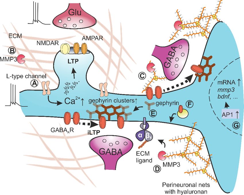
Cells 2021, 10, 2055                                                                                              2 of 25

                       that mediate the exchange of information between the inside and outside of the neuron
                       and thereby actively participate in regulating plastic changes at synapses [6,7]. Importantly,
                       both neurons and astrocytes use proteases anchored to the membrane or secreted into
                       the perisynaptic space to actively control transsynaptic adhesion and shape the structure
                       and composition of the ECM. Initially, extracellular proteases were believed to operate as
                       brain ECM “movers,” but presently, they are known to selectively cleave defined adhesion
                       proteins and structural or signaling molecules within ECM to trigger specific signaling
                       pathways that regulate the physiology of the synapse during learning [8]. In addition, an
                       essential role of extracellular proteases in pathological plasticity that is related, for example,
                       to addiction [5], epilepsy [9], or Alzheimer’s disease [10] has recently been reviewed.
                              The family of matrix metalloproteinases (MMPs) consists of more than 20 proteolytic
                       enzymes belonging to the metzincin superfamily. In terms of homology and substrate
                       specificity, MMPs are divided into several subgroups, including collagenases (MMP1, 8,
                       3), stromelysins (MMP3, 10, 11), gelatinases (MMP2, 9), and proteases anchored in the cell
                       membrane (MMP14, 15, 16, 17, 24, 25). Among all MMPs encoded in mammalian genomes,
                       the majority of them was detected at the level of mRNA or protein in the healthy brain or in
                       numerous pathological conditions [10,11]. All MMPs are synthesized as inactive zymogens
                       (proMMPs) with a propeptide sequence blocking the catalytic domain. In addition, most
                       MMP family members also contain a proline-rich hinge region and the hemopexin domain,
                       which is responsible for interaction with other proteins, substrate recognition, binding of
                       endogenous inhibitors, and proper localization in the extracellular space [12]. Even though
                       not all MMPs are equipped with transmembrane domains, most of them operate in the
                       vicinity of the cell surface by associating with membrane proteins that concentrate active
                       forms of MMPs. Additionally, the proteolytic activity of MMPs is temporally and spatially
                       restricted by tissue inhibitors of metalloproteinases (TIMP1, 2, 3, 4), with TIMP1 mainly
                       blocking the activity of MMP2, 3, 9, and TIMP3 of almost every metzincin [13]. Finally, as
                       with the majority of extracellular proteases, MMPs are internalized upon binding to the
                       LRP1 (low-density lipoprotein receptor-related protein 1) [14]. For a detailed description of
                       MMP molecular biology, with the emphasis on MMP9, we direct the reader to excellent
                       reviews by Gomis-Rüth [15] and Vandooren et al. [16].
                              As already mentioned, due to the activity of MMPs, the synaptic adhesome and the
                       extracellular matrix constituents are processed continuously upon induction of synaptic
                       plasticity and, consequently, MMP-dependent proteolysis actively participates in both
                       the structural remodeling of synaptic structures and functional changes in synaptic effi-
                       cacy [5,17,18]. MMPs, secreted and activated upon increased neuronal activity, can locally
                       loosen ECM to define hotspots of structural plasticity on a fragment of dendritic tree [17].
                       Simultaneously, active MMPs may activate other proteins through proteolysis, release new
                       peptides from full-length proteins, and process adhesion molecules to trigger or terminate
                       intracellular signal transduction pathways that would modulate the abundance of synaptic
                       receptors and gene expression in the nucleus. Therefore, MMP-mediated proteolysis, with
                       its irreversible nature, is an excellent candidate for a mechanism that ties the functional and
                       structural component of synaptic plasticity to produce long-lasting changes underlying
                       cognitive processes.
                              In this review, we discuss recent studies elucidating the roles of different MMPs in
                       multifarious forms of synaptic plasticity, learning, and memory. Since MMP9 is the most
                       widely studied MMP in the brain, we briefly outline its perisynaptic activation, interaction
                       with local structures, and endogenous inhibition to explore the current knowledge on its
                       role in neuroplasticity phenomena and cognitive processes in this context. Additionally,
                       special attention is given to the latest studies investigating the role of MMP3 in molecular
                       mechanisms governing the excitatory synaptic plasticity that depends on L-type calcium
                       channels [18]. For years, research into the inhibitory synaptic plasticity was overshadowed
                       by investigations into the plasticity of excitatory synapses, but this situation started to
                       change a decade ago or so. In this report, we thus take advantage of recent progress in
                       studies on mechanisms of GABAergic synaptic plasticity. A particular emphasis is placed
Cells 2021, 10, 2055 3 of 25
on the key role of MMP3 in this type of plasticity, which was investigated by our group
systematically in the hippocampus [19]. Finally, based on the latest findings, we review
and discuss the increasing number of behavioral studies linking the activity of specific
MMPs in excitatory and inhibitory neuronal networks to learning and memory.
2. Regulation of Synaptic Plasticity in Excitatory Synapses and Neuronal Excitability
by MMPs
2.1. Background Considerations on the Involvement of MMPs in Synaptic Plasticity
Early evidence that metalloproteinases might play a central role in synaptic plasticity
was derived from experiments on an epileptic mouse model, which showed elevated MMP2
and MMP9 in the hippocampus a few hours after injection of kainate [20]. Previously, the
T. Bliss group reported increased proteolytic activity in hippocampal homogenates after
LTP induction in perforant path synapses [21]. In particular, they described an increase in
the activity of 80 kDa protease, which is capable of digesting the gelatin in gel zymography
which can now be, with high confidence, ascribed to MMP9 [22]. In the follow-up study,
the same group reported increased extracellular proteolytic processing of amyloid-beta
precursor protein (APP) and neural cell adhesion molecule (NCAM) after LTP [23]. These
results, together with the discovery of the role of tissue plasminogen activator (tPA) [24] and
TIMP1 in LTP [25], provided the first direct evidence that elevated extracellular proteolysis
can be a hallmark of long-term synaptic plasticity.
Having established the involvement of proteolysis in the phenomena of synaptic
plasticity, the major question to be answered was which specific proteases were involved.
Unfortunately, this issue proved difficult as a precise identification of MMPs involved in
plastic changes is hampered by the lack of specific inhibitors of individual MMPs and
compensatory effects in mouse gene knockouts. Indeed, available MMP inhibitors are not
fully specific and selective, and they usually block two or more metzincins with a similar
affinity. Thus, for this reason, it is advisable to use a wide battery of different inhibitors with
a non-overlapping spectrum of blocked proteases to identify the involvement of individual
MMP in the studied process and to exclude other proteases concurrently. Additionally,
studies using genetic knockout animals should validate the synaptic role of a given protease,
but caution should be exercised because of compensatory changes that may occur in
MMP-deficient mice. For example, MMP9 deficiency leads to a compensatory increase
in MMP8 expression [26]; similarly, MMP3 genetic knockout mice show elevated MMP7
and MMP12 [27]. Furthermore, genetically modified mice created using 129-derived
embryonic stem cells often contain passenger mutations in sequences flanking targeted
genes, hence contaminating the phenotypic outcome of genetic manipulation. For example,
knockouts of several MMP genes located on mouse chromosome 9 (mmp3, mmp7, mmp8)
contain passenger mutation that inactivates the gene encoding caspase 11, which is also
a protease [28]. As a result, partial inactivation of caspase 11 may confound phenotypic
interpretation of MMP genetic knockout models or perturb the identification of MMP
substrates by mass spectrometry-assisted degradomic studies. Importantly, for some
strains of MMP9 genetic knockout mice, the residual leakiness at the protein level was
reported [29]. This problem, especially in the field of neuroscience, was not commonly
realized until recently and it would be advantageous to confirm previous results on animal
strains in which MMP (especially MMP9 which was shown to be problematic with this
respect) deficiency has been unequivocally confirmed. Taking into account the important
evidence presented by de Bruyn et al. [29], it seems necessary to accept as a prerequisite in
these studies to demonstrate by means of e.g., gelatin gel zymography, that knockout mice
do not show any gelatin zymolytic MMP9 levels whatsoever (aside from WT controls).
Given the multitude of proteins involved in synaptic plasticity, specific criteria were
proposed to determine whether a given protein plays an active role in the molecular
mechanisms of plasticity maintenance. When exploring the potential role of a new protein
in synaptic plasticity or learning, three types of tests should be used, as it was summarized
initially by John Lisman [30]. The first and the most common test, referred to as the necessity
test, determines whether a protein is required for plasticity by pharmacological inhibitionCells 2021, 10, 2055 4 of 25
or genetic knockout. Next, the occlusion test, in which the active form of investigated
protein is introduced, and the effect should occlude the induction of synaptic plasticity.
Finally, the erasure test examines whether manipulation of analyzed protein at some time
point after the induction of synaptic plasticity can reduce its maintenance [30]. We use
these experimental criteria to summarize available information on the roles of specific
MMPs in the molecular mechanisms of neuroplasticity.
Among all metalloproteinases, the MMP9 is the most extensively studied in the con-
text of synaptic plasticity, learning and memory [31]. MMP9-deficient mice show impaired
LTP in the hippocampal CA3-CA1 [18,32] and mossy fiber-CA3 projections [33,34] as well
as in projections within the amygdala from the lateral to the basal nucleus and from the
basal to the medial section of the central amygdala [35]. Furthermore, the administration of
the active form of MMP9 spontaneously increases the efficacy of excitatory synaptic trans-
mission [32,36] and occludes the induction of LTP [33,37]. In addition, the administration
of MMP9 inhibitor up to 30 min after LTP induction disrupts the maintenance phase of
induced plasticity [38,39]. Altogether, MMP9 passed the necessity, occlusion, and erasure
tests, which explicitly proved the engagement of this protease in the consolidation phase
of long-term potentiation.
2.2. The Role of MMP9 in LTP
2.2.1. The Mechanism of MMP9 Synthesis and Release in the Perisynaptic Environment
In general, the functioning of metalloproteinases in the synaptic and perisynaptic
space can be divided into the following well-defined stages: (1) The induction of synaptic
plasticity caused by a temporary change in the neural network activity leads to the synthesis
and secretion of a protease into the extracellular space. (2) Once activated and properly
located, it cleaves other proteins and peptides according to its substrate spectrum. (3) After
a short period of activity, a protease can be inactivated due to an autocatalytic mechanism,
blocked by endogenous inhibitors or internalized through the interaction with membrane
receptors. The role of MMP9 in excitatory synapses is described in reference to these
three stages.
The mRNA encoding MMP9 is transported into dendrites, where it is locally trans-
lated, and the protease is released into the perisynaptic area in an activity-dependent
manner [40]. MMP9 mRNA is a part of the FMRP (fragile X mental retardation protein)
complex that controls its dendritic transport and translation [41]. Additionally, miR-131,
an activity-regulated microRNA, binds to the 30 untranslated region of MMP9 mRNA
and negatively regulates its expression in neurons. Stimulation of excitatory synapses
(e.g., by activating mGluRs) drives MMP9 translation due to the dissociation of miR-131
from polyribosomes [42]. Next, after the synthesis, the zymogen of proMMP9 is stored in
Golgi-derived vesicles associated with the dendritic cytoskeleton [43].
The molecular mechanisms of MMP9 secretion into extracellular space upon increased
neuronal activity are unclear. Therefore, future studies are needed to describe this process
precisely, as was the case, for instance, with BDNF [44,45]. Nevertheless, at this point, it is
important to note that MMP9-deficient mice show normal long-term depression (LTD) at
excitatory synapses in the hippocampal CA1 field [32], suggesting that the frequency of
synaptic stimulation or the timing of postsynaptic action potentials determines whether
MMP9 is involved in a given type of long-term synaptic plasticity. Moreover, calcium influx
through the NMDA receptor appears to be involved in MMP9 secretion because when
NMDARs are blocked, the residual LTP recorded in the hippocampal CA1 is insensitive to
MMP9 inhibition [18], and the endogenous protease is not activated [37].
The exocytosis of postsynaptic MMP9-containing vesicles occurs at membrane local-
izations lateral to the postsynaptic density either within or outside dendritic spines [46]. It
is interesting how far from the synapse proMMP9 is released and how distant its diffusion
is following activation during the induction of synaptic plasticity. The hemopexin domain
of MMP9 is known to play a role of a multifunctional ligand for numerous membrane
receptors and ECM proteins, such as β5-integrin, Ku70/Ku80 heterodimer, LRP1, chon-Cells 2021, 10, 2055 5 of 25
droitin sulfate proteoglycans [12,47], or CD44 [48]. These interactions are expected to limit
the diffusion process favoring the concentration of this protease in the vicinity of synapses.
MMP9 is well-known to participate in the mechanism of structural plasticity of dendritic
spines [49] and control the enlargement of small, but not large, dendritic spines in neuronal
cell cultures after LTP induction [50]. However, the mechanisms underlying this selectivity
are not clear: proMMP9 could be released selectively at small synapses prone to structural
plasticity, or big spines could lack specific receptors for MMP9 that would concentrate it,
enabling the protease to trigger the plasticity mechanisms.
2.2.2. The Mechanisms of MMP9 Activation
Once released into the extracellular space, brain MMP9 proform is activated by at least
two different processes: (1) the proteolytic removal of inhibitory prodomain that in vitro
can be accomplished by several proteases, such as tPA, uPA (urokinase-type plasminogen
activator), MMP3 [16,51], or (2) through S-nitrosylation, which requires the synthesis
of nitric oxide [52–55]. Nevertheless, the exact mechanism of MMP9 activation in the
perisynaptic region in vivo or ex vivo in brain slices is far from being understood. The
activation by MMP3 is rather unlikely because the effects of MMP3 and MMP9 inhibition
on LTP induction do not occlude reciprocally [18]. However, an interesting mechanism
of MMP9 activation in neurons was discovered by Padamsey et al. [56]. It was shown
that the backpropagation of action potentials in a postsynaptic neuron elicited calcium
influx that caused the secretion of the content of lysosomes. Consequently, lysosomal
proteases, e.g., cathepsin B, were released into extracellular space, where they activated
proMMP9 required for long-term structural plasticity of dendritic spines in hippocampal
CA1 neurons [56]. Additionally, the activation of serotonergic receptors 5-HT7R stimulated
the activity of MMP9 in the vicinity of dendritic spines and shafts in neuronal cultures [57].
Future studies should shed more light on the timing of secretion and spatial localization
of exocytosis spots for lysosomes and proMMP9-containing vesicles during long-term
synaptic plasticity (Figure 1B).
2.2.3. Putative Targets of MMP9 Proteolytic Activity in the Perisynaptic Area
Active MMP9 in extracellular space can cleave numerous substrates that act as ad-
hesion receptors, soluble signaling proteins, or structural ECM constituents (we refer the
readers to excellent reviews on this topic [58–60]). However, considering the low speci-
ficity and pleiotropic nature of MMP inhibitors, we focus on neuronal MMP9 substrates
validated in MMP9-deficient animals in vivo or ex vivo in brain slices.
A majority of well-documented synaptic substrates of MMP9 belong to different
families of adhesion proteins. During the critical period, vision restoration after monocular
deprivation induces massive synaptic plasticity in the visual cortex, accompanied by an
increase in MMP9 activity [61]. This plasticity requires MMP9-dependent proteolytic
processing of adhesion protein, neuroligin-1 (NLGN1), in a process that relies on NMDA
receptors and CaMKII [62]. Similarly, stimulation of synaptoneurosomes with NMDA
results in the MMP-dependent cleavage of neuroligin-1 [63]. This process destabilizes the
transsynaptic neurexin-neuroligin complex, reducing the frequency of miniature EPSCs,
presynaptic probability of neurotransmitter release, and the amplitude of evoked EPSC [62].
These results support a model where the time-limited extracellular proteolysis of NLGN1
transiently disrupts synaptic adhesive apparatus that leads to functional changes in the
excitatory synapses and structural remodeling of the dendritic spines (Figure 1G). The
crucial role of MMP9 in the timing of the visual critical period was also recently observed
in a study in which astrocytic connexin signaling decreased the expression of MMP9
through RhoA signaling. This process resulted in the stabilization of perineuronal nets
and the closure of a critical period in visual cortex [64]. This observation underscores the
potential of both astrocytic and neuronal MMP9 in controlling plasticity phenomena in the
developing brain.Cells 2021, 10, 2055 6 of 25
Cells 2021, 10, x FOR PEER REVIEW 8 of 25
Figure 1. Functions of MMP9 in long-term potentiation at excitatory synapses. (A) High frequency
Figure 1. Functions of MMP9 in long-term potentiation at excitatory synapses. (A) High frequency
or spike-timing-dependent stimulation leads to calcium influx in dendritic spine that triggers the
or spike-timing-dependent stimulation leads to calcium influx in dendritic spine that triggers the
exocytosis of lysosomes and secretory vesicles with proMMP9. (B) In extracellular space, proMMP9
exocytosis of lysosomes
becomes activated and secretory
by lysosomal vesicles with
cathepsin-B. proMMP9.
(C) Active MMP9 (B)cleaves
In extracellular space,
unidentified ECM proMMP9
constitu-
becomes activated by lysosomal cathepsin-B. (C) Active MMP9 cleaves unidentified
ents to release matricryptins, which activate integrins containing β1 subunit. (D) Integrin-depend- ECM constituents
toent
release matricryptins,
signaling pathways which
activate activate
kinase integrins containing
Src and CaMKII β1 subunit.
to change (D) Integrin-dependent
the lateral diffusion of AMPA
signaling
receptors,pathways activate
which results in kinase Src and at
their trapping CaMKII to change
synapses and LTP theinduction.
lateral diffusion of AMPA receptors,
(E) Simultaneously, MMP9
activity
which increases
results thetrapping
in their membrane diffusion of
at synapses and NMDA receptors (E)
LTP induction. thatSimultaneously,
probably resultsMMP9in an alteration
activity
of the subunit
increases composition
the membrane of synaptic
diffusion of NMDANMDARs. (F) Active
receptors MMP9 may
that probably cleave
results proBDNF
in an alterationto of
itsthe
ma-
ture form, which activates TrkB receptor in an autocrine manner. (G) Additionally,
subunit composition of synaptic NMDARs. (F) Active MMP9 may cleave proBDNF to its mature form, the proteolysis
of neuroligin-1, transiently destabilizes synaptic adhesome during the induction of LTP. (H) CD44
which activates TrkB receptor in an autocrine manner. (G) Additionally, the proteolysis of neuroligin-
anchors brain ECM to neuronal membrane through hyaluronan binding; MMP9-dependent cleav-
1, transiently destabilizes synaptic adhesome during the induction of LTP. (H) CD44 anchors brain
age of CD44 activates neuronal cdc42 that is crucial for structural plasticity of dendritic spines. (I)
ECM to neuronal
Signaling pathwaysmembrane
activatedthrough
during LTP hyaluronan
induction binding; MMP9-dependent
reach the nucleus to induce cleavage of CD44of
the expression
activates neuronal cdc42 that is crucial for structural plasticity of dendritic spines.
immediate early genes, e.g., a c-fos, which give rise to appearance of the AP1 transcription factor (I) Signaling
pathways
and driveactivated during LTP
the transcription induction
of mmp9 andreach
bdnf the
genes nucleus
among toothers.
induceTheir
the expression
mRNA is of immediateto
transported
early genes,tree
dendritic e.g.,
inaac-fos, which
complex withgive
FMRPrise to appearance
protein. of the
(J) After AP1period
a short transcription factor and
of proteolytic driveactive
activity, the
MMP9 is blocked by endogenous inhibitor form TIMP family.
transcription of mmp9 and bdnf genes among others. Their mRNA is transported to dendritic tree in
a complex with FMRP protein. (J) After a short period of proteolytic activity, active MMP9 is blocked
by2.2.4. The Crucial
endogenous Roleform
inhibitor of Endogenous
TIMP family. MMP9 Inhibition
After vesicular release followed by activation, MMP9 exerts its proteolytic activity
It was demonstrated
for a relatively short timethat MMP9
to be can also
eventually cleave and
inhibited, activate
avoiding proBDNF
excessive to its mature
proteolysis (Fig-
form (mBDNF). The downregulation of mBDNF was observed in MMP9-deficient
ure 1J). This endogenous inhibition is an essential step in MMP9 functioning and ensuing mice
after kindling-induced
regulatory phenomena, epilepsy [65].
as it was Additionally,that
demonstrated during development
the outcome and
of the upon learning,
short-term MMP9
the strengthened synapses are often co-arranged on the same dendritic branch. This
activity might dramatically differ from the effects of its long-term presence. For example,
synaptic clustering in hippocampal CA3 neurons requires MMP9 activity and mBDNF
the brief treatment with active MMP9 followed by protease inhibition or washout 10–20
production that activates TrkB receptors [66]. It is noteworthy in this context that the
min later increased the efficacy of CA1 excitatory synapses [32] and induced changes in
enriched environment that stimulates massive synaptic plasticity also upregulates MMP9
the morphology of dendritic spines toward mushroom-like shape [36,85]. In contrast, the
and mBDNF in the hippocampus [67]. Moreover, a positive feedback loop was proposed in
long-lasting presence of exogenous MMP9 or its overexpression caused the transfor-
which mBDNF activates the TrkB receptor, that through ERK signaling stimulates formation
mation of dendritic spines into a filopodial shape [75] and impaired LTP [34,83,84]. The
of AP-1 transcription factor, and finally leads to MMP9 synthesis [68]. Altogether, these
crucial role of MMP9 inhibition in its synaptic functioning is further supported by the
observations may indicate that MMP9 cleaves proBDNF to drive structural plasticity and
evidence of enhanced LTP in slices isolated from mice treated with intravenous injections
proMMP9 restocking (Figure 1F).
of tissue inhibitor of metalloproteinases-2 (TIMP2) [94]. Additionally, the involvement ofCells 2021, 10, 2055 7 of 25
In line with the crucial functions of MMP9 in the structural plasticity of dendritic
spines, recent work demonstrated a key role for MMP9-dependent cleavage of CD44 in this
process [57]. In this study, the stimulation of 5-HT7 receptors upregulated MMP9 activity,
which resulted in CD44 cleavage and the activation of Cdc42 signaling that finally triggered
dendritic spine remodeling and synaptic pruning (Figure 1H). Given the reported link
between MMP9 and CD44, it is of note that CD44 operates as a receptor for hyaluronan,
the main constituent of brain ECM and perineuronal nets. Significantly, while Fmr1 genetic
knockout mice, a model of fragile X syndrome, are characterized by elevated MMP9 activity
and loss of perineuronal nets around parvalbumin-expressing interneurons in the auditory
cortex of juvenile mice, the MMP9 deficiency restored the proper PNN formation [69].
Additionally, light reintroduction after dark exposure can trigger a massive degradation of
ECM accompanied by changes in neuronal excitability, synchrony, and enhanced structural
plasticity of dendritic spines. These effects are blocked by genetic ablation of MMP9,
indicating that increased activity of this protease reduced constraints on structural and
functional plasticity in the mature cortex [70,71]. In this regard, while additional studies
are needed to elucidate the impact of MMP9 on ECM development fully, it is increasingly
evident that MMP9 activity is intimately associated with ECM macroscopic integrity [64,72].
Interestingly, in most cases in which MMP9 was implicated in the plasticity of excita-
tory synapses, its action required integrin β1 in the hippocampus or cortex [11,33,72] or
integrin β3 in nucleus accumbens [73]. Indeed, the potentiation of synaptic transmission
after treatment with active MMP9 is completely abolished by integrin inhibitors [32] or by
antibody blocking integrin β1 functions [36]. Furthermore, it was also shown that active
MMP9 applied exogenously increased the lateral diffusion of NMDA receptors containing
GluN1 subunit, both in synaptic and extrasynaptic localization, through a process that is
blocked by β1 integrin inhibition [74]. Additionally, the application of exogenous active
MMP9 caused changes in the morphology of dendritic spines, and this process also de-
pends on functional β1 integrins whose signaling drives cofilin phosphorylation and actin
polymerization [36,75].
One of the most critical questions in the research on the role of MMP9 in synapses
is the identification of MMP9 substrates, which, after cleavage in perisynaptic space, can
activate β1 integrin (Figure 1C). The term matricryptin was proposed to describe peptides
resulting from extracellular proteolysis that show new properties compared to full-length
parent protein [76]. In this scenario, extracellular proteases decode and unveil the hidden
cryptome of adhesion and ECM proteins. However, the identification of matricryptins
released by MMP9 is still a matter of debate. It was shown that the extracellular domain of
postsynaptic adhesion receptor ICAM-5 (a well-known MMP9 substrate) might bind and
activate presynaptic β1 integrin in cultured hippocampal neurons [77], but this process
is unlikely to be responsible for LTP maintenance as mature spines lack ICAM-5 [77,78].
Thus, it seems that ICAM-5 is important for synaptogenesis and spine maturation [79,80]
rather than plasticity at mature synapses.
Overall, new findings concerning the role of β1 integrins in synaptic plasticity would
also shed some light on the functioning of neuronal MMP9. For example, interference with
the integrin β1-dependent adhesion, similar to MMP9 inhibition, blocked the long-term
structural plasticity of dendritic spines and the reorganization of actin microfilaments
inside spines in hippocampal slices [81]. In addition, after the induction of LTP in the
hippocampus, integrin β1 became activated only for a few minutes, shortly after the
application of the plasticity-inducing protocol [82]. This result suggests that MMP9, located
upstream in the signaling pathway, also should be activated at early stages of LTP, which
agrees with the time window of MMP9 requirement for LTP, inferred from pharmacological
experiments [38,83]. This also implies that the presence of active integrin β1 in the synapse
may be regarded as a marker of recent MMP9 activity. The comparison of the time scale
of synaptic transmission with the timing of MMP9 involvement in plastic changes within
synapses is intriguing. While millisecond precision is required for the induction of spike-
timing-dependent plasticity at excitatory synapses [84], it takes a few minutes from theCells 2021, 10, 2055 8 of 25
application of LTP induction protocol to MMP9 release and activation [32]. Then, after
10–20 min, the protease undergoes endogenous inhibition and/or internalization [85].
Figure 1 schematically summarizes the synaptic functioning of MMP9.
The development of high-throughput degradomic techniques has resulted in the
discovery of many potential MMP9 substrates in different tissues in physiological and in
pathological conditions [86–88]. Among putative MMP9 substrates described in this way,
several are expressed in the brain, where they can be found in intracellular, membrane
or extracellular compartments of the synapse. Tumor Necrosis Factor-α (TNFα) is note-
worthy [86] as it was shown to control synaptic efficacy [89] and to regulate homeostatic
plasticity [90]. Similarly, MMP9 may cleave and activate ADAM Metallopeptidase With
Thrombospondin Type 1 Motif 4 (ADAMTS4) [86], a metzincin able to cleave brevican dur-
ing homeostatic synaptic up-scaling [91]. These two examples show that among putative
MMP9 substrates discovered in vitro outside the brain, many may play an important role
in synapse physiology and during synaptic plasticity but these scenarios still remain to
be demonstrated. Finally, neuronal MMP9 may cleave and degrade β-amyloid peptide
in brains of patients suffering from the Alzheimer’s disease [92]. Interestingly, endoge-
nous amyloid-beta peptide controls presynaptic release probability in healthy brain [93],
suggesting that putative MMP9-dependent degradation may also control presynaptic
neurotransmitter release.
2.2.4. The Crucial Role of Endogenous MMP9 Inhibition
After vesicular release followed by activation, MMP9 exerts its proteolytic activity for
a relatively short time to be eventually inhibited, avoiding excessive proteolysis (Figure 1J).
This endogenous inhibition is an essential step in MMP9 functioning and ensuing regula-
tory phenomena, as it was demonstrated that the outcome of the short-term MMP9 activity
might dramatically differ from the effects of its long-term presence. For example, the
brief treatment with active MMP9 followed by protease inhibition or washout 10–20 min
later increased the efficacy of CA1 excitatory synapses [32] and induced changes in the
morphology of dendritic spines toward mushroom-like shape [36,85]. In contrast, the
long-lasting presence of exogenous MMP9 or its overexpression caused the transformation
of dendritic spines into a filopodial shape [75] and impaired LTP [34,83,84]. The crucial
role of MMP9 inhibition in its synaptic functioning is further supported by the evidence
of enhanced LTP in slices isolated from mice treated with intravenous injections of tissue
inhibitor of metalloproteinases-2 (TIMP2) [94]. Additionally, the involvement of endoge-
nous MMP inhibition in neuronal plasticity and learning is further confirmed by spatial
memory deficits in TIMP3-deficient mice [95], impaired fear-potentiated startle response
in TIMP2-null mice [96], and impaired LTP observed after TIMP1 overexpression in the
prefrontal cortex [97].
Increased level of MMP9 activity was reported in numerous brain regions after in-
ducing long-term plasticity and after learning. For example, the proteolysis mediated by
MMP9 is augmented in the perisynaptic region of hippocampal neurons after LTP induc-
tion in different projections [32,36,98]. Similarly, increased gelatinolytic activity, ascribed
to MMP9, was reported after contextual fear conditioning [99], after classical condition-
ing in a paradigm of pairing whisker stroking with tail shock [84], after chronic restraint
stress [100], after a period of breeding in an enriched environment [101], or after appetitive
learning [102]. Moreover, learning-induced augmentation of MMP9 activity in a consid-
ered brain region can be constrained to specific types of neurons. For example, learning in
cue-induced heroin seeking paradigm increased MMP9 activity only around D1 receptor-
expressing medium spiny neurons in the nucleus accumbens, while subsequent extinction
training augmented MMP2 activity around contiguous D2 medium spiny neurons [103].
This compartmentalization of the involvement of MMP9 and MMP2 in synaptic plasticity
suggests that different neurons could, in principle, express and use distinct MMPs during
plastic changes occurring at the synapse.Cells 2021, 10, 2055 9 of 25
In physiological conditions, the relative timing of MMP9 activation and the secretion
of TIMPs during LTP or learning is unknown. Interestingly, both proteins were spotted
in the same dendritic secretory vesicles in vitro [43], suggesting that the time window of
MMP9 activity may not be determined by the delayed release of TIMPs. Additionally,
the requirement of short-lasting MMP9 activity upon LTP induction, inferred from phar-
macological experiments [38,83], appears to disagree with the observation of increased
MMP9 activity for hours after LTP induction or even days after learning. This apparent
inconsistency may be explained by the limitations of in situ gelatin zymography, the most
common technique used to visualize MMP9 activity in the tissue. Before staining, the
tissue is usually fixated in alcohol (usually methanol with ethanol) that reversibly denature
proteins which are then renatured during the stage of hydration [22]. This process disrupts
the interaction of MMP and TIMP. Consequently, after the fixation in alcohol, the active
form of MMP9 that is blocked in the tissue by TIMP may dissociate from the inhibitor
and show in situ activity. Similarly, during the procedure of tissue homogenization before
gelatin gel zymography, the complexes of active MMP9 and TIMP are broken; therefore,
the intensity of gelatinolytic band ascribed to active MMP9 may not precisely correspond
to the level of MMP9 activity in the tissue [22]. In summary, the active MMP9 assessed
using zymography corresponds to the combined level of the protease that is indeed active
in the neuropil and the component of MMP9 that dissociates from TIMPs due to in vitro
processing. Additionally, natural proMMP9 occurs as monomers or stable homotrimers
with different binding affinity to TIMP1 [104] and distinct clearance mechanism [105].
Future studies should shed more light onto the role of trimeric MMP9 and its inhibition in
the brain.
2.2.5. MMP9 in Learning
Given the unique MMP9 function in synaptic plasticity, significant efforts were under-
taken to elucidate MMP9 roles in different types of learning and memory. Again, because
of the low specificity of available inhibition or insufficient testing against a wide range of
MMPs, we limited our considerations to studies in which genetic knockout models were
used. Early work in mice showed that MMP9-deficient mice are characterized by impaired
contextual fear conditioning that depends on the cortex and hippocampus [32,106]. Ad-
ditionally, Nagy et al. observed normal amygdala-dependent cued fear conditioning in
MMP9 mouse gene knockouts [32], whereas another group reported impaired learning in
this paradigm [106]. The unique MMP9 function in the amygdala may explain this discrep-
ancy, where it is required for learning in the conditioning paradigms that use appetitive
but not aversive motivation [102]. MMP9-deficient mice also show impairments in novel
object recognition and decreased anxiety [106]. Moreover, MMP9 is also required for the
reorganization of the somatosensory cortex after sensory deprivation [107] and for massive
synaptic plasticity after light reintroduction in binocular adult mice [70,71].
In line with reported changes in gelatinolytic activity in different models of addic-
tion [108], numerous studies demonstrated compromised substance abuse craving or
relapse in MMP9-deficient models [109,110]. For example, injection of MMP9 inhibitor
into the nucleus accumbens reduced cue-induced reinstatement of cocaine-seeking be-
havior [111]. Another report linked MMP9 function to alcohol addiction [112]. This
study demonstrated that mouse MMP9 knockouts had decreased motivation for alcohol
after withdrawal in a process that requires synapse silencing in the central nucleus of
the amygdala.
Despite the crucial role of MMP9 in synaptic plasticity, learning, and memory, the high-
throughput mass spectrometry proteomic studies aimed at finding learning-associated
proteins rarely reported learning-driven up- or downregulation of MMP9. This pecu-
liarity may be explained by a low level of MMP9 expression in the naïve brain [31] or
the cell specificity of MMP9 operation. Interestingly, MMP9 was reported to be strongly
upregulated in cortical somatostatin-containing interneurons in dark-housed mice after
exposure to light that activates substantial cortical plasticity [113]. A similar line of researchCells 2021, 10, 2055 10 of 25
was recently pursued by Salamian et al., who demonstrated that in slice cultures, MMP9
inhibition impairs carbachol-induced plasticity in excitatory synapses on CA1 fast-spiking
GABAergic interneurons [114]. This result opens new avenues for studies of MMPs in the
context of excitatory synaptic transmission onto interneurons during learning.
2.3. The Role of MMP3 in LTP
Generally, LTP can be divided into two phases, the early one that depends on post-
translational modification of existing synaptic proteins and the late-phase that relies on
transcription in the nucleus and translation of new proteins. Determining which LTP phase
is affected by interference with a given protein thus provides essential information on its
role in the signaling pathway underlying the plasticity. Interestingly, in studies addressing
the involvement of MMP9 in plasticity phenomena, the extent of LTP impairment observed
in MMP9-deficient mice [18,35] was found to show apparent differences with respect to
LTP recorded in the presence of MMP inhibitors, such as FN439 or NNGH [32,36,83,85].
However, in studies in which different MMPs blockers were used, their impact on LTP
showed differences, which eventually proved insightful. Indeed, some reports indicated
the LTP impairment from the very beginning after induction in the presence of FN-439
or NNGH [36,39,83,115,116]. However, other studies in which more specific MMP9 in-
hibitors were used (e.g., SB-3CT, MMP9 inhibitor I or S24994) reported a reduction in the
consolidation phase of LTP that started from ~1 h after induction in hippocampal CA3-CA3
or CA3-CA1 projections and the prefrontal cortex [32,35,37,97,117]. Subsequent studies
attributed this discrepancy to the unspecific activity of commonly used MMP9 inhibitors,
which blocked other MMPs engaged in synaptic plasticity. Indeed, early impairment of LTP
in the presence of broad-spectrum MMP inhibitors was explained by the activity of two
metalloproteinases, namely MMP9 and MMP3. While the classical form of LTP, which is
dependent on NMDA receptor (NMDA-LTP), was compromised in MMP9-deficient mice,
the form of LTP that requires the activity of L-type voltage-dependent calcium channels
(VDCC-LTP) was abolished in MMP3-deficient slices [18]. The crucial role of MMP3 in LTP
in the hippocampal CA1 region was subsequently confirmed in the necessity, occlusion, and
erasure tests. MMP3-deficient mice showed impaired LTP in the hippocampal CA3-CA1
projection but not in mossy fiber-CA3 pathway [18]. Furthermore, the administration of
the active MMP3 increased the glutamatergic synaptic currents [118]. In addition, the ad-
ministration of MMP3 inhibitor up to 15 min after LTP induction disrupts the maintenance
phase of induced plasticity [18]. Figure 2A,B presents schematically the current knowledge
on the role of MMP3 in VDCC-LTP.
In comparison to NMDA-LTP, molecular mechanisms of VDCC-LTP induction and
maintenance have not been thoroughly investigated. VDCC-LTP was shown to have a
clear dependence on ECM integrity because digestion of hyaluronan, the main component
of the brain’s ECM, completely abolished the induction of this form of LTP [119]. Inter-
estingly, MMP3 inhibition did not affect LTP recorded in hippocampal slices treated with
hyaluronidase that digests hyaluronan. It suggests that proteolysis of ECM elements by
MMP3 promotes VDCC-LTP, possibly through the generation of new matricryptins. More-
over, evidence from follow-up studies lends further support for an association between
ECM proteins and VDCC-LTP, as electrophysiological analyses in tenascins-C-deficient
mice found impairment of this form of LTP [120]. Furthermore, tenascins-C can be cleaved
by MMP3 [121], and this cleavage may act as a permissive factor upon induction of
VDCC-LTP. Additionally, VDCC-LTP also requires nitric oxide synthesis that operates
as a transsynaptic retrograde messenger [122,123], and nitric oxide may be necessary for
activation of synaptic or perisynaptic MMP3. Finally, the proteases responsible for the
activation of MMP3 in perisynaptic space are not known. In vitro, MMP3 pro-enzyme can
be activated in extracellular space by the tPA/plasmin system [124].Cells 2021, 10, x FOR PEER REVIEW 11 of 25
Cells 2021, 10, 2055 11 of 25
Figure 2A,B presents schematically the current knowledge on the role of MMP3 in VDCC-
LTP.
Figure2.2.MMP3
Figure MMP3ininlong-term
long-termpotentiation
potentiationatatexcitatory
excitatoryand
andinhibitory
inhibitorysynapses.
synapses.(A)(A)High-frequency
High-frequency
stimulation of excitatory synapse leads to calcium influx into the postsynaptic cell through L-type
stimulation of excitatory synapse leads to calcium influx into the postsynaptic cell through L-type
voltage-dependent calcium channels and NMDA receptors. (B) MMP3 by cleaving unknown ECM
voltage-dependent calcium channels and NMDA receptors. (B) MMP3 by cleaving unknown ECM
constituent controls L-type-dependent LTP but not NMDA-LTP. (C–G) Stimulation of excitatory
constituent controls
synapses leads L -type-dependent LTP but not NMDA-LTP. (C–G) Stimulation of excitatory
to the heterosynaptic long-term potentiation of nearby inhibitory synapses located
synapses
on the dendritic treeheterosynaptic
leads to the long-term
and soma. (C) Active MMP3potentiation of nearby inhibitory
cleaves unidentified synapses to
ECM constituents located
release
onmatricryptins,
the dendritic which
tree and soma. (C) Active MMP3 cleaves unidentified ECM constituents
activate signaling pathways in a postsynaptic neuron, resulting in decreased to release
matricryptins, which
lateral diffusion andactivate signaling
increased trapping pathways
of GABAin a postsynaptic
A receptors neuron,synapses.
at inhibitory resultingThisin decreased
causes the
lateral diffusion and increased trapping of GABAA receptors at inhibitory synapses. efficacy
induction of iLTP. (D) Activation of integrins containing β1 subunit potentiates the of in-
This causes
hibitory synaptic transmission. Matricryptins engaged in this process may be released
the induction of iLTP. (D) Activation of integrins containing β1 subunit potentiates the efficacy of by MMP3
from perineuronal
inhibitory nets. (E) MMP3
synaptic transmission. activity augments
Matricryptins engaged theinsize
thisof synaptic
process mayclusters of gephyrin—the
be released by MMP3
main structural protein of postsynaptic density at inhibitory synapses (F) iLTP induction is accom-
from perineuronal nets. (E) MMP3 activity augments the size of synaptic clusters of gephyrin—
panied by increased exocytosis of GABAA receptors. (G) Signaling pathways activated during a pe-
the main structural protein of postsynaptic density at inhibitory synapses (F) iLTP induction is
riod of increased synaptic activity induce the transcription of new genes in the nucleus. The activity
accompanied
of c-fos, whichby increased
forms theexocytosis of GABAAfactor,
AP1 transcription receptors. (G) Signaling
is required pathways
to drive activated during
the transcription of new
a mRNAs
period ofencoding
increased synaptic activity induce the transcription of new genes in
proteins crucial for iLTP such as MMP3, proBDNF or different integrins. the nucleus. The
activity of c-fos, which forms the AP1 transcription factor, is required to drive the transcription of
new mRNAs encoding proteins
In comparison crucial formolecular
to NMDA-LTP, iLTP such as MMP3, proBDNF
mechanisms or differentinduction
of VDCC-LTP integrins. and
maintenance have not been thoroughly investigated. VDCC-LTP was shown to have a
MMP3 was also implicated in long-term plasticity in the cerebral cortex; however,
clear dependence on ECM integrity because digestion of hyaluronan, the main component
differences between cortical regions were observed in this respect. In the somatosensory
of the brain’s ECM, completely abolished the induction of this form of LTP [119]. Interest-
cortex, LTP induced by the spike-timing-dependent paradigm in synapses from layer V
ingly, MMP3 inhibition did not affect LTP recorded in hippocampal slices treated with
to II/III relies on MMP9 but not MMP3 [84]. In contrast, LTP induced with a similar
hyaluronidase that digests hyaluronan. It suggests that proteolysis of ECM elements by
protocol in the anterior cingulate cortex depends on MMP3 but not MMP9 [125]. It was
MMP3 promotes VDCC-LTP, possibly through the generation of new matricryptins.
also demonstrated that MMP3 deficiency led to impaired plasticity in the visual cortex after
Moreover, evidence from follow-up studies lends further support for an association be-
monocular enucleation [126]. In this study, aberrant neuronal morphology was reported in
tween ECM proteins and VDCC-LTP, as electrophysiological analyses in tenascins-C-de-
the visual cortex of MMP3-deficient mice [126]. Similar morphological impairments were
ficient mice found impairment of this form of LTP [120]. Furthermore, tenascins-C can be
described in the cerebellum [127] but not in CA1 hippocampal pyramidal cells [128].
cleaved by MMP3 [121], and this cleavage may act as a permissive factor upon induction
While extensive MMP9 activity profiling was performed in different brain regions
of VDCC-LTP.
using Additionally,
in situ gelatin VDCC-LTP
zymography, also requires
the activity of other nitric
MMPs oxide
maysynthesis that operates
also be investigated
as a transsynaptic retrograde messenger [122,123], and nitric oxide may be necessary
using different fluorogenic substrates. For instance, casein in situ zymography carried for
activation of synaptic or perisynaptic MMP3. Finally, the proteases responsible
out in the presence of serine protease inhibitors allowed for the visualization of MMP3 for the
activity in the hippocampus [18]. Interestingly, this method showed that LTP induction ledCells 2021, 10, 2055 12 of 25
to increased MMP3 activity in the CA1 stratum radiatum. Notably, while MMP3 protein
was detected in neurons and astrocytes, the active MMP3 colocalized with the marker
of synapses, suggesting a prominent synaptic and perisynaptic locus of this protease
when activated [18].
In the hippocampus of adult mice, the level of MMP3 mRNA is very low [124].
Nevertheless, changes in MMP3 expression were reported after learning. An increase in
the level of hippocampal MMP3 mRNA and protein was observed during spatial learning
in the Morris water maze [39]. Additionally, an elevated level of active MMP3 in the
hippocampus was detected from 1 to 4 h after passive avoidance conditioning [129] and
head-shake response habituation [130].
Additionally, some evidence suggests that MMP3 may also affect NMDA receptors.
Massive calcium influx through NMDAR in cultured spinal cord neurons caused an
increase in the activity of MMP3, which in turn cleaved the GluN1 subunit of the NMDA
receptor and alleviated further calcium influx [131]. In addition, Brzdak et al. recently
demonstrated that MMP3 controls long-term potentiation of NMDAR activity in apical
but not basal dendrites of pyramidal neurons in the hippocampal CA1 region [118]. The
activity of MMP3 is also crucial for the plasticity of neuronal excitability [116]. Nevertheless,
additional research is needed to address the mechanism by which MMP3 controls neuronal
excitability and its possible role in LTD.
2.4. Other MMPs in Synaptic Plasticity
Considering the multitude of MMPs expressed in the brain (reviewed in [10]), it
is very likely that the current list of MMPs involved in synaptic plasticity is far from
complete. Indirect evidence that points to the role of MMPs other than MMP9 and MMP3
in long-term synaptic plasticity comes from the studies of long-term depression. Despite
the compromised LTD in the presence of a broad-spectrum MMP inhibitor FN439 in
CA1 hippocampus [83], it is noteworthy that electrophysiological recordings in slices
from MMP9-deficient mice showed unchanged LTD [32]. Additional experiments are
required to identify protease necessary for LTD. Nevertheless, preliminary analysis of
FN439 specificity shows that, at commonly used concentrations, this compound blocks—in
addition to MMP9—MMP1, 2, 3, 8, and partially tumor necrosis factor-alpha converting
enzyme (TACE), also known as ADAM17 [132,133]. Further evidence for the participation
of unidentified metalloproteinases in the molecular mechanisms of synaptic plasticity
comes from the study of the proteolysis of Netrin-G ligand-3 (NGL3). It is a postsynaptic
adhesion protein that interacts with presynaptic receptor tyrosine phosphatases from the
LAR family. Interestingly, NGL3 undergoes proteolytic processing mediated by unspecified
metalloproteinase during the induction of mGluR-dependent LTD in hippocampal slices.
The identity of responsible protease is not known, but the cleavage is blocked by a pan-
MMP inhibitor GM6001 [134].
Epilepsy and seizures in the brain are often associated with hyperplasticity of synapses,
changes in neuronal excitability, and massive morphological alterations (e.g., aberrant
sprouting of mossy fibers). Several MMPs were upregulated in animal models of epilepsy,
including MMP9 [135], MMP2, 14 [136], MMP3, and MMP13 [137]. It seems thus that in
epilepsy not only some specific proteases are affected but rather an interdependent network
of proteases is compromised. Similarly, the elevated expression of MMP1, 2, 8, 9, 10, and
13 was reported in epileptic patients [138]. Additionally, the mRNA of MMP2, 3, 7, 9, and
24 binds to the FMRP in dendrites and undergoes activity-dependent translation [139].
Among proteases listed above, synaptic functions were also found to be modified by
MMP7. Administration of exogenous active MMP7 in cultured hippocampal neurons alters
the morphology of dendritic spines towards elongated filopodia, indicative of immature
spines [140]. Furthermore, MMP7 may cleave GluN1 and GluN2A subunits of the NMDA
receptor diminishing calcium influx in hippocampal pyramidal neurons [141]. Thus, MMP7
may control the level of calcium in dendritic spine during synaptic plasticity and learning.
However, the mechanism whereby MMP7 participates in these processes awaits detailedCells 2021, 10, 2055 13 of 25
investigation. To conclude, the findings discussed so far emphasize the pleiotropic roles of
MMPs in excitatory synapses and raise the interesting question regarding the identity of
MMPs, which, in addition to MMP3 and MMP9, regulate long-term plasticity at the level
of single synapses, neuronal excitability, and neuronal networks.
3. Extracellular Proteolysis in the Plasticity of Inhibitory Synapses
3.1. Synaptic Plasticity of Inhibitory Synapses
Cortical and hippocampal inhibitory interneurons comprise a heterogeneous group of
neurons featuring diverse morphology, synaptic targets, and network properties [142]. This
variety also becomes apparent at the level of GABAergic synapses that are either formed at
excitatory principal cells or other interneurons [143]. Despite the numerous vital functions
that synaptic inhibition performs during the development, in rhytmogenesis, or during
learning, the rules and molecular mechanisms underlying the plasticity of GABAergic
synapses remained elusive for decades. They only started to be unveiled recently, taking
advantage of novel methods that allow tracking the activity of specific interneurons.
The most commonly used nomenclature divides all interneurons into three major
groups that express the calcium-binding protein parvalbumin (PV), neuropeptide so-
matostatin (SST), and vasoactive intestinal peptide (VIP). In addition, these groups are
subdivided based on the coexpression of other markers (like cholecystokinin CCK or
neuropeptide Y) and electrophysiological or morphological properties. In recent years,
inhibitory long-term plasticity in the form of iLTP (GABAergic inhibitory long-term poten-
tiation) and iLTD (inhibitory long-term depression) was described in several brain regions,
including the hippocampus, cortex, nucleus accumbens and cerebellum. For an in-depth
review of GABAergic plasticity and its learning-related functions, we direct the reader to
recent excellent reviews [144–147].
The long-term plasticity mechanisms at GABAergic synapses can be divided by con-
sidering the locus of expression as either pre- or postsynaptic. Presynaptic inhibitory
plasticity is mainly associated with the changes in GABA release, and the underlying
mechanism is most commonly specific to presynaptic interneurons. This plasticity requires
the communication between the postsynaptic site and the presynaptic terminal through
retrograde signals such as endocannabinoids or nitric oxide [148]. In many brain regions,
repetitive afferent stimulation triggers the synthesis of endocannabinoids in the postsynap-
tic cell, their trans-synaptic diffusion to the presynaptic terminal to suppress GABA release
in a short- or long-term manner [149].
Postsynaptic plasticity of inhibition depends either on the changes in Cl− equilibrium
or the dynamically regulated abundance of GABAA receptors in the postsynapse. The
density of synaptic GABAA receptors and thus the efficacy of inhibitory transmission is
determined by the balance between the exocytosis of receptors, their membrane lateral
diffusion, synaptic trapping, and endocytosis. At GABAergic synapses, heterosynaptically
induced iLTP or iLTD depends on a postsynaptic calcium influx through NMDA receptors
altering synaptic immobilization of GABAA receptors [150–152]. Similarly, homosynaptic
forms of iLTP and iLTD that rely on postsynaptic calcium influx through T-type voltage-
gated calcium channels were described [153–155]. Another type of postsynaptic GABAergic
plasticity depends on the changes in the intraneuronal Cl− concentration. In the adult
brain, the concentration of intracellular chloride ions is low due to the extrusion of these
anions by the KCC2 transporter. Increased neuronal activity can downregulate KCC2
leading to increased intracellular Cl− and thus more depolarized Cl− equilibrium potential.
As a result, this leads to the reduced amplitude of GABAergic synaptic currents [156].
In comparison with excitatory synapses, GABAergic synapses contain a distinctive set
of adhesion proteins that control synapse properties, development, and plasticity [157,158].
Additionally, the subset of inhibitory synapses, but not excitatory, is located on neuronal
soma where exceptionally dense ECM structures, called perineuronal nets, are formed [17].
It suggests a putative involvement of PNNs in the inhibitory plasticity of GABAergic
synapses located on neuronal soma. These features collectively support the hypothesisYou can also read