From Systemic Inflammation to Neuroinflammation: The Case of Neurolupus - MDPI
←
→
Page content transcription
If your browser does not render page correctly, please read the page content below
International Journal of
           Molecular Sciences

Review
From Systemic Inflammation to Neuroinflammation:
The Case of Neurolupus
Mykolas Bendorius 1 , Chrystelle Po 2 , Sylviane Muller 1,3       and Hélène Jeltsch-David 1, *
 1    UMR 7242 Biotechnologie et Signalisation Cellulaire, École Supérieure de Biotechnologie de
      Strasbourg (ESBS), Laboratoire d’Excellence Médalis, Université de Strasbourg/CNRS, 67412 Illkirch,
      France; mbendorius@unistra.fr (M.B.); sylviane.muller@unistra.fr (S.M.)
 2    ICube UMR 7357, Université de Strasbourg/CNRS, Fédération de Médecine Translationnelle de Strasbourg,
      67000 Strasbourg, France; chrystelle.po@unistra.fr
 3    University of Strasbourg Institute for Advanced Study (USIAS), 67000 Strasbourg, France
 *    Correspondence: hdavid@unistra.fr; Tel.: +33-368-855-103

 Received: 12 October 2018; Accepted: 9 November 2018; Published: 13 November 2018

 Abstract: It took decades to arrive at the general consensus dismissing the notion that the immune
 system is independent of the central nervous system. In the case of uncontrolled systemic
 inflammation, the relationship between the two systems is thrown off balance and results in cognitive
 and emotional impairment. It is specifically true for autoimmune pathologies where the central
 nervous system is affected as a result of systemic inflammation. Along with boosting circulating
 cytokine levels, systemic inflammation can lead to aberrant brain-resident immune cell activation,
 leakage of the blood–brain barrier, and the production of circulating antibodies that cross-react
 with brain antigens. One of the most disabling autoimmune pathologies known to have an effect
 on the central nervous system secondary to the systemic disease is systemic lupus erythematosus.
 Its neuropsychiatric expression has been extensively studied in lupus-like disease murine models
 that develop an autoimmunity-associated behavioral syndrome. These models are very useful
 for studying how the peripheral immune system and systemic inflammation can influence brain
 functions. In this review, we summarize the experimental data reported on murine models developing
 autoimmune diseases and systemic inflammation, and we explore the underlying mechanisms
 explaining how systemic inflammation can result in behavioral deficits, with a special focus on
 in vivo neuroimaging techniques.

 Keywords: autoimmunity; systemic lupus erythematosus (SLE); neuropsychiatric lupus (NPSLE);
 murine model; magnetic resonance imaging (MRI); blood–brain barrier; behavior

1. Introduction
     The effects of systemic inflammation on the central nervous system (CNS) are quite well illustrated
in neuropsychiatric systemic lupus erythematosus (NPSLE), a poorly understood, severe form of
systemic lupus erythematosus (SLE) disease that can affect up to 75% of SLE patients. This dramatic
form of lupus disease covers a wide range of manifestations that are divided into focal and diffuse
ones. Focal symptoms (e.g., seizures, cerebrovascular disease, aseptic meningitis) usually result from a
stroke occurring in a specific structure and can be readily detected by magnetic resonance imaging
(MRI), while diffuse symptoms (e.g., depression, cognitive dysfunction, mood and anxiety disorders,
acute confusional state, psychosis), however, are much harder to identify accurately by MRI and can
have debilitating consequences for patients as well. It is thought that diffuse symptoms proceed due
to neuroinflammatory processes both in the periphery and the CNS, which we seek to explore in this
review. After summarizing the main features of the pathology and the mouse models that recapitulate

Int. J. Mol. Sci. 2018, 19, 3588; doi:10.3390/ijms19113588                       www.mdpi.com/journal/ijms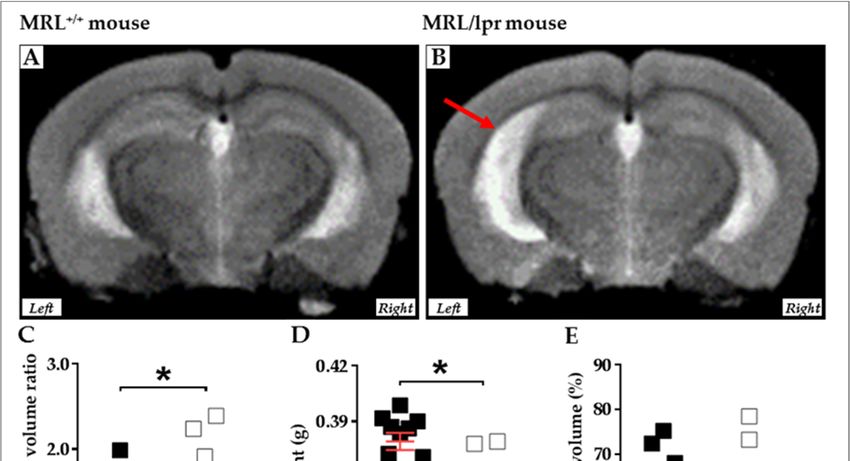
Int. J. Mol. Sci. 2018, 19, 3588                                                                                               2 of 34

the human disease in terms of peripheral inflammation and behavioral deficiencies, we inquire into
how advanced MRI techniques can be used non-invasively in mice to identify NPSLE symptoms and
how this might translate to the human disease. At last, the way in which neuroinflammation may
cause central nervous symptoms in these murine lupus-prone models is explored.

2. Systemic and NP Aspects of Lupus Disease

2.1. General Presentation of SLE
      SLE is a chronic relapsing-remitting autoimmune disease characterized by a rupture of
self-tolerance and systemic inflammation mainly resulting from the hyperactivation of peripheral B and
T cells [1], resulting in high levels of pathogenic autoantibodies (autoAbs), tissue deposition of immune
complexes, and, ultimately, multiple and various organ injuries (e.g., skin, kidneys, heart, lungs,
brain) [2–4]. The disease primarily affects females of childbearing age (90% of patients) [5], and its
etiology, which appears multigenic, is not fully understood due to it being also influenced by hormonal
and environmental factors (e.g., UV radiation, diet, smoke, infections, pollutants, stress) [6–8].

2.2. CNS Involvement in Human SLE: The Neuropsychiatric Lupus Disease (NPSLE)
     Depending on the study, a varying number of SLE sufferers (from 15% to 75%) present with
neuropsychiatric (NP) symptoms that cover the whole spectrum of psychiatric dysfunction [9,10].
These NP manifestations are associated with a reduced quality of life [11–13] and, when severe, they
substantially contribute to the morbidity and mortality rates of SLE patients [14]. Nowadays, the etiology
of NPSLE remains poorly understood, as is also the case for SLE and most autoimmune diseases.
     In 1999, the American College of Rheumatology (ACR) recognized 19 wide-ranging NP
manifestations related to SLE [15]: some affect the CNS, in which case they can be focal or diffuse [16],
and the others affect the peripheral nervous system (Table 1). This widely adopted classification
was meaningful from a clinical point of view but presents some limitations with respect to the
variety of symptoms and their specific attribution to SLE [11]. In 2001, Ainiala et al. revised
this original categorization and discarded certain minor NP symptoms, such as headaches and
anxiety disorders [17,18]. When compiling recently published data, it clearly appears that this
novel classification has affected the NPSLE prevalence values, pointing to the lack of consensus
on straightforward and uncontroversial NPSLE diagnosis (e.g., definition of impairment, selection of
patients and of cognitive tests used). Since then, various NPSLE diagnostic criteria have been proposed
but none of them have achieved both high sensitivity and specificity [19]. In fine, there is currently no
consensus about inclusion or exclusion criteria for NPSLE, resulting commonly in an overdiagnosis of
NPSLE and the administration of unnecessary immunosuppressive treatments.

                    Table 1. ACR case classification of the NP manifestations described in SLE [15].

   Central Nervous System                                                          Peripheral Nervous System
   Focal manifestations                    Diffuse manifestations
   Cerebrovascular disease                 Depression                              Cranial neuropathy
   Seizures                                Cognitive dysfunction                   Autonomic neuropathy
   Aseptic meningitis                      Mood and anxiety disorders 1            Mononeuropathy (single/multiplex)
   Movement disorder                       Psychosis                               Polyneuropathy
   Myelopathy                              Acute confusional state                 Plexopathy
   Demyelinating syndrome                  Headaches 1                             Myasthenia gravis
                                                                                   Acute inflammatory demyelinating
                                                                                   polyradiculoneuropathy
                                                                                   (Guillain-Barré syndrome)
      1 Anxiety disorders and headaches have been removed in the revised classification suggested by Ainiala et al. (2001) [18].

      According to Hanly et al. (2018) [11], posterior reversible encephalopathy syndrome, neuromyelitis optica spectrum
      disorder, and small fiber neuropathy should be included in a future revision of the ACR classification. Abbreviations:
      ACR, American College of Rheumatology; NP, neuropsychiatric; SLE, systemic lupus erythematosus. Adapted from
      Jeltsch-David and Muller (2014) [10].
Int. J. Mol. Sci. 2018, 19, 3588                                                                   3 of 34

     One critical problem with the diagnosis of NPSLE remains the lack of objective and specific
biomarkers. Therefore, this aspect remains eminently challenging. Today, NPSLE is essentially
clinically defined by physical examination, serological measures, psychological and neurological
evaluations, and brain imaging. Hopefully, rapidly advancing progress in live imaging technology
will help and allow the establishment of reliable and specific diagnostic criteria for NPSLE.
     Twenty autoAbs (11 brain-specific and 9 systemic) [20] and several cytokines found in the serum,
but more specifically in the cerebrospinal fluid (CSF) of patients, have been linked to NPSLE [10].
Focal manifestations can be detected by MRI techniques and reflect, usually, cerebral vasculopathy,
thrombosis, and complement activation [10,11,16]. The presence of antiphospholipid (aPL) autoAbs
in the CSF correlates with some of these events [16]. Diffuse symptoms are harder to identify [21]
and seem related, instead, to inflammation elicited by several mediators (e.g., interferon (IFN)α,
anti-N-methyl-D-aspartate receptor (NMDAR and anti-ribosomal P autoAbs), which induce not only
leakage of the blood–brain barrier (BBB) but also other barriers, as briefly addressed below [22–27].
     The brain is protected from the periphery by three distinct structural and functional interfaces
with systemic circulation: the BBB, the blood–CSF barrier (BCSFB), and the meningeal barrier [28].
These entities comprise endothelial cells (ECs) connected by tight junctions that prevent free passage
of soluble macromolecules and cells, control the influx of nutrients and efflux of toxic molecules,
and maintain a regulated microenvironment optimal for neuronal signaling [29,30]. The BBB has been
proposed to be leaky both in SLE patients and lupus-prone mice [31], resulting in the deleterious
diffusion of proinflammatory factors. In humans, this alteration has been evidenced mainly by the
presence of serum albumin and immunoglobulins G (IgGs) in the CSF [26,32,33] and through the use
of MRI [34]. Previously, it was claimed that BBB disruption was directly responsible for the passage
of peripheral molecules to the brain, initiating NPSLE disease. However, recent data obtained from
experiments with mice argue that the peripheral molecules reach the brain through the BCSFB rather
than via the BBB [22], providing interesting hypotheses in favor of BCSFB leakage in human patients
as well [35], and refining the BBB leakage dogma.

2.3. Modelization of the Disease in Mice
     There are obvious limits to the search for mechanisms of CNS disease in human patients.
Thus, murine models offer several advantages for elucidating the early mechanisms of NP
manifestations of SLE and help to distinguish between CNS-specific and -nonspecific mechanisms [36].
Neuroinflammatory mechanisms of NPSLE are quite well recapitulated in lupus-prone murine models
that develop a lupus-like disease [37], including NP events, manifesting through the production of
autoAbs in the serum [38] and CSF [39,40]. Some animal models are spontaneous, as the BXSB mice,
the Murphy Roths Large (MRL) mice, and the F1 hybrid of the New Zealand Black (NZB) and New
Zealand White (NZW) mouse strain (called (NZB × NZW) F1) [41]. Others are genetically engineered,
such as the 564Igi mouse [42,43]. All these murine models help to provide valuable insights into how
the CNS can be affected by systemic inflammation. Nevertheless, after the identification of autoAbs
targeting the brain, NPLSE-like inducible NPSLE models have also been developed. In these induced
models, human autoAbs are passively injected or, alternatively, mice are immunized against these
specific brain self-antigens, leading to the development of NPLSE-like disease without the severe
peripheral autoimmune damage [44–47]. Even if none of these animal models reflect the human disease
perfectly, they all provide some key elements involved in the disease’s pathogenesis, leading to the
development of safer and more efficient treatments, as was very well depicted in a recent review [48].
     The MRL/lpr strain is one of the best-established spontaneous models of SLE and is the
most commonly investigated in lupus-related NP studies. This strain displays an autoimmune
phenotype, and its composite genome is derived from LG/J (75%), AKR/J (12.6%), C3H/HeDi
(12.1%), and C57BL/6J (0.3%) mice [49]. MRL/lpr mice spontaneously develop an autosomal recessive
lymphoproliferation (lpr) mutation affecting the Fas gene [50]. Identified as an intron deletion in the
Fas gene, this mutation leads to aberrant Fas mRNA splicing [50] and the loss of protein expression [51].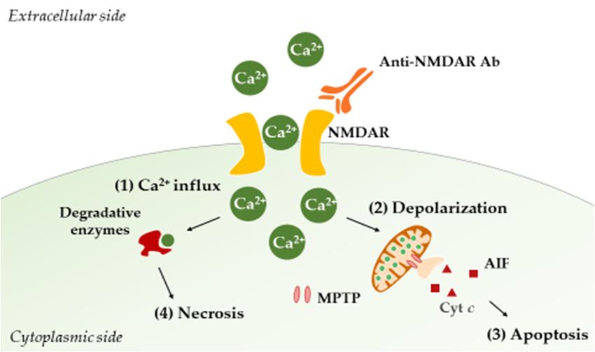
Int. J. Mol. Sci. 2018, 19, 3588 4 of 34
Interestingly, this mutation does not necessarily increase the proliferative capacity of lymphocytes
Int. J. Mol. Sci. 2018, 19, x FOR PEER REVIEW 4 of 33
but rather allows them to escape the negative and positive selection processes [50,51]. The parental
lymphoproliferation
strain of MRL/lpr mice, the(lpr) mutation affecting
MRL/MpJ mousethe Fas gene
(also named MRL+/+
[50]. Identified as later
an intron in deletion
the text),in the
does not carry
Fas gene, this mutation leads to aberrant Fas mRNA splicing [50] and the loss of protein expression
the lpr mutation and develops the autoimmune syndrome, albeit weaker
[51]. Interestingly, this mutation does not necessarily increase the proliferative capacity of
and delayed in life, thus
representinglymphocytes
a naturalbut andrather allows them to escape the negative and positive selection processes [50,51]. exhibited a
adequate control [52]. Over the past decade, the MRL/lpr
“fortuitous attenuation” ofofsymptoms
The parental strain MRL/lpr mice,of the MRL/MpJ
still unknown mouse (also named
origin, andMRL the+/+ later in the text), does
MRL/lpr original stock was
not carry the lpr mutation and develops the autoimmune syndrome, albeit weaker and delayed in
re-established in 2008 (http://jaxmice.jax.org/strain/006825.html) [36,41].
life, thus representing a natural and adequate control [52]. Over the past decade, the MRL/lpr
The disease
exhibited developed
a “fortuitousby MRL/lpr
attenuation” mice mimics
of symptoms humanorigin,
of still unknown SLE.and Intheparticular, in both settings,
MRL/lpr original
a leaky BBB stock
and was there-established
presence ofincirculating
2008 (http://jaxmice.jax.org/strain/006825.html)
autoAbs directed against [36,41]. dsDNA and Smith (Sm) antigen
The disease developed by MRL/lpr mice mimics human SLE. In particular, in both settings, a
are observed, the only serological biomarkers of SLE in humans. As in human SLE, where a strong
leaky BBB and the presence of circulating autoAbs directed against dsDNA and Smith (Sm) antigen
gender preference is usually
are observed, the only described
serological (9:1 female
biomarkers of to
SLEmale ratio),Asfemale
in humans. in human MRL/lpr
SLE, wheremice develop a more
a strong
severe diseasegender
[41].preference
Finally, iscomparable
usually described
to some(9:1 female to male ratio),evidenced
NP symptoms female MRL/lpr in SLEmicepatients,
develop a autoimmune
more severe disease [41]. Finally, comparable to some NP symptoms evidenced in SLE patients,
MRL/lpr mice spontaneously develop pathological changes in the brain and an autoimmunity-associated
autoimmune MRL/lpr mice spontaneously develop pathological changes in the brain and an
behavioral syndrome (e.g., depression,
autoimmunity-associated emotional
behavioral syndrome and cognitive
(e.g., dysfunction)
depression, emotional that and can be characterized
cognitive
with a batterydysfunction)
of behavioral that cantests,
be characterized
summarized with abelow
battery of behavioral
(Figure 1). tests, summarized
However, it is below (Figureto emphasize
important
1). However, it is important to emphasize that, in this murine model, the disease-associated brain
that, in this murine model, the disease-associated brain atrophy remains poorly understood.
atrophy remains poorly understood.
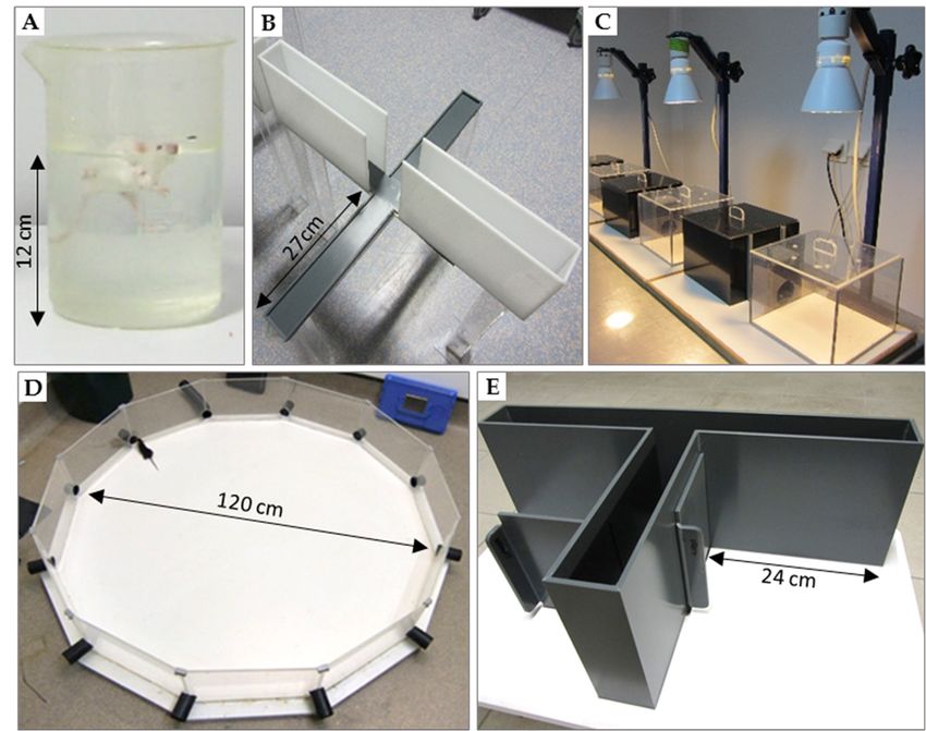
Figure 1. Tests used for behavioral evaluation of mice. (A) Forced-swim test (or Porsolt test) used to
Figure 1. Tests used for behavioral evaluation of mice. (A) Forced-swim test (or Porsolt test) used to
assess depressive-like behavior; (B) elevated plus-maze and (C) dark/light preference test to measure
assess depressive-like behavior;
anxiety-like behavior; (D) (B) elevated
Morris plus-maze
water maze and
(or paddling test)(C) dark/light
[53]; preference
(E) T-maze that test to measure
is considered
anxiety-like very
behavior;
sensitive(D) Morrisinwater
to alteration maze pathways.
hippocampal (or paddling test) [53]; (E) T-maze that is considered very
sensitive to alteration in hippocampal pathways.
2.3.1. Tests of Depression
• Depression
2.3.1. Tests of Forced-swim test (Porsolt test; Figure 1A)
In this experimental procedure, a mouse is forced to swim in a small container without the
• Forced-swim test (Porsolt test; Figure 1A)
possibility to escape. Quickly, after a time of struggling and swimming, the animal stops any attempt
In this of escape and becomes almost motionless (e.g., floating on the water surface and moving its limbs
experimental procedure, a mouse is forced to swim in a small container without the
possibility to escape. Quickly, after a time of struggling and swimming, the animal stops any attempt
of escape and becomes almost motionless (e.g., floating on the water surface and moving its limbs only
to rebalance itself). Periods of activity alternate with periods of immobility, and the duration of the
latter increases with time. Within this framework, immobility objectivizes behavioral helplessness and
is interpreted as behavioral “distress” that ensues when the animal realizes that it cannot escape [54].
This model of behavioral despair is repeatedly validated by the pharmacological plan to detect the
efficacy of antidepressants. The quicker the animal becomes immobile and stops trying to escape,
i.e., the longer it stays immobile, the greater extent to which it is thought to display depressive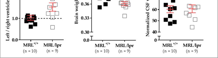
Int. J. Mol. Sci. 2018, 19, 3588                                                                      5 of 34

symptoms. As compared to their parental MRL+/+ littermates used as controls, MRL/lpr mice exhibit
defects [55–57] implying an autoimmunity-associated syndrome.
• Sucrose preference test
     This test evaluates deficiencies in motivated and goal-directed behavior and anhedonia, i.e.,
a reduced sensitivity to positive stimuli. Here, MRL/lpr mice manifest a reduced preference for
palatable drinking solutions with sucrose compared to control MRL+/+ mice. This observation is well
established and can be detected both early in life and later during disease progression [57–61].
• Open-field locomotor activity test
     Depressive-like manifestations also include apathy and fatigue, which are reflected as a decrease
in spontaneous exploration of a novel environment, like an open field. Generally, as compared to
control mice, MRL/lpr mice show such a decrease in spontaneous locomotion [55,56,62].

2.3.2. Anxiety Tests
• Elevated plus-maze and dark/light preference tests (Figure 1B,C)
     The elevated plus-maze and the dark/light preference tests are usually implemented in
pharmacology for the screening of anxiolytic molecules [63–65]. The plus-maze test consists of a cross
structure with two arms with walls (closed arms) and two wall-free arms (open arms). The dark/light
preference test consists of two boxes, one is dark and the other is well lit, and an opaque tunnel that
connects the dark box with the lit one. Both tests create an approach-avoidance conflict between the
natural tendency of mice to explore and their aversion to open or brightly lit spaces. Generally, the
more anxious the animal is, the less it will venture and stay in the open or lit compartments. Some
debatable results have been reported in MRL/lpr mice with the elevated plus-maze test. Sakić and
colleagues observed anxiety in MRL/lpr mice, i.e., diminution of the time spent in the open arms [55],
while other groups noted the opposite result, with MRL/lpr mice being significantly less anxious than
their MRL+/+ littermates [57,66,67]. An important experimental difference between these two sets of
studies is that some experiments were conducted with males [55] and others with females [57,66,67].
• Open-field test
      The open-field test is sometimes used to measure anxiety-related behavior, as quantified by the
duration for which the mouse avoids the central part of a novel enclosed arena and remains in close
proximity of the walls (thigmotaxis) [68,69]. In this test, as compared to control MRL+/+ mice, MRL/lpr
mice disclose anxiety-like behavior, as evidenced by increased thigmotaxis and impaired exploration
of space [55,57].
      Controversial results have been reported for open-field and elevated plus-maze tests that might
be explained by the use of different strains and sex of mice [70,71]. To discuss the absence of
consensus in detail is beyond the scope of this essay but, as well depicted in an exhaustive review, it is
worth mentioning that independent of the strain, male and female mice display inherently different
anxiety-coping mechanisms [72]. Potential processes underlying sex differences in anxiety states
include emerging evidence supporting the existence of two anatomically and functionally distinct
serotonergic circuits that modulate conflict anxiety and panic-like anxiety, respectively. Regarding
the MRL/lpr strain, female MRL/lpr mice seem to display, as in human SLE, a strong bias in severity
(e.g., higher levels of IgG in the CSF) [40] and rate of progression of the autoimmune disease (e.g., earlier
apparition of serum autoAbs) [57], which manifest in stronger cerebral pathology [56,66,73]. Therefore,
sex differences in the expression of anxiety behavior are not surprising or unexpected in MRL/lpr mice.

2.3.3. Cognitive Tests
    The detection of cognitive deficits in animals seems to be task-dependent. This finding is hardly
surprising with regard to the fact that there are multiple “types” of memory, which are differently
Int. J. Mol. Sci. 2018, 19, 3588 6 of 34
sensitive to brain damages [74]. Even if the three tests listed below are classified as “cognitive” tasks,
they do not measure the same “cognitive processes” (working (or short-term) visual memory in
the novel object recognition test [75], spatial working and reference (or long-term) memory in the
Morris water maze, spatial working memory in the T-maze that is also sensitive to nonspatial learning
aspects (e.g., temporal discriminations) [76]). Furthermore, these tests differ in terms of perceptual
stimuli cueing choice behavior, and their accomplishments rely on different mechanisms, some being
noncognitive (e.g., motivational factors), subserved by the activity of many brain regions.
• Novel object recognition test
This test is based on the tendency of rodents to preferentially explore novel objects. First, a mouse
is placed into an arena and allowed to explore two identical novel objects. After a fixed period of
exploration (defined as rearing on the object, whisking, sniffing, touching with nose and/or forepaws),
the mouse is removed. Next, one of the objects is changed to a novel object and the mouse is
reintroduced into the arena. Mice naturally tend to explore novel objects, so the duration of exploring
the novel object is taken as the measure of visual working memory. In this paradigm of testing,
MRL/lpr mice perform as well as MRL+/+ controls [66] or even outperform them [57].
• Morris water maze (Figure 1D)
The Morris water maze test evaluates spatial memory in rodents [77]. The animal is placed into
a tank filled with opaque water in which a platform is hidden beneath the surface. In this aversive
situation, mice must learn using spatial cues placed in the testing room to navigate and find the
platform. Longer latencies to find the platform reflect poorer performances. In this test, MRL/lpr mice
show pronounced thigmotaxic swimming but no clear-cut impairment in learning/memory abilities
compared to MRL+/+ littermates [56,78,79].
• The T-maze alternation test (Figure 1E)
This test, shaped like the letter T, is based on the willingness of rodents to explore a new
environment, i.e., they prefer to visit a new arm of the maze rather than the familiar one. The solving
of this task consists of two turns, i.e., right and left. Mice are first placed in the start arm of the
T-maze. Upon leaving the start arm, they choose between entering either the left or the right goal arm.
With repeated trials, the animals show less tendency to enter a previously visited arm. The percentage
of alternation (number of turns in each goal arm) is recorded. This test is currently used to evaluate
cognitive deficits in mice and test novel chemical entities for their effects on cognition. The T-maze test
is well known as particularly sensitive for detecting hippocampal dysfunction [80–83]. In MRL/lpr
mice, alternation impairments have been evidenced as compared to the MRL+/+ counterparts [84,85],
strongly supporting cognitive and, more specifically, hippocampal dysfunction in these mice.
2.3.4. Locomotor function
• Beam-walking test
The beam-walking test assesses the capacity of mice to coordinate movement on a narrow beam [67].
Mice are placed on the beam and the latency to cross and the number of paw slips are recorded.
Interestingly, female MRL/lpr mice do not display locomotor deficits compared to their control MRL+/+
littermates [49,50]. However, males perform significantly worse [50]. The precise mechanisms of this
sex difference remain unclear but might be due to different hormonal backgrounds [86].
Other tests assessing locomotor functions have been described, such as the pole test and string
agility test, for example [68,69]. These tests, which require a good driving agility, give an idea of the
integrity of proprioceptive and vestibular pathways. Generally, they are used in order to quantify the
effects of either alterations in the CNS (motor cortex), those of various neuromuscular pathologies,
or those of fatigue on the fine motricity and proprioceptive functions. These tests also allow checking for
the presence of possible sensory-motor biases, which could affect observations obtained in other tests.Int. J. Mol. Sci. 2018, 19, 3588 7 of 34
The characterization of specific inflammatory factors giving rise to an autoimmunity-associated
behavioral syndrome has contributed to the development of NPSLE inducible murine models.
For example, some NP features have been observed in C3H/HeJ mice after ribonucleoprotein P human
autoAb intracerebroventricular injection [87]. Anti-NMDAR Abs effects have been studied following
injection of human anti-dsDNA Abs into the hippocampi of C57BL/6 mice [46] or immunization of
BALB/c mice with the DWEYS peptide [44,45,88,89]. To date, only a few inducible models have been
yet developed, but it is anticipated that a growing number of relatively specific models of NPSLE will
be generated in the nearest future. Taken together, we can conclude that spontaneous and inducible
NPSLE models fairly well recapitulate the human disease and provide important insights into how
systemic inflammation can induce CNS damage. This central aspect is discussed below.
3. Neuroimaging in NPSLE
3.1. Magnetic Resonance Imaging Modalities
As already mentioned, two distinct, but potentially complementary, pathogenic mechanisms
are distinguished in NPSLE: (i) vascular/thrombotic injury, likely implicating aPL Abs; and (ii)
inflammation-mediated injury, implicating other pathogenic autoAbs, proinflammatory cytokines
(e.g., interleukin (IL)-6, IFNα), and disruption of the BBB, the latter being favored, among other
factors, by the activation of the complement system and binding of immune complexes to ECs [90,91].
Nowadays, implementation of noninvasive anatomical and functional neuroimaging modalities is
highly recommended for the identification of outcomes of pathogenic mechanisms that impact the
structure, metabolism, and functionality of the brain.
MRI detects signals thanks to the properties of hydrogen nuclei in water and fat molecules, which
are found in biological tissues, including the brain. It is the most relevant neuroimaging technique
for the detection of structural alterations in the CNS (e.g., cortical atrophy, focal periventricular
and subcortical white matter lesions, diffuse gray matter changes, reduced volume of the corpus
callosum or the hippocampus) [92]. It is thus particularly sensitive to detecting acute focal NP
manifestations [93–101]. Unfortunately, its diagnostic value remains limited since, in the case of diffuse
manifestations, MRI usually gives unremarkable results or shows nonspecific abnormalities. To make
matters worse, cerebral MRI abnormalities can be observed in SLE patients without NP symptoms and
are even sometimes detected in healthy individuals [102–104].
Today, several advanced imaging modalities exist. These include diffusion-weighted imaging
(DWI) [105], magnetic transfer imaging (MTI) [106], magnetic resonance angiography (MRA) [107],
magnetic resonance spectroscopy (MRS) [108], diffusion tensor imaging (DTI) [109], and blood oxygen
level-dependent functional MRI (BOLD-fMRI) [93], and they ideally could be implemented for identifying
functional hallmarks of NPSLE and allow its diagnosis [11,110]. These methods are not detailed here,
as this review rather focuses on data obtained in preclinical research performed on murine NPSLE models.
3.2. Brain Imaging and MRL/lpr Mice
In NPSLE preclinical research, MRI remains, unfortunately, rarely used, largely due to the high
cost necessary for its implementation. However, some data in relation with anatomical findings have
been generated and are summarized below.
• MRI
Atrophied cerebral structures in MRL/lpr mice were identified several years ago using
paraformaldehyde (PFA)-fixed MRL/lpr brain [111]. Atrophy was noticeable in the superior colliculus,
periaqueductal gray matter, pons, and midbrain. However, as PFA fixation could affect both brain
morphology and MRI data [112], the conclusions raised from these studies remain to be confirmed.
We recently compared the brain morphology of 16-week-old female MRL+/+ (n = 10) and MRL/lpr
(n = 9) mice based on transverse relaxation time (T2)-weighted imaging (T2WI) MRI. MRI wasInt. J. Mol. Sci. 2018, 19, x FOR PEER REVIEW 8 of 33
Int. J. Mol. Sci. 2018,
colliculus, 19, 3588
periaqueductal gray matter, pons, and midbrain. However, as PFA fixation could affect8 of 34
both brain morphology and MRI data [112], the conclusions raised from these studies remain to be
confirmed.
performed Weusing
recentlya 7/30 Biospec
compared system
the brain (Bruker Biospin,
morphology Ettlingen,
of 16-week-old female Germany)
MRL+/+ (n =with ParaVision
10) and MRL/lpr 6.0.1
software.
(n = 9) mice based on transverse relaxation time (T2)-weighted imaging (T2WI) MRI. MRIofwas
Transmission was achieved with a quadrature volume resonator (inner diameter 86 mm),
and aperformed
standardusingmouse brain
a 7/30 quadrature
Biospec surfaceBiospin,
system (Bruker coil (~19 × 19 mm
Ettlingen, 2 ) was used
Germany) for signal reception
with ParaVision 6.0.1
software.
(Bruker Transmission
BioSpin, Ettlingen, wasGermany).
achieved with A aT2WI
quadrature
axialvolume resonator
anatomical (innerwas
dataset diameter of 86 mm),
acquired using the
RARE and a standard
sequence mouse
(256 × 256 brain quadrature
acquisition surface
matrix, 23coil (~190.5
slices, × 19mmmmslice
2) was used for signal reception
thickness, in-plane resolution
(Bruker BioSpin,
2 Ettlingen, Germany). A T2WI axial anatomical
of 78 × 78 µm , repetition time (TR) of 3 s, echo time (TE) of 30.6 ms, RARE-Factor dataset was acquired usingof the8).
RAREAnimals
sequence (256 × 256 acquisition matrix, 23 slices, 0.5 mm slice thickness, in-plane resolution of 78 × 78
were anesthetized with 2% isoflurane, the respiration was noninvasively monitored using a magnetic
μm2, repetition time (TR) of 3 s, echo time (TE) of 30.6 ms, RARE-Factor of 8). Animals were
resonance-compatible system, and the body temperature was maintained constant at 37–38 ◦ C. In this
anesthetized with 2% isoflurane, the respiration was noninvasively monitored using a magnetic
study,resonance-compatible
an asymmetrical enlargement
system, and was noticeable
the body in MRL/lpr
temperature brains (Figure
was maintained constant2A,B) with
at 37–38 °C.significantly
In this
increased ventricular volume on the left side, as demonstrated by
study, an asymmetrical enlargement was noticeable in MRL/lpr brains (Figure 2A,B) with the increased left/right ratio for
ventricular volume
significantly (p = 0.035;
increased Figure
ventricular 2C). Interestingly,
volume on the left side,aassignificant
demonstrated lossbyofthe
brain weight
increased was noted
left/right
in diseased MRL/lpr mice
ratio for ventricular volume (p(p== 0.022; Figure
0.035; Figure 2D),
2C). but no difference
Interestingly, a significant concerning
loss of brainthe CSFwas
weight volume
couldnoted in diseased
be noted betweenMRL/lpr
MRLmice+/+ and(p = 0.022;
MRL/lpr Figure 2D),(p
mice but
= no difference
0.842; Figureconcerning the CSF dilation
2E). Ventricular volume and
could
cerebral be noted
atrophy between
have MRL+/+
also been and MRL/lpr
previously mice (p
reported in=human
0.842; Figure
NPLSE 2E). Ventricular
patients [94]. dilation and
The meaningful
cerebral atrophy have also been previously reported in human NPLSE
enlargement of the left ventricle in MRL/lpr mice remains largely unstudied but might reveal patients [94]. The meaningful
enlargement of the left ventricle in MRL/lpr mice remains largely unstudied but might reveal
lateralized brain damage [113] that, for example, has already been observed in neurodegenerative
lateralized brain damage [113] that, for example, has already been observed in neurodegenerative
diseases [114]. Such asymmetry might be viewed as a major feature of the disease, which also raises
diseases [114]. Such asymmetry might be viewed as a major feature of the disease, which also raises
questions on the
questions nature
on the of this
nature of thislateralization.
lateralization.
FigureFigure 2. Cerebral
2. Cerebral abnormalitiesmeasured
abnormalities measured inin MRL
MRL+/+ andand
+/+ MRL/lpr mice. mice.
MRL/lpr Conventional mid-axialmid-axial
Conventional T2-
weighted MRI revealed a dilation of ventricles (red arrow) in 16-week-old female MRL/lpr mice (B)
T2-weighted MRI revealed a dilation of ventricles (red arrow) in 16-week-old female MRL/lpr mice
as compared to the age-matched counterparts MRL+/+mice (A). In the MRL/lpr strain, this enlargement
(B) as compared to the age-matched counterparts MRL+/+ mice (A). In the MRL/lpr strain, this
is more remarkable on the left side, as revealed by analysis of the right/left ventricle volume ratio (C).
enlargement is more remarkable on the left side, as revealed by analysis of the right/left ventricle
volume ratio (C). Furthermore, a significant loss of brain weight was detected in these mice (D), even if
the normalized volume of CSF did not show significant difference between both strains (E). Statistics:
All data were analyzed with unpaired t test, and significance was defined as p < 0.05 (*) Errors bars
are mean standard deviation. Sample size is indicated as n. Abbreviations: CSF, cerebrospinal fluid;
lpr, lymphoproliferation; MRI, magnetic resonance imaging; MRL, Murphy Roths Large.Int. J. Mol. Sci. 2018, 19, 3588 9 of 34
• MEMRI
MEMRI is a relatively new imaging technique that is not dependent on blood flow. As a
paramagnetic contrast agent, Mn2+ is of special interest in neuroimaging, as it enhances MRI contrast
in vivo by shorting both T1 (longitudinal) and T2 (transverse) relaxation times. Mn2+ ions enter neurons
and excitable cells during the excitation phase through voltage-gated calcium channels. As such, MEMRI
has unique capabilities for defining cerebral architecture, mapping neuronal pathways, and studying
connectivity in morphological and functional imaging studies [115].
This MRI modality was applied to explore the olfactory pathway in mice with experimental
NPSLE induced after intracerebroventricular injection of anti-ribosomal-P Abs [116]. Passive transfer of
anti-ribosomal-P Abs induced a depressive-like behavior with a significant deficit in olfactory function.
MEMRI of these mice demonstrated significant reduction in normalized Mn2+ enhancement ratios
of olfactory structures, as compared to control mice. Thus, an impaired olfactory neuronal function
in mice with experimental depression, mediated by passive transfer of human-anti-ribosomal-P Abs,
can be spotted by MEMRI.
• 1 H-MRS
MRS uses the principles of MRI and, as with nuclear magnetic resonance spectroscopy,
allows identification of specific metabolites in vivo at concentrations ranging from 0.5 to 10 mM.
1 H-MRS determines the 1 H spectra of molecules [117]. The molecules that can be studied include
choline-containing compounds (markers of cellular membrane turnover), creatine (involved in energy
metabolism), N-acetylaspartate (NAA; marker of neuronal viability), and lactate (marker of anaerobic
metabolism, necrosis, and/or infections). 1 H-MRS is more sensitive than MRI, evidencing lesions in
white and gray matter that appear normal in a conventional MRI of SLE patients.
1 H-MRS studies have shown significant metabolic differences between MRL+/+ and lupus-prone
mice in the hippocampus and the cortex [66], structures that, as we already emphasize in this
review, are involved in the regulation of cognition and memory. In MRL/lpr mice, these metabolic
abnormalities were shown to reflect dysfunction of neuronal (and/or glial) activity, but they did not
correlate to structural changes that were detectable by MRI [66]. Both methods are, therefore, necessary
to generate a complete picture.
MRI data evidencing an altered cerebral metabolic state in MRL/lpr mice confirm insights on
the neuroinflammatory mechanisms of NPSLE in these mice. One major advantage of MRI is its
applicability in vivo, allowing the real-time study of the disease progression. At this stage, additional
investigations have to be carried out to understand the reasons for the ventricular system asymmetry
in MRL/lpr mice, a phenomenon never described until now.
4. Neuroinflammation in Lupus-Prone Mice
4.1. Characteristic Elements Occurring in the Brain of Lupus-Prone Mice
A summary of the different autoAbs and cytokines detected in MRL/lpr mice and suspected to
play a role in NPSLE is shown in Table 2.
4.1.1. Pathogenic AutoAbs
Evidence supporting the role of autoAbs in the CNS pathology and subsequent negative
behavioral outcomes includes the higher levels of autoAbs in the serum of diseased MRL/lpr mice
that develops earlier in females [57,66] (for extensive reviews, see [36,41]).
There is further demonstration that some of these serum autoAbs recognize brain antigens, such as
a subtype of NMDAR glutamate [118,119], an excitatory neurotransmitter. When injected directly
into the brain of healthy mice, or when injected peripherally into animals with a breached BBB, these
autoAbs are neurotoxic and induce deficits in cognition and emotional behavior [46,120]. We focus
attention on this issue in greater detail in Section 4.3.Int. J. Mol. Sci. 2018, 19, 3588 10 of 34
Intrathecal administration of anti-ribosomal P Abs induces depression-like behavior in the forced
swim test [87]. The relation between serum or CSF levels of autoAbs and NPSLE disease processes is
complex, but it is likely that intrathecal CSF brain-reactive autoAbs titers [39] may be more critically
related to NPSLE pathogenesis and symptoms than serum ones [121].
Table 2. Potential NPSLE biological hallmarks found in MRL/lpr mice.
Hallmarks Location Levels/Expression References
AutoAbs
aPL (e.g., anticardiolipin) Serum Increased levels [122]
Anti-dsDNA Serum Increased levels [123]
Anti-nucleosome * Serum Increased levels [124]
Anti-ribosomal P protein Serum Increased levels [125]
Anti-Sm Serum Increased levels [125]
Anti-ribosomal S10 Serum Increased levels [126]
Anti-NMDAR Serum Increased levels [57,66]
Cytokines
Serum
IL-1β Increased levels [127–130]
CNS
IL-2 T cells Decreased expression [131,132]
Serum
IL-6 Increased levels [127,133–135]
CSF
IL-9 Serum Increased levels [128,130,136]
B cells
IL-10 Dysregulation [128,130,137,138]
CNS
IL-12 Serum Increased levels [139]
IL-17 Serum Increased levels [140]
IL-18 Serum Increased levels [141,142]
IL-21 Serum Dysregulation [143]
IL-22 Serum Increased levels [144]
M-CSF Serum Increased levels [145]
MIF Serum Increased levels [146]
Splenocytes
IFNγ Dysregulation [128,147,148]
CNS
TNFα Serum Increased levels [127,129,149]
TWEAK CNS Tendency to increase [150]
Receptors
sIL-6R Serum Increased levels [133]
Fn14 (TWEAK receptor) CNS Increased levels [150]
* Molecules indicated in italics are those for which relevance in NP manifestations remains to be confirmed.
Abbreviations: aPL, antiphospholipid; CSF, cerebrospinal fluid; ds, double-stranded; IFN, interferon; IL, interleukin;
lpr, lymphoproliferation; M-CSF, macrophage colony-stimulating factor; MIF, macrophage migration inhibitory
factor; MRL, Murphy Roths Large; NMDAR, N-methyl-D-aspartate receptor; sIL-6R, soluble IL-6 receptor; Sm, Smith;
TNF, tumor necrosis factor; TWEAK, TNF-like weak inducer of apoptosis.
4.1.2. Cytokines
As it is not necessary that they pass the BBB to regulate neural function, cytokines and chemokines
are critical early factors involved in behavioral defects [151]. The role of several cytokines in behavioral
disturbances in MRL/lpr mice is supported by numerous studies (see [36]). More specifically, the early
dysregulation of cytokine production, especially IL-1, IL-2, IL-6, and tumor necrosis factor (TNF)α,
corresponds to the onset of symptoms of depressive-like behavior, such as anhedonia and behavioral
despair. Interestingly, anhedonia is ameliorated by cyclophosphamide, which suppresses the typically
early and significant rise of IL-6, and can be replicated by exogenous IL-6 [58]. Furthermore, high levels
of proinflammatory cytokines may also impair the function of the BBB and, thus, may be permissive to
the deleterious effects of certain autoAbs and lymphocytes.Int. J. Mol. Sci. 2018, 19, 3588 11 of 34
In MRL/lpr mice, the CNS actively responds to the systemic production of several cytokines,
as an upregulation of adhesion molecule expression is observed in the brain of diseased mice [129,152].
Upregulated in the peripheral blood of MRL/lpr mice, TNFα, IL-1α, and IL-1β are able to diffuse into
the CSF and brain parenchyma [153,154] where they increase the expression of ICAM-1 and VCAM-1,
leading to the recruitment of immune cells and subsequently to damage by either direct cytotoxicity
or exacerbation of neuroinflammation. As suggested by the observation that inhibition of ICAM-1
in MRL/lpr mice prevents peripheral nerve damage, inhibiting cell adhesion molecule expression
in NPSLE could be a promising therapeutic option. However, this line of treatment did not reduce
infiltration to the choroid plexus (CP) [155], indicating that, though potentially beneficial, this therapy
alone is not sufficient to allow complete resolution of NPSLE signs.
Of the various cytokines suspected to be involved in NPSLE pathogenesis, we make special
mention the possible role of macrophage colony-stimulating factor (MIF), which is a proinflammatory
cytokine displaying multifunctional properties. Operative in innate and adapted immunity [156,157],
it plays an important role in regulating macrophage effector functions and T cell division [158].
MIF has moreover been highlighted in different structures of the CNS (e.g., hippocampus, cortex,
cerebellum, pons) [159,160]. Its implication in the pathogenesis of many autoimmune/inflammatory
diseases (e.g., multiple sclerosis (MS), Guillain Barré syndrome) is quite well described both in
experimental and clinical studies [161–163]. MIF contributes also, with sex-specific regulation,
to the emergence of depression and psychiatric disorders, likely via the dysregulation of the
hypothalamic–pituitary–adrenal (HPA) axis and glucocorticoid secretion [164,165]. MIF is upregulated
in SLE patients, where its level correlates positively with disease progression [166], as well as in
MRL/lpr mice [146]. Moreover, the deletion of the MIF gene protects MRL/lpr mice from renal and
skin manifestations of the disease [167] and reduces depressive symptoms in C57BL/6 mice [164].
Even more, as specific inhibitors of MIF attenuate the clinical course of SLE, therapeutic antagonism
of MIF may be investigated as an opportunity for targeted therapy [167]. All these data, as well as
the fact that MIF is also involved in autophagy [166], emphasize the important role played by MIF in
the effector pathways of immune-mediated inflammatory damage both in SLE patients and murine
lupus models [146,166,168–170]. They also point to its potential therapeutic use for treating NPSLE. In
agreement with this hypothesis, an immunomodulatory peptide, hCDR1, which reduces NPSLE-like
symptoms in lupus-prone mice [171], has been found to act on the MIF pathway by reducing its
overexpression in the hippocampus [172]. Unfortunately, the effects of MIF and its inhibitors have not
yet been further investigated in human NPSLE.
4.1.3. Peripheral Immune Cell Infiltration
Some years ago, the discovery of lymphatic vessels within the dura mater surrounding the brain
provoked the dismissal of the existence of CNS immune privilege [173–175]. Regarding SLE and
NPSLE, the access of peripheral immune cells, of which the best studied are lymphocytes, to the CNS
through the CP has now been demonstrated in MRL/lpr mice [22]. Indeed, CD3+ T cells are detected
in various areas of the MRL/lpr brain (e.g., ventricles, CP, interhemispheric fissure, hippocampus,
meninges, stria medullaris, cerebellar parenchyma), while CD19+ B cells are only found in the CP and
the interhemispheric fissure [176]. Among the CD3+ T cells, brain parenchyma is largely infiltrated
by inactive CD8+ T cells [177], while, in the CP, the larger fraction of infiltrating cells are effector
CD4+ T cells, which were further identified as T follicular helper (Tfh) cells [73]. Whereas a specific Tfh
subset promotes B cell differentiation in the CP, CD4+ cells are probably responsible for the increased
production of proinflammatory cytokines upon detection of brain-derived self-antigens, leading to
the recruitment of other immune cells and parenchymal infiltration [178]. Deletion of the CD4+ T
cell phenotype has proven to be a successful therapy in mice developing CNS disease, indicating
that targeting this cell phenotype might be an approach for treating NPSLE [179]. Unfortunately, this
therapeutical option induces deleterious effects in other organs, probably as a result of the deletion of
CD4+ T regulatory cells, a cell phenotype that attenuates inflammation [180].Int. J. Mol. Sci. 2018, 19, 3588 12 of 34
4.1.4. Glial Cells
Although poorly explored and not well understood, the role of glial cells, the “sentinels
of the brain”, is expected to be central during the neuroinflammatory process of lupus [84].
Some authors reported the upregulation of ionized calcium binding adaptor molecule 1 (Iba1;
a microglia/macrophage-specific calcium-binding protein) and of CD68 (glycoprotein expressed
by macrophages) in microglia of lupus-prone mice [43,44]. When activated, these cells express
proinflammatory molecules and, as mentioned below, perform “synaptic pruning” [43]. Moreover,
they probably incur damage by yet unidentified mechanisms. On the other hand, astrocytes are also
activated during the disease, as evidenced by elevated astrogliosis in MRL/lpr mouse brain [181].
Further investigations focusing on the function of these cells in the pathogenic mechanisms of NPSLE
are needed to better understand their precise role.
4.2. Hippocampus as the Possible Primary Target of Neuroinflammatory Lupus
NP symptoms observed in mouse models of lupus are commonly attributed to the dysfunction
of the hippocampus, an observation also seen in human patients in whom reduced volumes of the
hippocampal CA1 and CA4 regions have been associated with worse cognitive performances [182].
Accordingly, hippocampal neurons in MRL/lpr mice show signs of degeneration when stained by
Fluoro-Jade B [153] that are backed by increased levels of proinflammatory cytokines (e.g., IFN-γ,
IL-1β, IL-6). Nonetheless, the concomitant presence of anti-inflammatory and immunomodulatory
cytokines, for example, IL-10, highlights the complexity of the inflammatory context that occurs in
the brains of these mice. In a comparable line, the simultaneous dysregulated production of both
proinflammatory and anti-inflammatory cytokines has also been observed in SLE patients at the
periphery [183]. This apparently common immunopathogenic aspect strengthens even more the
immunopathogenic relevance of the murine models of lupus used to approach human SLE.
Several important properties of the hippocampus may explain both the neuroinflammatory
and neurodegenerative processes affecting this cerebral structure during lupus disease. Firstly,
the hippocampus is located next to the CP that, in addition to the meninges, as already pointed out
above, is a common site of immune peripheral infiltration [176]. Furthermore, immunohistochemistry
techniques have revealed that some peripheral immune cells infiltrate the hippocampus itself,
homing in on structures that are much beyond their initial site of penetration. Thus, hippocampal
proximity to immune infiltration sites could partially explain the susceptibility to NPSLE in this
setting. Secondly, the hippocampus is a zone of prominent neurogenesis containing proliferating
and maturing cell populations [184–186]. In MRL/lpr mice, replicating neuronal cells may display
abnormal distribution throughout the CNS [187]. Moreover, the CSF of diseased mice evidences
cytotoxicity toward proliferating neuronal cells [121,188]. Two general lines of hypotheses might
explain why neurogenesis renders the hippocampus particularly sensitive to autoimmune diseases.
One assumption is that newly forming neurons express surface markers different from those
of differentiated neurons. Maturing neurons would express NMDAR [189] that is targeted by
brain-reactive autoAbs. When it occurs, such binding elicits immediate excitotoxic neuronal death [190].
The second line of assumption questions the involvement of complement factors in neurogenesis and
disease pathogenesis [191,192]. The complement components C1q and C3, produced by microglia, can
be considered proinflammatory factors, as they play effector roles in a range of functions, including T
cell activation and survival, chemotaxis, mast cell degranulation, and macrophage activation. Some of
these activities participate in the synaptic loss and death of developing neurons [43,193]. In addition
to neurogenesis, the hippocampus is a site of intense synaptic plasticity during ontogenesis and
the learning phase [194]. Synaptic plasticity requires the trimming of specific neuronal circuits and
reinforcement of others. Normally, this trimming, called “synaptic pruning”, occurs during early fetal
development and adolescence in humans (corresponding to 4 weeks post-natally in mice), but it can
also be observed during adulthood [195]. Synaptic pruning is performed by microglia in the CNS [196]
and also depends on complement factors [194]. Its overactivation results in injury in NPSLE [43].Int. J. Mol. Sci. 2018, 19, x FOR PEER REVIEW 13 of 33
Int. J. Mol. Sci. 2018, 19, 3588 13 of 34
natally in mice), but it can also be observed during adulthood [195]. Synaptic pruning is performed
by microglia in the CNS [196] and also depends on complement factors [194]. Its overactivation
results in injury
Although furtherinresearch
NPSLE is [43]. Although
needed, this further research
observation is needed,
suggests this observation
that some zones of highersuggests that
synaptic
some zones
plasticity of higher
where synaptic plasticity
new neuronal where
circuits are new neuronal
developing, circuitsinare
for instance, thedeveloping,
hippocampus for[195],
instance, in
could
theparticularly
be hippocampus [195],tocould
sensitive be particularly
autoimmune sensitive
dysfunction. to treatments
Overall, autoimmune dysfunction.
promoting Overall,
hippocampal
treatments promoting
neurogenesis hippocampal
and inhibiting neurogenesis
the complement and might
cascade inhibiting the complement
be regarded cascade
as promising might be
therapeutic
regarded as against
approaches promising therapeutic
NPSLE approaches against NPSLE [193,197,198].
[193,197,198].
Though the hippocampus seems to be centrally involved in NPSLE, other cerebral areas are
affected in this disease, such as the cortex [199], the paraventricular nucleusnucleus (PVN)
(PVN) [39,193,200,201],
[39,193,200,201],
the cerebellum
cerebellum [130], and probably a few others. For example, damage to the PVNthe
[130], and probably a few others. For example, damage to the PVN alters sucrose
alters the
preference
sucrose behavior
preference [58,60] [58,60]
behavior and increases anxiety.
and increases PVNPVN
anxiety. damages
damages areare
also
alsolinked
linkedto to systemic
inflammation as excessive proinflammatory cytokines inhibit the negative negative feedback
feedback of of the
the HPA
HPA axis,
axis,
increase the permeability of the BBB, and disturb the glutamatergic balance [200,202]. At At this stage,
stage,
however, the
however, the mechanisms
mechanisms underlying
underlying these
these processes
processes are
arenot
notclear.
clear.
4.3. Potential Mechanisms of Neuroinflammation in NPSLE
In lupus-prone
lupus-pronemice,mice,different
different studies
studies usingusing
TUNEL TUNEL and Fluoro-Jade
and Fluoro-Jade staining staining have
have reported
reported neurodegeneration and apoptosis phenomena in the hippocampus,
neurodegeneration and apoptosis phenomena in the hippocampus, the PVN, and the cortex the PVN, and the
cortex [153,193,201,203,204].
[153,193,201,203,204]. Today,Today, a causal
a causal relationship
relationship between
between such
such damagesand
damages andCNSCNSexposure
exposure to
autoAbs is fairly well documented, and the anti-NMDAR
anti-NMDAR autoAbs
autoAbs are
are of
of special
special interest
interest in
in NPSLE.
NPSLE.
These Abs specifically bind the NMDAR and lock it in an open position, leading to the uncontrolled
entry of calcium ions into thethe cell. This passage
cell. This passage eventually
eventually causes neuronal cell death through a
mechanism known
known asas “excitotoxicity”
“excitotoxicity”[190]
[190](Figure
(Figure3).
3).
Acuteexcitotoxicity
Figure 3. Acute excitotoxicityininNPSLE.
NPSLE. The
The binding
binding of of anti-NMDAR
anti-NMDAR AbsAbs to NMDAR
to NMDAR allows
allows the
the free
free
entryentry of calcium
of calcium ionsions
(1).(1). Intracellularly,
Intracellularly, thethe ions
ions areare takenup
taken upbybymitochondria
mitochondriain inorder
order to
to buffer
incoming calcium, leading to increased cellular respiration and ROS ROS production.
production. Concomitant with
the increase in calcium
calcium concentration,
concentration, the mitochondrial
mitochondrial membrane potential collapses, and MPTPs
open (2). Consequently,
Consequently, proapoptotic
proapoptotic molecules
molecules (e.g.,
(e.g., Cyt c, AIF)
Cyt c, AIF) are released, and apoptosis
(controlled neuronal death) occurs (3). On the other hand, calcium can activate activate cytosolic enzymes
(e.g., phospholipases, proteases, endonucleases) that will damage neurons intracellularly, leading to
necrosis (4). Abbreviations: Ab, Ab, antibody;
antibody; AIF,
AIF, apoptosis-inducing
apoptosis-inducing factor;
factor; Cyt
Cyt c, cytochrome c;c;MPTP,
c, cytochrome MPTP,
mitochondrial permeability transition pore; NMDAR, N-methyl- D -aspartate receptor;
mitochondrial permeability transition pore; NMDAR, N-methyl-D-aspartate receptor; ROS, reactive ROS, reactive
oxygen
oxygen species.
species.
Of
Of interest
interest is
is excitotoxicity-related
excitotoxicity-related dysfunction,
dysfunction, such
such as
as calcium
calcium overload
overload inin mitochondria,
mitochondria, as as it
it
can result in dendrite degeneration [205]. This process could explain how long-term excitotoxicity
can result in dendrite degeneration [205]. This process could explain how long-term excitotoxicity may
lead
may not
leadonly
not to neuronal
only death death
to neuronal but also toalso
but dendritic spine degeneration
to dendritic and retraction
spine degeneration [206] in [206]
and retraction NPSLE.in
Neuronal damage can also be induced by these same Abs through the overactivation
NPSLE. Neuronal damage can also be induced by these same Abs through the overactivation of the of the already
mentioned mechanism
already mentioned of synaptic
mechanism pruningpruning
of synaptic [43]. Here, anti-NMDAR
[43]. autoAbsautoAbs
Here, anti-NMDAR present present
on the surface
on the
of neurons are recognized by the complement factor C1q [44]. This binding leads
surface of neurons are recognized by the complement factor C1q [44]. This binding leads to the recruitment
to theYou can also read


