Recent Understandings of Biology, Prophylaxis and Treatment Strategies for Hypertrophic Scars and Keloids - MDPI
←
→
Page content transcription
If your browser does not render page correctly, please read the page content below
International Journal of
           Molecular Sciences

Review
Recent Understandings of Biology, Prophylaxis and
Treatment Strategies for Hypertrophic Scars
and Keloids
Ho Jun Lee 1      ID
                       and Yong Ju Jang 2, *
 1    Department of Otorhinolaryngology-Head and Neck Surgery, Chuncheon Sacred Heart Hospital,
      College of Medicine, Hallym University, Chuncheon 24253, Korea; leehj@hallym.or.kr
 2    Department of Otolaryngology, Asan Medical Center, University of Ulsan College of Medicine,
      Seoul 05505, Korea
 *    Correspondence: jangyj@amc.seoul.kr; Tel.: +82-2-3010-3712

 Received: 22 November 2017; Accepted: 8 January 2018; Published: 2 March 2018

 Abstract: Hypertrophic scars and keloids are fibroproliferative disorders that may arise after any
 deep cutaneous injury caused by trauma, burns, surgery, etc. Hypertrophic scars and keloids are
 cosmetically problematic, and in combination with functional problems such as contractures and
 subjective symptoms including pruritus, these significantly affect patients’ quality of life. There have
 been many studies on hypertrophic scars and keloids; but the mechanisms underlying scar formation
 have not yet been well established, and prophylactic and treatment strategies remain unsatisfactory.
 In this review, the authors introduce and summarize classical concepts surrounding wound healing
 and review recent understandings of the biology, prevention and treatment strategies for hypertrophic
 scars and keloids.

 Keywords: keloid; hypertrophic scar; scar biology; scar prevention; scar treatment

1. Introduction
     Many life situations result in injury to the skin. Physical trauma, surgical incisions, burn injuries,
vaccinations, skin piercings, herpes infection and even insect bites can cause skin injury and resultant
scar problems. Each year in the developed world, approximately 100 million people suffer from
scar-related issues [1]. Most superficial injuries do not leave significant scars, but deep cutaneous
injuries occasionally produce serious problems, hypertrophic scars and keloids [2]. Cosmetic problems,
functional problems such as contractures and patients’ subjective symptoms including pruritus and
pain can cause hypertrophic scars and keloids to dramatically affect patients’ quality of life, physical
status and psychological health [3]. Hypertrophic scars and keloids are fibroproliferative disorders
that result from abnormal wound healing, defined as increased or decreased regulation of certain
wound healing processes. Understanding the major mechanisms underlying abnormal wound healing
and correcting them will benefit numerous patients, like the wide-spread public health effects of
antibiotics in the twentieth century. Many studies on hypertrophic scars and keloids have been
reported, and our understanding of these conditions is improving. However, the pathophysiology
remains extremely complex. In this review, we introduce and summarize the classical concepts of
wound healing and review the recent biological advances in treatment, as well as the manner in which
these advances translate into preventive and treatment strategies for hypertrophic scars and keloids.
This review included the basic knowledge on scar biology any kind of physician should know and
may be appropriate for general physicians rather than scar specialists.

Int. J. Mol. Sci. 2018, 19, 711; doi:10.3390/ijms19030711                        www.mdpi.com/journal/ijms
Int. J. Mol. Sci. 2018, 19, 711 2 of 19
2. Methods
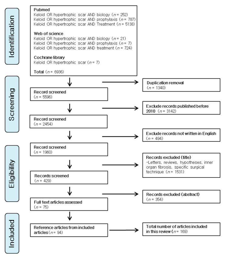
The original articles dealing with the biology, prophylaxis and treatment strategies for
hypertrophic scars and keloids were searched and reviewed. PubMed, Web of Science and Cochrane
library databases were searched on the keywords: hypertrophic scar OR keloid AND biology;
hypertrophic scar OR keloid AND prophylaxis; hypertrophic scar OR keloid AND treatment.
Time limits were from 1 January 2010 to the present. In addition, important reference articles from
the included articles were also reviewed. Several meta-analysis were also reviewed to estimate
the outcome of a certain treatment modality. Duplicates, letters, reviews, hypotheses articles dealing
with the fibrotic disorders on internal organs, studies dealing with specific surgical techniques and
studies published in a language other than English were excluded. Figure 1 shows the flowchart of
the literature search for this review.
Figure 1. The flowchart of the literature search for this review.
3. Classical Concepts of Wound Healing
The classical model of wound healing involves three distinct, but overlapping phases that follow
a time sequence: the inflammatory phase, the proliferative phase and the remodeling phase. The first
phase of wound healing is the inflammatory phase that starts immediately after tissue injury and lasts
for approximately 2–3 days after injury. Coagulation cascades, complement activation and platelet
degranulation prevent further fluid and blood losses by creating platelet plugs and a fibrin matrix [4].
The immune system and inflammatory reactions are activated to prevent infection and removingInt. J. Mol. Sci. 2018, 19, 711 3 of 19
devitalized tissues [5]. Neutrophils are recruited by chemotactic factors produced by platelet and
bacterial degranulations [6], and monocytes are recruited and differentiated into macrophages 2–3 days
after injury.
The second phase of wound healing is the proliferative phase. This phase of new tissue formation
occurs approximately 2–3 days after tissue damage and may last for 3–6 weeks. Active cellular
proliferation and migration characterize this phase. Keratinocytes migrate to the damaged dermis;
new blood vessels grow inward within the damaged tissue; and new capillaries replace the fibrin
matrix with granulation tissue via the actions of macrophages and fibroblasts. Granulation tissue
forms a new substrate for keratinocyte migration. Keratinocytes proliferate and mature within
granulation tissue along the wound margin, restoring the protective function of the epithelium.
In the late proliferative phase, a portion of the fibroblasts differentiates into myofibroblasts in
association with macrophages. Fibroblasts and myofibroblasts produce extracellular matrix (ECM),
mainly in the form of collagen; this accumulated collagen forms most of the eventual scar [7].
Other constituents of ECM include elastin, hyaluronic acids and proteoglycans. Myofibroblasts,
which contain actin filaments, have contractile properties and help bring the edges of the wound
together over time [8]. Once wound closure is accomplished, the final remodeling phase commences.
This phase is characterized by degradation of excessive tissue, transforming immature healing products
into a mature form. Remodeling may last for a year or more. Excessive ECM is degraded and remodeled
from type III collagen, the main component of ECM present during the early wound healing process,
to mature type I collagen.
4. Important Proteins and Cytokines in the Wound Healing Processes
It is important to achieve a proper balance between these wound healing phases. Synthesis
and degradation of ECM should be balanced, otherwise wound healing may be delayed or result in
excessive scars. Important proteins and cytokines that influence balanced wound healing processes
are summarized herein (Figure 2) and are important for understanding current investigations in
keloid treatment.
Figure 2. Important proteins and cytokines in the wound healing processes. The classical model of wound
healing involves three distinct, but overlapping phases that follow a time sequence: the inflammatory,
proliferative and remodeling phases. Important cells, proteins and cytokines in each phase are listed.Int. J. Mol. Sci. 2018, 19, 711 4 of 19
4.1. Inflammatory and Proliferative Phase
Prolonged and excessive inflammatory reactions result within the context of increased fibroblast
activity, which in turn produces excessive ECM. In this phase, degranulation of platelets releases
and activates transforming growth factor β (TGF-β), particularly TGF-β1, TGF-β2, platelet-derived
growth factor (PDGF), insulin-like growth factor (IGF-1) and epidermal growth factor (EGF). Vascular
endothelial growth factor (VEGF), which is produced by epidermal cells, is a positive regulator of
angiogenesis. Because of this, overexpression of VEGF is related to excessive capillary formation,
collagen type I production and overall scar volume increase [9]. These cytokines are not only
fibrogenic growth factors, but also chemotactic agents for epithelial cells, endothelial cells, neutrophils,
macrophages, mast cells and fibroblasts [4,8]. Fibroblasts originating in keloid tissues show increased
receptors to these growth factors and demonstrate increased responsiveness compared with fibroblasts
from normal tissues [10–13]. Tissue inhibitors of metalloproteinases (TIMPs) are endogenous inhibitors
of matrix metalloproteinases (MMPs); thus, increased levels of TIMPs, especially TIMP-1 and
TIMP-2, are associated with hypertrophic scar formation [9]. Tumor necrosis factor-α (TNF-α)
is an inflammatory cytokine produced by monocytes and macrophages during the inflammatory
phase. It has been known that this cytokine induces collagen degranulation and contributes to
minimizing excessive scarring. One suggested mechanism is that TNF-α increases the MMP1/TIMP3,
MMP2/TIMP3 ratios [14]. However, other studies showed that the biologic effect of TNF-α was not
the same on the fibroblasts from lung and skin tissues showing tissue specificity [15] and TNF-α
induced epithelial-mesenchymal transition in human skin wound healing [16]. Therefore, it is still
unclear whether TNF-α would promote or attenuate scar formation.
Immune responses are also related to wound healing processes. T helper CD4 cells are thought
to be major immunoregulatory cells during wound healing processes. CD4 T cells express Th1 or
Th2 responses [17]. Th1 responses produce interferon-γ and interleukin (IL)-12 and are thought to
be related to the attenuation of fibrogenesis. Th2 responses of CD4 cells are generally likely related
to fibrogenesis. IL-4, IL-5, IL-6 and IL-13 are thought to be related to pro-fibrosis [18,19], but IL-10 is
thought to be related to anti-fibrosis [20–22]. These cytokines are essential for promoting or impeding
the fibroblast recruitment and proliferation, ECM deposition, angiogenesis and re-epithelialization [4].
Endothelial cytokines including IL-8, IGF-1, fibroblast growth factor (FGF)-β and heparin promote
angiogenesis. Wound re-epithelialization is enhanced by EGF, TGF-α and IGF-1 [4].
4.2. Remodeling Phase
During the remodeling phase, excessive ECM is degraded, and collagen type III, an immature
collagen form, is converted to mature collagen type I. TGF-β3 is considered to play a role in reducing
the newly-synthesized ECM [23]. Significantly lower TGF-β3 mRNA expression was found in keloid
tissues [24,25]. However, TGF-β isoforms (TGF-β1, TGF-β2 and TGF-β3) do not present its activity as
isolated ligands, but are also associated with receptors and activity modulators; therefore, the mere
presence or absence of TGF-β may not fully explain abnormal wound healing [26]. Members of
the MMP family have major effects on ECM degradation and remodeling and mediate the degradation
of type III and type I collagens, the major components of ECM [27,28]. MMP-2 and MMP-9 are
active during the remodeling phase. MMP-9 is involved in degradation of type IV and V collagens,
fibronectin and elastin. MMP-2 plays an important role in ECM remodeling by degrading denatured
collagen [29,30]. MMPs have a downregulating effect on inflammation by decreasing and antagonizing
chemokines [31,32]. Immunity, cell migration and angiogenesis are also influenced by MMPs [33].
MMP activities are regulated by TIMPs. Decorin is a proteoglycan component of dermal connective
tissue that binds to type I collagen fibrils and influences TGF-β [34]. This protein is decreased in keloids
and hypertrophic scars [35]. By binding and neutralizing TGF-β, decorin decreases the stimulatory
effects of TGF-β on collagen, fibronectin and glycosaminoglycan synthesis [17]. Decorin also inhibits
angiogenesis by interacting with VEGF receptors (VEGFR2) and by inhibiting hepatocyte growthInt. J. Mol. Sci. 2018, 19, 711 5 of 19
factors and PDGF [36]. Decorin’s antifibrotic properties are receiving attention as a future therapeutic
agent [37,38].
5. Recent Findings of Scar Biology
Here, we introduce some recent findings on scar biology. These consist of some factors that
influence pro-fibrotic or anti-fibrotic pathways.
5.1. Hypoxia
Oxygen has long been known to be an important factor in wound healing [39,40]. There have been
many reports suggesting that a hypoxic environment is associated with keloid formation [41,42].
Zhao et al. measured the quantity of hypoxia inducible factor (HIF)-1α in keloid and normal
tissues and reported that keloid tissues are relatively hypoxic tissues compared to normoxic tissues,
and hypoxia induces a pro-fibrotic state in dermal fibroblasts via the TGF-β1/SMAD3 pathway [43].
5.2. Periostin
Periostin is a secreted extracellular matrix (ECM) protein, which was originally identified in
osteoblast, periodontal ligament and periosteum [44]. This matricellular protein is expressed in
the basement membrane, dermis and hair follicle [45]. Periostin is induced by TGF-β in human dermal
fibroblast and has an important role in wound healing and scar pathogenesis by inducing angiogenesis,
fibroblast proliferation and myofibroblast persistence [45–47]. It starts to increase its expression from
a few days after injury, peaking after about seven days after injury and decreasing afterwards [48,49].
Many authors have reported that periostin is abnormally elevated in hypertrophic scars and keloids
compared to normal tissues [45,46,50,51] and implicates periostin as a possible therapeutic target in
the treatment of hypertrophic scars and keloid.
5.3. MicroRNAs
MicroRNAs (miRNAs) are a group of short noncoding RNAs that pair complementarily with
target genes and silence that genes post-transcriptionally. It thereby regulates negatively the expression
of their target genes. miRNAs are thought to be deregulated in many skin diseases such as malignant
skin diseases and keloids [52–55]. Some researchers performed miRNA expression microarrays in
keloids and normal tissues [55,56] and reported upregulated or downregulated miRNAs in keloid
tissues compared to normal tissues. miRNA-199a-5p [57], miRNA-21 [58–61], miRNA-146a [62],
miRNA-1224-5p [56], miRNA-31 [63], and so forth, were investigated and showed potential in
the treatment of hypertrophic scars and keloids.
6. Preventions and Treatment Strategies for Hypertrophic Scars and Keloids
Because the processes are so complicated, the definitive processes that underlie excessive scar
formation are yet to be elucidated. So far, preventions and treatment strategies mainly focus on
reducing inflammation. Other therapies, targeting genes and molecules, require more study prior
to being introduced in clinical practice. The current treatment strategies for hypertrophic scars and
keloids are listed below and summarized in Table 1.Int. J. Mol. Sci. 2018, 19, 711 6 of 19
Table 1. Current treatment strategies for hypertrophic scars and keloids.
Categories Modalities Suggested Mechanisms Use
-Debridement of inviable tissues,
-Reduce inflammation by reducing
Tension-free closure adequate hemostasis
mechanotransduction
-Rapid tension free primary closure
-Reduce inflammation by reducing -Start 2 weeks after primary
Taping or silicone sheeting mechanotransduction: occlusion wound treatment
and hydration -12 h a day for at least 2 months
Prophylaxis
-Start 2 weeks after primary
-Induction of MMPs
Flavonoids wound treatment
-Inhibition of SMADs expression
-Generally twice daily for 4 to 6 months
-Occlusion of blood vessels -Pressure of 15 to 40 mmHg
Pressure therapy
-Inducing apoptosis -More than 23 h a day for at least 6 months
-Intralesional injection: triamcinolone
10 to 40 mg/mL
-Reducing inflammation and proliferation -1 to 2 sessions a month (2 to 3 sessions,
Corticosteroids
-Vasoconstriction but can be extended)
-Tapes/plasters, ointments are available
-Combination is common
-At least 1 year after primary
Scar revision -Direct reduction of scar volume wound treatment
-Combination is recommended
-Deliver liquid nitrogen using spray,
contact or intralesional needle cryoprobe
Treatment Cryotherapy -Scar tissue necrosis
-10 to 20 s freeze-thaw cycles
(current)
-Combination is common
-Adjuvant after scar revision
-Anti-angiogenesis -24–48 h after scar revision surgery
Radiotherapy
-Anti-inflammation -Total of 40 Gray or less, over several
divided sessions
-585-nm pulsed dye laser: 6.0–7.5 J/cm2
(7 mm spot) or 4.5–5.5 J/cm2 (10 mm spot)
-Vaporize blood vessel
Laser therapy -1064-nm Nd:YAG laser: 14 J/cm2
-Anti-inflammation
(5 mm spot)
-2 to 6 sessions, every 3–4 weeks
-Intralesional injection: 50 mg/mL
-Anti-angiogenesis
5-Fluorouracil -Weekly for 12 weeks
-Anti-inflammation
-Combination is common
-Modulation of proinflammatory
-Systemic injection
cell activity
MSC * therapy -Local injection (at the wound)
-Anti-fibrosis
-Engineered MSC-seeded tissue scaffold
-Promote normal angiogenetic activity
Treatment -Fat injection or fat tissue grafting
Fat grafting -Deliver adipose-tissue derived MSCs
(Emerging) underneath or into the wound
-Downregulating TGF-β1
-Intralesional injection: 1.5 × 10⁶ IU,
Interferon -Attenuates collagen synthesis and
twice daily over 4 days
fibroblast proliferation
Human recombinant
TGF-β3/TGF-β1 or -Adjust TGF-β3: TGF-β1 or 2 ratio Not available currently
2 neutralizing antibody
-Reduce muscle tension during
wound healing -Intralesional injection: 70~140 U,
Botulinum toxin type A
-Arrest cell cycle in non-proliferative stage 1 or 3 months interval, 3 sessions
-Influence TGF-β1 expression
-Decreasing collagen synthesis
-Intralesional injection: 1.5 IU/mL,
Bleomycin -Reduce lysyl-oxidase levels
2 to 6 sessions at monthly interval
-Induce apoptosis
* MSC: mesenchymal stem cell; MMPs: matrix metalloproteinases; TGF: transforming growth factor.
6.1. Prevention
6.1.1. Tension-Free Primary Closure
Regardless of a patient’s tendency to exhibit bad scars (or not), (1) debridement of inviable or
severely contaminated tissues, (2) adequate hemostasis to prevent hematoma, seroma or abscess
formation and (3) rapid primary closure using tension-free techniques are wound care basics and
are very important for minimizing the effects of bad scars. Wound epithelialization that is delayed
beyond 10–14 days increases the risk of hypertrophic scars, and quick primary closure to induce rapidInt. J. Mol. Sci. 2018, 19, 711 7 of 19
epithelialization is necessary to achieve good scarring [64]. The importance of tension-free closure
techniques cannot be overstated. Wounds that are subject to tension tend to develop into bad scars [65].
The exact molecular mechanisms that govern how our skin responds to physical tension remain
uncertain; however, several pathways that convert mechanical forces into biochemical responses
have been investigated and reported. This process is called mechanotransduction [66]. Gurtner et al.
reported on the fibrotic effects of mechanical tension and described the preventive effect of offloading
wound tension on scar formation [67].
6.1.2. Passive Mechanical Stabilization
To prevent wound stretching and consequential mechanotransduction, prolonged passive
mechanical wound stabilization has been applied [68–71] using paper tapes or silicone sheets.
Paper tapes help alleviate scar formation, and silicone sheeting is superior to paper tapes because it
avoids repeated epidermal avulsion.
Other mechanisms of silicone sheets include occlusion and hydration of the scar surface.
The inherent antifibrotic properties of silicone are not definite [72]. Silicone sheeting is recommended
for use from two weeks after primary wound treatment for more than 12 h a day for at least two months.
For body areas where silicone sheets do not easily fit, silicone gel can be applied.
6.1.3. Flavonoids
Flavonoids (or bioflavonoids) are naturally-derived substances from various plants and have
been used for preventing severe scar formation. Several studies have reported the efficacy of
flavonoid scar gels like Contractubex Gel (Merz Pharma, Frankfurt, Germany) or Mederma Skin
Care Gel (Merz Pharmaceuticals, Greensboro, NC, USA). The efficacy of these gel products is
controversial [73–77], but other flavonoids like quercetin exert antifibrotic actions. These actions may be
mediated through induction of MMP-1 or inhibition of SMAD2, SMAD3 or SMAD4 expression [77,78].
The instructions of flavonoids, for instance, Contractubex Gel, is as follows: (1) start two weeks after
primary wound treatment; and (2) twice daily for four to six months.
6.1.4. Pressure Therapy
Cutaneous wound compression has been used not only for prevention, but also for treatment
of hypertrophic scars and keloids. Although pressure therapy reduces the subjective and objective
signs and symptoms of hypertrophic scars and keloids, the scientific evidence supporting their use
is weak, and their clinical efficacy is also controversial [79]. The suggested mechanisms underlying
pressure therapy include occlusion of blood vessels and limiting the delivery of inflammatory
cytokines, nutrients and oxygen from blood vessels to scar tissue [80–84]. Increasing apoptosis may be
another mechanism of pressure therapy [85]. There are no comparative analyses of pressure amount,
and the pressure amount that is used clinically relies on empirical reports. Currently, the recommended
amount is 15–40 mm Hg for more than 23 h a day for at least six months [83,86].
6.2. Current Treatment Strategies
6.2.1. Corticosteroids
Intralesional steroid injection, steroid tapes/plasters and steroid ointments have been used
to treat hypertrophic scars and keloids. Intralesional injection is the most popular method for
steroid administration, although steroid tapes/plasters are gaining popularity [87]. The mechanism
underlying this therapy is attributed to its anti-inflammatory effect [72]. In addition, steroid therapy
seems to reduce collagen synthesis, glycosaminoglycan production, fibroblast proliferation and
degeneration of collagen and fibroblasts [88,89]. Another suggested mechanism is induction of
vasoconstriction mediated by binding of the topical steroid to classical glucocorticoid receptors [2].
Resolution rates for keloids treated with intralesional steroid injections are variable and range fromInt. J. Mol. Sci. 2018, 19, 711 8 of 19
50% to 100% and recur in 9% to 50% [90]. Most previous studies used triamcinolone acetonide (TAC),
injected alone or in combination with other treatment modalities such as 5-FU, verapamil, cryotherapy
or surgery. The concentrations of injectable TAC vary from 10 to 40 mg/mL, but the recommended
concentration of TAC in monotherapy is 40 mg/mL for keloid resolution [91]. The injection is
performed 1–2 times a months until the scar has flattened. Intralesional steroid injections could
cause side effects such as skin atrophy or telangiectasia.
6.2.2. Scar Revision Surgery
Surgical excision is a traditional treatment for hypertrophic scars and keloids. The remodeling
phase of classical wound healing may last for more than one year; therefore, excision of hypertrophic
scars or keloids should be considered after at least one year of primary wound treatment therapy.
As time goes by, hypertrophic scars tend to regress naturally or with conservative treatment such
as steroid injections. Therefore, in many cases, there is no need to perform scar revision surgery.
For keloids, surgical excision alone frequently results in disappointing outcomes. To improve
postoperative surgical outcomes, multimodal combination therapy such as postoperative steroid
application or radiotherapy might be added. When surgeons perform scar revision surgery, they should
establish tension-free wound closure in order to decrease tension-related inflammation and thereby
reduce recurrence. Various techniques including three-layered sutures, subcutaneous/fascial tensile
reduction sutures, Z-plastics or local flap reconstruction can be utilized on a case-by-case basis [92,93].
Recurrence rates of hypertrophic scars after scar revision surgery are low, but the recurrence rate of
keloids after scar revision surgery is 45% to 100% [94–96].
6.2.3. Cryotherapy
Cryotherapy has been used to treat hypertrophic scars or keloids as a monotherapy or in
conjunction with other therapies such as intralesional steroid injections [97]. Treatments that combine
cryotherapy and intralesional triamcinolone injections significantly improve hypertrophic scars and
keloids [98–100]. Delivery methods for cryotherapy are variable and include sprays, contact or
the intralesional-needle cryoprobe method. The intralesional-needle cryoprobe method shows better
results than the spray or contact method, producing rapid re-epithelialization [101]. The suggested
mechanism underlying cryotherapy is tissue necrosis induced by vascular damage. It seems that
necrotized tissues induced by frostbite (as opposed to burn injury) secrete unique inflammatory
cytokines; therefore, the responses of fibroblasts may differ [2]. Cryotherapy success rates range from
32 to 74% after several sessions [102–104].
6.2.4. Radiotherapy
Several studies have shown the effectiveness of radiotherapy on keloid treatment. Both external
beam therapy and brachytherapy (or internal radiation therapy) have been used and studied for
treatment of keloids. Radiotherapy is generally conducted as an adjuvant treatment 24 to 48 h after
scar revision surgery, and the recommend radiation dose is 40 Gray over several divided sessions
to minimize adverse effects [105]. The suggested mechanism of radiotherapy for treating keloids
is anti-angiogenesis and successive anti-fibroblast activity. Suppression of angiogenesis decreases
delivery of inflammatory cytokines, and successive inhibition of fibroblast activity results in decreased
collagen synthesis, thus suppressing keloid development [106,107]. Radiotherapy carries an inherent
risk of carcinogenesis; therefore, even though the risk is low [108,109], radiation-vulnerable areas,
including the thyroid and breast, should be treated after achieving informed consent and with abundant
cautions. Shen et al. reported the recurrence rate of 9.59% [109]. Recently, radioactive skin patches have
been used for localized skin diseases like skin cancers or keloids [110,111]. Radioactive skin patches
use various kinds of radionuclides and have variable effectiveness for treating keloids. These patches
are frequently used in combination with other available treatment.Int. J. Mol. Sci. 2018, 19, 711 9 of 19
6.2.5. Laser Therapy
Laser therapy was introduced for keloid treatment in the 1980s [112], and several kinds of lasers
with various wavelengths were investigated and reported. Among these, the most popular laser used
to treat hypertrophic scars and keloids is the 585-nm pulsed dye laser (PDL) [113]. The recommended
energy is 6.0 to 7.5 J/cm2 (7-mm spot) or 4.5 to 5.5 J/cm2 (10-mm spot) [114], and two to six sessions
of treatment may be needed [113]. The 1064-nm Nd:YAG laser is another popular laser for treating
hypertrophic scars and keloids. For this laser, the recommended energy is 14 J/cm2 (5-mm spot),
with the procedure being repeated every three to four weeks [115,116]. These laser treatments vaporize
blood vessels. By doing this, inflammatory cytokines are limited in their ability to reach hypertrophic
scars and keloids, thereby suppressing the development of aberrant scars. Possible side effects of
laser therapy include hyperpigmentation, hypopigmentation, blister formation and postoperative
purpura [117–120].
6.2.6. 5-Fluorouracil
5-FU is a medication mainly used to treat cancer. By injecting it into a vein, it can be used for
the treatment of esophageal, stomach, pancreatic, colon, breast and cervical cancers. It can also be used
topically for actinic keratosis and basal cell carcinoma in a cream or solution formulation [87]. 5-FU has
also been used to treat keloids [121]. The suggested mechanism is anti-angiogenesis, anti-fibroblast
proliferation and anti-collagen Type I expression induced by TGF-β [122–124]. This therapy is used
solely or in combination with another treatment, and intralesional injection is the preferred method of
delivery. Nanda et al. reported scar size reduction in a majority of patients in whom 5-FU was injected
intralesionally weekly for 12 weeks in a concentration of 50 mg/mL [122]. Possible side effects include
pain and ulceration. A systematic review reported 45% to 96% of effectiveness [125].
6.3. Emerging Therapies
6.3.1. Mesenchymal Stem Cell Therapy
Mesenchymal stem cells (MSCs) have immunomodulatory and antifibrotic effects by secreting
paracrine growth factors [126–129]. The antifibrotic effects of MSC on fibrotic diseases such as
myocardial infarctions, renal fibrosis or liver cirrhosis have been investigated and reported [130–136].
MSCs are also used to prevent or attenuate excessive inflammatory processes that are characteristic
of hypertrophic scars and keloids. MSC treatments have variable delivery methods and doses [137].
Delivery is conducted via systemic injections, local injections (at the wound, intradermal or
subcutaneously) or via an engineered MSC-seeded tissue scaffold [138–141]. The possible mechanisms
underlying MSC treatment include: (1) modulation and inhibition of proinflammatory cell
activity; (2) antifibrotic activity via downregulation of myofibroblast differentiation and collagen
type I and III production; and (3) promotion of normal angiogenetic activity that aids in normal
wound healing [137,142]. Even though many researchers have reported anti-inflammatory and
anti-fibrotic effects of MSC, there are reports of possible proinflammatory actions of MSC [143–145].
More investigations and long-term preclinical studies should be conducted to apply this method to
clinical practice.
6.3.2. Fat Grafting
Autologous fat grafting or lipotransfer, underneath or into the wound, has been performed for
patients with hypertrophic scars or keloids. Several studies have reported the effectiveness of fat
grafting on severely-scarred lesions [146–148]. These reports showed beneficial effects on excessive
scar lesions, and side effects were rarely reported. The mechanism underlying fat injections is believed
to be that transferred fat tissues deliver adipose-tissue derived MSCs to the wound.Int. J. Mol. Sci. 2018, 19, 711 10 of 19
6.3.3. Interferon
Interferon (IFN) is comprised of cytokines that have anti-proliferative and anti-fibrotic effects.
As mentioned earlier, IFNs attenuate collagen synthesis and fibroblast proliferation by downregulating
TGF-β1. Although adverse effects including pain at the injection site and flu-like symptoms are
relatively common in IFN treatment, some authors reported a good outcome of combination therapy
of IFN α-2b with TAC injection [149,150].
6.3.4. Transforming Growth Factor-β
TGF-β isoforms (TGF-β1,2,3) had long been a target of anti-keloid therapy. Several studies showed
that the ratio of TGF-β3 and TFG-β1 and 2 is important in scar progression or remission [151,152].
Many studies had been performed to investigate the effect of exogenous TGF-β1 and 2 neutralizing
antibodies and exogenous TFG-β3 and had proven the effect of TGF-β isoforms; TGF-β1 and 2 increase
fibrosis, and TGF-β3 attenuates fibrosis [153]. Recombinant human TGF-β3, avotermin (planned trade
name Juvista) showed successful results in phase I/II clinical trials [154–156], but failed in phase III
clinical trials.
6.3.5. Botulinum Toxin A
Botulinum toxin, which is derived from Clostridium botulinum, is a potent neurotoxin that
blocks neuromuscular transmission. Some authors have reported that botulinum toxin type
A can minimize scar formation by reducing muscle tension during wound healing, causing
the fibroblast cell cycle to be paused in a non-proliferative state, G0 or G1, and influencing TGF-β1
expression [157–161]. Intralesional injection was the preferred delivery method, and 70–140 U of
Type A botulinum toxin was delivered per sessions at one- or three-month intervals for three or
nine months (three sessions) [160,162–164]. Treatment outcomes were generally favorable, and patient
satisfaction was high. Improvement was also reported regarding pain, tenderness and itching
sensation [160,162,163].
6.3.6. Bleomycin
Bleomycin is a cytotoxic, antineoplastic, antiviral and antibacterial agent [165], derived from
Streptomyces verticillus, and has been used for dermatologic diseases such as warts. This agent has
also been used for hypertrophic scars and keloids. Several studies have found that bleomycin-treated
human dermal fibroblasts showed diminished collagen synthesis, even with the co-existence of
TFG-β1, and a reduction in the levels of lysyl-oxidase, which is involved in the maturation of collagen.
In addition, apoptosis was also induced by bleomycin treatment [166–169]. Intralesional injection
is the preferred delivery method, and 1.5 IU/mL of bleomycin were injected two to six sessions at
monthly intervals. Several studies reported that complete flattening was achieved in 54% to 73% of
keloid patients [166,167] and other symptoms like itching and pain were also resolved. Possible side
effects include injection site pain, ulceration, atrophy and hyperpigmentation, but systemic side effects
were not observed [165,167].
7. Conclusions
Hypertrophic scars and keloids result from abnormal wound healing. Excessive ECM deposition
is characteristic of these lesions. Increased inflammatory and proliferative processes and decreased
remodeling processes cause excessive ECM deposition. Genetic and systemic factors are also related to
these excessively scarring lesions. Although encouraging results of molecular- or cytokine-targeting
therapies are being continuously reported, current prophylaxis and treatment strategies still mainly
focus on decreasing inflammatory processes. Further understanding of the mechanisms underlying
excessive scarring is needed to develop more effective prophylaxis and treatment strategies.Int. J. Mol. Sci. 2018, 19, 711 11 of 19
Acknowledgments: No funding was received for this study.
Author Contributions: Ho Jun Lee: conception of the work, acquisition and analysis of data, drafting the work,
approved the submitted version, agreed to be personally accountable for the author’s own contributions and for
ensuring that questions related to the accuracy or integrity of any part of the work; Yong Ju Jang: conception of
the work, substantively revised the draft, approved the submitted version, agreed to be personally accountable
for the author’s own contributions and for ensuring that questions related to the accuracy or integrity of any part
of the work.
Conflicts of Interest: The authors declare no conflict of interest.
References
1. Sund, B. New Development in Wound Care; PJB Publications: London, UK, 2000; pp. 1–255.
2. Ogawa, R. Keloid and Hypertrophic Scars Are the Result of Chronic Inflammation in the Reticular Dermis.
Int. J. Mol. Sci. 2017, 18, 606. [CrossRef] [PubMed]
3. Chiang, R.S.; Borovikova, A.A.; King, K.; Banyard, D.A.; Lalezari, S.; Toranto, J.D.; Paydar, K.Z.; Wirth, G.A.;
Evans, G.R.; Widgerow, A.D. Current concepts related to hypertrophic scarring in burn injuries. Wound Repair
Regen. 2016, 24, 466–477. [CrossRef] [PubMed]
4. Tredget, E.E.; Nedelec, B.; Scott, P.G.; Ghahary, A. Hypertrophic scars, keloids, and contractures. The cellular
and molecular basis for therapy. Surg. Clin. N. Am. 1997, 77, 701–730. [CrossRef]
5. Imhof, B.A.; Jemelin, S.; Ballet, R.; Vesin, C.; Schapira, M.; Karaca, M.; Emre, Y. CCN1/CYR61-mediated
meticulous patrolling by Ly6Clow monocytes fuels vascular inflammation. Proc. Natl. Acad. Sci. USA 2016,
113, E4847–E4856. [CrossRef] [PubMed]
6. Grose, R.; Werner, S. Wound-healing studies in transgenic and knockout mice. Mol. Biotechnol. 2004, 28,
147–166. [CrossRef]
7. Werner, S.; Krieg, T.; Smola, H. Keratinocyte-fibroblast interactions in wound healing. J. Investig. Dermatol.
2007, 127, 998–1008. [CrossRef] [PubMed]
8. Zhu, Z.; Ding, J.; Tredget, E.E. The molecular basis of hypertrophic scars. Burns Trauma 2016, 4, 2. [CrossRef]
[PubMed]
9. Wang, P.; Jiang, L.Z.; Xue, B. Recombinant human endostatin reduces hypertrophic scar formation in rabbit
ear model through down-regulation of VEGF and TIMP-1. Afr. Health Sci. 2016, 16, 542–553. [CrossRef]
[PubMed]
10. Tuan, T.L.; Nichter, L.S. The molecular basis of keloid and hypertrophic scar formation. Mol. Med. Today
1998, 4, 19–24. [CrossRef]
11. Ishihara, H.; Yoshimoto, H.; Fujioka, M.; Murakami, R.; Hirano, A.; Fujii, T.; Ohtsuru, A.; Namba, H.;
Yamashita, S. Keloid fibroblasts resist ceramide-induced apoptosis by overexpression of insulin-like growth
factor I receptor. J. Investig. Dermatol. 2000, 115, 1065–1071. [CrossRef] [PubMed]
12. Butler, P.D.; Longaker, M.T.; Yang, G.P. Current progress in keloid research and treatment. J. Am. Coll. Surg.
2008, 206, 731–741. [CrossRef] [PubMed]
13. Ladak, A.; Tredget, E.E. Pathophysiology and management of the burn scar. Clin. Plast. Surg. 2009, 36,
661–674. [CrossRef] [PubMed]
14. Chen, X.; Thibeault, S.L. Role of tumor necrosis factor-α in wound repair in human vocal fold fibroblasts.
Laryngoscope 2010, 120, 1819–1825. [CrossRef] [PubMed]
15. Mariani, T.J.; Sandefur, S.; Roby, J.D.; Pierce, R.A. Collagenase-3 induction in rat lung fibroblasts
requires the combined effects of tumor necrosis factor-α and 12-lipoxygenase metabolites: A model of
macrophage-induced, fibroblast driven extracellular matrix remodeling during inflammatory lung injury.
Mol. Biol. Cell 1998, 9, 1411–1424. [CrossRef] [PubMed]
16. Yan, C.; Grimm, W.A.; Garner, W.L.; Qin, L.; Travis, T.; Tan, N.; Han, Y.P. Epithelial to mesenchymal transition
in human skin wound healing is induced by tumor necrosis factor-α through bone morphogenic protein-2.
Am. J. Pathol. 2010, 176, 2247–2258. [CrossRef] [PubMed]
17. Armour, A.; Scott, P.G.; Tredget, E.E. Cellular and molecular pathology of HTS: Basis for treatment.
Wound Repair Regen. 2007, 15 (Suppl. S1), S6–S17. [CrossRef] [PubMed]
18. Doucet, C.; Brouty-Boye, D.; Pottin-Clemenceau, C.; Canonica, G.W.; Jasmin, C.; Azzarone, B. Interleukin
(IL) 4 and IL-13 act on human lung fibroblasts. Implication in asthma. J. Clin. Investig. 1998, 101, 2129–2139.
[CrossRef] [PubMed]Int. J. Mol. Sci. 2018, 19, 711 12 of 19
19. Wynn, T.A. Fibrotic disease and the Th1/Th2 paradigm. Nat. Rev. Immunol. 2004, 4, 583–594. [CrossRef]
[PubMed]
20. Van den Broek, L.J.; van der Veer, W.M.; de Jong, E.H.; Gibbs, S.; Niessen, F.B. Suppressed inflammatory
gene expression during human hypertrophic scar compared to normotrophic scar formation. Exp. Dermatol.
2015, 24, 623–629. [CrossRef] [PubMed]
21. Namazi, M.R.; Fallahzadeh, M.K.; Schwartz, R.A. Strategies for prevention of scars: What can we learn from
fetal skin? Int. J. Dermatol. 2011, 50, 85–93. [CrossRef] [PubMed]
22. Liechty, K.W.; Kim, H.B.; Adzick, N.S.; Crombleholme, T.M. Fetal wound repair results in scar formation in
interleukin-10-deficient mice in a syngeneic murine model of scarless fetal wound repair. J. Pediatr. Surg.
2000, 35, 866–872. [CrossRef] [PubMed]
23. Bock, O.; Yu, H.; Zitron, S.; Bayat, A.; Ferguson, M.W.; Mrowietz, U. Studies of transforming growth factors
β 1–3 and their receptors I and II in fibroblast of keloids and hypertrophic scars. Acta Derm. Venereol. 2005,
85, 216–220. [CrossRef] [PubMed]
24. Lee, T.Y.; Chin, G.S.; Kim, W.J.; Chau, D.; Gittes, G.K.; Longaker, M.T. Expression of transforming growth
factor β 1, 2, and 3 proteins in keloids. Ann. Plast. Surg. 1999, 43, 179–184. [CrossRef] [PubMed]
25. Xia, W.; Phan, T.T.; Lim, I.J.; Longaker, M.T.; Yang, G.P. Complex epithelial-mesenchymal interactions
modulate transforming growth factor-β expression in keloid-derived cells. Wound Repair Regen. 2004, 12,
546–556. [CrossRef] [PubMed]
26. Lu, L.; Saulis, A.S.; Liu, W.R.; Roy, N.K.; Chao, J.D.; Ledbetter, S.; Mustoe, T.A. The temporal effects of anti-TGF-β1,
2, and 3 monoclonal antibody on wound healing and hypertrophic scar formation. J. Am. Coll. Surg. 2005, 201,
391–397. [CrossRef] [PubMed]
27. Fujiwara, M.; Muragaki, Y.; Ooshima, A. Keloid-derived fibroblasts show increased secretion of factors
involved in collagen turnover and depend on matrix metalloproteinase for migration. Br. J. Dermatol. 2005,
153, 295–300. [CrossRef] [PubMed]
28. Ghahary, A.; Ghaffari, A. Role of keratinocyte-fibroblast cross-talk in development of hypertrophic scar.
Wound Repair Regen. 2007, 15 (Suppl. S1), S46–S53. [CrossRef] [PubMed]
29. Mauviel, A. Cytokine regulation of metalloproteinase gene expression. J. Cell. Biochem. 1993, 53, 288–295.
[CrossRef] [PubMed]
30. Zhang, Y.; McCluskey, K.; Fujii, K.; Wahl, L.M. Differential regulation of monocyte matrix metalloproteinase
and TIMP-1 production by TNF-α, granulocyte-macrophage CSF, and IL-1 β through prostaglandin-dependent
and -independent mechanisms. J. Immunol. 1998, 161, 3071–3076. [PubMed]
31. McQuibban, G.A.; Gong, J.H.; Tam, E.M.; McCulloch, C.A.; Clark-Lewis, I.; Overall, C.M. Inflammation
dampened by gelatinase A cleavage of monocyte chemoattractant protein-3. Science 2000, 289, 1202–1206.
[CrossRef] [PubMed]
32. McQuibban, G.A.; Gong, J.H.; Wong, J.P.; Wallace, J.L.; Clark-Lewis, I.; Overall, C.M. Matrix
metalloproteinase processing of monocyte chemoattractant proteins generates CC chemokine receptor
antagonists with anti-inflammatory properties in vivo. Blood 2002, 100, 1160–1167. [PubMed]
33. Rohani, M.G.; Parks, W.C. Matrix remodeling by MMPs during wound repair. Matrix Biol. 2015, 44, 113–121.
[CrossRef] [PubMed]
34. Krumdieck, R.; Hook, M.; Rosenberg, L.C.; Volanakis, J.E. The proteoglycan decorin binds C1q and inhibits
the activity of the C1 complex. J. Immunol. 1992, 149, 3695–3701. [PubMed]
35. Scott, P.G.; Dodd, C.M.; Tredget, E.E.; Ghahary, A.; Rahemtulla, F. Chemical characterization and quantification
of proteoglycans in human post-burn hypertrophic and mature scars. Clin. Sci. 1996, 90, 417–425. [CrossRef]
[PubMed]
36. Jarvelainen, H.; Sainio, A.; Wight, T.N. Pivotal role for decorin in angiogenesis. Matrix Biol. 2015, 43, 15–26.
[CrossRef] [PubMed]
37. Zhang, Z.; Garron, T.M.; Li, X.J.; Liu, Y.; Zhang, X.; Li, Y.Y.; Xu, W.S. Recombinant human decorin inhibits
TGF-β1-induced contraction of collagen lattice by hypertrophic scar fibroblasts. Burns 2009, 35, 527–537.
[CrossRef] [PubMed]
38. Mukhopadhyay, A.; Wong, M.Y.; Chan, S.Y.; Do, D.V.; Khoo, A.; Ong, C.T.; Cheong, H.H.; Lim, I.J.; Phan, T.T.
Syndecan-2 and decorin: Proteoglycans with a difference—Implications in keloid pathogenesis. J. Trauma
2010, 68, 999–1008. [CrossRef] [PubMed]Int. J. Mol. Sci. 2018, 19, 711 13 of 19
39. Sen, C.K.; Roy, S. Oxygenation state as a driver of myofibroblast differentiation and wound contraction:
Hypoxia impairs wound closure. J. Investig. Dermatol. 2010, 130, 2701–2703. [CrossRef] [PubMed]
40. Nauta, T.D.; van Hinsbergh, V.W.; Koolwijk, P. Hypoxic signaling during tissue repair and regenerative
medicine. Int. J. Mol. Sci. 2014, 15, 19791–19815. [CrossRef] [PubMed]
41. Ueda, K.; Yasuda, Y.; Furuya, E.; Oba, S. Inadequate blood supply persists in keloids. Scand. J. Plast. Reconstr.
Surg. Hand Surg. 2004, 38, 267–271. [CrossRef] [PubMed]
42. Steinbrech, D.S.; Mehrara, B.J.; Chau, D.; Rowe, N.M.; Chin, G.; Lee, T.; Saadeh, P.B.; Gittes, G.K.;
Longaker, M.T. Hypoxia upregulates VEGF production in keloid fibroblasts. Ann. Plast. Surg. 1999,
42, 514–519. [CrossRef] [PubMed]
43. Zhao, B.; Guan, H.; Liu, J.Q.; Zheng, Z.; Zhou, Q.; Zhang, J.; Su, L.L.; Hu, D.H. Hypoxia drives the transition of
human dermal fibroblasts to a myofibroblast-like phenotype via the TGF-β1/Smad3 pathway. Int. J. Mol. Med.
2017, 39, 153–159. [CrossRef] [PubMed]
44. Horiuchi, K.; Amizuka, N.; Takeshita, S.; Takamatsu, H.; Katsuura, M.; Ozawa, H.; Toyama, Y.; Bonewald, L.F.;
Kudo, A. Identification and characterization of a novel protein, periostin, with restricted expression to
periosteum and periodontal ligament and increased expression by transforming growth factor β. J. Bone
Miner. Res. 1999, 14, 1239–1249. [CrossRef] [PubMed]
45. Zhou, H.M.; Wang, J.; Elliott, C.; Wen, W.; Hamilton, D.W.; Conway, S.J. Spatiotemporal expression of
periostin during skin development and incisional wound healing: Lessons for human fibrotic scar formation.
J. Cell Commun. Signal. 2010, 4, 99–107. [CrossRef] [PubMed]
46. Crawford, J.; Nygard, K.; Gan, B.S.; O’Gorman, D.B. Periostin induces fibroblast proliferation and
myofibroblast persistence in hypertrophic scarring. Exp. Dermatol. 2015, 24, 120–126. [CrossRef] [PubMed]
47. Elliott, C.G.; Wang, J.; Guo, X.; Xu, S.W.; Eastwood, M.; Guan, J.; Leask, A.; Conway, S.J.; Hamilton, D.W.
Periostin modulates myofibroblast differentiation during full-thickness cutaneous wound repair. J. Cell Sci.
2012, 125, 121–132. [CrossRef] [PubMed]
48. Conway, S.J.; Izuhara, K.; Kudo, Y.; Litvin, J.; Markwald, R.; Ouyang, G.; Arron, J.R.; Holweg, C.T.; Kudo, A.
The role of periostin in tissue remodeling across health and disease. Cell. Mol. Life Sci. 2014, 71, 1279–1288.
[CrossRef] [PubMed]
49. Jackson-Boeters, L.; Wen, W.; Hamilton, D.W. Periostin localizes to cells in normal skin, but is associated with
the extracellular matrix during wound repair. J. Cell Commun. Signal. 2009, 3, 125–133. [CrossRef] [PubMed]
50. Zhang, Z.; Nie, F.; Kang, C.; Chen, B.; Qin, Z.; Ma, J.; Ma, Y.; Zhao, X. Increased periostin expression affects
the proliferation, collagen synthesis, migration and invasion of keloid fibroblasts under hypoxic conditions.
Int. J. Mol. Med. 2014, 34, 253–261. [CrossRef] [PubMed]
51. Zhang, Z.; Nie, F.; Chen, X.; Qin, Z.; Kang, C.; Chen, B.; Ma, J.; Pan, B.; Ma, Y. Upregulated periostin promotes
angiogenesis in keloids through activation of the ERK 1/2 and focal adhesion kinase pathways, as well
as the upregulated expression of VEGF and angiopoietin1. Mol. Med. Rep. 2015, 11, 857–864. [CrossRef]
[PubMed]
52. Kashiyama, K.; Mitsutake, N.; Matsuse, M.; Ogi, T.; Saenko, V.A.; Ujifuku, K.; Utani, A.; Hirano, A.;
Yamashita, S. miR-196a downregulation increases the expression of type I and III collagens in keloid
fibroblasts. J. Investig. Dermatol. 2012, 132, 1597–1604. [CrossRef] [PubMed]
53. Liu, Y.; Yang, D.; Xiao, Z.; Zhang, M. miRNA expression profiles in keloid tissue and corresponding normal
skin tissue. Aesthet. Plast. Surg. 2012, 36, 193–201. [CrossRef] [PubMed]
54. Lovendorf, M.B.; Skov, L. miRNAs in inflammatory skin diseases and their clinical implications. Expert Rev.
Clin. Immunol. 2015, 11, 467–477. [CrossRef] [PubMed]
55. Luan, Y.; Liu, Y.; Liu, C.; Lin, Q.; He, F.; Dong, X.; Xiao, Z. Serum miRNAs Signature Plays an Important Role
in Keloid Disease. Curr. Mol. Med. 2016, 16, 504–514. [CrossRef] [PubMed]
56. Yao, X.; Cui, X.; Wu, X.; Xu, P.; Zhu, W.; Chen, X.; Zhao, T. Tumor suppressive role of miR-1224-5p in keloid
proliferation, apoptosis and invasion via the TGF-β1/Smad3 signaling pathway. Biochem. Biophys. Res. Commun.
2017, 495, 713–720. [CrossRef] [PubMed]
57. Wu, Z.Y.; Lu, L.; Liang, J.; Guo, X.R.; Zhang, P.H.; Luo, S.J. Keloid microRNA expression analysis and
the influence of miR-199a-5p on the proliferation of keloid fibroblasts. Genet. Mol. Res. 2014, 13, 2727–2738.
[CrossRef] [PubMed]Int. J. Mol. Sci. 2018, 19, 711 14 of 19
58. Liu, Y.; Wang, X.; Yang, D.; Xiao, Z.; Chen, X. MicroRNA-21 affects proliferation and apoptosis by regulating
expression of PTEN in human keloid fibroblasts. Plast. Reconstr. Surg. 2014, 134, 561e–573e. [CrossRef]
[PubMed]
59. Li, Y.; Zhang, J.; Lei, Y.; Lyu, L.; Zuo, R.; Chen, T. MicroRNA-21 in Skin Fibrosis: Potential for Diagnosis and
Treatment. Mol. Diagn. Ther. 2017, 21, 633–642. [CrossRef] [PubMed]
60. Zhou, R.; Wang, C.; Wen, C.; Wang, D. miR-21 promotes collagen production in keloid via Smad7. Burns
2017, 43, 555–561. [CrossRef] [PubMed]
61. Liu, Y.; Li, Y.; Li, N.; Teng, W.; Wang, M.; Zhang, Y.; Xiao, Z. TGF-β1 promotes scar fibroblasts proliferation
and transdifferentiation via up-regulating microRNA-21. Sci. Rep. 2016, 6, 32231. [CrossRef] [PubMed]
62. Liu, Z.; Lu, C.L.; Cui, L.P.; Hu, Y.L.; Yu, Q.; Jiang, Y.; Ma, T.; Jiao, D.K.; Wang, D.; Jia, C.Y.
MicroRNA-146a modulates TGF-β1-induced phenotypic differentiation in human dermal fibroblasts by
targeting SMAD4. Arch. Dermatol. Res. 2012, 304, 195–202. [CrossRef] [PubMed]
63. Zhang, J.; Xu, D.; Li, N.; Li, Y.; He, Y.; Hu, X.; Lyu, L.; He, L. Downregulation of microRNA-31 inhibits
proliferation and induces apoptosis by targeting HIF1AN in human keloid. Oncotarget 2017, 8, 74623–74634.
[CrossRef] [PubMed]
64. Bond, J.S.; Duncan, J.A.; Mason, T.; Sattar, A.; Boanas, A.; O’Kane, S.; Ferguson, M.W. Scar redness in humans:
How long does it persist after incisional and excisional wounding? Plast. Reconstr. Surg. 2008, 121, 487–496.
[CrossRef] [PubMed]
65. Mutalik, S. Treatment of keloids and hypertrophic scars. Indian J. Dermatol. Venereol. Leprol. 2005, 71, 3–8.
[CrossRef] [PubMed]
66. Wong, V.W.; Akaishi, S.; Longaker, M.T.; Gurtner, G.C. Pushing back: Wound mechanotransduction in repair
and regeneration. J. Investig. Dermatol. 2011, 131, 2186–2196. [CrossRef] [PubMed]
67. Gurtner, G.C.; Dauskardt, R.H.; Wong, V.W.; Bhatt, K.A.; Wu, K.; Vial, I.N.; Padois, K.; Korman, J.M.;
Longaker, M.T. Improving cutaneous scar formation by controlling the mechanical environment: Large
animal and phase I studies. Ann. Surg. 2011, 254, 217–225. [CrossRef] [PubMed]
68. Atkinson, J.A.; McKenna, K.T.; Barnett, A.G.; McGrath, D.J.; Rudd, M. A randomized, controlled trial to
determine the efficacy of paper tape in preventing hypertrophic scar formation in surgical incisions that
traverse Langer’s skin tension lines. Plast. Reconstr. Surg. 2005, 116, 1648–1656. [CrossRef] [PubMed]
69. Daya, M.; Nair, V. Traction-assisted dermatogenesis by serial intermittent skin tape application. Plast. Reconstr.
Surg. 2008, 122, 1047–1054. [CrossRef] [PubMed]
70. Fulton, J.E., Jr. Silicone gel sheeting for the prevention and management of evolving hypertrophic and keloid
scars. Dermatol. Surg. 1995, 21, 947–951. [CrossRef] [PubMed]
71. Sawada, Y.; Sone, K. Hydration and occlusion treatment for hypertrophic scars and keloids. Br. J. Plast. Surg.
1992, 45, 599–603. [CrossRef]
72. Reish, R.G.; Eriksson, E. Scar treatments: Preclinical and clinical studies. J. Am. Coll. Surg. 2008, 206, 719–730.
[CrossRef] [PubMed]
73. Beuth, J.; Hunzelmann, N.; van Leendert, R.; Basten, R.; Noehle, M.; Schneider, B. Safety and efficacy of local
administration of contractubex to hypertrophic scars in comparison to corticosteroid treatment. Results of
a multicenter, comparative epidemiological cohort study in Germany. In Vivo 2006, 20, 277–283. [PubMed]
74. Chung, V.Q.; Kelley, L.; Marra, D.; Jiang, S.B. Onion extract gel versus petrolatum emollient on new surgical
scars: Prospective double-blinded study. Dermatol. Surg. 2006, 32, 193–197. [CrossRef] [PubMed]
75. Ho, W.S.; Ying, S.Y.; Chan, P.C.; Chan, H.H. Use of onion extract, heparin, allantoin gel in prevention of
scarring in chinese patients having laser removal of tattoos: A prospective randomized controlled trial.
Dermatol. Surg. 2006, 32, 891–896. [CrossRef] [PubMed]
76. Jackson, B.A.; Shelton, A.J. Pilot study evaluating topical onion extract as treatment for postsurgical scars.
Dermatol. Surg. 1999, 25, 267–269. [CrossRef] [PubMed]
77. Phan, T.T.; Lim, I.J.; Sun, L.; Chan, S.Y.; Bay, B.H.; Tan, E.K.; Lee, S.T. Quercetin inhibits fibronectin production
by keloid-derived fibroblasts. Implication for the treatment of excessive scars. J. Dermatol. Sci. 2003, 33,
192–194. [CrossRef] [PubMed]
78. Cho, J.W.; Cho, S.Y.; Lee, S.R.; Lee, K.S. Onion extract and quercetin induce matrix metalloproteinase-1
in vitro and in vivo. Int. J. Mol. Med. 2010, 25, 347–352. [PubMed]
79. Atiyeh, B.S. Nonsurgical management of hypertrophic scars: Evidence-based therapies, standard practices,
and emerging methods. Aesthet. Plast. Surg. 2007, 31, 468–492. [CrossRef] [PubMed]Int. J. Mol. Sci. 2018, 19, 711 15 of 19
80. Baur, P.S.; Larson, D.L.; Stacey, T.R.; Barratt, G.F.; Dobrkovsky, M. Ultrastructural analysis of pressure-treated
human hypertrophic scars. J. Trauma 1976, 16, 958–967. [CrossRef] [PubMed]
81. Kelly, A.P. Medical and surgical therapies for keloids. Dermatol. Ther. 2004, 17, 212–218. [CrossRef] [PubMed]
82. Macintyre, L.; Baird, M. Pressure garments for use in the treatment of hypertrophic scars—An evaluation of
current construction techniques in NHS hospitals. Burns 2005, 31, 11–14. [CrossRef] [PubMed]
83. Macintyre, L.; Baird, M. Pressure garments for use in the treatment of hypertrophic scars—A review of
the problems associated with their use. Burns 2006, 32, 10–15. [CrossRef] [PubMed]
84. Macintyre, L.; Ferguson, R. Pressure garment design tool to monitor exerted pressures. Burns 2013, 39,
1073–1082. [CrossRef] [PubMed]
85. Reno, F.; Sabbatini, M.; Lombardi, F.; Stella, M.; Pezzuto, C.; Magliacani, G.; Cannas, M. In vitro mechanical
compression induces apoptosis and regulates cytokines release in hypertrophic scars. Wound Repair Regen.
2003, 11, 331–336. [CrossRef] [PubMed]
86. Van den Kerckhove, E.; Stappaerts, K.; Fieuws, S.; Laperre, J.; Massage, P.; Flour, M.; Boeckx, W. The assessment
of erythema and thickness on burn related scars during pressure garment therapy as a preventive measure for
hypertrophic scarring. Burns 2005, 31, 696–702. [CrossRef] [PubMed]
87. Rauscher, G.E.; Kolmer, W.L. Treatment of recurrent earlobe keloids. Cutis 1986, 37, 67–68. [PubMed]
88. Boyadjiev, C.; Popchristova, E.; Mazgalova, J. Histomorphologic changes in keloids treated with Kenacort.
J. Trauma 1995, 38, 299–302. [CrossRef] [PubMed]
89. Cruz, N.I.; Korchin, L. Inhibition of human keloid fibroblast growth by isotretinoin and triamcinolone
acetonide in vitro. Ann. Plast. Surg. 1994, 33, 401–405. [CrossRef] [PubMed]
90. Robles, D.T.; Berg, D. Abnormal wound healing: Keloids. Clin. Dermatol. 2007, 25, 26–32. [CrossRef]
[PubMed]
91. Wong, T.S.; Li, J.Z.; Chen, S.; Chan, J.Y.; Gao, W. The Efficacy of Triamcinolone Acetonide in Keloid Treatment:
A Systematic Review and Meta-analysis. Front. Med. 2016, 3, 71. [CrossRef] [PubMed]
92. Ogawa, R.; Akaishi, S.; Huang, C.; Dohi, T.; Aoki, M.; Omori, Y.; Koike, S.; Kobe, K.; Akimoto, M.;
Hyakusoku, H. Clinical applications of basic research that shows reducing skin tension could prevent
and treat abnormal scarring: The importance of fascial/subcutaneous tensile reduction sutures and flap
surgery for keloid and hypertrophic scar reconstruction. J. Nippon Med. Sch. 2011, 78, 68–76. [CrossRef]
[PubMed]
93. Ogawa, R.; Akaishi, S.; Kuribayashi, S.; Miyashita, T. Keloids and Hypertrophic Scars Can Now Be Cured
Completely: Recent Progress in Our Understanding of the Pathogenesis of Keloids and Hypertrophic Scars
and the Most Promising Current Therapeutic Strategy. J. Nippon Med. Sch. 2016, 83, 46–53. [CrossRef]
[PubMed]
94. Leventhal, D.; Furr, M.; Reiter, D. Treatment of keloids and hypertrophic scars: A meta-analysis and review
of the literature. Arch. Facial Plast. Surg. 2006, 8, 362–368. [CrossRef] [PubMed]
95. Muir, I.F. On the nature of keloid and hypertrophic scars. Br. J. Plast. Surg. 1990, 43, 61–69. [CrossRef]
96. Mustoe, T.A.; Cooter, R.D.; Gold, M.H.; Hobbs, F.D.; Ramelet, A.A.; Shakespeare, P.G.; Stella, M.; Teot, L.;
Wood, F.M.; Ziegler, U.E.; et al. International clinical recommendations on scar management. Plast. Reconstr. Surg.
2002, 110, 560–571. [CrossRef] [PubMed]
97. Har-Shai, Y.; Zouboulis, C.C. Intralesional Cryotherapy for the Treatment of Keloid Scars: A Prospective
Study. Plast. Reconstr. Surg. 2015, 136, 397e–398e. [CrossRef] [PubMed]
98. Boutli-Kasapidou, F.; Tsakiri, A.; Anagnostou, E.; Mourellou, O. Hypertrophic and keloidal scars:
An approach to polytherapy. Int. J. Dermatol. 2005, 44, 324–327. [CrossRef] [PubMed]
99. Jaros, E.; Priborsky, J.; Klein, L. Treatment of keloids and hypertrophic scars with cryotherapy. Acta Med.
1999, 42, 61–63.
100. Yosipovitch, G.; Widijanti Sugeng, M.; Goon, A.; Chan, Y.H.; Goh, C.L. A comparison of the combined
effect of cryotherapy and corticosteroid injections versus corticosteroids and cryotherapy alone on keloids:
A controlled study. J. Dermatol. Treat. 2001, 12, 87–90. [CrossRef] [PubMed]
101. Har-Shai, Y.; Amar, M.; Sabo, E. Intralesional cryotherapy for enhancing the involution of hypertrophic scars
and keloids. Plast. Reconstr. Surg. 2003, 111, 1841–1852. [CrossRef] [PubMed]
102. Rusciani, L.; Paradisi, A.; Alfano, C.; Chiummariello, S.; Rusciani, A. Cryotherapy in the treatment of keloids.
J. Drugs Dermatol. 2006, 5, 591–595. [CrossRef] [PubMed]You can also read