ESCAPE OF SARS-COV-2 501Y.V2 VARIANTS FROM NEUTRALIZATION BY CONVALESCENT PLASMA - AHRI
←
→
Page content transcription
If your browser does not render page correctly, please read the page content below
Escape of SARS-CoV-2 501Y.V2 variants
from neutralization by convalescent plasma

Sandile Cele1,2 , Inbal Gazy2,3,4 , Laurelle Jackson1 , Shi-Hsia Hwa1,5 , Houriiyah Tegally3 , Gila Lustig6 ,
Jennifer Giandhari3 , Sureshnee Pillay3 , Eduan Wilkinson3 , Yeshnee Naidoo3 , Farina Karim1,2 , Yashica
Ganga1 , Khadija Khan1 , Alejandro B. Balazs7 , Bernadett I. Gosnell8 , Willem Hanekom1,5 ,
Mahomed-Yunus S. Moosa8 , NGS-SA§ , COMMIT-KZN Team§§ , Richard J. Lessells2,3,6 , Tulio de
Oliveira2,3,6,9* , Alex Sigal1,2,10*

1
 Africa Health Research Institute, Durban 4001, South Africa. 2 School of Laboratory Medicine and
Medical Sciences, University of KwaZulu-Natal, Durban 4001, South Africa. 3 KwaZulu-Natal Research
Innovation and Sequencing Platform, Durban 4001, South Africa. 4 Department of Biochemistry and
Molecular Biology, The Institute for Medical Research Israel-Canada, Hadassah Medical School, The
Hebrew University of Jerusalem, 91120, Jerusalem, Israel. 5 Division of Infection and Immunity,
University College London, London WC1E 6BT, UK. 6 Centre for the AIDS Programme of Research in
South Africa, Durban 4001, South Africa. 7 Ragon Institute of MGH, Harvard, and MIT, Cambridge,
USA. 8 Department of Infectious Diseases, Nelson R. Mandela School of Clinical Medicine, University
of KwaZulu-Natal, Durban 4001, South Africa. 9 Department of Global Health, University of
Washington, Seattle, USA. 10 Max Planck Institute for Infection Biology, Berlin 10117, Germany.

∗
    Corresponding authors. Email: deoliveira@ukzn.ac.za, alex.sigal@ahri.org

                                                     1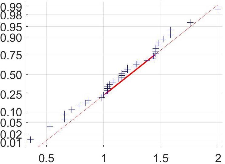
1    Abstract
2    New SARS-CoV-2 variants with mutations in the spike glycoprotein have arisen inde-
3    pendently at multiple locations and may have functional significance. The combination
4    of mutations in the 501Y.V2 variant first detected in South Africa include the N501Y,
5    K417N, and E484K mutations in the receptor binding domain (RBD) as well as muta-
6    tions in the N-terminal domain (NTD). Here we address whether the 501Y.V2 variant
7    could escape the neutralizing antibody response elicited by natural infection with ear-
8    lier variants. We were the first to outgrow two variants of 501Y.V2 from South Africa,
9    designated 501Y.V2.HV001 and 501Y.V2.HVdF002. We examined the neutralizing ef-
10   fect of convalescent plasma collected from six adults hospitalized with COVID-19 using
11   a microneutralization assay with live (authentic) virus. Whole genome sequencing of the
12   infecting virus of the plasma donors confirmed the absence of the spike mutations which
13   characterize 501Y.V2. We infected with 501Y.V2.HV001 and 501Y.V2.HVdF002 and com-
14   pared plasma neutralization to first wave virus which contained the D614G mutation but
15   no RBD or NTD mutations. We observed that neutralization of the 501Y.V2 variants was
16   strongly attenuated, with IC50 6 to 200-fold higher relative to first wave virus. The de-
17   gree of attenuation varied between participants and included a knockout of neutralization
18   activity. This observation indicates that 501Y.V2 may escape the neutralizing antibody
19   response elicited by prior natural infection. It raises a concern of potential reduced pro-
20   tection against re-infection and by vaccines designed to target the spike protein of earlier
21   SARS-CoV-2 variants.

22       Through genomic surveillance of the severe acute respiratory syndrome-related coronavirus 2 (SARS-
23   CoV-2), a number of new variants have recently been identified with multiple mutations in the spike
24   glycoprotein [1, 2, 3]. We recently described the emergence of the N501Y.V2 variant in South Africa,
25   characterised by the K417N, E484K, and N501Y mutations in the spike receptor binding domain (RBD)
26   as well as four substitutions and a deletion in the N-terminal domain (NTD) [1]. This variant was first
27   detected in October 2020, and has rapidly become the dominant variant in several parts of the country
28   at a time of a rapid resurgence in infections.
29       The RBD is the main target of neutralizing antibodies (NAbs) elicited by SARS-CoV-2 infection,
30   with the remaining activity directed at the NTD [4, 5, 6]. All three amino acid residues in the RBD that
31   carry mutations in 501Y.V2 interact directly with the human angiotensin-converting enzyme 2 (hACE2)
32   receptor and form part of the epitopes for hACE2-blocking NAbs [7]. The E484 residue specifically is a
33   hotspot for binding of highly potent NAbs [7]. In a number of separate in vitro studies using monoclonal
34   antibodies (mAbs), mutations at E484 have emerged as immune escape mutations, often conferring broad
35   cross-resistance to panels of mAbs [8, 9, 10, 11]. E484K also emerged during passage with convalescent
36   plasma, leading to substantial drops in neutralization with convalescent plasma samples [12, 13]. Using
37   a deep mutation scanning approach to determine the effect of individual mutations on neutralization by
38   polyclonal serum, mutations at E484 were associated with the largest drops in neutralization [14].
39       Here, using a microneutralization assay with authentic virus, we address the question of whether
40   501Y.V2 variants can escape the neutralizing response elicited by natural infection with previous vari-
41   ants. We outgrew and compared the neutralization of two SARS-CoV-2 501Y.V2 variants to a previously
42   circulating variant derived from South Africa which does not have the 501Y.V2 defining mutations.
43       For neutralization, we used plasma samples from our ongoing longitudinal cohort study that tracks
44   COVID-19 cases enrolled at two hospitals in Durban, South Africa [15]. We sampled participants
45   weekly for the first month post-enrollment, and at each timepoint a blood draw and combined nasopha-
46   ryngeal/oropharyngeal swab was performed to obtain both the plasma and the infecting virus.

                                                        2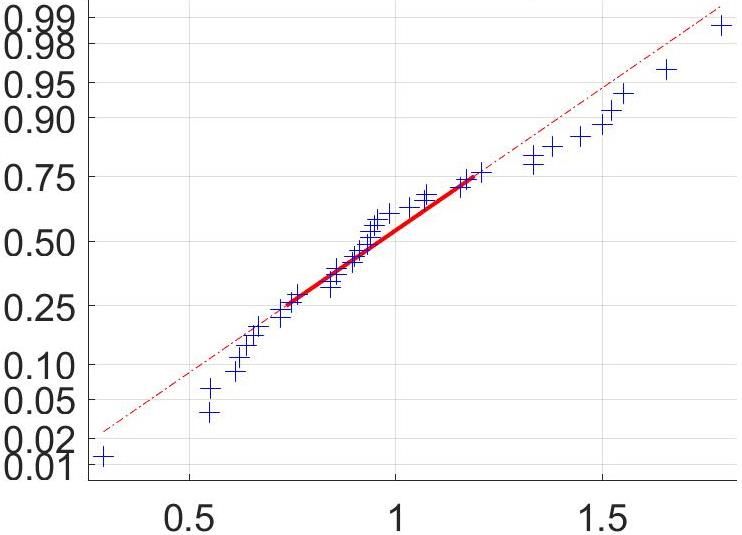
A

B
Dec-19
Jan-20
                                                                                                                  Sampling
Feb-20
                                                                                                                     Global
Mar-20                                                                                                               South Africa
Apr-20
                                       501Y.V2

May-20                                                                                                                Viruses and plasma
Jun-20                                                                                                                in this study

 Jul-20                                                                                                                  B.1.140
                                                                                                                         B.1.1
Aug-20
                                                                                                                         B.1.1.1
Sep-20                                                                                                                   B.1.351 (501Y.V2)
Oct-20                                                                                                                   B.1.5
Nov-20
Dec-20

                                                       Plasma used in challenge
                                                           Virus outgrown
                       Outgrown viral variants                               Infecting variant sequences of blood plasma donors

    Lineage        B.1.1         501Y.V2 (B.1.351)               B.1.1         B.1.1         B.1.5        B.1.5       B.1.140     B.1.1.1
Sequence ID       K002868      K005321            K005325       K002868     K004289     K004285     K004291     K004295     K004302
  Plasma ID     039-13-0013                                   039-13-0013 039-02-0014 039-13-0015 039-13-0033 039-02-0017 039-13-0062
    Isolate                   501Y.V2.            501Y.V2.
                CoV2.V003
 designation                  HVdF002              HV001
                                                    L18F
                               D80A
                                                   D80A
                               D215G
                                                   D215G
                               K417N
   Spike          D614G                            K417N        D614G
                               E484K                                          D614G         D614G        D614G        D614G       D614G
  mutations       A688V                            E484K        A688V
                               N501Y
                                                   N501Y
                               D614G
                                                   D614G
                               A701V
                                                   A701V
 Spike indels                 242-244del         242-244del

                                                                         3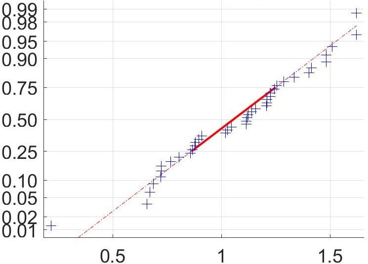
Figure 1: Study design and sequences of SARS-CoV-2 variants. (A) We obtained convalescent plasma
     and detected the matching infecting variant in the first SARS-CoV-2 infection wave in South Africa. A blood
     draw and nasopharyngeal/oropharyngeal was performed on study participants. First wave virus was outgrown
     from one of the participants and compared to two viruses outgrown from the second wave, which were 501Y.V2
     variants. A focus forming microneutralization assay was used to quantify neutralization. (B) Phylogenetic tree
     and mutations of variant sequences. Variants which infected the study participants who were plasma donors only
     for this study are marked in blue. Sequences of variants which were outgrown are marked in yellow. Participant
     039-13-0013 was both a plasma donor and the donor from whom the first wave virus was outgrown. Y-axis
     denotes time of sampling for viral sequencing. Table shows mutations present in Spike for the 501Y.V2 variants
     and the first wave virus used in the study. See Table S2 for a complete list of mutations in the viral genomes.

47       We chose plasma from participants from the first infection wave where the infecting virus was suc-
48   cessfully sequenced (Table S1) and where RBD binding was detected by ELISA. These viruses were
49   from a variety of B.1 lineages circulating in South Africa and contained the D614G mutation but none
50   of the spike mutations defining 501Y.V2 (Figure 1, see Table S2 for whole genome mutations). Plasma
51   samples were from blood drawn approximately 1 month post-symptom onset (Table S1), shown to be
52   close to the antibody response peak [16, 17].
53       We outgrew first wave virus (Materials and methods) from a sample obtained from a cohort par-
54   ticipant (039-13-0013) in July 2020, and second wave 501Y.V2 virus from two samples obtained in
55   November 2020 through our genomic surveillance program. We used a microneutralization live virus
56   focus forming assay (FFA) [18]. This relies on a methylcellulose overlay to limit cell-free viral spread,
57   resulting in a local infection focus then detected by an anti-SARS-CoV-2 Spike antibody (Materials and
58   methods). Re-sequencing of the first 501Y.V2 variant after outgrowth revealed no changes in the RBD
59   or NTD but a deletion in the furin cleavage site (Table S3) commonly observed after in vitro culture
60   in Vero E6 cells [19, 20]. We designated this variant 501Y.V2.HVdF002. HV represents the outgrowth
61   protocol which included initial outgrowth in a human H1299 cell line derivative overexpressing the ACE2
62   receptor, followed by a cell-to-cell infection of Vero E6 cells (Materials and methods). dF represents
63   the deletion of the furin cleavage site. Deletion of the furin cleavage site may not affect neutralization
64   [19]. However, we proceeded to test an additional 501Y.V2 variant. This variant, which we designated
65   501Y.V2.HV001, had an additional mutation, L18F, in the NTD prior to outgrowth and showed no
66   changes in spike sequence after outgrowth.
67       We mixed the virus with serially diluted participant plasma, then added the mixture to Vero E6
68   cells and counted infection foci after 28 hours (Figure 2A, Materials and methods). There was a clear
69   visual difference in the number of foci as a function of plasma dilution. 501Y.V2.HV001 also showed
70   dramatically larger foci (Figure 2A).
71       We normalized the number of foci to the number of foci in the absence of plasma on the same plate
72   to obtain the transmission index (Tx, [21]). In this context, it is the number of foci in the presence of
73   plasma inhibition divided by the number of foci in the absence of plasma. This controls for experiment
74   variability between plates and experiments. The data from the FFA approximated a normal distribution
75   (Figure S1) and we therefore used parametric statistics to describe it. We observed neutralization of the
76   first wave virus which varied between plasma samples (Figure 2B). To obtain the IC50 , we fitted the data
77   for each participant to a sigmoidal function [22] with IC50 as the only free parameter (Materials and
78   methods). Fitted IC50 values (Figure 2D) varied between 4×10⁻³ for participant 039-13-0013 to 1×10⁻⁴
79   for participants 039-13-0033 and 039-02-0015, consistent with the previously observed heterogeneity in
80   neutralization between individuals [16, 17].
81       We next determined neutralization of 501Y.V2. A decline in plasma neutralization was clearly ob-
82   served (Figure 2A). T501Y.V2.HV001 also showed attenuated neutralization likely greater than that
83   of 501Y.V2.HVdF002 (Figure S2), ruling out the in vitro generated deletion in the furin cleavage site
84   as being responsible for escape. We combined the data for both 501Y.V2 variants. Fitted IC50 values
85   varied between 1 × 10⁻³ (1:100 dilution) for plasma from participant 039-13-0033 to a complete knock-
86   out of activity for plasma from participant 039-13-0013 (Figure 2D). The 501Y.V2 to first wave IC50
87   ratio ranged from 6 to 200-fold (Figure 2D). Averaging across all participants highlighted the dramatic
88   decrease in sensitivity to neutralization of authentic 501Y.V2 variants (Figure 2E).

                                                           4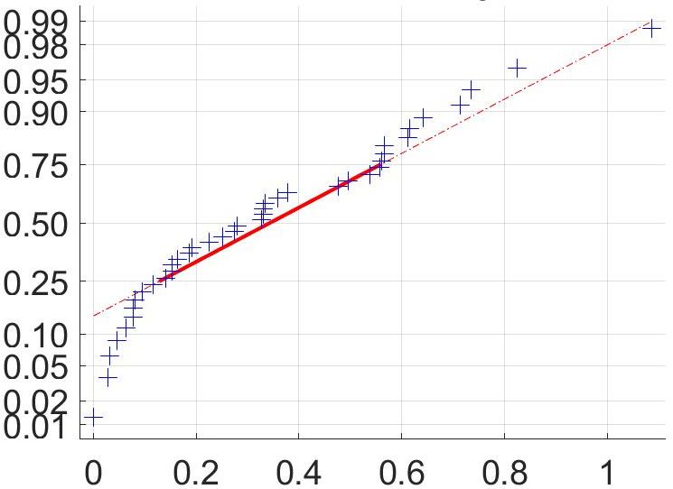
A                                         Dilution of PID 039-13-0015 plasma
                              1:100          1:200          1:400          1:800           1:1600      no plasma
          Viral variant

           1st wave

            501Y.V2
            HVdF002

             501Y.V2
             HV001

      B                                                      C
                              1st Wave                                               501Y.V2
           1.2                                                       1.2
           1.0                                                       1.0
           0.8                                                       0.8
                                                             Tx
      Tx

                                                                           Control pool
           0.6                                                       0.6   039-13-0013
                                                                           039-02-0014
           0.4                                                       0.4   039-02-0017
                                                                           039-13-0015
           0.2                                                       0.2   039-13-0062
                                                                           039-13-0033
             0                                                        0
                      10-3       Dilution            10-2                  10-3           Dilution              10-2

     D                            IC50                       E
                        1st wave 501Y.V2    Ratio
      Plasma donor:                                                                                   First Wave All
          039-13-0013
                                                                     1.2                              501Y.V2 All

                                                                     1.0
          039-02-0014
                                                                     0.8
                                                                Tx

          039-02-0015
                                                                     0.6
          039-13-0017
                                                                     0.4
          039-13-0033
                                                                     0.2
          039-13-0062
                                                                      0
                                                                           10-3            Dilution             10-2

Figure 2: Neutralization of first wave and 501Y.V2 variants by convalescent plasma from first
wave infections. (A) A representative focus forming assay using plasma from participant 039-13-0015. Plasma
neutralization of (B) first wave virus and (C) the combined results from the two 501Y.V2 variants. Colored
circles represent means and standard errors from 8 independent neutralization experiments using plasma from
6 convalescent participants who were infected by first wave variants in the first peak of the pandemic in South
Africa. Correspondingly colored lines are fits of the sigmoidal equation with IC50 as the fitted parameter. Black
points represent a pool of plasma from three uninfected controls. The transmission index (Tx) is the number
of foci in the presence of the plasma dilution normalized by the number of foci in the absence of plasma. (D)
Plasma IC50 values and ratios for first wave and 501Y.V2 variants. Knockout (KO) was scored as IC50 > 1.
ND, not defined. (E) Mean and standard error across all plasma donors.

                                                            5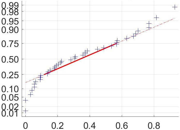
89       As we have entered the second year of the SARS-CoV-2 pandemic with high levels of transmission in
 90   many parts of the world, variants with mutations at key residues in the spike glycoprotein have emerged.
 91   Here we present clear evidence using authentic SARS-CoV-2 that the 501Y.V2 variant first detected
 92   in South Africa is associated with reduced neutralization by plasma collected from patients infected
 93   in the first wave with SARS-CoV-2 variants without the 501Y.V2 defining RBD and NTD mutations.
 94   While our findings are based on plasma samples from six convalescent study participants, the relative
 95   consistency of the effect argues that the potential to escape neutralizing antibodies elicited by prior
 96   SARS-CoV-2 infection may be widespread.
 97       The reduced neutralization is most likely related to the mutations in the spike RBD and NTD that
 98   characterize the 501Y.V2 variant. While the E484K mutation has the clearest association with immune
 99   escape, the other mutations in the RBD (K417N, N501Y) are also located within residues targeted
100   by some class 1 and class 2 NAbs [7]. Information about the significance of NTD mutations is also
101   emerging. NAbs targeting this site have been shown to be potent neutralizers of SARS-CoV-2 [5, 6].
102   The deletion at residues 242-244 is just outside an antigenic supersite loop (residues 245-264) and L18
103   also falls within the antigenic supersite. Furthermore, mutations at L18 and D80 have been selected
104   during passage with mAbs [5]. Our second variant contains the L18F mutation. This may be associated
105   with the trend to lower neutralization sensitivity relative to the first 501Y.V2 variant (Figure S2). This
106   variant also has strikingly larger foci (Figure 2A).
107       The reasons for the rapid emergence and fixation of potential immune escape mutations in South
108   Africa remain unclear. The 501Y.V2 variant was first detected in the Eastern Cape Province of South
109   Africa, in Nelson Mandela Bay, an urban municipality with a population of just over one million.
110   While we have no SARS-CoV-2 seroprevalence data from this area, there were 1909 excess natural
111   deaths (approximately 1600 per million population) by the end of the first wave in mid-September (
112   https://www.samrc.ac.za/reports/report-weekly-deaths-south-africa). In the context of a young popu-
113   lation (over 80 percent of the population under 50 years), this data would suggest a high attack rate
114   from the first wave. While circumstantial, this provides some support to a hypothesis of high levels of
115   population immunity driving the selection of variants with capacity to evade natural immunity. This
116   area also has high HIV prevalence, and has amongst the lowest proportions of people with HIV who have
117   viral suppression (http://www.hivdata.org.za/). We have not observed evidence of chronic SARS-CoV-2
118   infection in people living with HIV in our longitudinal cohort [15]. However, most cohort participants
119   had sustained virological suppression with antiretroviral therapy (ART). We did observe altered im-
120   mune dynamics after SARS-CoV-2 infection in HIV viremic participants relative to those who were
121   virologically suppressed, and we are currently enrolling additional participants to examine SARS-CoV-2
122   clearance in the HIV viremic subset.
123       The implications of these results for re-infection and vaccine efficacy are still unclear. Our findings
124   emphasize the need to understand whether the 501Y.V2 variant, and other similar variants, are associ-
125   ated with an increased rate of re-infection. Vaccines such as the Oxford/Astra Zeneca ChAdOx1 [23]
126   and the Pfizer-BioNTech BNT162b2 [24] elicit neutralization titers in a similar range to the convalescent
127   plasma in this study. However, these vaccines may elicit a broader antibody response and protective T
128   cell immunity [25]. Protective T cell immunity also likely occurs following natural infection. Further-
129   more, it is unclear what degree of neutralization mediates protection, and infection may be particularly
130   sensitive to inhibition at exposure [26].
131       In conclusion, we present data suggesting that the 501Y.V2 variant first detected in South Africa
132   is able to escape the neutralizing antibody response elicited by natural infection with earlier variants.
133   We expect data in the next weeks from phase 3 vaccine trials being conducted in South Africa. If the
134   variant does have an effect on vaccine efficacy, then there may be a signal in the data from these clinical
135   trials.

                                                          6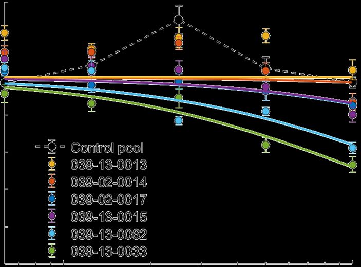
136   Material and methods
137   Ethical statement
138   Nasopharyngeal/oropharyngeal swab samples and plasma samples were obtained from six hospital-
139   ized adults with PCR-confirmed SARS-CoV-2 infection enrolled in a prospective cohort study ap-
140   proved by the Biomedical Research Ethics Committee (BREC) at the University of KwaZulu-Natal
141   (reference BREC/00001275/2020). The 501Y.V2 variants were obtained from residual nasopharyn-
142   geal/oropharyngeal samples used for routine SARS-CoV-2 diagnostic testing by the National Health
143   Laboratory Service, through our SARS-CoV-2 genomic surveillance program (BREC approval reference
144   BREC/00001510/2020).

145   Whole genome sequencing, genome assembly and phylogenetic analysis
146   cDNA synthesis was performed on the extracted RNA using random primers followed by gene specific
147   multiplex PCR using the ARTIC V3 protocol. Briefly, extracted RNA was converted to cDNA using the
148   Superscript IV First Strand synthesis system (Life Technologies, Carlsbad, CA) and random hexamer
149   primers. SARS-CoV-2 whole genome amplification was performed by multiplex PCR using primers de-
150   signed on Primal Scheme (http://primal.zibraproject.org/) to generate 400bp amplicons with an overlap
151   of 70bp that covers the 30Kb SARS-CoV-2 genome. PCR products were cleaned up using AmpureXP
152   purification beads (Beckman Coulter, High Wycombe, UK) and quantified using the Qubit dsDNA
153   High Sensitivity assay on the Qubit 4.0 instrument (Life Technologies Carlsbad, CA). We then used the
154   Illumina® Nextera Flex DNA Library Prep kit according to the manufacturer’s protocol to prepare
155   indexed paired end libraries of genomic DNA. Sequencing libraries were normalized to 4nM, pooled and
156   denatured with 0.2N sodium acetate. 12pM sample library was spiked with 1% PhiX (PhiX Control v3
157   adapter-ligated library used as a control). We sequenced libraries on a 500-cycle v2 MiSeq Reagent Kit
158   on the Illumina MiSeq instrument (Illumina, San Diego, CA). We have previously published full details
159   of the amplification and sequencing protocol [27].
160       We assembled paired-end fastq reads using Genome Detective 1.126 (https://www.genomedetective.com)
161   and the Coronavirus Typing Tool [28]. We polished the initial assembly obtained from Genome Detective
162   by aligning mapped reads to the references and filtering out low-quality mutations using bcftools 1.7-2
163   mpileup method. Mutations were confirmed visually with bam files using Geneious software (Biomatters
164   Ltd, Auckland, New Zealand). All of the sequences were deposited in GISAID (https://www.gisaid.org/).
165   We retrieved all South African SARS-CoV-2 genotypes from the GISAID database as of 11 January
166   2021 (N=2704). We initially analyzed South African genotypes against the global reference dataset
167   (N=2592) using a custom pipeline based on a local version of NextStrain. The pipeline contains several
168   python scripts that manage the analysis workflow. It performs alignment of genotypes in MAFFT [29],
169   phylogenetic tree inference in IQ-Tree20, tree dating and ancestral state construction and annotation
170   (https://github.com/nextstrain/ncov).

171   Cells
172   Vero E6 cells (ATCC CRL-1586, obtained from Cellonex) were propagated in complete DMEM with 10%
173   fetal bovine serum (Hylone) containing 1% each of HEPES, sodium pyruvate, L-glutamine, and non-
174   essential amino acids (Sigma-Aldrich). Cells were passaged every 3-4 days. H1299 cells were propagated
175   in complete RPMI with 10% fetal bovine serum containing 1% each of HEPES, sodium pyruvate, L-
176   glutamine, and non-essential amino acids and and passaged every second day.

177   H1299-E3 cell line for first passage SARS-CoV-2 outgrowth
178   The H1299-H2AZ clone with nuclear labelled YFP [30] was constructed to overexpress ACE2 as follows:
179   VSVG-pseudotyped lentivirus containing the human ACE2 was generated by co-transfecting 293T cells
180   with the pHAGE2-EF1alnt-ACE2-WT plasmid along with the lentiviral helper plasmids HDM-VSVG,

                                                        7
181   HDM-Hgpm2, HDM-tat1b and pRC-CMV-Rev1b using TransIT-LT1 (Mirus) transfection reagent. Su-
182   pernatant containing the lentivirus was harvested two days after infection, filtered through a 0.45µm
183   filter (Corning) and used to spinfect H1299-H2AZ at 1000 rcf for 2 hours at room temperature in the pres-
184   ence of 5 µg/mL polybrene (Sigma-Aldrich). ACE-2 transduced H1299-H2AZ cells were then subcloned
185   at the single cell density in 96-well plates (Eppendorf) in conditioned media derived from confluent cells.
186   After 3 weeks, wells were trypsinized (Sigma-Aldrich) and plated in two replicate plates, where the first
187   plate was used to determine infectivity and the second was stock. The first plate was screened for the
188   fraction of mCherry positive cells per cell clone upon infection with SARS-CoV-2 mCherry expressing
189   Spike pseudotyped lentiviral vector 1610-pHAGE2/EF1a Int-mCherry3-W produced by transfecting as
190   above. Screening was performed using a Metamorph-controlled (Molecular Devices, Sunnyvale, CA)
191   Nikon TiE motorized microscope (Nikon Corporation, Tokyo, Japan) with a 20x, 0.75 NA phase ob-
192   jective, 561 laser line, and 607 nm emission filter (Semrock, Rochester, NY). Images were captured
193   using an 888 EMCCD camera (Andor). Temperature (37°C), humidity and CO2 (5%) were controlled
194   using an environmental chamber (OKO Labs, Naples, Italy). The clone with the highest fraction of
195   mCherry expression was expanded from the stock plate and denoted H1299-E3. This clone was used in
196   the outgrowth.

197   Viral Outgrowth
198   All live virus work was performed in Biosafety level 3 containment using AHRI Institutional Biosafety
199   Committee approved protocols for SARS-CoV-2. For first wave virus, a T25 flask (Corning) was seeded
200   with Vero E6 cells at 2 × 10⁵ cells/ml and incubated for 18-20 hours. After 1 DPBS wash, the sub-
201   confluent cell monolayer was inoculated with 500µL universal transport medium (UTM) diluted 1:1
202   with growth medium and filtered through a 0.45µM filter. Cells were incubated for 1 hour. Flask was
203   then filled with 7mL of complete growth medium and checked daily for cytopathic effect (CPE). Four
204   days post infection, supernatants of the infected culture were collected, centrifuged at 300 rcf for 3
205   minutes to remove cell debris, and filtered using a 0.45µM filter. Viral supernatant was aliquoted and
206   stored at -80◦ C. For 501Y.V2 variants, we used H1299-ACE2-E3 cells for initial isolation followed by
207   passage into Vero E6 cells. H1299-ACE2-E3 cells were seeded at 1.5 × 10⁵ cells/ml and incubated for
208   18-20 hours. After 1 DPBS wash, the sub-confluent cell monolayer was inoculated with 500µL universal
209   transport medium (UTM) diluted 1:1 with growth medium and filtered through a 0.45µM filter. Cells
210   were incubated for 1 hour. Wells were then filled with 3mL of complete growth medium. 8 days post-
211   infection, cells were trypsinized, centrifuged at 300 rcf for 3 minutes and resuspended in 4mL growth
212   medium. 1mL was added to Vero E6 cells that had been seeded at t 2 × 10⁵ cells/ml 18-20 hours earlier
213   in a T25 flask (approximately 1:8 donor-to-target cell dilution ratio) for cell-to-cell infection. Coculture
214   of H1299-ACE2-E3 and Vero E6 cells was incubated for 1 hour and flask was then filled with 7mL of
215   complete growth medium and incubated for 6 days. Viral supernatant was aliquoted and stored at
216   -80◦ C or further passaged in Vero E6 cells as above.

217   Microneutralization using focus forming assay
218   Vero E6 cells were plated in an 96 well plate (Eppendorf) at 30,000 cells per well 1 day pre-infection.
219   Plasma was separated from EDTA-anticoagulated blood by centrifugation at 500 rcf for 10 minutes and
220   stored at -80◦ C. Aliquots of plasma samples were heat-inactivated at 56◦ C for 30 minutes, and clarified
221   by centrifugation at 10,000 rcf for 5 minutes, where the clear middle layer was used for experiments.
222   Inactivated plasma was stored in single use aliquots to prevent freeze-thaw cycles. For experiments,
223   plasma was serially diluted two-fold from 1:100 to 1:1600. Virus stocks were used at approximately 50
224   focus-forming units (FFU) per microwell and added to diluted plasma; antibody-virus mixtures were
225   incubated for 1 hour at 37◦ C, 5% CO2 . Cells were infected with 100µL of the virus-antibody mixtures,
226   to allow adsorption of virus. Subsequently, 100µL of a 1x RPMI 1640 (Sigma-Aldrich R6504), 1.5%
227   carboxymethylcellulose (Sigma-Aldrich C4888) overlay was added to the wells without removing the
228   inoculum. Cells were fixed at 28 hours post-infection using 4% paraformaldehyde (Sigma-Aldrich) for
229   20 minutes. For staining of foci, a rabbit anti-Spike monoclonal antibody (mAb BS-R2B12, GenScript

                                                           8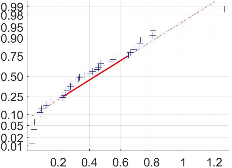
230   A02058) was used at 0.5µg/mL as the primary detection antibody. Antibody was resuspended in
231   a permiabilization buffer containing 0.1% saponin (Sigma-Aldrich), 0.1% BSA (Sigma-Aldrich), and
232   0.05% tween (Sigma-Aldrich) in PBS. Plates were incubated with primary antibody overnight at 4◦ C,
233   then washed with wash buffer containing 0.05% tween in PBS. Secondary goat anti-rabbit horseradish
234   peroxidase (Abcam ab205718) was added at 1 µg/mL and incubated for 2 hours at room temperature
235   with shaking. The TrueBlue peroxidase substrate (SeraCare 5510-0030) was then added at 50µL per
236   well and incubated for 20 minutes at room temperature. Plates were then dried for 2 hours and imaged
237   using a Metamorph-controlled Nikon TiE motorized microscope with a 2x objective. Automated image
238   analysis was performed using a Matlab2019b (Mathworks) custom script, where focus detection was
239   automated and did not involve user curation. Image segmentation steps were stretching the image from
240   minimum to maximum intensity, local Laplacian filtering, image complementation, thresholding and
241   binarization. For the second 501Y.V2 variant, a dilation/erosion step was introduced to prevent the
242   large foci from fragmenting into smaller objects.

243   Statistics and fitting
244   All statistics and fitting were performed using Matlab2019b. Neutralization data was fit to

                                              T x = 1/1 + (D/IC50 ).
245       Here Tx is the number of foci normalized to the number of foci in the absence of plasma on the same
246   plate at dilution D. Fit to a normal distribution using Matlab2019b function normplot, which compared
247   the distribution of the Tx data to the normal distribution (see https://www.mathworks.com/help/stats/normplot.html).

248   Acknowledgements
249   This work was supported by the Bill and Melinda Gates Investment INV-018944 (AS) and by the South
250   African Medical Research Council and the Department of Science and Innovation (TdO).

251   § Network for Genomic Surveillance in South Africa (NGS-SA)
252   Shareef Abrahams1 , Luiz Carlos Junior Alcantara2 , Arghavan Alisoltani-Dehkordi3,4 , Mushal Allam5 ,
253   Jinal N Bhiman5,6 , Mary-Ann Davies7,8 , Deelan Doolabh9 , Susan Engelbrecht10 , Vagner Fonseca11 ,
254   Marta Giovanetti2 , Allison J Glass6,12 , Adam Godzik4 , Dominique Goedhals13 , Diana Hardie14 , Mar-
255   vin Hsiao14 , Arash Iranzadeh4 , Arshad Ismail5 , Stephen Korsman14 , Sergei L Kosakovsky Pond15 ,
256   Oluwakemi Laguda-Akingba1,16 , Jose Lourenco17 , Gert Marais14 , Darren Martin9,18 , Caroline Maslo19 ,
257   Koleka Mlisana20,21 , Thabo Mohale5 , Nokukhanya Msomi22 , Innocent Mudau9 , Francesco Petruccione23,24 ,
258   Wolfgang Preiser10 , Emmanuel James San11 , Bryan Trevor Sewell25 , Lynn Tyers9 , Gert Van Zyl10 ,
259   Anne von Gottberg5,6 , Sibongile Walaza5,26 , Steven Weaver15 , Constantinos Kurt Wibmer5 , Carolyn
260   Williamson9,14,21 , Denis York27 .
      1
261     National Health Laboratory Service, Port Elizabeth, South Africa. 2 Laboratorio de Flavivirus, Fun-
262   dacao Oswaldo Cruz, Rio de Janeiro, Brazil. 3 Division of Medical Virology, Department of Pathology,
263   University of Cape Town, Cape Town, South Africa. 4 Division of Biomedical Sciences, University of
264   California Riverside School of Medicine, Riverside, California, USA. 5 National Institute for Commu-
265   nicable Diseases of the National Health Laboratory Service, Johannesburg, South Africa. 6 School of
266   Pathology, Faculty of Health Sciences, University of the Witwatersrand, Johannesburg, South Africa.
      7
267     Centre for Infectious Disease Epidemiology and Research, University of Cape Town, Cape Town, South
268   Africa. 8 Western Cape Government: Health, Cape Town, South Africa. 9 Division of Medical Virology,
269   Institute of Infectious Disease and Molecular Medicine, University of Cape Town, Cape Town, South
270   Africa. 10 Division of Medical Virology at NHLS Tygerberg Hospital and Faculty of Medicine and Health
271   Sciences, Stellenbosch University, Cape Town, South Africa. 11 KwaZulu-Natal Research Innovation and

                                                        9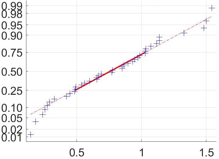
272   Sequencing Platform (KRISP), Department of Laboratory Medicine and Medical Sciences, University
273   of KwaZulu-Natal, Durban, South Africa. 12 Department of Molecular Pathology, Lancet Laboratories,
274   Johannesburg, South Africa. 13 Division of Virology at NHLS Universitas Academic Laboratories, Uni-
275   versity of The Free State, Bloemfontein, South Africa. 14 Division of Medical Virology at NHLS Groote
276   Schuur Hospital, University of Cape Town, Cape Town, South Africa. 15 Institute for Genomics and
277   Evolutionary Medicine, Temple University, Philadelphia, Pennsylvania, USA. 16 Department of Labo-
278   ratory Medicine and Pathology, Faculty of Health Sciences, Walter Sisulu University, Mthatha, South
279   Africa. 17 Department of Zoology, University of Oxford, Oxford, United Kingdom. 18 Computational Bi-
280   ology Division, Department of Integrative Biomedical Sciences, University of Cape Town, Cape Town,
281   South Africa. 19 Department of Quality Leadership, Netcare Hospitals, Johannesburg, South Africa.
      20
282      National Health Laboratory Service, Johannesburg, South Africa. 21 Centre for the AIDS Programme
283   of Research in South Africa (CAPRISA), Durban, South Africa. 22 Discipline of Virology, University of
284   KwaZulu-Natal, School of Laboratory Medicine and Medical Sciences and National Health Laboratory
285   Service, Durban, South Africa. 23 Centre for Quantum Technology, University of KwaZulu-Natal, Dur-
286   ban, South Africa 24 National Institute for Theoretical Physics (NITheP), KwaZulu-Natal, South Africa.
      25
287      Structural Biology Research Unit, Department of Integrative Biomedical Sciences, University of Cape
288   Town, Rondebosch, South Africa. 26 School of Public Health, Faculty of Health Sciences, University
289   of the Witwatersrand, Johannesburg, South Africa. 27 Molecular Diagnostics Services, Durban, South
290   Africa.

291   § § COMMIT-KZN Team
292   Moherndran Archary1 , Kaylesh J. Dullabh2 , Philip Goulder3,4 , Guy Harling3,5 , Rohen Harrichandparsad6 ,
293   Kobus Herbst3,7 , Prakash Jeena1 , Thandeka Khoza3 , Nigel Klein3,8 , Henrik Kløverpris3,9,10 , Alasdair
294   Leslie3,9 , Rajhmun Madansein2 , Mohlopheni Marakalala3,9 , Matilda Mazibuko3 , Mosa Moshabela11 ,
295   Ntombifuthi Mthabela3 , Kogie Naidoo12 , Zaza Ndhlovu3,13 , Thumbi Ndung’u3,9,14,15 , Kennedy Nyamande16 ,
296   Nesri Padayatchi12 , Vinod Patel17 , Theresa Smit3 , Adrie Steyn3,18 , Emily Wong3,18 .
      1
297    Department of Paediatrics and Child Health, University of KwaZulu-Natal, Durban, South Africa.
      2
298    Department of Cardiothoracic Surgery, University of KwaZulu-Natal, Durban, South Africa. 3 Africa
299   Health Research Institute, Durban, South Africa. 4 Department of Paediatrics, Oxford, UK. 5 Institute
300   for Global Health, University College London, UK. 6 Department of Neurosurgery, University of KwaZulu-
301   Natal, Durban, South Africa. 7 South African Population Research Infrastructure Network, Durban,
302   South Africa. 8 Institute of Child Health, University College London, UK. 9 Division of Infection and
303   Immunity, University College London, London, UK. 10 Department of Immunology and Microbiology,
304   University of Copenhagen, Copenhagen, Denmark. 11 College of Health Sciences, University of KwaZulu-
305   Natal, Durban, South Africa. 12 Centre for the AIDS Programme of Research in South Africa, Durban,
306   South Africa. 13 Ragon Institute of MGH, MIT and Harvard, Boston, USA. 14 HIV Pathogenesis Pro-
307   gramme, The Doris Duke Medical Research Institute, University of KwaZulu-Natal, Durban, South
308   Africa. 15 Max Planck Institute for Infection Biology, Berlin, Germany. 16 Department of Pulmonology
309   and Critical Care, University of KwaZulu-Natal, Durban, South Africa. 17 Department of Neurology,
310   University of KwaZulu-Natal, Durban, South Africa. 18 Division of Infectious Diseases, University of
311   Alabama at Birmingham.

312   References
313       [1] Houriiyah Tegally, Eduan Wilkinson, Marta Giovanetti, Arash Iranzadeh, Vagner Fonseca,
314           Jennifer Giandhari, Deelan Doolabh, Sureshnee Pillay, Emmanuel James San, Nokukhanya
315           Msomi, Koleka Mlisana, Anne von Gottberg, Sibongile Walaza, Mushal Allam, Arshad Ismail,
316           Thabo Mohale, Allison J Glass, Susan Engelbrecht, Gert Van Zyl, Wolfgang Preiser, Francesco
317           Petruccione, Alex Sigal, Diana Hardie, Gert Marais, Marvin Hsiao, Stephen Korsman, Mary-

                                                       10
318 Ann Davies, Lynn Tyers, Innocent Mudau, Denis York, Caroline Maslo, Dominique Goedhals,
319 Shareef Abrahams, Oluwakemi Laguda-Akingba, Arghavan Alisoltani-Dehkordi, Adam Godzik,
320 Constantinos Kurt Wibmer, Bryan Trevor Sewell, José Lourenço, Luiz Carlos Junior Alcantara,
321 Sergei L Kosakovsky Pond, Steven Weaver, Darren Martin, Richard J Lessells, Jinal N Bhiman,
322 Carolyn Williamson, and Tulio de Oliveira. Emergence and rapid spread of a new severe acute
323 respiratory syndrome-related coronavirus 2 (sars-cov-2) lineage with multiple spike mutations in
324 south africa. medRxiv, page 2020.12.21.20248640, 2020. doi: 10.1101/2020.12.21.20248640. URL
325 https://www.medrxiv.org/content/medrxiv/early/2020/12/22/2020.12.21.20248640.full.pdf.
326 [2] Erik Volz, Swapnil Mishra, Meera Chand, Jeffrey C. Barrett, Robert Johnson, Lily Geidel-
327 berg, Wes R Hinsley, Daniel J Laydon, Gavin Dabrera, Áine O’Toole, Roberto Amato, Manon
328 Ragonnet-Cronin, Ian Harrison, Ben Jackson, Cristina V. Ariani, Olivia Boyd, Nick Loman, John T
329 McCrone, Sónia Gonçalves, David Jorgensen, Richard Myers, Verity Hill, David K. Jackson, Katy
330 Gaythorpe, Natalie Groves, John Sillitoe, Dominic P. Kwiatkowski, Seth Flaxman, Oliver
331 Ratmann, Samir Bhatt, Susan Hopkins, Axel Gandy, Andrew Rambaut, and Neil M Ferguson.
332 Transmission of sars-cov-2 lineage b.1.1.7 in england: Insights from linking epidemiological and
333 genetic data. medRxiv, page 2020.12.30.20249034, 2021. doi: 10.1101/2020.12.30.20249034. URL
334 https://www.medrxiv.org/content/medrxiv/early/2021/01/04/2020.12.30.20249034.full.pdf.
335 [3] Carolina M Voloch, Ronaldo da Silva F, Luiz G P de Almeida, Cynthia C Cardoso, Otavio J.
336 Brustolini, Alexandra L Gerber, Ana Paula de C Guimarães, Diana Mariani, Raissa Mirella da
337 Costa, Orlando C. Ferreira, Adriana Cony Cavalcanti, Thiago Silva Frauches, Claudia Maria Braga
338 de Mello, Rafael Mello Galliez, Débora Souza Faffe, Terezinha M P P Castiñeiras, Amilcar Tanuri,
339 and Ana Tereza R de Vasconcelos. Genomic characterization of a novel sars-cov-2 lineage from rio de
340 janeiro, brazil. medRxiv, page 2020.12.23.20248598, 2020. doi: 10.1101/2020.12.23.20248598. URL
341 https://www.medrxiv.org/content/medrxiv/early/2020/12/26/2020.12.23.20248598.full.pdf.
342 [4] L. Piccoli, Y. J. Park, M. A. Tortorici, N. Czudnochowski, A. C. Walls, M. Beltramello, C. Silacci-
343 Fregni, D. Pinto, L. E. Rosen, J. E. Bowen, O. J. Acton, S. Jaconi, B. Guarino, A. Minola, F. Zatta,
344 N. Sprugasci, J. Bassi, A. Peter, A. De Marco, J. C. Nix, F. Mele, S. Jovic, B. F. Rodriguez,
345 S. V. Gupta, F. Jin, G. Piumatti, G. Lo Presti, A. F. Pellanda, M. Biggiogero, M. Tarkowski,
346 M. S. Pizzuto, E. Cameroni, C. Havenar-Daughton, M. Smithey, D. Hong, V. Lepori, E. Albanese,
347 A. Ceschi, E. Bernasconi, L. Elzi, P. Ferrari, C. Garzoni, A. Riva, G. Snell, F. Sallusto, K. Fink,
348 H. W. Virgin, A. Lanzavecchia, D. Corti, and D. Veesler. Mapping neutralizing and immunodom-
349 inant sites on the sars-cov-2 spike receptor-binding domain by structure-guided high-resolution
350 serology. Cell, 183(4):1024–1042 e21, 2020. ISSN 1097-4172 (Electronic) 0092-8674 (Linking). doi:
351 10.1016/j.cell.2020.09.037. URL https://www.ncbi.nlm.nih.gov/pubmed/32991844.
352 [5] Matthew McCallum, Anna De Marco, Florian Lempp, M. Alejandra Tortorici, Dora Pinto,
353 Alexandra C. Walls, Martina Beltramello, Alex Chen, Zhuoming Liu, Fabrizia Zatta, Samantha
354 Zepeda, Julia di Iulio, John E. Bowen, Martin Montiel-Ruiz, Jiayi Zhou, Laura E. Rosen, Siro
355 Bianchi, Barbara Guarino, Chiara Silacci Fregni, Rana Abdelnabi, Shi-Yan Caroline Foo, Paul W.
356 Rothlauf, Louis-Marie Bloyet, Fabio Benigni, Elisabetta Cameroni, Johan Neyts, Agostino Riva,
357 Gyorgy Snell, Amalio Telenti, Sean P.J. Whelan, Herbert W. Virgin, Davide Corti, Matteo Samuele
358 Pizzuto, and David Veesler. N-terminal domain antigenic mapping reveals a site of vulnerability
359 for sars-cov-2. bioRxiv, page 2021.01.14.426475, 2021. doi: 10.1101/2021.01.14.426475. URL
360 https://www.biorxiv.org/content/biorxiv/early/2021/01/14/2021.01.14.426475.full.pdf.
361 [6] Gabriele Cerutti, Yicheng Guo, Tongqing Zhou, Jason Gorman, Myungjin Lee, Micah Rapp,
362 Eswar R. Reddem, Jian Yu, Fabiana Bahna, Jude Bimela, Yaoxing Huang, Phinikoula S.
363 Katsamba, Lihong Liu, Manoj S. Nair, Reda Rawi, Adam S. Olia, Pengfei Wang, Gwo-Yu
364 Chuang, David D. Ho, Zizhang Sheng, Peter D. Kwong, and Lawrence Shapiro. Potent
365 sars-cov-2 neutralizing antibodies directed against spike n-terminal domain target a single
366 supersite. bioRxiv, page 2021.01.10.426120, 2021. doi: 10.1101/2021.01.10.426120. URL
367 https://www.biorxiv.org/content/biorxiv/early/2021/01/11/2021.01.10.426120.full.pdf.
11368 [7] C. O. Barnes, C. A. Jette, M. E. Abernathy, K. A. Dam, S. R. Esswein, H. B. Gristick, A. G.
369 Malyutin, N. G. Sharaf, K. E. Huey-Tubman, Y. E. Lee, D. F. Robbiani, M. C. Nussenzweig, Jr.
370 West, A. P., and P. J. Bjorkman. Sars-cov-2 neutralizing antibody structures inform therapeutic
371 strategies. Nature, 588(7839):682–687, 2020. ISSN 1476-4687 (Electronic) 0028-0836 (Linking). doi:
372 10.1038/s41586-020-2852-1. URL https://www.ncbi.nlm.nih.gov/pubmed/33045718.
373 [8] Alina Baum, Benjamin O Fulton, Elzbieta Wloga, Richard Copin, Kristen E Pascal, Vincenzo
374 Russo, Stephanie Giordano, Kathryn Lanza, Nicole Negron, and Min Ni. Antibody cocktail to
375 sars-cov-2 spike protein prevents rapid mutational escape seen with individual antibodies. Science,
376 369(6506):1014–1018, 2020. ISSN 0036-8075.
377 [9] A. J. Greaney, T. N. Starr, P. Gilchuk, S. J. Zost, E. Binshtein, A. N. Loes, S. K. Hilton, J. Hud-
378 dleston, R. Eguia, K. H. D. Crawford, A. S. Dingens, R. S. Nargi, R. E. Sutton, N. Suryadevara,
379 P. W. Rothlauf, Z. Liu, S. P. J. Whelan, R. H. Carnahan, Jr. Crowe, J. E., and J. D. Bloom.
380 Complete mapping of mutations to the sars-cov-2 spike receptor-binding domain that escape anti-
381 body recognition. Cell Host Microbe, 2020. ISSN 1934-6069 (Electronic) 1931-3128 (Linking). doi:
382 10.1016/j.chom.2020.11.007. URL https://www.ncbi.nlm.nih.gov/pubmed/33259788.
383 [10] Z. Liu, H. Zheng, H. Lin, M. Li, R. Yuan, J. Peng, Q. Xiong, J. Sun, B. Li, J. Wu, L. Yi,
384 X. Peng, H. Zhang, W. Zhang, R. J. G. Hulswit, N. Loman, A. Rambaut, C. Ke, T. A. Bowden,
385 O. G. Pybus, and J. Lu. Identification of common deletions in the spike protein of severe acute
386 respiratory syndrome coronavirus 2. J Virol, 94(17), 2020. ISSN 1098-5514 (Electronic) 0022-538X
387 (Linking). doi: 10.1128/JVI.00790-20. URL https://www.ncbi.nlm.nih.gov/pubmed/32571797.
388 [11] Y. Weisblum, F. Schmidt, F. Zhang, J. DaSilva, D. Poston, J. C. Lorenzi, F. Muecksch,
389 M. Rutkowska, H. H. Hoffmann, E. Michailidis, C. Gaebler, M. Agudelo, A. Cho, Z. Wang,
390 A. Gazumyan, M. Cipolla, L. Luchsinger, C. D. Hillyer, M. Caskey, D. F. Robbiani, C. M. Rice,
391 M. C. Nussenzweig, T. Hatziioannou, and P. D. Bieniasz. Escape from neutralizing antibodies by
392 sars-cov-2 spike protein variants. Elife, 9, 2020. ISSN 2050-084X (Electronic) 2050-084X (Linking).
393 doi: 10.7554/eLife.61312. URL https://www.ncbi.nlm.nih.gov/pubmed/33112236.
394 [12] E. Andreano, G. Piccini, D. Licastro, L. Casalino, N. V. Johnson, I. Paciello, S. D. Monego,
395 E. Pantano, N. Manganaro, A. Manenti, R. Manna, E. Casa, I. Hyseni, L. Benincasa, E. Mon-
396 tomoli, R. E. Amaro, J. S. McLellan, and R. Rappuoli. Sars-cov-2 escape in vitro from a highly
397 neutralizing covid-19 convalescent plasma. bioRxiv, 2020. doi: 10.1101/2020.12.28.424451. URL
398 https://www.ncbi.nlm.nih.gov/pubmed/33398278.
399 [13] Zhuoming Liu, Laura A. VanBlargan, Louis-Marie Bloyet, Paul W. Rothlauf, Rita E.
400 Chen, Spencer Stumpf, Haiyan Zhao, John M. Errico, Elitza S. Theel, Mariel J. Liebe-
401 skind, Brynn Alford, William J. Buchser, Ali H. Ellebedy, Daved H. Fremont, Michael S.
402 Diamond, and Sean P. J. Whelan. Landscape analysis of escape variants identifies
403 sars-cov-2 spike mutations that attenuate monoclonal and serum antibody neutraliza-
404 tion. bioRxiv, page 2020.11.06.372037, 2021. doi: 10.1101/2020.11.06.372037. URL
405 https://www.biorxiv.org/content/biorxiv/early/2021/01/11/2020.11.06.372037.full.pdf.
406 [14] Allison J Greaney, Andrea N Loes, Katharine HD Crawford, Tyler N Starr, Keara D Malone, He-
407 len Y Chu, and Jesse D Bloom. Comprehensive mapping of mutations to the sars-cov-2 receptor-
408 binding domain that affect recognition by polyclonal human serum antibodies. bioRxiv, page
409 2020.12. 31.425021, 2021.
410 [15] Farina Karim, Inbal Gazy, Sandile Cele, Yenzekile Zungu, Robert Krause, Mallory
411 Bernstein, Yashica Ganga, Hylton Rodel, Ntombifuthi Mthabela, Matilda Mazibuko,
412 Khadija Khan, Daniel Muema, Dirhona Ramjit, Gila Lustig, Thumbi Ndung’u, Willem
413 Hanekom, Bernadett I. Gosnell, Emily Wong, Tulio de Oliveira, Mahomed-Yunus S.
414 Moosa, Alasdair Leslie, Henrik Kløverpris, and Alex Sigal. Hiv infection alters sars-
415 cov-2 responsive immune parameters but not clinical outcomes in covid-19 disease.
12416 medRxiv, page 2020.11.23.20236828, 2020. doi: 10.1101/2020.11.23.20236828. URL
417 https://www.medrxiv.org/content/medrxiv/early/2020/11/24/2020.11.23.20236828.full.pdf.
418 [16] D. F. Robbiani, C. Gaebler, F. Muecksch, J. C. C. Lorenzi, Z. Wang, A. Cho, M. Agudelo, C. O.
419 Barnes, A. Gazumyan, S. Finkin, T. Hagglof, T. Y. Oliveira, C. Viant, A. Hurley, H. H. Hoffmann,
420 K. G. Millard, R. G. Kost, M. Cipolla, K. Gordon, F. Bianchini, S. T. Chen, V. Ramos, R. Patel,
421 J. Dizon, I. Shimeliovich, P. Mendoza, H. Hartweger, L. Nogueira, M. Pack, J. Horowitz, F. Schmidt,
422 Y. Weisblum, E. Michailidis, A. W. Ashbrook, E. Waltari, J. E. Pak, K. E. Huey-Tubman, N. Ko-
423 randa, P. R. Hoffman, Jr. West, A. P., C. M. Rice, T. Hatziioannou, P. J. Bjorkman, P. D. Bieniasz,
424 M. Caskey, and M. C. Nussenzweig. Convergent antibody responses to sars-cov-2 in convalescent
425 individuals. Nature, 584(7821):437–442, 2020. ISSN 1476-4687 (Electronic) 0028-0836 (Linking).
426 doi: 10.1038/s41586-020-2456-9. URL https://www.ncbi.nlm.nih.gov/pubmed/32555388.
427 [17] C. Gaebler, Z. Wang, J. C. C. Lorenzi, F. Muecksch, S. Finkin, M. Tokuyama, A. Cho, M. Jankovic,
428 D. Schaefer-Babajew, T. Y. Oliveira, M. Cipolla, C. Viant, C. O. Barnes, Y. Bram, G. Breton,
429 T. Hagglof, P. Mendoza, A. Hurley, M. Turroja, K. Gordon, K. G. Millard, V. Ramos, F. Schmidt,
430 Y. Weisblum, D. Jha, M. Tankelevich, G. Martinez-Delgado, J. Yee, R. Patel, J. Dizon, C. Unson-
431 O’Brien, I. Shimeliovich, D. F. Robbiani, Z. Zhao, A. Gazumyan, R. E. Schwartz, T. Hatziioannou,
432 P. J. Bjorkman, S. Mehandru, P. D. Bieniasz, M. Caskey, and M. C. Nussenzweig. Evolution of
433 antibody immunity to sars-cov-2, 2021. ISSN 1476-4687 (Electronic) 0028-0836 (Linking). URL
434 https://www.ncbi.nlm.nih.gov/pubmed/33461210.
435 [18] James Brett Case, Adam L Bailey, Arthur S Kim, Rita E Chen, and Michael S Diamond. Growth,
436 detection, quantification, and inactivation of sars-cov-2. Virology, 2020.
437 [19] W. B. Klimstra, N. L. Tilston-Lunel, S. Nambulli, J. Boslett, C. M. McMillen, T. Gilliland, M. D.
438 Dunn, C. Sun, S. E. Wheeler, A. Wells, A. L. Hartman, A. K. McElroy, D. S. Reed, L. J. Ren-
439 nick, and W. P. Duprex. Sars-cov-2 growth, furin-cleavage-site adaptation and neutralization
440 using serum from acutely infected hospitalized covid-19 patients. J Gen Virol, 101(11):1156–
441 1169, 2020. ISSN 1465-2099 (Electronic) 0022-1317 (Linking). doi: 10.1099/jgv.0.001481. URL
442 https://www.ncbi.nlm.nih.gov/pubmed/32821033.
443 [20] A. C. Walls, Y. J. Park, M. A. Tortorici, A. Wall, A. T. McGuire, and D. Veesler. Struc-
444 ture, function, and antigenicity of the sars-cov-2 spike glycoprotein. Cell, 181(2):281–292 e6,
445 2020. ISSN 1097-4172 (Electronic) 0092-8674 (Linking). doi: 10.1016/j.cell.2020.02.058. URL
446 https://www.ncbi.nlm.nih.gov/pubmed/32155444.
447 [21] A. Sigal, J. T. Kim, A. B. Balazs, E. Dekel, A. Mayo, R. Milo, and D. Baltimore. Cell-to-
448 cell spread of hiv permits ongoing replication despite antiretroviral therapy. Nature, 477(7362):
449 95–8, 2011. ISSN 1476-4687 (Electronic) 0028-0836 (Linking). doi: 10.1038/nature10347. URL
450 https://www.ncbi.nlm.nih.gov/pubmed/21849975.
451 [22] L. Shen, S. Peterson, A. R. Sedaghat, M. A. McMahon, M. Callender, H. Zhang, Y. Zhou,
452 E. Pitt, K. S. Anderson, E. P. Acosta, and R. F. Siliciano. Dose-response curve slope
453 sets class-specific limits on inhibitory potential of anti-hiv drugs. Nat Med, 14(7):762–
454 6, 2008. ISSN 1546-170X (Electronic) 1078-8956 (Linking). doi: 10.1038/nm1777. URL
455 https://www.ncbi.nlm.nih.gov/pubmed/18552857.
456 [23] M. N. Ramasamy, A. M. Minassian, K. J. Ewer, A. L. Flaxman, P. M. Folegatti, D. R. Owens,
457 M. Voysey, P. K. Aley, B. Angus, G. Babbage, S. Belij-Rammerstorfer, L. Berry, S. Bibi, M. Bit-
458 taye, K. Cathie, H. Chappell, S. Charlton, P. Cicconi, E. A. Clutterbuck, R. Colin-Jones, C. Dold,
459 K. R. W. Emary, S. Fedosyuk, M. Fuskova, D. Gbesemete, C. Green, B. Hallis, M. M. Hou,
460 D. Jenkin, C. C. D. Joe, E. J. Kelly, S. Kerridge, A. M. Lawrie, A. Lelliott, M. N. Lwin,
461 R. Makinson, N. G. Marchevsky, Y. Mujadidi, A. P. S. Munro, M. Pacurar, E. Plested, J. Rand,
462 T. Rawlinson, S. Rhead, H. Robinson, A. J. Ritchie, A. L. Ross-Russell, S. Saich, N. Singh,
13463 C. C. Smith, M. D. Snape, R. Song, R. Tarrant, Y. Themistocleous, K. M. Thomas, T. L. Vil-
464 lafana, S. C. Warren, M. E. E. Watson, A. D. Douglas, A. V. S. Hill, T. Lambe, S. C. Gilbert,
465 S. N. Faust, A. J. Pollard, and Covid Vaccine Trial Group Oxford. Safety and immunogenic-
466 ity of chadox1 ncov-19 vaccine administered in a prime-boost regimen in young and old adults
467 (cov002): a single-blind, randomised, controlled, phase 2/3 trial. Lancet, 396(10267):1979–1993,
468 2021. ISSN 1474-547X (Electronic) 0140-6736 (Linking). doi: 10.1016/S0140-6736(20)32466-1.
469 URL https://www.ncbi.nlm.nih.gov/pubmed/33220855.
470 [24] E. E. Walsh, Jr. Frenck, R. W., A. R. Falsey, N. Kitchin, J. Absalon, A. Gurtman, S. Lock-
471 hart, K. Neuzil, M. J. Mulligan, R. Bailey, K. A. Swanson, P. Li, K. Koury, W. Kalina,
472 D. Cooper, C. Fontes-Garfias, P. Y. Shi, O. Tureci, K. R. Tompkins, K. E. Lyke, V. Raabe,
473 P. R. Dormitzer, K. U. Jansen, U. Sahin, and W. C. Gruber. Safety and immunogenic-
474 ity of two rna-based covid-19 vaccine candidates. N Engl J Med, 383(25):2439–2450, 2020.
475 ISSN 1533-4406 (Electronic) 0028-4793 (Linking). doi: 10.1056/NEJMoa2027906. URL
476 https://www.ncbi.nlm.nih.gov/pubmed/33053279.
477 [25] Alba Grifoni, Daniela Weiskopf, Sydney I Ramirez, Jose Mateus, Jennifer M Dan, Carolyn Ry-
478 dyznski Moderbacher, Stephen A Rawlings, Aaron Sutherland, Lakshmanane Premkumar, and
479 Ramesh S Jadi. Targets of t cell responses to sars-cov-2 coronavirus in humans with covid-19
480 disease and unexposed individuals. Cell, 2020. ISSN 0092-8674.
481 [26] A. Moyano, G. Lustig, H. E. Rodel, T. Antal, and A. Sigal. Interference with hiv infection of
482 the first cell is essential for viral clearance at sub-optimal levels of drug inhibition. PLoS Comput
483 Biol, 16(2):e1007482, 2020. ISSN 1553-7358 (Electronic) 1553-734X (Linking). doi: 10.1371/jour-
484 nal.pcbi.1007482. URL https://www.ncbi.nlm.nih.gov/pubmed/32017770.
485 [27] Sureshnee Pillay, Jennifer Giandhari, Houriiyah Tegally, Eduan Wilkinson, Benjamin Chimukan-
486 gara, Richard Lessells, Stacey Mattison, Yunus Moosa, Inbal Gazy, Maryam Fish, et al. Whole
487 genome sequencing of sars-cov-2: Adapting illumina protocols for quick and accurate outbreak
488 investigation during a pandemic. bioRxiv, 2020.
489 [28] Sara Cleemput, Wim Dumon, Vagner Fonseca, Wasim Abdool Karim, Marta Giovanetti, Luiz Car-
490 los Alcantara, Koen Deforche, and Tulio De Oliveira. Genome detective coronavirus typing tool
491 for rapid identification and characterization of novel coronavirus genomes. Bioinformatics, 36(11):
492 3552–3555, 2020.
493 [29] Kazutaka Katoh, Kazuharu Misawa, Kei-ichi Kuma, and Takashi Miyata. Mafft: a novel method
494 for rapid multiple sequence alignment based on fast fourier transform. Nucleic acids research, 30
495 (14):3059–3066, 2002.
496 [30] A. Sigal, R. Milo, A. Cohen, N. Geva-Zatorsky, Y. Klein, I. Alaluf, N. Swerdlin, N. Perzov,
497 T. Danon, Y. Liron, T. Raveh, A. E. Carpenter, G. Lahav, and U. Alon. Dynamic proteomics
498 in individual human cells uncovers widespread cell-cycle dependence of nuclear proteins. Nat Meth-
499 ods, 3(7):525–31, 2006. ISSN 1548-7091 (Print) 1548-7091 (Linking). doi: 10.1038/nmeth892. URL
500 https://www.ncbi.nlm.nih.gov/pubmed/16791210.
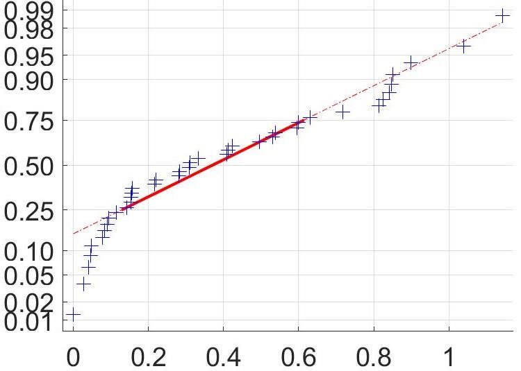
141st Wave 501Y.V2
Dilution
1:100
1:200
Cumulative probability
1:400
1:800
1:1600
Tx
Figure S 1: Fit of combined data for each plasma dilution to a normal distribution. The Matlab2019b
function normplot was used to assess the fit of the data (blue crosses) to a normal distribution (solid red line).
Lack of pronounced curvature of the data in the range of the solid line indicates that a the data is a reasonably
good fit to a normal distribution. see https://www.mathworks.com/help/stats/normplot.html for additional
information.A 1st Wave B 501Y.V2.HV001
1.2 1.2
1.0 1.0
0.8 0.8
Tx
Tx
0.6 0.6 Control pool
039-13-0013
0.4 0.4 039-02-0014
039-02-0017
039-13-0015
0.2 0.2 039-13-0062
039-13-0033
0 0
10-3 Dilution 10-2 10-3 Dilution 10-2
C 1st Wave D 501Y.V2.HVdF002
1.2 1.2
1.0 1.0
0.8 0.8
Tx
Tx
0.6 0.6
0.4 0.4
0.2 0.2
0 0
10-3 Dilution 10-2 10-3 Dilution 10-2
Figure S 2: Neutralization of first wave and 501Y.V2 by convalescent plasma from first wave
infections separated by variant. Four sets of independent experiments were performed per 501Y.V2 - first
wave pair, where the matched first wave variant results are shown to the left of the 501Y.V2 neutralization
results. 501Y.V2 variant 2 contained the L18F mutation in addition to the mutations of variant 1 , and did
not have the furin cleavage site deletion from outgrowth in Vero E6 cells. Colored points represent means
and standard errors from 4 independent experiments for each 501Y.V2 variant/first wave pair of neutralization
activity of plasma from 6 convalescent participants infected by first wave viruses. Corresponding lines are fits of
the sigmoidal equation with IC50 as the fitted parameter. Black points represent a pool of plasma from three
uninfected controls. The transmission index (Tx) is the number of foci in the presence of the plasma dilution
normalized by the number of foci in the absence of plasma.Table S 1: Plasma donor characteristics
Date of Days between Days between
Supplemental
Cohort ID Sex Age symptom symptom onset and symptom onset and
oxygen
onset diagnostic swab plasma collection
039-02-0014 F 66 No 01-Jul-2020 13 27
039-02-0017 F 66 Yes 21-Jul-2020 7 28
039-13-0013 F 54 No 29-Jun-2020 3 30
039-13-0015 F 42 No 21-Jun-2020 12 26
039-13-0033 F 37 No 24-Jun-2020 23 30
039-13-0062 M 67 No 06-Aug-2020 12 26Table S 2: Mutation profile for the genomes of the outgrown viruses and for the infecting viruses of convalescent plasma donors
Supplementary Table 2. Mutation profile for the genomes of the outgrown viruses and for the infecting viruses of convalescent plasma donors
Outgrown virus Infecting virus from plasma donors
Lineage B.1.1 B.1.351 (510Y.V2) B.1.351 (501Y.V2) B.1.1 B.1.1 B.1.5 B.1.5 B.1.140 B.1.1.1
Sequence ID K002868 K005321 K005325 K002868 K004289 K004285 K004291 K004295 K004302
Accession ID EPI_ISL_602622 EPI_ISL_678570 EPI_ISL_678615 EPI_ISL_602622 EPI_ISL_660170 EPI_ISL_660167 EPI_ISL_660172 EPI_ISL_660167 EPI_ISL_660181
Cohort ID 039-13-0013 - - 039-13-0013 039-02-0014 039-13-0015 039-13-0033 039-02-0017 039-13-0062
Spike amino acid S:D614G S:D80A S:L18F S:D614G S:D614G S:D614G S:D614G S:D614G S:D614G
substitutions S:A688V S:D215G S:D80A S:A688V
S:K417N S:D215G
S:E484K S:K417N
S:N501Y S:E484K
S:D614G S:N501Y
S:A701V S:D614G
S:A701V
Spike deletions S:242-244del S:242-244del
Other amino acid N:L139F E:P71L E:P71L N:L139F E:L73P E:L73P N:T148A ORF1a:F1178S N:R203K
substitutions N:R203K N:T205I N:T205I N:R203K N:R203K ORF1a:D3728N ORF10:A28V ORF1b:P314L N:G204R
N:G204R ORF14:L52F ORF14:L52F N:G204R N:G204R ORF1b:P314L ORF1a:K2511R ORF14:G50N
ORF14:G50N ORF1a:T265I ORF1a:T265I ORF14:G50N ORF14:G50N ORF1a:V3858I ORF1a:T1246I
ORF1a:D1481N ORF1a:K1655N ORF1a:K1655N ORF1a:D1481N ORF1b:P314L ORF1b:P314L ORF1a:G3278S
ORF1b:P314L ORF1a:K3353R ORF1a:K3353R ORF1b:P314L ORF1b:T1522I ORF1b:P314L
ORF1b:P314L ORF1b:P314L
ORF3a:Q57H ORF3a:Q57H
ORF3a:S171L ORF3a:W131L
ORF3a:S171L
ORF7a:V93F
Other deletions orf1ab:3675-3677del orf1ab:3675-3677del
Lineage classification was performed by Pangolin software application version v2.1.7 (https://cov-lineages.org/pangolin.html).
Accession ID refers to GISAID EpiCoV™ database (www.gisaid.org)
Amino acid mutation nomenclature includes open reading frame, wild-type amino acid, ORF position and amino-acid mutation (e.g. S:D80A, Spike D to A substitution at position 80). del refers to deletion
between stated positions. Amino acid mutations are annotated based on mature protein region of coding sequence (CDS) of SARS-CoV-2 reference sequence NC_045512.2.Table S 3: Mutation profile for the genomes of the outgrown 501Y.V2 viruses, showing the original genome produced from the
nasopharyngeal swab specimen and the genomes generated following passage in VeroE6 cells
Supplementary Table 3. Mutation profile for the genomes of the outgrown 501Y.V2 viruses, showing the original genome produced from the nasopharyngeal swab specimen and the genomes
generated following passage in VeroE6 cells
Outgrown 501Y.V2 Outgrown 501Y.V2 Outgrown 501Y.V2 Outgrown 501Y.V2 Outgrown 501Y.V2
Original After passage 2 After passage 3 Original After passage 3
Sequence ID K005321 K007776 K007624 K005325 K007621
Spike amino acid S:D80A S:D80A S:D80A S:L18F S:L18F
substitutions S:D215G S:D215G S:D215G S:D80A S:D80A
S:K417N S:K417N S:K417N S:D215G S:D215G
S:E484K S:E484K S:E484K S:K417N S:K417N
S:N501Y S:N501Y S:N501Y S:E484K S:E484K
S:D614G S:D614G S:D614G S:N501Y S:N501Y
S:A701V S:A701V S:A701V S:D614G S:D614G
S:A701V S:A701V
Spike deletions S:242-244del S:242-244del S:242-244del S:242-244del S:242-244del
S:677-681del S:677-681del
Other amino acid E:P71L E:P71L E:P71L E:P71L E:P71L
substitutions N:T205I N:T205I N:T205I N:T205I N:R32H
ORF14:L52F ORF14:L52F ORF14:L52F ORF14:L52F N:T205I
ORF1a:T265I ORF1a:T265I ORF1a:T265I ORF1a:T265I ORF14:L52F
ORF1a:K1655N ORF1a:K1655N ORF1a:K1655N ORF1a:K1655N ORF1a:T265I
ORF1a:K3353R ORF1a:K3353R ORF1a:K3353R ORF1a:K3353R ORF1a:K1655N
ORF1b:P314L ORF1a:Q3878R ORF1a:Q3878R ORF1b:P314L ORF1a:K3353R
ORF3a:Q57H ORF1b:P314L ORF1b:P314L ORF3a:Q57H ORF1a:N4358K
ORF3a:S171L ORF3a:Q57H ORF3a:Q57H ORF3a:W131L ORF1b:P314L
ORF3a:S171L ORF3a:S171L ORF3a:S171L ORF3a:Q57H
ORF7a:V93F ORF3a:W131L
ORF3a:S171L
ORF7a:V93F
ORF9b:A29T
Other deletions orf1ab:3675-3677del orf1ab:3675-3677del orf1ab:3675-3677del orf1ab:3675-3677del orf1ab:3675-3677del
Amino acid mutation nomenclature includes open reading frame, wild-type amino acid, ORF position and amino-acid mutation (e.g. S:D80A, Spike D to A substitution at position 80). del
refers to deletion between stated positions. Amino acid mutations are annotated based on mature protein region of coding sequence (CDS) of SARS-CoV-2 reference sequence NC_045512.2.
Substitutions and deletions in bold are those emerging during passageYou can also read