Skin Pigmentation Abnormalities and Their Possible Relationship with Skin Aging - MDPI
←
→
Page content transcription
If your browser does not render page correctly, please read the page content below
International Journal of
Molecular Sciences
Review
Skin Pigmentation Abnormalities and Their Possible
Relationship with Skin Aging
Ai-Young Lee
Department of Dermatology, College of Medicine, Dongguk University Ilsan Hospital, 814 Siksa-dong,
Ilsandong-gu, Goyang-si 410-773, Gyeonggi-do, Korea; lay5604@naver.com; Tel.: +82-319617250;
Fax: +82-319-617-695
Abstract: Skin disorders showing abnormal pigmentation are often difficult to manage because
of their uncertain etiology or pathogenesis. Abnormal pigmentation is a common symptom ac-
companying aging skin. The association between skin aging and skin pigmentation abnormalities
can be attributed to certain inherited disorders characterized by premature aging and abnormal
pigmentation in the skin and some therapeutic modalities effective for both. Several molecular
mechanisms, including oxidative stress, mitochondrial DNA mutations, DNA damage, telomere
shortening, hormonal changes, and autophagy impairment, have been identified as involved in
skin aging. Although each of these skin aging-related mechanisms are interconnected, this review
examined the role of each mechanism in skin hyperpigmentation or hypopigmentation to propose
the possible association between skin aging and pigmentation abnormalities.
Keywords: skin pigmentation abnormalities; skin aging; oxidative stress; DNA damage; telomere
shortening; hormonal change; autophagy impairment
Citation: Lee, A.-Y. Skin 1. Introduction
Pigmentation Abnormalities and
Melanin pigmentation plays a critical role in protecting the skin from the harmful
Their Possible Relationship with Skin
effects of ultraviolet (UV) radiation. A variety of genetic and environmental factors are
Aging. Int. J. Mol. Sci. 2021, 22, 3727.
involved in the regulation of pigmentation. Excessive or deficient amounts of melanin
https://doi.org/10.3390/ijms22073727
cause disorders showing abnormal skin pigmentation. Although wrinkling and laxity are
Academic Editor: Andrzej Slominski
well-identified symptoms [1], abnormal pigmentation is also a common symptom in aging
skin [2–5]. Pigmentation abnormalities associated with skin aging have recently drawn
Received: 22 February 2021 attention, however, not many reports have described the changes in melanin pigmentation
Accepted: 1 April 2021 in aged human skin, and the role of skin aging in pigmentation abnormalities remains to
Published: 2 April 2021 be clarified.
Human skin undergoes chronological aging and environmental aging. Chronological
Publisher’s Note: MDPI stays neutral aging is dependent upon the passage of time. Ultraviolet (UV) irradiation is the primary
with regard to jurisdictional claims in factor in environmental aging, which is also called photoaging, although other factors such
published maps and institutional affil- as tobacco smoking and air pollution are involved in environmental aging [6,7]. With age,
iations. senescent cells accumulate in human skin, which can compromise skin function and in-
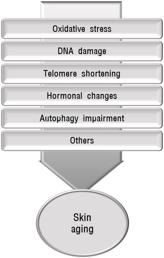
tegrity. Chronological aging and photoaging share certain molecular mechanisms (Figure 1).
Skin aging is influenced by several factors including oxidative stress, mitochondrial DNA
mutations, DNA damage, telomere shortening, and hormonal changes [8–10]. Autophagy
Copyright: © 2021 by the author. impairment is also involved in aging and the senescence of skin cells [11,12]. After a brief
Licensee MDPI, Basel, Switzerland. description of skin pigmentation and skin conditions showing abnormal pigmentation
This article is an open access article accompanied by aging, this review covers the association between each of the mechanisms
distributed under the terms and implicated in skin aging and skin hyperpigmentation or hypopigmentation to provide a
conditions of the Creative Commons basis for their possible relationship.
Attribution (CC BY) license (https://
creativecommons.org/licenses/by/
4.0/).
Int. J. Mol. Sci. 2021, 22, 3727. https://doi.org/10.3390/ijms22073727 https://www.mdpi.com/journal/ijmsInt. J. Mol. Sci. 2021, 22, 3727 2 of 19
Figure 1. Schematic view of causative factors involved in skin aging. Skin aging is influenced by
several factors including oxidative stress, mitochondrial DNA mutations, DNA damage, telomere
shortening, hormonal changes, and autophagy impairment.
2. Melanin Pigmentation and Its Abnormalities Accompanied by Skin Aging
Skin pigmentation is largely determined by melanin synthesis in melanocytes, mela-
nosome transfer to keratinocytes, and melanosome degradation. Melanin synthesis is
restricted to melanosomes, which contain tyrosinase, the key regulatory enzyme of melano-
genesis, and tyrosine-related proteins (TRPs). Tyrosinase is involved in the early steps of
melanin synthesis to dopaquinone, which are common to two types of melanin, brown-
black eumelanin and yellow-red pheomelanin. Eumelanin is photoprotective by scattering
and absorbing UV light. In contrast, pheomelanin is photo-unstable and phototoxic, pro-
moting UV-induced damage including photoaging [13]. Eumelanin and pheomelanin
can be produced at different proportions within the same cell. A paucity of eumelanin
production makes the skin vulnerable to UV-induced damage [14]. The biogenesis of
melanin is controlled by complex regulatory mechanisms. The tyrosinase and TRPs genes
have binding sites for microphthalmia-associated transcription factor (MITF), which plays
a fundamental role in the transcriptional regulation of melanogenesis via the cAMP, protein
kinase C, and nitrogen oxide intracellular signal transduction pathways [15].
Abnormalities in melanin pigmentation are divided into two types, hypermelanosis
and hypomelanosis, based on the amount of melanin in the skin. Reduced melanin synthe-
sis with the downregulation of TRP-1, TRP-2, and MITF in senescent melanocytes [16], and
decreased melanosome degradation by declining autophagy activity in aging [17] indicate
the association of pigmentation abnormalities with skin aging. Their association has also
been suggested in inherited and acquired disorders. Inherited disorders showing acceler-Int. J. Mol. Sci. 2021, 22, 3727 3 of 19
ated aging and skin hyperpigmentation or hypopigmentation provide strong evidence that
skin aging can be associated with abnormalities in melanin pigmentation. The favorable
therapeutic effects of chemicals and lasers such as polyphenol compounds and fractional
photothermolysis, respectively, on both skin aging and pigmentation abnormalities [18,19]
also provide reliable evidence for their association. Among the acquired skin disorders of
hypermelanosis, melasma is a common skin condition. Although a complex interaction of
causative factors is likely to be involved in melasma development [20], a certain proportion
of melasma cases are associated with skin aging, either UV-related or UV-unrelated [21,22].
Seborrheic keratosis, which is a benign skin tumor presenting as sharply demarcated light
brown-to-black papules containing a greater amount of melanin, develops in elderly in-
dividuals, mainly in sun-exposed areas. Senile lentigo is another example of a common
benign skin tumor with hyperpigmentation in aged people associated with chronic UV
irradiation. As for hypomelanosis, vitiligo is a representative skin condition associated
with premature senescence of the skin cells [22]. The role of melanocyte senescence has
also been suggested in idiopathic guttate hypomelanosis [23].
3. Association of Mechanisms Implicated in Skin Aging with Skin
Pigmentation Abnormalities
3.1. Role of Oxidative Stress in Skin Pigmentation Abnormalities
Oxidative stress is considered one of the most important mechanisms involved in
skin aging [24–28]. Whenever the cellular accumulation of reactive oxygen species (ROS),
a family of oxygen-based free radicals including hydrogen peroxide (H2 O2 ), overcomes
the cellular antioxidant capacity, oxidative stress develops. ROS are generated by ex-
ogenous stimuli such as UV irradiation [29] and endogenous stimuli including cellular
metabolism [30]. The skin is a high turnover, metabolically active organ, requiring energy.
Because ROS are produced as the by-products of energy generation by mitochondrial respi-
ration, mitochondria can play a critical role in oxidative stress [28,31]. Conversely, ROS
can cause mitochondrial DNA mutations [32], leading to mitochondrial dysfunction. This
interconnection provides a reasonable basis for considering mitochondrial DNA mutations
in conjunction with oxidative stress. The skin is rich in potential biological targets for
oxidative damage such as lipids, proteins, and DNA. Oxidative damage leads to cellular
senescence, and thus, skin aging [33,34], therefore, the use of antioxidants can induce
anti-aging effects [35–37].
3.1.1. Oxidative Stress and Skin Hypermelanosis
The regulatory role of H2 O2 in melanocyte tyrosinase and the amelioration of hyper-
pigmentation by antioxidants support the effect of oxidative damage in skin hyperpig-
mentation [38–41]. The role of oxidative stress in skin pigmentation abnormalities can be
predicted by the clinical findings of abnormal skin pigmentation in certain congenital skin
diseases caused by mutations in DNA affecting mitochondrial function (Table 1). Mutation
of the cytochrome c oxidase subunit 7B (COX7B) induces oxidative phosphorylation defects
and mitochondrial respiration deficiency [42]. Patients with COX7B mutations show eye
symptoms and linear skin defects with hyperpigmented streaks [28,43]. Mutations in
excision repair cross-complementing group 6 (ERCC6) cause defective mitophagy and the
accumulation of damaged mitochondria [44]. The ERCC6 gene encoding a DNA excision re-
pair protein is responsible for Cockayne syndrome group B and can also cause UV-sensitive
syndrome, characterized by photosensitivity, hyperpigmentation, freckling, and dryness in
sun-exposed areas [45]. A mutation in Fanconi anemia complementation group A (FANCA)
can reduce the electron transfer between respiratory complex I-III and ROS detoxifica-
tion enzymes [46,47] and impair mitophagy [48]. Patients with FANCA mutation show
morphological abnormalities of the skin, including hyperpigmentation [28]. Mutations in
the human Harvey rat sarcoma viral oncogene homolog (HRAS) can cause mitochondrial
dysfunction and defective oxidative phosphorylation [49,50]. HRAS mutations and skin
hyperpigmentation have been anecdotally associated with the presence of nevi and pha-Int. J. Mol. Sci. 2021, 22, 3727 4 of 19
comatosis pigmentokeratotica in patients with the mutation [51–53]. Holocytochrome c
synthase (HCCS) is an enzyme located in the inner mitochondrial membrane, which adds
heme to apocytochrome c and c1. Mutations in HCCS mutation reduce oxidative phos-
phorylation efficiency [54] and cause microphthalmia with linear skin defects syndrome
exhibiting skin pigmentation [55]. Protoporphyrinogen oxidase (PPOX), the penultimate
enzyme in heme biosynthesis, is a nucleus-encoded flavoprotein associated with the outer
surface of the inner mitochondrial membrane. PPOX mutations cause mitochondrial
abnormalities [56–58] and variegate porphyria, which has cutaneous manifestations char-
acterized by photosensitivity, skin fragility, hypertrichosis, and hyperpigmentation [59].
Mutations in BRAF, NRAS, KIT, GNAQ, CDK4, PTK2B, ERBB4, GNA11, MEK, MITF, AKT3,
MMP, CXCR4, EPHA3, FAS, PIK3CA, MET, CTNNB1, NEK10, and PDGFRA have been re-
ported to induce mitochondrial dysfunction, mitochondrial respiratory complex deficiency,
and decrease mitochondrial oxidative phosphorylation [60,61], which are associated with
skin pigmentation [28]. Mitochondrial DNA polymerase gamma (POLG1) is responsible
for mitochondrial DNA replication and repair. POLG1 mutations result in the depletion of
mitochondrial DNA and can cause Alpers-Huttenlocher syndrome, which is characterized
by the classic triad of seizures, cognitive degeneration, and hepatopathy. Most patients
with this syndrome develop other system involvement [62], such as hyperpigmentation in
the antecubital and popliteal fossae and dorsa of the feet [63].
Acquired abnormal skin pigmentation can be related to oxidative stress (Table 2). Sun-
light stimulates melanin production for protection against the harmful effects from UV radi-
ation such as from tanning. The substantial role of oxidative stress in UV-induced melano-
genesis and the alleviation of UV-induced melanogenesis by antioxidant therapy [64–66]
indicates an association between oxidative stress and UV-induced hyperpigmentation. UV
irradiation can generate the peroxidation products of sebum, squalene monohydroper-
oxides. The hyperpigmentation of reconstructed human skin equivalents by exposure
to squalene monohydroperoxides and the amelioration of squalene monohydroperoxide-
induced hyperpigmentation by co-treatment with 12-hydroxystearic acid support a pivotal
role of antioxidant mechanisms in UV-induced hyperpigmentation [65]. Although the
exact pathogenesis of melasma is uncertain, oxidative stress has been implicated based
on increased serum malondialdehyde but reduced serum catalase levels in the patient
groups [67,68] and the efficacy of antioxidant drugs identified in a clinical trial [69,70]. The
role of oxidative stress has also been identified in seborrheic keratosis. Guanine deaminase,
shown to be upregulated in seborrheic keratosis, stimulated UV-induced keratinocyte
senescence via ROS generation [71].
3.1.2. Oxidative Stress and Skin Hypomelanosis
Oxidative stress caused by mitochondrial redox imbalance or antioxidant enzyme
deficiency is also associated with skin hypopigmentation (Table 1). The loss of ATPase
copper-transporting alpha (ATP7A) activity, which causes Menkes disease, results in the
accumulation of copper in cellular organelles, including mitochondria, thereby disrupting
the mitochondrial redox balance [72]. Tyrosinase is a copper-dependent enzyme involved
in melanogenesis. Because ATP7A protein is required for tyrosinase activity, the loss
of ATP7A activity by gene mutations impairs melanogenesis [73]. Mutations in SURF1
cytochrome C oxidase assembly factor (SURF1) are associated with Leigh syndrome, a
rare progressive neurodegenerative disorder caused by mitochondrial cytopathy. Skin and
hair abnormalities, including hypopigmentation are often accompanying symptoms [74].
The deletion of mitochondrial DNA induces Kearns-Sayre syndrome, a mitochondrial
multisystem disorder characterized by the clinical symptoms of ophthalmoplegia and
pigmentary retinopathy. Hypopigmented patches like Hypomelanosis of Ito have been
reported as a possible symptom [75]. Piebaldism, characterized by depigmented patches
presenting at birth, is caused by KIT gene mutations. An association between H2 O2 -
mediated stress and piebaldism has been identified, although its mechanism in oxidative
stress differs from that in vitiligo [76].Int. J. Mol. Sci. 2021, 22, 3727 5 of 19
Concerning the role of oxidative stress in acquired hypomelanosis, most data are
related to vitiligo (Table 2). Oxidative stress caused by ROS production and low levels
of antioxidants are considered the pivotal mechanisms in vitiligo development [77,78],
although the therapeutic effects of antioxidants have been controversial [79]. Elevations
in superoxide dismutase and malondialdehyde [80], reductions in catalase or catalase
gene polymorphism [81–83], increases in advanced oxidation protein products [84], nuclear
factor erythroid 2-like 2 (Nrf2) gene polymorphism [85], and reduced Nrf2 activation [86,87]
are associated with vitiligo susceptibility. Increased mitochondrial DNA copy numbers [88],
mitochondrial dysfunction by sirtuin3 (SIRT3) deficiency [89], and increased calcium influx
by the upregulation of transient receptor potential cation channel subfamily M member 2
(TRPM2), a calcium channel sensitive to oxidative stress [90], contribute to oxidative
stress-induced melanocyte damage. Autophagy impairment also plays a role in oxidative
stress by disrupting the antioxidant defense system [91,92]. ROS accumulation damages
melanocytes by the generation of autoantigens, which leads to the CD8+ T cell-mediated
destruction of melanocytes.
Table 1. Abnormal skin pigmentation in congenital disorders related to oxidative stress.
Pigment Change Mutated Gene Dysfunction Skin Symptom Reference
Oxidative phosphorylation defect,
Linear skin defects with
COX7B Mitochondrial respiration [28,42,43]
hyperpigmented streaks
deficiency
Accumulation of damaged Photosensitivity,
ERCC6 mitochondria, Hyperpigmentation, freckling, and [44,45]
Mitophagy defect dryness in sun-exposed areas
Reduction of electron transfer
between respiratory complex I-III Morphological abnormalities
FANCA [28,46–48]
and ROS detoxification enzymes, including hyperpigmentation
Mitophagy impairment
Hyper Mitochondrial dysfunction, Nevi
HRAS [49–53]
Oxidative phosphorylation defect Phacomatosis pigmentokeratotica
Reduction of oxidative Linear skin defects syndrome
HCCS [54,55]
phosphorylation efficiency exhibiting skin pigmentation
Variegate porphyria characterized
by photosensitivity, skin fragility,
PPOX Mitochondrial abnormalities [56–59]
hypertrichosis, and
hyperpigmentation
Hyperpigmentation in the
POLG1 Mitochondrial DNA depletion antecubital and popliteal fossae [62,63]
and dorsa of the feet
Mitochondrial redox imbalance by
ATP7A Melanogenesis impairment [72,73]
copper accumulation
Skin and hair abnormalities
SURF1 Mitochondrial cytopathy [74]
Hypo including hypopigmentation
Mitochondrial Mitochondrial multisystem Hypopigmented patches like
[75]
DNA deletion disorder hypomelanosis of Ito
KIT H2 O2 -mediated stress Piebaldism [76]Int. J. Mol. Sci. 2021, 22, 3727 6 of 19
Table 2. Acquired abnormal skin pigmentation related to oxidative stress.
Pigment Change Disease/Condition Evidence for Oxidative Stress-Related Reference
Improvement of hyperpigmentation by
[64–66]
antioxidant therapy
UV-induced melanogenesis
Hyperpigmentation on skin equivalents by
squalene monohydroperoxides, which is [65]
ameliorated by 12-hydroxystearic acid
Hyper
Increased malondialdehyde with decreased
[67,68]
Melasma catalase in serum
Clinical trial using antioxidant drugs [69,70]
Role of guanine deaminase in UV-induced
Seborrheic keratosis [71]
keratinocyte senescence via ROS generation
Elevated superoxide dismutase
[80]
and malondialdehyde
Reduced catalase or catalase
[81–83]
gene polymorphism
Nrf2 gene polymorphism or reduced
Hypo Vitiligo [85–87]
Nrf2 activation
Increased mitochondrial DNA copy numbers
[88,89]
or mitochondrial dysfunction
Upregulated TRPM2 [90]
Autophagy impairment [91,92]
3.2. Role of DNA Damage in Skin Pigmentation Abnormalities
DNA repair mechanisms such as the repair of nucleotide excision and double-strand
breaks should be activated to maintain homeostasis whenever DNA damage occurs. The
downregulation of DNA repair and the accumulation of DNA damage drives the aging
process [93,94]. Impaired DNA replication and repair by mutations in a group of premature
aging syndromes known as progeroid syndromes provide strong evidence for the role of
DNA damage in aging [95]. UV radiation is a notorious inducer of DNA damage in all skin
types despite its inverse relationship with constitutive skin pigmentation [96].
3.2.1. DNA Damage and Skin Hypermelanosis
The association between DNA damage and hyperpigmentation, particularly aging-
related effects, has been identified in certain DNA repair syndromes caused by pathogenic
variants encoding proteins in DNA replication and cellular responses to DNA damage
(Table 3). Mutations in RecQ protein-like 4 (RECQL4) belonging to the RECQ DNA helicase
family, which participate in many aspects of DNA metabolism [97], cause Rothmund-
Thomson syndrome [98]. The syndrome shows clinical features of accelerated aging, such
as atrophic skin and pigment changes [97]. Mutations in one of the eight genes involved
in nucleotide excision repair and DNA polymerase cause Xeroderma pigmentosum [99].
Xeroderma pigmentosum is characterized by the extreme sensitivity to sunlight, resulting
in sunburn, unusually increased numbers of lentigines in sun-exposed areas, areas contain-
ing both hyperpigmentation and hypopigmentation in the absence of rigorous sunlight
protection, accelerated photoaging, and increased skin cancer risk over time [98]. Muta-
tions in several genes involved in DNA repair, particularly interstrand crosslink repair and
telomere maintenance, cause Fanconi anemia. A poikilodermatic change with hypopigmen-
tation, hyperpigmentation, and telangiectasia appears in the skin of patients with Fanconi
anemia [100,101]. Inactivating mutations of the tumor suppressor serine-threonine kinase
11/liver kinase B1 (STK11/LKB1), which can play a role in the regulation of the UV-induced
DNA damage response in mice skin [102], underlie Peutz-Jeghers syndrome. Hyperpig-Int. J. Mol. Sci. 2021, 22, 3727 7 of 19
mentation of mucous membranes and the skin is one of the characteristic symptoms of
Peutz-Jeghers syndrome.
Acquired abnormal skin pigmentation can be related to DNA damage (Table 4).
Skin keratinocytes with DNA damage induced by UV exposure secrete α–melanocyte-
stimulating hormone (α-MSH) [103]. α-MSH interacts with melanocortin 1 receptor
(MC1R), which enhances nucleotide excision repair in melanocytes [104]. The role of
α-MSH in melanogenesis suggests an association between DNA damage and melano-
genesis. Defects in DNA repair have not been reported in melasma, seborrheic keratosis,
and senile lentigo, although microsatellite instability indicating DNA repair defects was
detected in seborrheic keratosis developed in a patient with hereditary nonpolyposis
colorectal cancer [105].
Table 3. Abnormal skin pigmentation in congenital disorders related to DNA damage.
Pigment Change Mutated Gene Dysfunction Skin Symptom Reference
Abnormal regulation of
STK11 Hyperpigmentation of mucous
Hyper UV-induced DNA [102]
/LKB1 membranes and the skin
damage response
Accelerated aging such as
Defects in many aspects of
RECQL4 atrophic skin and [97,98]
DNA metabolism
Hyper and hypo pigment changes
Photosensitivity, Lentigines,
XPA XPB, XPC, XPD, Defects in damaged Hyperpigmentation and
[98,99]
XPE, XPF, XPG, or XPV DNA repair hypopigmentation,
Accelerated photoaging
Poikilodermatic change with
Defects in interstrand
hypopigmentation,
Fanconi anemia crosslink repair and [100,101]
hyperpigmentation,
telomere maintenance
and telangiectasia
Leukocyte telomere
Deletion on 15q11.2-q13 Hypopigmentation [106,107]
Hypo length shortening
Oculocutaneous albinism Increased tyrosinase Oculocutaneous
and Hermansky-Pudlak degradation through Albinism, [108–111]
syndrome ubiquitin-proteasome system Photoaging
Table 4. Acquired abnormal skin pigmentation related to DNA damage.
Pigment Change Disease/Condition Evidence for DNA Damage-Related Reference
Role of α-MSH secreted from UV-damaged
Hyper UV-induced melanogenesis [103,104]
keratinocytes in melanogenesis
Hypo Leukotrichia Associated with APE1 polymorphism [112]
3.2.2. DNA Damage and Skin Hypomelanosis
Certain inherited disorders caused by DNA damage can exhibit aging-related skin
hypopigmentation alone or both hypopigmentation and hyperpigmentation (Table 3). As
described above, xeroderma pigmentosum patients have hypopigmented areas in addition
to hyperpigmented skin [98]. Fanconi anemia shows poikilodermatic changes consisting of
hypopigmentation, hyperpigmentation, atrophy, and telangiectasia as the characteristic
skin symptoms [100,101]. Poikilodermatic changes also appear in the skin of Rothmund-
Thomson syndrome patients [98]. A chromosomal deletion on 15q11.2-q13, which induces
shortening of leukocyte telomere length, a marker of biological age, causes Prader-Willi
syndrome [106]; as one of the premature aging syndromes, Prader-Willi syndrome patients
can exhibit hypopigmentation [107]. Oculocutaneous albinism and Hermansky-PudlakInt. J. Mol. Sci. 2021, 22, 3727 8 of 19
syndrome are hereditary diseases with DNA mutations [108,109]. Both diseases exhibit
oculocutaneous albinism caused by the increased degradation of tyrosinase through the
ubiquitin-proteasome system [110]. Photoaging is accelerated in albino hairless mice by
exposure to solar simulation [111].
DNA damage and defects in DNA repair have not been identified in vitiligo, although
an association between apurinic/apyrimidinic endonuclease 1 (APE1) polymorphism with
leukotrichia suggests the role of this polymorphism in vitiligo [112] (Table 4).
3.3. Role of Telomere Shortening in Skin Pigmentation Abnormalities
Telomeres are dynamic nucleoprotein-DNA structures composed of long tandem
TTAGGG repeat, guanine-rich sequences located at the end of chromosomes to cap and
protect the linear chromosome ends. Although the length of telomeres differs between
chromosome arms, telomeres shorten by cell division, and the initial length of telomeres
correlates with the cellular replicative capacity. Accelerated aging in short telomere syn-
dromes caused by inheritable gene mutations resulting in decreased telomere lengths [113]
indicates that telomere shortening can be one of the primary hallmarks of aging [114].
3.3.1. Telomere Shortening and Skin Hypermelanosis
The genetically inherited diseases with much shorter telomeres compared to age-
matched controls are referred to as telomeropathies. Primary telomeropathies are defined
as disorders caused by mutations in the telomere maintenance machinery. Secondary
telomeropathies are caused by mutations in genes encoding proteins involved in telomere
DNA repair, leading to telomere aberrations and loss [115]. However, overlap exists
between telomeropathies, particularly secondary telomeropathies, and DNA mutations.
The role of telomeres in skin pigmentation has been identified in telomeropathies
(Table 5). The first disorder identified as a primary telomeropathy was dyskeratosis con-
genita, which is caused by mutations in the DKC1 gene encoding the dyskerin protein, or
one of the core telomerase genes encoding a protein involved in telomere maintenance [116],
resulting in telomere shortening, and thereby, premature aging [117,118]. Reticular hy-
perpigmentation is a symptom of the dyskeratosis congenita diagnostic triad along with
oral leukoplakia and nail dystrophy. Xeroderma pigmentosum and Fanconi anemia are
included in secondary telomeropathies, indicating an overlap between telomere shortening
and DNA repair syndromes. Among the eight complementation groups in xeroderma
pigmentosum, mutations in complementation group C (XPC), can cause the instability
of telomere upon UV exposure [117]. Fanconi anemia is caused by genetic mutations
involved in DNA repair for telomere maintenance. As described before, patients with
these disorders manifest abnormal skin pigmentation [100,101]. A point mutation in the
LMNA gene, which produces abnormal lamin A protein associated with rapid telom-
ere erosion, can cause Hutchinson-Gilford progeria syndrome [119,120]. As one of the
most severe segmental progeroid syndromes, patients with Hutchinson-Gilford syndrome
demonstrate premature aging with distinct dermatologic features including hyper- and
hypopigmentation over areas of sclerodermoid changes [95,121].
Although telomere attrition contributes to certain age-related disorders, the associa-
tion of melasma, seborrheic keratosis, or senile lentigo with telomere shortening has not
been reported.
3.3.2. Telomere Shortening and Skin Hypomelanosis
Telomere shortening can be associated with skin hypomelanosis, although hypopig-
mentation usually coexists with hyperpigmentation as shown in patients with xeroderma
pigmentosum and Hutchinson-Gilford progeria syndrome [95,98] (Table 5).
In contrast, in vitiligo, telomerase activity is lower in lesional skin compared to non-
lesional skin and healthy control skin, which was proposed as a reason for the low incidence
of sun damage and cancer in the lesional skin of vitiligo patients [122].Int. J. Mol. Sci. 2021, 22, 3727 9 of 19
Table 5. Abnormal skin pigmentation in congenital disorders related to telomere shortening.
Pigment Change Mutated Gene Dysfunction Skin Symptom Reference
Reticular hyperpigmentation,
Hyper DKC1 Telomere maintenance defect Oral leukoplakia, [116–118]
Nail dystrophy
Photosensitivity,
Defect in telomere stability Lentigines,
XPC and recognition of DNA Hyperpigmentation and [99,117]
photoproducts hypopigmentation,
Accelerated photoaging
Hyper and hypo Poikilodermatic change with
Defect in DNA repair for hypopigmentation,
Fanconi anemia [100,101]
telomere maintenance hyperpigmentation,
and telangiectasia
Hyperpigmentation and
LMNA Rapid telomere erosion hypopigmentation over areas of [95,119–121]
sclerodermoid change
3.4. Role of Hormones in Skin Pigmentation Abnormalities
Cutaneous neuroendocrine systems can regulate skin function in response to environ-
mental stresses, such as UV radiation and pollutants. Melatonin belongs to the family of
neurohormones [123]. The pineal gland is the main organ of melatonin synthesis and secre-
tion. However, human skin can produce and rapidly metabolize melatonin to maintain
homeostasis against environmental stresses [124]. Melatonin is involved in the regulation
of various physiological activities, including circadian rhythm, immune responses, oxida-
tive process, apoptosis, and mitochondrial homeostasis. Antioxidant actions of melatonin
protect skin from UVB-induced harm [125,126], indicating the critical role of melatonin
in photoaging.
Receptors for sex steroids, particularly estrogen receptors, are present in human skin,
including epidermal keratinocytes and dermal fibroblasts. Many findings, such as the
gradual decline of these receptors with chronological and environmental aging [127,128],
skin aging accelerated by the loss of estradiol production after menopause, and UV-induced
collagen degradation reversed by estradiol replacement [128–130], indicate the role of
estrogens in protecting skin from aging.
3.4.1. Hormones and Skin Hypermelanosis
Oxidative stress is the main mechanism involved in skin aging and pigmentation.
The antioxidant actions of melatonin [125,126] are implicated in the association between
melatonin and skin pigmentation. Although significantly lower serum levels of melatonin
have been seen in melasma patients compared to controls [68], relevant reports are rare.
Sex hormones, particularly estrogens, play a role, which is stimulatory but not in-
hibitory, in pigmentation in cultured skin cells [131] and in melasma [20,132]. These
results suggest that estrogens enhance skin pigmentation and delay rather than accelerate
skin aging.
3.4.2. Hormones and Skin Hypomelanosis
The association of melatonin with vitiligo has been described [133]. A connection
between reduced testosterone due to secondary hypogonadism and vitiligo has also been
suggested [134].
Above all, vitiligo patients frequently suffer from serious psychological problems
such as depression and anxiety. The levels of dehydroepiandrosterone sulfate, which has
antioxidant properties, were lower but the ratios of cortisol to dehydroepiandrosterone
sulfate were higher in vitiligo patients compared to healthy controls [135]. The resultsInt. J. Mol. Sci. 2021, 22, 3727 10 of 19
suggest the role of dehydroepiandrosterone sulfate in vitiligo development and provide
reliable evidence for the association of vitiligo with stress-related hormones.
3.5. Role of Autophagy in Skin Pigmentation Abnormalities
Autophagy is an essential cellular process for homeostasis accomplished by degrading
and recycling damaged cellular components. Autophagic activity declines with aging [136].
However, defects in autophagy can also cause skin aging with an accumulation of damaged
cellular components [137]. The association between reduced autophagy and skin aging
has mostly been investigated in photoaging. The phenotype of skin aging induced by
UVA irradiation in human fibroblasts and mice with Cockayne syndrome type B defi-
ciency is accompanied by reduced autophagic activity and is rescued by the improvement
in autophagic function [138]. Repeated UVA irradiation results in the accumulation of
autophagosomes and lysosomes, suggesting that photoaging can reduce autophagic ac-
tivity at the degradation stage [139,140]. Oxidative stress-induced cellular senescence in
murine melanocytes and keratinocytes associated with autophagy-related 7 (Atg7) defi-
ciency [12,141] and the protective role of caffeine in oxidative stress-induced senescence of
human epidermal keratinocytes through autophagy induction [142] suggest that autophagy
defects can accelerate skin aging by increasing oxidative stress.
3.5.1. Autophagy and Skin Hypermelanosis
The melanosome is a lysosome-related organelle. Autophagy-related regulators are in-
volved in the formation, maturation, and degradation of melanosomes in melanocytes [143].
In vitro and in vivo results have demonstrated the association between autophagy and
skin pigmentation abnormalities (Table 6).
Table 6. Abnormal skin pigmentation in disorders related to autophagy impairment.
Pigment Change Causative Factor Dysfunction Skin Symptom Reference
Decrease in autophagic
Reduced Hsp70-1A Skin color [144]
melanosome degradation
Accelerated keratinocyte
Decrease in autophagic Hyperpigmented lesion
senescence caused by [145]
melanosome degradation of melasma
arginase-2 upregulation
Hyper
Significant decreases Hyperpigmented lesion
Premature skin aging [17]
in autophagy of senile lentigo
Lightening of skin
Chemical agents Autophagy activation [146–149]
hyperpigmentation
Lightening of skin
Light-emitting diodes Autophagy activation [150]
hyperpigmentation
Autophagosome-lysosome
fusion defect, leading to Oculocutaneous
EPG5 mutations [151,152]
antioxidant defense hypopigmentation
system disruption
Autophagic dysregulation
Hypo TSC mutations by mTOR signaling Hypopigmented macules [153,154]
pathway activation
Defect in melanosome
Hypopigmented macules [155–157]
formation and autophagy
Downregulated GPNMB
Reduced melanocyte
Vitiligo [158]
survivalInt. J. Mol. Sci. 2021, 22, 3727 11 of 19
The diversity of skin color was affected by epidermal heat shock 70 kDa protein 1A
(Hsp70-1A), which modulates autophagic melanosome degradation in keratinocytes [144].
Accelerated keratinocyte senescence by arginase-2 upregulation impairs autophagy and
reduces melanosome degradation, resulting in hyperpigmentation in melasma [145]. As-
sociation between premature skin aging with autophagy reduction was also detected in
the hyperpigmented lesions of patients with senile lentigo, whereas autophagy activation
ameliorated pigmentation in ex vivo lesional skin [17]. Chemical agents such as tranex-
amic acid, β–mangostin, 3-O-glyceryl-2-O-hexyl ascorbate, and Melasolv induced skin
lightening through autophagy activation [146–149]. Light-emitting diodes can also activate
autophagy, resulting in skin lightening [150].
3.5.2. Autophagy and Skin Hypomelanosis
Recessive mutations in EPG5 encoding the ectopic P-granules autophagy protein 5
homolog, a key regulator of autophagy, cause Vici syndrome. As one of the most typical ex-
amples of congenital autophagy disorders, patients with the syndrome exhibit characteristic
features that include oculocutaneous hypopigmentation [151]. Although the mechanism of
hypopigmentation remains to be clarified, defective autophagosome-lysosome fusion in
EPG5-depleted melanocytes [152] may disrupt the antioxidant defense system. Autophagic
dysregulation by activation of the mTOR signaling pathway in melanocytes has been
identified as a mechanism of reduced pigmentation in hypopigmented macules in patients
with tuberous sclerosis complex (TSC) gene mutations [153,154]. The loss of glycoprotein
nonmetastatic melanoma protein B (GPNMB) causes amyloidosis cutis dyschromica, which
is characterized by generalized hyperpigmentation mottled with hypopigmented mac-
ules [155]. GPNMB is a melanocytic cell marker involved in multiple functions, including
melanosome formation and autophagy [156]. GPNMB knockdown reduced the number of
melanosomes in melanocytes [157].
GPNMB has also been downregulated in vitiligo keratinocytes, causing the loss of
melanocytes [158]. The finding indicates the regulatory role of GPNMB in skin hypome-
lanosis by reducing melanocyte survival in addition to reducing melanogenesis.
4. Conclusions
Skin diseases with pigmentation abnormalities are difficult to treat primarily due
to unknown causes or pathogenesis. The pathomechanisms in individual patients can
be different even in the same disease. It is important to identify the cause to manage
skin pigmentation abnormalities. To propose the possible role of skin aging in abnormal
pigmentation, the association between the identified mechanisms involved in skin aging
and skin pigmentation abnormalities in various inherited and acquired disorders were
reviewed. The mechanisms implicated in skin aging include oxidative stress, which is
the most pivotal cause of skin aging, DNA damage, telomere shortening, decreased mela-
tonin, and autophagy impairment. Both skin aging and pigmentation abnormalities in
various inherited disorders caused by DNA damage or telomere shortening indicate the
relevant relationship between skin aging and pigmentation abnormalities. However, other
mechanisms may not yet sufficiently support the relationship between skin aging and
pigmentation abnormalities (Figure 2). Epigenetic changes, particularly DNA methyla-
tion [159,160] and microRNAs [161], are also proposed to be involved in skin aging without
an identified role in skin pigmentation. More studies are needed to prove the reliable role
of skin aging in various conditions showing abnormal skin pigmentation.Int. J. Mol. Sci. 2021, 22, 3727 12 of 19
Figure 2. Schematic view of the possible association between abnormal pigmentation and skin
aging. Some causative mechanisms, such as DNA damage and telomere shortening, are involved in
hereditary disorders that show accelerated aging and skin hyperpigmentation or hypopigmentation,
providing reliable evidence for the association between skin aging and pigmentation abnormalities.
However, oxidative stress, hormonal changes, and autophagy impairment, may not yet sufficiently
support the relationship between skin aging and pigmentation abnormalities.
Funding: This research was supported by a grant of the Korea Health Technology R&D Project
through the Korea Health Industry Development Institute (KHIDI), funded by the Ministry of Health
& Welfare, Republic of Korea (grant number: HP20C0131). This work was supported by the National
Research Foundation of Korea (NRF) grant funded by the Korea government (MSIT) (No. NRF
2018R1A5A2023127).
Conflicts of Interest: The author states no conflict of interest.
Abbreviations
α-MSH α–melanocyte-stimulating hormone
APE1 apurinic/apyrimidinic endonuclease 1
Atg autophagy-related
ATP7A ATPase copper transporting alpha
CAT catalase
COX7B cytochrome c oxidase subunit 7B
DKC1 dyskeratosis congenita 1
EPG5 ectopic P-granules autophagy protein 5 homolog
ERCC6 excision repair cross-complementing group 6
FANCA Fanconi anemia complementation group A
GPNMB glycoprotein nonmetastatic melanoma protein B
H2 O2 hydrogen peroxide
HCCS holocytochrome c synthase
HRAS Harvey rat sarcoma viral oncogene homolog
Hsp70-1A heat shock 70kDa protein 1AInt. J. Mol. Sci. 2021, 22, 3727 13 of 19
LMNA lamin A
MC1R melanocortin 1 receptor
MITF microphthalmia-associated transcription factor
Nrf2 nuclear factor erythroid 2-like 2
POLG1 polymerase gamma
PPOX protoporphyrinogen oxidase
RECQL4 RecQ protein-like 4
ROS reactive oxygen species
SIRT3 sirtuin3
STK11/LKB1 serine-threonine kinase 11/liver kinase B1
SURF1 SURF1 cytochrome C oxidase assembly factor
TERT telomerase reverse transcriptase
TRP tyrosine-related protein
TRPM2 transient receptor potential cation channel subfamily M member 2
TSC tuberous sclerosis complex
UV ultraviolet
XPC Xeroderma pigmentosum complementation group C
References
1. McCullough, J.L.; Kelly, K.M. Prevention and treatment of skin aging. In Aging Interventions and Therapies; World Scientific
Publishing: Singapore, 2006; Volume 1067, pp. 323–331.
2. Okazaki, M.; Yoshimura, K.; Uchida, G.; Harii, K. Correlation between age and the secretions of melanocyte-stimulating cytokines
in cultured keratinocytes and fibroblasts. Br. J. Dermatol. 2005, 153 (Suppl. 2), 23–29. [CrossRef] [PubMed]
3. Nakama, M.; Murakami, Y.; Tanaka, H.; Nakata, S. Decrease in nicotinamide adenine dinucleotide dehydrogenase is related to
skin pigmentation. J. Cosmet. Dermatol. 2012, 11, 3–8. [CrossRef] [PubMed]
4. Duval, C.; Cohen, C.; Chagnoleau, C.; Flouret, V.; Bourreau, E.; Bernerd, F. Key regulatory role of dermal fibroblasts in
pigmentation as demonstrated using a reconstructed skin model: Impact of photo-aging. PLoS ONE 2014, 9, e114182. [CrossRef]
[PubMed]
5. Lee, E.J.; Kim, J.Y.; Oh, S.H. Advanced glycation end products (AGEs) promote melanogenesis through receptor for AGEs. Sci.
Rep. 2016, 6, 27848. [CrossRef]
6. Krutmann, J.; Bouloc, A.; Sore, G.; Bernard, B.A.; Passeron, T.J. The skin aging exposome. J. Dermatol. Sci. 2017, 85, 152–161.
[CrossRef]
7. Schikowski, T.; Hüls, A. Air Pollution and Skin Aging. Curr. Environ. Health Rep. 2020, 7, 58–64. [CrossRef]
8. Fisher, G.J.; Kang, S.; Varani, J.; Bata-Csorgo, Z.; Wan, Y.; Datta, S.; Voorhees, J.J. Mechanisms of photoaging and chronological
skin aging. Arch. Dermatol. 2002, 138, 1462–1470. [CrossRef]
9. Tobin, D.J. Introduction to skin aging. J. Tissue Viability 2017, 26, 37–46. [CrossRef]
10. Schuch, A.P.; Moreno, N.C.; Schuch, N.J.; Menck, C.F.M.; Garcia, C.C.M. Sunlight damage to cellular DNA: Focus on oxidatively
generated lesions. Free Radic. Biol. Med. 2017, 107, 110–124. [CrossRef]
11. Deruy, E.; Nassour, J.; Martin, N.; Vercamer, C.; Malaquin, N.; Bertout, J.; Chelli, F.; Pourtier, A.; Pluquet, O.; Abbadie, C. Level of
macroautophagy drives senescent keratinocytes into cell death or neoplastic evasion. Cell Death Dis. 2014, 5, e1577. [CrossRef]
12. Zhang, C.F.; Gruber, F.; Ni, C.; Mildner, M.; Koenig, U.; Karner, S.; Barresi, C.; Rossiter, H.; Narzt, M.S.; Nagelreiter, I.M.; et al.
Suppression of autophagy dysregulates the antioxidant response and causes premature senescence of melanocytes. J. Investig.
Dermatol. 2015, 135, 1348–1357. [CrossRef]
13. Ito, S.; Wakamatsu, K.; Sarna, T. Photodegradation of Eumelanin and Pheomelanin and Its Pathophysiological Implications.
Photochem. Photobiol. 2018, 94, 409–420. [CrossRef]
14. Nasti, T.H.; Timares, L. MC1R, eumelanin and pheomelanin: Their role in determining the susceptibility to skin cancer. Photochem.
Photobiol. 2015, 91, 188–200. [CrossRef]
15. Slominski, A.; Tobin, D.J.; Shibahara, S.; Wortsman, J. Melanin pigmentation in mammalian skin and its hormonal regulation.
Physiol. Rev. 2004, 84, 1155–1228. [CrossRef]
16. Ko, H.; Kim, M.M. H2 O2 promotes the aging process of melanogenesis through modulation of MITF and Nrf2. Mol. Biol. Rep.
2019, 46, 2461–2471. [CrossRef]
17. Murase, D.; Kusaka-Kikushima, A.; Hachiya, A.; Fullenkamp, R.; Stepp, A.; Imai, A.; Ueno, M.; Kawabata, K.; Takahashi, Y.;
Hase, T.; et al. Autophagy Declines with Premature Skin Aging resulting in Dynamic Alterations in Skin Pigmentation and
Epidermal Differentiation. Int. J. Mol. Sci. 2020, 21, 5708. [CrossRef]
18. Narurkar, V.A.; Alster, T.S.; Bernstein, E.F.; Lin, T.J.; Loncaric, A. Safety and Efficacy of a 1550 nm/1927 nm Dual Wavelength
Laser for the Treatment of Photodamaged Skin. J. Drugs Dermatol. 2018, 17, 41–46.
19. Boo, Y.C. Human Skin Lightening Efficacy of Resveratrol and Its Analogs: From in Vitro Studies to Cosmetic Applications.
Antioxidants 2019, 8, 332. [CrossRef]Int. J. Mol. Sci. 2021, 22, 3727 14 of 19
20. Lee, A.Y. Recent progress in melasma pathogenesis. Pigment Cell Melanoma Res. 2015, 28, 648–660. [CrossRef]
21. Kim, N.H.; Choi, S.H.; Lee, T.R.; Lee, C.H.; Lee, A.Y. Cadherin 11 involved in basement membrane damage and dermal changes
in melasma. Acta Derm. Venereol. 2016, 96, 635–640. [CrossRef]
22. Bellei, B.; Picardo, M. Premature cell senescence in human skin: Dual face in chronic acquired pigmentary disorders. Ageing Res.
Rev. 2020, 57, 100981. [CrossRef] [PubMed]
23. Rani, S.; Kumar, R.; Kumarasinghe, P.; Bhardwaj, S.; Srivastava, N.; Madaan, A.; Parsad, D. Melanocyte abnormalities and
senescence in the pathogenesis of idiopathic guttate hypomelanosis. Int. J. Dermatol. 2018, 57, 559–565. [CrossRef] [PubMed]
24. Bogdan, A.I.; Baumann, L. Antioxidants used in skin care formulations. Skin Therapy Lett. 2008, 13, 5–9.
25. Sasaki, M.; Kajiya, H.; Ozeki, S.; Okabe, K.; Ikebe, T. Reactive oxygen species promotes cellular senescence in normal human
epidermal keratinocytes through epigenetic regulation of p16(INK4a.). Biochem. Biophys. Res. Commun. 2014, 452, 622–628.
[CrossRef]
26. Kammeyer, A.; Luiten, R.M. Oxidation events and skin aging. Ageing Res. Rev. 2015, 21, 16–29. [CrossRef]
27. De Jager, T.L.; Cockrell, A.E.; Du Plessis, S.S. Ultraviolet Light Induced Generation of Reactive Oxygen Species. Adv. Exp. Med.
Biol. 2017, 996, 15–23.
28. Sreedhar, A.; Aguilera-Aguirre, L.; Singh, K.K. Mitochondria in skin health, aging, and disease. Cell Death Dis. 2020, 11, 444.
[CrossRef]
29. Zhang, X.; Rosenstein, B.S.; Wang, Y.; Lebwohl, M.; Wei, H. Identification of possible reactive oxygen species involved in
ultraviolet radiation-induced oxidative DNA damage. Free Radic. Biol. Med. 1997, 23, 980–985. [CrossRef]
30. Trouba, K.J.; Hamadeh, H.K.; Amin, R.P.; Germolec, D.R. Oxidative stress and its role in skin disease. Antioxid. Redox Signal. 2002,
4, 665–673. [CrossRef]
31. Hudson, L.; Bowman, A.; Rashdan, E.; Birch-Machin, M.A. Mitochondrial damage and ageing using skin as a model organ.
Maturitas 2016, 93, 34–40. [CrossRef]
32. Shen, J.; Wan, J.; Huff, C.; Fang, S.; Lee, J.E.; Zhao, H. Mitochondrial DNA 4977-base pair common deletion in blood leukocytes
and melanoma risk. Pigment Cell Melanoma Res. 2016, 29, 372–378. [CrossRef]
33. Birch-Machin, M.A.; A Bowman, A. Oxidative stress and ageing. Br. J. Dermatol. 2016, 175 (Suppl. 2), 26–29. [CrossRef]
34. Gu, Y.; Han, J.; Jiang, C.; Zhang, Y. Biomarkers, oxidative stress and autophagy in skin aging. Ageing Res. Rev. 2020, 59, 101036.
[CrossRef]
35. Masaki, H. Role of antioxidants in the skin: Anti-aging effects. J. Dermatol. Sci. 2010, 58, 85–90. [CrossRef]
36. Lephart, E.D. Skin aging and oxidative stress: Equol’s anti-aging effects via biochemical and molecular mechanisms. Ageing Res.
Rev. 2016, 31, 36–54. [CrossRef]
37. Zouboulis, C.C.; Ganceviciene, R.; Liakou, A.I.; Theodoridis, A.; Elewa, R.; Makrantonaki, E. Aesthetic aspects of skin aging,
prevention, and local treatment. Clin. Dermatol. 2019, 37, 365–372. [CrossRef]
38. Schallreuter, K.U.; Kothari, S.; Chavan, B.; Spencer, J.D. Regulation of melanogenesis–controversies and new concepts. Exp.
Dermatol. 2008, 17, 395–404. [CrossRef]
39. Park, D.J.; Sekhon, S.S.; Yoon, J.; Kim, Y.H.; Min, J. Color reduction of melanin by lysosomal and peroxisomal enzymes isolated
from mammalian cells. Mol. Cell Biochem. 2016, 413, 119–125. [CrossRef]
40. Jiang, R.; Xu, X.H.; Wang, K.; Yang, X.Z.; Bi, Y.F.; Yan, Y.; Liu, J.Z.; Chen, X.N.; Wang, Z.Z.; Guo, X.L.; et al. Ethyl acetate extract
from Panax ginseng C.A. Meyer and its main constituents inhibit α-melanocyte-stimulating hormone-induced melanogenesis by
suppressing oxidative stress in B16 mouse melanoma cells. J. Ethnopharmacol. 2017, 208, 149–156. [CrossRef]
41. Rodboon, T.; Okada, S.; Suwannalert, P. Germinated Riceberry Rice Enhanced Protocatechuic Acid and Vanillic Acid to Suppress
Melanogenesis through Cellular Oxidant-Related Tyrosinase Activity in B16 Cells. Antioxidants 2020, 9, 247. [CrossRef]
42. Gronow, S.; Noah, C.; Blumenthal, A.; Lindner, B.; Brade, H. Construction of a deep-rough mutant of Burkholderia cepacia ATCC
25416 and characterization of its chemical and biological properties. J. Biol. Chem. 2003, 278, 1647–1655. [CrossRef] [PubMed]
43. Temple, I.K.; Hurst, J.A.; Hing, S.; Butler, L.; Baraitser, M. De novo deletion of Xp22.2-pter in a female with linear skin lesions of
the face and neck, microphthalmia, and anterior chamber eye anomalies. J. Med. Genet. 1990, 27, 56–58. [CrossRef] [PubMed]
44. Scheibye-Knudsen, M.; Ramamoorthy, M.; Sykora, P.; Maynard, S.; Lin, P.-C.; Minor, R.K.; Wilson, D.M., 3rd; Cooper, M.;
Spencer, R.; de Cabo, R.; et al. Cockayne syndrome group B protein prevents the accumulation of damaged mitochondria by
promoting mitochondrial autophagy. J. Exp. Med. 2012, 209, 855–869. [CrossRef] [PubMed]
45. Sin, Y.; Makimura, F.; Saijo, M.; Obika, S. Generation of splice switching oligonucleotides targeting the Cockayne syndrome group
B gene product in order to change the diseased cell state. Biochem. Biophys. Res. Commun. 2018, 500, 163–169. [CrossRef]
46. Cappelli, E.; Ravera, S.; Vaccaro, D.; Cuccarolo, P.; Bartolucci, M.; Panfoli, I.; Dufour, C.; Degan, P. Mitochondrial respiratory
complex I defects in Fanconi anemia. Trends Mol. Med. 2013, 19, 513–514. [CrossRef]
47. Ravera, S.; Vaccaro, D.; Cuccarolo, P.; Columbaro, M.; Capanni, C.; Bartolucci, M.; Panfoli, I.; Morelli, A.; Dufour, C.; Cap-
pelli, E.; et al. Mitochondrial respiratory chain Complex I defects in Fanconi anemia complementation group A. Biochimie 2013,
95, 1828–1837. [CrossRef]
48. Sumpter, R., Jr.; Sirasanagandla, S.; Fernández, Á.F.; Wei, Y.; Dong, X.; Franco, L.; Zou, Z.; Marchal, C.; Lee, M.Y.; Clapp, D.W.; et al.
Fanconi Anemia Proteins Function in Mitophagy and Immunity. Cell 2016, 165, 867–881. [CrossRef]
49. Aeby, A.; Sznajer, Y.; Cavé, H.; Rebuffat, E.; Coster, R.V.; Rigal, O.; Bogaert, P.V. Cardiofaciocutaneous (CFC) syndrome associated
with muscular coenzyme Q10 deficiency. J. Inherit. Metab. Dis. 2007, 30, 827. [CrossRef]Int. J. Mol. Sci. 2021, 22, 3727 15 of 19
50. Champion, K.J.; Bunag, C.; Estep, A.L.; Jones, J.R.; Bolt, C.H.; Rogers, R.C.; Rauen, K.A.; Everman, D.B. Germline mutation in
BRAF codon 600 is compatible with human development: De novo p.V600G mutation identified in a patient with CFC syndrome.
Clin. Genet. 2011, 79, 468–474. [CrossRef]
51. Groesser, L.; Herschberger, E.; Sagrera, A.; Shwayder, T.; Flux, K.; Ehmann, L.; Wollenberg, A.; Torrelo, A.; Bagazgoitia, L.;
Blanca Diaz-Ley, B.; et al. Phacomatosis pigmentokeratotica is caused by a postzygotic HRAS mutation in a multipotent progenitor
cell. J. Investig. Dermatol. 2013, 133, 1998–2003. [CrossRef]
52. Sarin, K.Y.; McNiff, J.M.; Kwok, S.; Kim, J.; Khavari, P.A. Activating HRAS mutation in nevus spilus. J. Investig. Dermatol. 2014,
134, 1766–1768. [CrossRef]
53. Martin, R.J.; Arefi, M.; Splitt, M.; Redford, L.; Moss, C.; Rajan, N. Phacomatosis pigmentokeratotica and precocious puberty
associated with HRAS mutation. Br. J. Dermatol. 2018, 178, 289–291. [CrossRef]
54. Amary, M.F.; Damato, S.; Halai, D.; Eskandarpour, M.; Berisha, F.; Bonar, F.; McCarthy, S.; Fantin, V.R.; Straley, K.S.; Lobo, S.; et al.
Ollier disease and Maffucci syndrome are caused by somatic mosaic mutations of IDH1 and IDH2. Nat. Genet. 2011, 43, 1262–1265.
[CrossRef]
55. Slavotinek, A.M. Eye development genes and known syndromes. Mol. Genet. Metab. 2011, 104, 448–456. [CrossRef]
56. Lange, H.; Mühlenhoff, U.; Denzel, M.; Kispal, G.; Lill, R. The heme synthesis defect of mutants impaired in mitochondrial
iron-sulfur protein biogenesis is caused by reversible inhibition of ferrochelatase. J. Biol. Chem. 2004, 279, 29101–29108. [CrossRef]
57. Nilsson, R.; Schultz, I.J.; Pierce, E.L.; Soltis, K.A.; Naranuntarat, A.; Ward, D.M.; Baughman, J.M.; Paradkar, P.N.; Kingsley, P.D.;
Culotta, V.C.; et al. Discovery of genes essential for heme biosynthesis through large-scale gene expression analysis. Cell Metab.
2009, 10, 119–130. [CrossRef]
58. Bareth, B.; Dennerlein, S.; Mick, D.U.; Nikolov, M.; Urlaub, H.; Rehling, P. The heme a synthase Cox15 associates with cytochrome
c oxidase assembly intermediates during Cox1 maturation. Mol. Cell Biol. 2013, 33, 4128–4137. [CrossRef]
59. Dawe, R. An overview of the cutaneous porphyrias. F1000Reserach 2017, 6, 1906. [CrossRef]
60. Deichmann, M.; Kahle, B.; Benner, A.; Thome, M.; Helmke, B.; Helmut Näher, H. Somatic mitochondrial mutations in melanoma
resection specimens. Int. J. Oncol. 2004, 24, 137–141. [CrossRef]
61. Dutton-Regester, K.; Irwin, D.; Hunt, P.; Aoude, L.G.; Tembe, V.; Pupo, G.M.; Lanagan, C.; Carter, C.D.; O’Connor, L.;
O’Rourke, M.; et al. A high-throughput panel for identifying clinically relevant mutation profiles in melanoma. Mol. Cancer Ther.
2012, 11, 888–897. [CrossRef]
62. Saneto, R.P. Alpers-Huttenlocher syndrome: The role of a multidisciplinary health care team. J. Multidiscip. Healthc. 2016,
9, 323–333. [CrossRef]
63. Campuzano-García, A.E.; Rodríguez-Arámbula, A.; Torres-Alvarez, B.; Castanedo-Cázares, J.P. Hyperpigmentation and atrophy
in folds as cutaneous manifestation in a case of mitochondrial myopathy. Dermatol. Online J. 2015, 21, 5.
64. Onkoksoong, T.; Jeayeng, S.; Poungvarin, N.; Limsaengurai, S.; Thamsermsang, O.; Tripatara, P.; Akarasereenont, P.; Panich, U.
Thai herbal antipyretic 22 formula (APF22) inhibits UVA-mediated melanogenesis through activation of Nrf2-regulated antioxi-
dant defense. Phytother. Res. 2018, 32, 1546–1554. [CrossRef]
65. Mi, T.; Dong, Y.; Santhanam, U.; Huang, N. Niacinamide and 12-hydroxystearic acid prevented benzo(a)pyrene and squalene
peroxides induced hyperpigmentation in skin equivalent. Exp. Dermatol. 2019, 28, 742–746. [CrossRef]
66. Nahhas, A.F.; Abdel-Malek, Z.A.; Kohli, I.; Braunberger, T.L.; Lim, H.W.; Hamzavi, I.H. The potential role of antioxidants
in mitigating skin hyperpigmentation resulting from ultraviolet and visible light-induced oxidative stress. Photodermatol.
Photoimmunol. Photomed. 2019, 35, 420–428. [CrossRef]
67. Choubey, V.; Sarkar, R.; Garg, V.; Kaushik, S.; Ghunawat, S.; Sonthalia, S. Role of oxidative stress in melasma: A prospective study
on serum and blood markers of oxidative stress in melasma patients. Int. J. Dermatol. 2017, 56, 939–943. [CrossRef]
68. Sarkar, R.; Devadasan, S.; Choubey, V.; Goswami, B. Melatonin and oxidative stress in melisma—An unexplored territory; a
prospective study. Int. J. Dermatol. 2020, 59, 572–575. [CrossRef]
69. Nofal, A.; Ibrahim, A.M.; Nofal, E.; Gamal, N.; Osman, S. Topical silymarin versus hydroquinone in the treatment of melasma: A
comparative study. J. Cosmet. Dermatol. 2019, 18, 263–270. [CrossRef] [PubMed]
70. Babbush, K.M.; Babbush, R.A.; Khachemoune, A. The Therapeutic Use of Antioxidants for Melasma. J. Drugs Dermatol. 2020,
19, 788–792. [CrossRef] [PubMed]
71. Cheong, K.A.; Lee, A.Y. Guanine Deaminase Stimulates Ultraviolet-induced Keratinocyte Senescence in Seborrhoeic Keratosis via
Guanine Metabolites. Acta. Derm. Venereol. 2020, 100, adv00109. [CrossRef] [PubMed]
72. Bhattacharjee, A.; Yang, H.; Duffy, M.; Robinson, E.; Conrad-Antoville, A.; Lu, Y.-W.; Capps, T.; Braiterman, L.; Wolfgang, M.;
Murphy, M.P.; et al. The Activity of Menkes Disease Protein ATP7A Is Essential for Redox Balance in Mitochondria. J. Biol. Chem.
2016, 291, 16644–16658. [CrossRef]
73. Petris, M.J.; Strausak, D.; Mercer, J.F. The Menkes copper transporter is required for the activation of tyrosinase. Hum. Mol. Genet.
2000, 9, 2845–2851. [CrossRef]
74. Sonam, K.; Khan, N.A.; Bindu, P.S.; Taly, A.B.; Gayathri, N.; Bharath, M.M.S.; Govindaraju, C.; Arvinda, H.R.; Nagappa, M.;
Sinha, S.; et al. Clinical and magnetic resonance imaging findings in patients with Leigh syndrome and SURF1 mutations. Brain
Dev. 2014, 36, 807–812. [CrossRef]Int. J. Mol. Sci. 2021, 22, 3727 16 of 19
75. Kakourou, T.; Garoufi, A.; Nicolaidou, P.; Dafni, E.; Tsamouri, M.; Papadimitriou, A.; Karpathios, T. Kearns Sayre syndrome
initially presenting as hypomelanosis of Ito. Arch. Dis. Child. 1999, 81, 280–281.
76. Vafaee, T.; Rokos, H.; Salem, M.M.; Schallreuter, K.U. In vivo and in vitro evidence for epidermal H2O2-mediated oxidative stress
in piebaldism. Exp. Dermatol. 2010, 19, 883–887. [CrossRef]
77. Xie, H.; Zhou, F.; Liu, L.; Zhu, G.; Li, Q.; Li, C.; Gao, T. Vitiligo: How do oxidative stress-induced autoantigens trigger
autoimmunity? J. Dermatol. Sci. 2016, 81, 3–9. [CrossRef]
78. Wang, Y.; Li, S.; Li, C. Perspectives of New Advances in the Pathogenesis of Vitiligo: From Oxidative Stress to Autoimmunity.
Med. Sci. Monit. 2019, 25, 1017–1023. [CrossRef]
79. Speeckaert, R.; Dugardin, J.; Lambert, J.; Lapeere, H.; Verhaeghe, E.; Speeckaert, M.M.; van Geel, N. Critical appraisal of the
oxidative stress pathway in vitiligo: A systematic review and meta-analysis. J. Eur. Acad. Dermatol. Venereol. 2018, 32, 1089–1098.
[CrossRef]
80. Shi., M.-H.; Wu, Y.; Li, L.; Cai, Y.-F.; Liu, M.; Gao, X.-H.; Chen, H.-D. Meta-analysis of the association between vitiligo and the
level of superoxide dismutase or malondialdehyde. Clin. Exp. Dermatol. 2017, 42, 21–29. [CrossRef]
81. Mansuri, M.S.; Jadeja, S.D.; Singh, M.; Laddha, N.C.; Dwivedi, M.; Begum, R. The catalase gene promoter and 50 -untranslated
region variants lead to altered gene expression and enzyme activity in vitiligo. Br. J. Dermatol. 2017, 177, 1590–1600. [CrossRef]
82. Caputo, V.; Niceta, M.; Fiorella, S.; Vecchia, M.L.; Bastonini, E.; Bongiorno, M.R.; Pistone, G. Vitiligo susceptibility and catalase
gene polymorphisms in Sicilian population. G. Ital. Dermatol. Venereol. 2018, 153, 619–623. [CrossRef] [PubMed]
83. Ochoa-Ramírez, L.A.; Díaz-Camacho, S.P.; Becerra-Loaiza, D.S.; Verdugo-Nieto, L.; Muñoz-Estrada, V.F.; Servín-Vázquez, L.A.;
Osuna-Ramírez, I.; Rodríguez-Millán, J.; Velarde-Félix, J.S. Catalase but not vitamin D receptor gene polymorphisms are associated
with nonsegmental vitiligo in Northwestern Mexicans. Int. J. Dermatol. 2019, 58, 1264–1269. [CrossRef] [PubMed]
84. Vaccaro, M.; Bagnato, G.; Cristani, M.; Borgia, F.; Spatari, G.; Tigano, V.; Saja, A.; Guarneri, F.; Cannavò, S.P.; Gangemi, S.
Oxidation products are increased in patients affected by non-segmental generalized vitiligo. Arch. Dermatol. Res. 2017,
309, 485–490. [CrossRef] [PubMed]
85. Song, P.; Li, K.; Liu, L.; Wang, X.; Jian, Z.; Zhang, W.; Wang, G.; Li, C.; Gao, T. Genetic polymorphism of the Nrf2 promoter region
is associated with vitiligo risk in Han Chinese populations. J. Cell Mol. Med. 2016, 20, 1840–1850. [CrossRef]
86. Kim, H.; Park, C.S.; Lee, A.Y. Reduced Nrf2 activation in PI3K phosphorylation-impaired vitiliginous keratinocytes increases
susceptibility to ROS-generating chemical-induced apoptosis. Environ. Toxicol. 2017, 32, 2481–2491. [CrossRef]
87. Arowojolu, O.A.; Orlow, S.J.; Elbuluk, N.; Manga, P. The nuclear factor (erythroid-derived 2)-like 2 (NRF2) antioxidant response
promotes melanocyte viability and reduces toxicity of the vitiligo-inducing phenol monobenzone. Exp. Dermatol. 2017, 26, 637–644.
[CrossRef]
88. Vaseghi, H.; Houshmand, M.; Jadali, Z. Increased levels of mitochondrial DNA copy number in patients with vitiligo. Clin. Exp.
Dermatol. 2017, 42, 749–754. [CrossRef]
89. Yi, X.; Guo, W.; Shi, Q.; Yang, Y.; Zhang, W.; Chen, X.; Kang, P.; Chen, J.; Cui, T.; Ma, J.; et al. SIRT3-Dependent Mitochondrial Dy-
namics Remodeling Contributes to Oxidative Stress-Induced Melanocyte Degeneration in Vitiligo. Theranostics 2019, 9, 1614–1633.
[CrossRef]
90. Kang, P.; Zhang, W.; Chen, X.; Yi, X.; Song, P.; Chang, Y.; Zhang, S.; Gao, T.; Li, C.; Li, S. TRPM2 mediates mitochondria-dependent
apoptosis of melanocytes under oxidative stress. Free Radic. Biol. Med. 2018, 126, 259–268. [CrossRef]
91. Qiao, Z.; Wang, X.; Xiang, L.; Zhang, C. Dysfunction of Autophagy: A Possible Mechanism Involved in the Pathogenesis of
Vitiligo by Breaking the Redox Balance of Melanocytes. Oxid. Med. Cell Longev. 2016, 2016, 3401570. [CrossRef]
92. He, Y.; Li, S.; Zhang, W.; Dai, W.; Cui, T.; Wang, G.; Gao, T.; Li, C. Dysregulated autophagy increased melanocyte sensitivity to
H2O2-induced oxidative stress in vitiligo. Sci. Rep. 2017, 7, 42394. [CrossRef]
93. Moriwaki, S.; Takahashi, Y. Photoaging and DNA repair. J. Dermatol. Sci. 2008, 50, 169–176. [CrossRef]
94. Ashapkin, V.V.; Kutueva, L.I.; Kurchashova, S.Y.; Kireev, I.I. Are There Common Mechanisms Between the Hutchinson-Gilford
Progeria Syndrome and Natural Aging? Front. Genet. 2019, 10, 455. [CrossRef]
95. Foo, M.X.R.; Ong, P.F.; Dreesen, O. Premature aging syndromes: From patients to mechanism. J. Dermatol. Sci. 2019, 96, 58–65.
[CrossRef]
96. Cestari, T.; Buster, K. Photoprotection in specific populations: Children and people of color. J. Am. Acad. Dermatol. 2017,
76, S110–S121. [CrossRef]
97. Lu, L.; Jin, W.; Wang, L.L. Aging in Rothmund-Thomson syndrome and related RECQL4 genetic disorders. Ageing Res. Rev. 2017,
33, 30–35. [CrossRef]
98. Walsh, M.F.; Chang, V.Y.; Kohlmann, W.K.; Scott, H.S.; Cunniff, C.; Bourdeaut, F.; Molenaar, J.J.; Porter, C.C.; Sandlund, J.T.;
Plon, S.E.; et al. Recommendations for Childhood Cancer Screening and Surveillance in DNA Repair Disorders. Clin. Cancer Res.
2017, 23, e23–e31. [CrossRef]
99. Lehmann, A.R.; McGibbon, D.; Stefanini, M. Xeroderma pigmentosum. Orphanet. J. Rare Dis. 2011, 6, 70. [CrossRef]
100. Che, R.; Zhang, J.; Nepal, M.; Han, B.; Fei, P. Multifaceted Fanconi Anemia Signaling. Trends Genet. 2018, 34, 171–183. [CrossRef]
101. Ruggiero, J.L.; Dodds, M.; Freese, R.; Polcari, I.C.; Maguiness, S.; Hook, K.P.; Boull, C. Cutaneous Findings in Fanconi Anemia.
J. Am. Acad. Dermatol. 2020. [CrossRef]You can also read