Oxidative stress mediates thalidomide-induced pain by targeting peripheral TRPA1 and central TRPV4
←
→
Page content transcription
If your browser does not render page correctly, please read the page content below
De Logu et al. BMC Biology      (2020) 18:197
https://doi.org/10.1186/s12915-020-00935-9

 RESEARCH ARTICLE                                                                                                                                     Open Access

Oxidative stress mediates thalidomide-
induced pain by targeting peripheral
TRPA1 and central TRPV4
Francesco De Logu1, Gabriela Trevisan2, Ilaria Maddalena Marone1, Elisabetta Coppi3, Diéssica Padilha Dalenogare2,
Mustafa Titiz1, Matilde Marini1, Lorenzo Landini1, Daniel Souza Monteiro de Araujo1, Simone Li Puma1,
Serena Materazzi1, Gaetano De Siena1, Pierangelo Geppetti1 and Romina Nassini1*

  Abstract
  Background: The mechanism underlying the pain symptoms associated with chemotherapeutic-induced peripheral
  neuropathy (CIPN) is poorly understood. Transient receptor potential ankyrin 1 (TRPA1), TRP vanilloid 4 (TRPV4),
  TRPV1, and oxidative stress have been implicated in several rodent models of CIPN-evoked allodynia. Thalidomide
  causes a painful CIPN in patients via an unknown mechanism. Surprisingly, the pathway responsible for such
  proalgesic response has not yet been investigated in animal models.
  Results: Here, we reveal that a single systemic administration of thalidomide and its derivatives, lenalidomide
  and pomalidomide, elicits prolonged (~ 35 days) mechanical and cold hypersensitivity in C57BL/6J mouse hind
  paw. Pharmacological antagonism or genetic deletion studies indicated that both TRPA1 and TRPV4, but not
  TRPV1, contribute to mechanical allodynia, whereas cold hypersensitivity was entirely due to TRPA1.
  Thalidomide per se did not stimulate recombinant and constitutive TRPA1 and TRPV4 channels in vitro, which,
  however, were activated by the oxidative stress byproduct, hydrogen peroxide. Systemic treatment with an
  antioxidant attenuated mechanical and cold hypersensitivity, and the increase in oxidative stress in hind paw,
  sciatic nerve, and lumbar spinal cord produced by thalidomide. Notably, central (intrathecal) or peripheral
  (intraplantar) treatments with channel antagonists or an antioxidant revealed that oxidative stress-dependent
  activation of peripheral TRPA1 mediates cold allodynia and part of mechanical allodynia. However, oxidative
  stress-induced activation of central TRPV4 mediated the residual TRPA1-resistant component of mechanical
  allodynia.
  Conclusions: Targeting of peripheral TRPA1 and central TRPV4 may be required to attenuate pain associated
  with CIPN elicited by thalidomide and related drugs.
  Keywords: Thalidomide, Oxidative stress, TRPA1, TRPV4, Chemotherapeutic-induced peripheral neuropathy

* Correspondence: romina.nassini@unifi.it
1
 Department of Health Sciences, Section of Clinical Pharmacology and
Oncology, University of Florence, Viale Pieraccini 6, 50139 Florence, Italy
Full list of author information is available at the end of the article

                                          © The Author(s). 2020 Open Access This article is licensed under a Creative Commons Attribution 4.0 International License,
                                          which permits use, sharing, adaptation, distribution and reproduction in any medium or format, as long as you give
                                          appropriate credit to the original author(s) and the source, provide a link to the Creative Commons licence, and indicate if
                                          changes were made. The images or other third party material in this article are included in the article's Creative Commons
                                          licence, unless indicated otherwise in a credit line to the material. If material is not included in the article's Creative Commons
                                          licence and your intended use is not permitted by statutory regulation or exceeds the permitted use, you will need to obtain
                                          permission directly from the copyright holder. To view a copy of this licence, visit http://creativecommons.org/licenses/by/4.0/.
                                          The Creative Commons Public Domain Dedication waiver (http://creativecommons.org/publicdomain/zero/1.0/) applies to the
                                          data made available in this article, unless otherwise stated in a credit line to the data.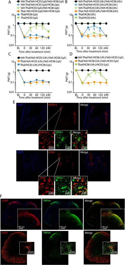
De Logu et al. BMC Biology   (2020) 18:197                                                                    Page 2 of 17

Background                                                   oxidative stress [26]. This response contributes to their
Thalidomide is an old sedative, anti-emetic, and anxio-      anticancer action, but seems to be responsible for signifi-
lytic drug, withdrawn from the market because it causes      cant side effects, including CIPN [27]. In line with the
teratogenicity [1]. Its clinical use has been repurposed     assumption that ROS contribute to CIPN, several pre-
for the treatment of complications of leprosy [2] and, as    clinical findings have shown that mechanical and ther-
an alternative to bortezomib, for multiple myeloma,          mal      hypersensitivity    evoked     in    rodents    by
other hematological malignancies, and solid tumors [3].      chemotherapeutics is attenuated by antioxidants [28].
However, like other chemotherapeutics, the anticancer        However, these positive results have not been replicated
action of thalidomide is associated with the development     by clinical studies [29, 30]. Failure of antioxidants to al-
of a painful peripheral neuropathy that may result in        leviate CIPN might be attributed to their rapidly
delay or even premature termination of an otherwise          exhausted antioxidant activity. For this reason, the iden-
successful treatment [4]. The thalidomide derivatives,       tification of the possible targets that mediate the proal-
lenalidomide and pomalidomide, have also been re-            gesic action of ROS in CIPN is of marked interest.
ported to cause painful neuropathy [5, 6]. Paradoxically,       The aim of the present study was twofold. First, we ex-
several studies have shown analgesic properties of thal-     plored whether thalidomide, lenalidomide, and pomalido-
idomide in mouse models of inflammatory [7], cancer          mide evoked pain-like responses in mice. Second, as
[8], and neuropathic [9] pain. A variety of mechanisms       thalidomide administration is known to generate oxidative
have been proposed to explain the analgesic activity of      stress in mice, rats, and humans [31, 32], we explored the
thalidomide, including downregulation of the tumor ne-       role of oxidative stress and TRP channels sensitive to oxi-
crosis factor-α (TNF-α) [7, 9], and inhibition of nuclear    dants in mechanical and thermal hypersensitivities evoked
factor kappa B (NF-κB) expression [8]. A paper [10] re-      by thalidomide, lenalidomide, and pomalidomide. Via
ported electrophysiological conduction abnormalities in      pharmacological and genetic tools, we revealed that the
primary sensory neurons of thalidomide-treated rats.         three drugs produce mechanical and cold hypersensitivity.
However, to the best of our knowledge, there are no          We also found that oxidative stress generated in periph-
established animal models of thalidomide-induced neur-       eral tissues targets TRPA1 to signal cold allodynia, and
opathy which reproduce painful responses in rodents,         part of the mechanical allodynia, whereas oxidative stress
mimicking those that they cause in patients.                 generated in the central nervous system (CNS) targets
  The transient receptor potential ankyrin 1 (TRPA1)         central TRPV4 to mediate the TRPA1-resistant compo-
channel is abundantly expressed by a subpopulation of        nent of mechanical allodynia.
primary sensory neurons [11]. Pharmacological blockade
and genetic deletion of TRPA1 completely abrogated           Results
mechanical and cold hypersensitivity induced by the          Thalidomide evokes mechanical and cold hypersensitivity
proteasome inhibitor, bortezomib, and platinum-based         mediated by TRPA1 and TRPV4
anticancer drugs (cisplatin and oxaliplatin) in rodents      To explore whether thalidomide elicited sensory hyper-
[12, 13]. Among the additional TRP channels expressed        sensitivities in mice, we administered a single i.p. injec-
in primary sensory neurons, vanilloid 4 (TRPV4) [14]         tion of increasing doses (1, 10, 50, and 100 mg/kg) of the
has been implicated in mechanical hypersensitivity pro-      drug, or its vehicle, in C57BL/6J mice. We observed a
duced by paclitaxel in mice [15] and TRPV1 has been          dose-dependent, early (3 h after administration), and
found to contribute to cisplatin-induced thermal hyper-      prolonged (~ 35 days) mechanical and cold allodynia
algesia [16]. Although TRPA1 is a major oxidant sensor       (Fig. 1a, b). In contrast, any dose of thalidomide failed to
[17], as it is activated by an unprecedented series of re-   evoke hypersensitivity to thermal (hot) stimuli (Fig. 1c).
active and electrophilic substances, including hydrogen      Further mechanistic studies were performed in mice
peroxide (H2O2) and 4-hydroxynonenal (4-HNE) [18,            treated with a single dose (50 mg/kg, i.p.) of thalidomide,
19], TRPV1 and TRPV4 are also sensitive to the redox         which, after the man to mouse conversion [33], approxi-
potential of the milieu [17].                                mates the starting therapeutic dose (200 mg) used in pa-
  Ever-increasing evidence indicates that reactive oxygen    tients [34].
species (ROS) sustain pain hypersensitivity in a variety        To test the implication of TRP channels, both pharma-
of neuropathic pain models, including diabetic neur-         cological and genetic tools were used. Seven days after
opathy [20], alcohol-related peripheral neuropathy [21],     thalidomide administration, when the allodynia plat-
peripheral nerve injury [22, 23], and chemotherapeutic-      eaued, systemic (i.p.) administration of the selective
induced peripheral neuropathy (CIPN) [12, 13, 15, 24,        TRPA1 antagonist, HC-030031 (100 mg/kg) [35], par-
25]. Treatment with different classes of anticancer drugs,   tially reversed mechanical allodynia (Fig. 1d), without af-
including platinum salts, bortezomib, and spindle poi-       fecting the basal threshold value in naive animals
sons (vinca alkaloids, taxanes, epothilones), produces       (Fig. 1d). Because of the incomplete inhibition produced
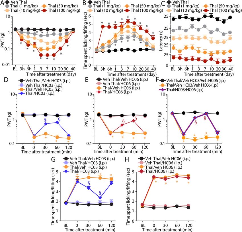
De Logu et al. BMC Biology       (2020) 18:197                                                                                            Page 3 of 17

 Fig. 1 Thalidomide elicits mechanical and cold, but not heat, hypersensitivity that is dependent on TRPA1 and TRPV4. a–c Dose- and time-
 dependent mechanical and cold allodynia and heat hypersensitivity following intraperitoneal (i.p.) injection of thalidomide (Thal, 1, 10, 50, and
 100 mg/kg) or Veh. d–f Mechanical allodynia at day 7 following Thal (50 mg/kg, i.p.) and Veh and after the administration of HC-030031 (HC03,
 100 mg/kg, i.p.), HC-067047 (HC06, 10 mg/kg, i.p.), a combination of HC03 (100 mg/kg, i.p.) and HC06 (10 mg/kg, i.p.), or Veh. g, h Cold allodynia at
 day 7 following Thal (50 mg/kg, i.p.) or Veh and after the administration of HC03 (100 mg/kg, i.p.), HC-067047 (HC06, 10 mg/kg, i.p.), or Veh. BL,
 baseline. Data are mean ± SEM, n = 6 mice. *P < 0.05 vs. Veh Thal or Veh Thal/Veh HC03/Veh HC06; §P < 0.05 vs. Thal/Veh HC03/Veh HC06. Two-
 way ANOVA followed by Bonferroni’s post hoc test

by TRPA1 antagonism, the role of TRPV4 and TRPV1                             animals (Fig. 1e), partially attenuated mechanical allody-
was explored. The TRPV1 antagonist, capsazepine (4                           nia (Fig. 1e). However, a combination of TRPA1 and
mg/kg, i.p.), given at day 7 after thalidomide or vehicle,                   TRPV4 antagonists completely reversed thalidomide-
failed to affect mechanical allodynia (Additional file 1:                    evoked mechanical allodynia (Fig. 1f). Cold allodynia, in-
Fig. S1A). Conversely, systemic (i.p.) administration of                     duced by thalidomide, resulted to be exclusively
the selective TRPV4 antagonist, HC-067047 (10 mg/kg,                         dependent on TRPA1 as HC-030031 administered at
at day 7 after thalidomide or vehicle) [36], which did not                   day 7 after thalidomide completely attenuated the re-
affect the baseline threshold value in vehicle-treated                       sponse to the cold stimulation, while administration of
De Logu et al. BMC Biology   (2020) 18:197                                                                       Page 4 of 17

HC-067047 or capsazepine was ineffective (Fig. 1g, h            reversed mechanical allodynia (Additional file 1: Fig. S2F
and Additional file 1: Fig. S1B).                               and S2G), which was, however, completely attenuated in
  To further prove the contribution of TRP channels,            mice receiving a combination of HC-030031 and HC-
mice with genetic deletion of TRPA1, TRPV4, or TRPV1            067047 (Additional file 1: Fig. S2H). Cold allodynia
were used. Trpa1+/+, Trpv4+/+, and Trpv1+/+ mice devel-         evoked by pomalidomide and lenalidomide was entirely
oped mechanical and cold hypersensitivity with time             inhibited by HC-030031 and a combination of HC-
courses similar to those observed in C57BL/6J mice,             030031 and HC-067047, whereas HC-067047 was inef-
starting 3 h and lasting ~ 35 days after thalidomide ad-        fective (Additional file 1: Fig. S2J-S2L). Present pharma-
ministration (Fig. 2a, b and Additional file 1: Fig. S1C).      cological and genetic findings indicate that TRPA1 and
While Trpv1−/− mice showed unchanged mechanical hy-             TRPV4 channels contribute to the mechanical allodynia
persensitivities (Additional file 1: Fig. S1C), in Trpa1−/−     induced by thalidomide and its derivatives, but only
and Trpv4−/− mice, thalidomide-evoked mechanical allo-          TRPA1 mediates cold hypersensitivity caused by these
dynia was significantly, but not completely, reduced            drugs.
(Fig. 2a, b). The relative contribution of TRPA1 and
TRPV4 to thalidomide-evoked mechanical allodynia was            Thalidomide and its derivatives elicit hypersensitivity via
further investigated by evaluating the combined effect of       oxidative stress generation that targets TRPA1 and TRPV4
channel pharmacological antagonism and genetic dele-            To test the hypothesis that thalidomide and its deriva-
tion. Thus, mechanical allodynia at day 7 after thalido-        tives directly activate both the TRPA1 and TRPV4 re-
mide injection was completely attenuated in Trpa1−/−            ceptors, we studied the ability of the drugs to elicit an
treated with HC-067047 (10 mg/kg, i.p.) and in Trpv4−/−         inward current in cultured rat DRG neurons. Thalido-
mice treated with HC-030031 (100 mg/kg, i.p.) (Fig. 2c,         mide, pomalidomide, and lenalidomide (all 100 μM)
d). Cold allodynia observed in Trpa1+/+, Trpv4+/+, and          failed to evoke any inward current in capsaicin-sensitive
Trpv1+/+ was completely abolished in Trpa1−/− mice and          DRG neurons (Fig. 3a), which otherwise responded to
unaffected in both Trpv4−/− and Trpv1−/− mice (Fig. 2e, f       the TRPA1 and TRPV4 agonist, AITC (100 μM) and 4-
and Additional file 1: Fig. S1D). Similar results, for either   αPDD (100 μM), respectively. Like other anticancer
mechanical or cold allodynia, were obtained if a lower          drugs, thalidomide and its derivatives are known to gen-
dose of thalidomide (1 mg/kg) was tested in the three           erate oxidative stress [39, 40]. Thus, we hypothesized
strains of mice (Additional file 1: Fig. S1E-G).                that oxidative stress burst, and its reactive byproducts
                                                                generated by thalidomide, pomalidomide, and lenalido-
Pomalidomide and lenalidomide evoke mechanical and              mide, could be implicated in mechanical and cold allo-
cold allodynia similar to thalidomide                           dynia evoked by the anticancer drug. Systemic (i.p.)
The two newer derivatives of thalidomide, pomalidomide          administration of the ROS scavenger, PBN (100 mg/kg),
and lenalidomide, used for the treatment of multiple            at day 7 after the administration of the three drugs
myeloma and other hematological conditions [37], have           abated mechanical and cold allodynia (Fig. 3b, c and
been reported to evoke CIPN [5, 6]. Thus, the ability of        Additional file 1: Fig. S2I and S2M), thus supporting a
pomalidomide and lenalidomide to induce mechanical              role of oxidative stress. PBN did not affect the basal
and thermal hypersensitivity was explored in mice. Sys-         threshold value in naive animals. Furthermore, by using
temic administration of amounts of pomalidomide (1              calcium imaging assay, we showed that PBN had no dir-
mg/kg, i.p.) and lenalidomide (5 mg/kg, i.p.), equivalent       ect effect on TRPA1 and TRPV4 channel activity, since
in mice to the respective therapeutic doses [6, 38], in-        the calcium response evoked by channel-selective ago-
duced a time-dependent mechanical and cold allodynia            nists, AITC and 4-αPDD, was unaffected after preincu-
that initiated 3 h and lasted 35 days after drug adminis-       bation with PBN in hTRPA1- and hTRPV4-HEK293
tration (Additional file 1: Fig. S2A and B). The two drugs      cells (Fig. 3d).
did not affect the threshold value to heat stimuli (Add-           While strong evidence has been accumulated on the
itional file 1: Fig. S2C). Thalidomide, pomalidomide, and       ability of H2O2 to activate TRPA1 [18], only H2O2-medi-
lenalidomide did not affect motor coordination and bal-         ated activation of TRPV4 was reported in human and
ance in mice, which were evaluated by using the rotarod         rodent (rat and mouse) lung microvascular endothelial
and the balance beam walk tests (Additional file 1: Fig.        cells [41] and indirectly in rat brain slices [42]. Here, we
S2D and S2E). In addition, mice did not exhibit writhing        show that H2O2 elicited concentration-dependent Ca2+
or other stereotypic behaviors, such as freezing or hyper-      responses either in hTRPV4- or in mTRPV4-HEK293
activity, curling, grooming, or biting/licking, after drug      cells with an EC50 of 5 mM and 3 mM, respectively,
injection. Seven days after pomalidomide and lenalido-          which resulted 10 times higher than that observed in
mide administration, treatment with HC-030031 (100              hTRPA1- and mTRPA1-HEK293 cells (EC50 500 μM
mg/kg, i.p.) or HC-067047 (10 mg/kg, i.p.) partially            and 300 μM, respectively) (Fig. 3e). The Ca2+ response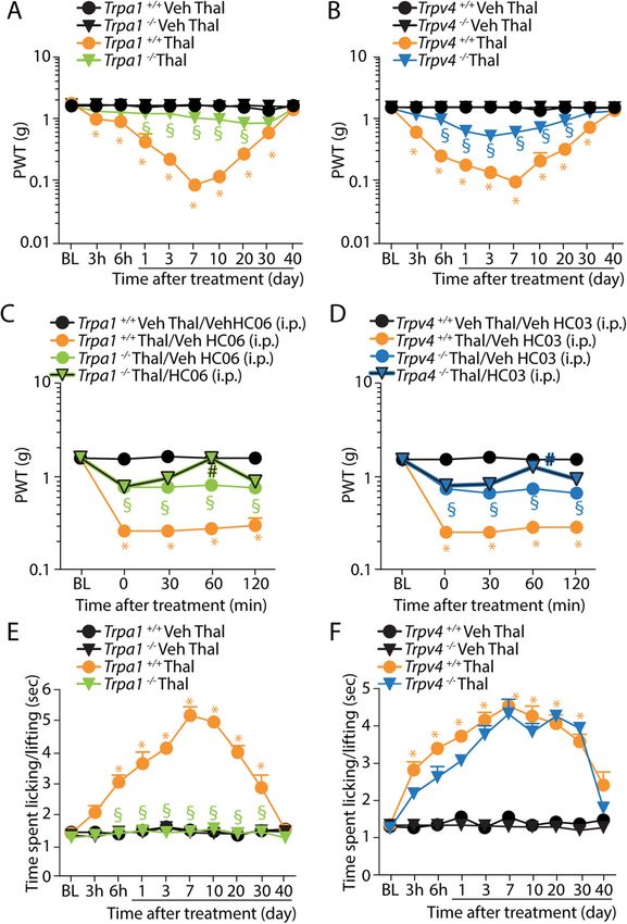
De Logu et al. BMC Biology       (2020) 18:197                                                                                            Page 5 of 17

 Fig. 2 Genetic deletion of TRPA1 and TRPV4 attenuates mechanical and cold hypersensitivity evoked by thalidomide. a, b Time-dependent
 mechanical allodynia following intraperitoneal (i.p.) injection of thalidomide (Thal, 50 mg/kg) or Veh in Trpa1+/+, Trpa1−/−, Trpv4+/+, and Trpv4−/−
 mice. c, d Mechanical allodynia in Trpa1+/+ and Trpa1−/−, Trpv4+/+ and Trpv4−/− mice at day 7 following Thal (50 mg/kg, i.p.) or Veh and after the
 administration of HC-067047 (HC06, 10 mg/kg, i.p.) or Veh in Trpa1−/− mice and HC-030031 (HC03, 100 mg/kg, i.p.) or Veh in Trpv4−/− mice. e, f
 Time-dependent cold allodynia following Thal (50 mg/kg, i.p.) or Veh in Trpa1+/+, Trpa1−/−, Trpv4+/+, or Trpv4−/− mice. BL, baseline. Data are
 mean ± SEM, n = 6 mice. *P < 0.05 vs. Trpa1+/+/Veh Thal or Trpv4+/+/Veh Thal or Trpa1+/+/Veh Thal/Veh HC06 or Trpv4+/+/Veh Thal/Veh HC03; §P <
 0.05 vs. Trpa1+/+/Thal or Trpv4+/+/Thal or Trpa1+/+/Thal/Veh HC06 or Trpv4+/+/Thal/Veh HC03; #P < 0.05 vs. Trpa1−/−/Thal/Veh HC06 or Trpv4−/−/
 Thal/Veh HC03. Two-way ANOVA followed by Bonferroni’s post hoc test
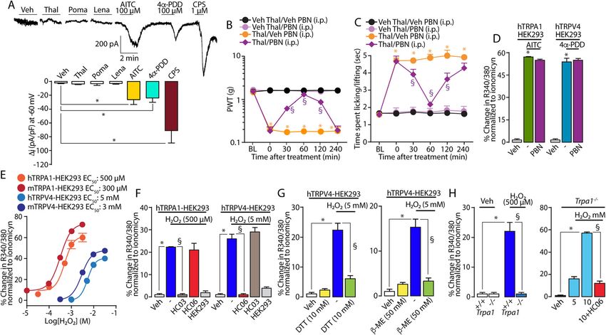
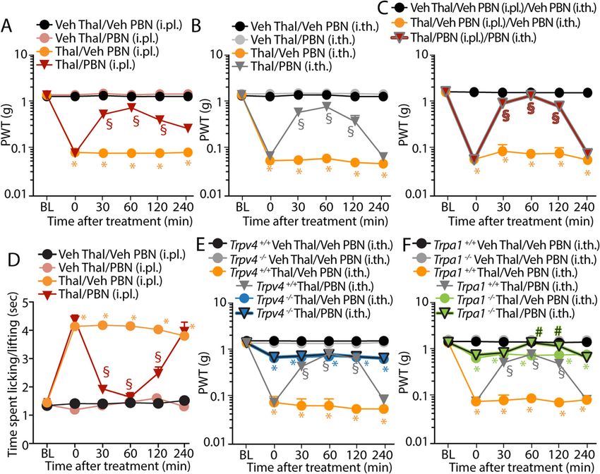
De Logu et al. BMC Biology      (2020) 18:197                                                                                         Page 6 of 17

 Fig. 3 Oxidative stress targets TRPA1 and TRPV4. a Typical traces and pooled data of patch-clamp inward currents elicited by thalidomide (Thal),
 pomalidomide (Poma) and lenalidomide (Lena) (all, 100 μM), allyl isothiocyanate (AITC, 100 μM), 4α-phorbol 12,13-didecanoate (4α-PDD, 100 μM),
 capsaicin (CPS, 1 μM), or Veh in mouse dorsal root ganglion (DRG) neurons. Data are mean ± SEM, n = 4–5 cells. *P < 0.05 vs. Veh. One-way
 ANOVA followed by Bonferroni’s post hoc test. b, c Mechanical and cold allodynia at day 7 following the intraperitoneal (i.p.) injection of
 thalidomide (Thal, 50 mg/kg) or Veh, and after the administration of phenyl-α-tert-butyl nitrone (PBN, 100 mg/kg, i.p.) or Veh. Data are mean ±
 SEM, n = 6 mice. *P < 0.05 vs. Veh Thal/Veh PBN; §P < 0.05 vs. Thal/Veh PBN. Two-way ANOVA followed by Bonferroni’s post hoc test. d Pooled
 data of Ca2+ response to AITC (10 μM), 4α-PDD (1 μM), or Veh hTRPA1-HEK293 and hTRPV4-HEK293 cells in the presence of PBN (30 μM). e
 Concentration response (Ca2+ mobilization)-curve to H2O2 in cultured hTRPA1- and mTRPA1-HEK293 and hTRPV4- and mTRPV4-HEK293
 transfected cells. f Pooled data of Ca2+ response to H2O2 (500 μM and 5 mM) or Veh in untransfected HEK293 or hTRPA1-HEK293, hTRPV4-HEK293
 cells in the presence of HC-030031 (HC03, 30 μM) or HC-067047 (HC06, 10 μM). g Pooled data of the Ca2+ response to H2O2 (5 mM) in hTRPV4-
 HEK293 cells in the presence of dithiothreitol (DTT, 10 mM) or 2-mercaptoethanol (β-ME, 50 mM). h Pooled data of the Ca2+ response to H2O2
 (500 μM) in DRG neurons from Trpa1+/+ and Trpa1−/− mice and H2O2 (5 and 10 mM) in DRG neurons from Trpa1−/− mice in the presence of HC06
 (10 μM). Data are mean ± SEM, n = 20–25 neurons or 80–100 cells. *P < 0.05 vs. Veh; §P < 0.05 vs. H2O2 (500 μM, 5 mM, or 10 mM). One-way
 ANOVA followed by Bonferroni’s post hoc test

evoked by a lower H2O2 concentration (500 μM) in                           those from Trpa1−/− mice (Fig. 3h). The residual calcium
hTRPA1-HEK293 was inhibited in the presence of HC-                         response to a higher concentration of H2O2 (10 mM) ob-
030031, but not of HC-067047 (Fig. 3f). However, the                       served in DRG neurons from Trpa1−/− mice was abated
Ca2+ response evoked by H2O2 (5 mM) in hTRPV4-                             in the presence of HC-067047 (Fig. 3h). Thus, in vitro
HEK293 was attenuated by HC-067047, but not by HC-                         data confirmed the ability of H2O2 to target the TRPV4
030031. H2O2 (500 μM or 5 mM) was ineffective in                           channel, provided that the concentration/dose of H2O2
untransfected HEK293 cells (Fig. 3f). Considering that                     is sufficiently high.
H2O2 caused TRPA1 activation via the oxidization of
cysteine residues [18], we tested the hypothesis that oxi-                 Peripheral and central (spinal) TRPA1 and TRPV4
dation by H2O2 of TRPV4 implicates cysteine residues.                      activation differentially contributes to thalidomide-
In hTRPV4-HEK293, exposure to two cysteine-reducing                        induced mechanical allodynia
agents, DTT and β-ME [18], attenuated the Ca2+ re-                         One major issue raised by the present data is that, while
sponse evoked by H2O2 in hTRPV4-HEK293 cells                               oxidative stress inhibition completely attenuated mech-
(Fig. 3g). Finally, we tested low (500 μM) and high (5                     anical allodynia, TRPA1 or TRPV4 pharmacological an-
and 10 mM) H2O2 concentrations in cultured mouse                           tagonism or gene deletion provided partial reduction,
DRG neurons taken from Trpa1+/+ and Trpa1−/− mice.                         and total reduction was attained solely by the simultan-
The lower H2O2 concentration (500 μM) elicited a Ca2+                      eous inhibition of both channels. A recent study re-
response in neurons from Trpa1+/+ mice, but not in                         ported that oxidative stress generated at central or
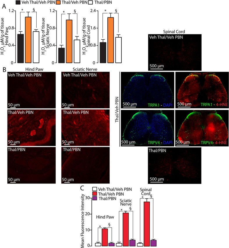
De Logu et al. BMC Biology   (2020) 18:197                                                                       Page 7 of 17

peripheral sites may contribute differently to cisplatin-      study with double immunofluorescence staining was
and paclitaxel-evoked hypersensitivity [25]. Thus, we hy-      undertaken. In slices that contain bundles of the plantar
pothesized whether oxidative stress activates TRPA1 and        nerve, TRPA1 expression was detected in PGP9.5+ (pro-
TRPV4 at different anatomical sites to mediate                 tein gene product) nerve fibers and S100+ Schwann cells
thalidomide-evoked mechanical allodynia. To test this hy-      (Fig. 5e). At the central level, TRPV4 staining exhibited
pothesis, we measured two oxidative stress biomarkers,         a well-matched colocalization with CGRP+ (calcitonin
H2O2 and the more stable peroxidation product of plasma        gene-related peptide) nerve fibers and GFAP+ (glial fi-
membrane phospholipid peroxidation, 4-HNE [19]. H2O2           brillary acidic protein) astrocytes (Fig. 5f).
levels (Fig. 4a) and 4-HNE staining (Fig. 4b, c) were in-        To understand how the increased oxidative stress could
creased in homogenates or tissue slices, respectively, of      engage the peripheral TRPA1 and the central TRPV4,
the hind paw, sciatic nerve, and lumbar spinal cord, taken     PBN was given to mice by either i.pl. or i.th. administra-
from mice at day 7 after thalidomide, compared to its ve-      tion. At day 7 after thalidomide, i.pl. (100 μg) or i.th.
hicle. Systemic treatment with a dose of PBN that reversed     (100 μg) PBN injection partially inhibited mechanical allo-
thalidomide-evoked allodynia reduced H2O2 levels and 4-        dynia (Fig. 6a, b), while a combination of i.pl. and i.th.
HNE staining in all three tissues (Fig. 4a–c). Notably, 4-     PBN completely attenuated the response (Fig. 6c). How-
HNE staining in the spinal cord revealed that the oxidative    ever, cold allodynia, which is entirely TRPA1-dependent,
stress marker does not localize to a specific site (e.g.,      was completely reversed by i.pl. PBN (Fig. 6d). PBN (i.pl.
superficial lamina) where TRPA1 and TRPV4 are mainly           or i.th.) did not affect the basal threshold value in vehicle-
expressed. The 4-HNE staining was evenly distributed in        treated mice. Similar results were obtained with the
the entire tissue slice of the lumbar spinal cord, thus sug-   H2O2-detoxifying enzyme, catalase. Either i.pl. or i.th.
gesting the possibility that thalidomide generates oxidative   catalase administration partially inhibited mechanical allo-
stress in a non-specific manner from several cell types.       dynia, which, however, resulted completely reduced by a
Thus, while oxidative stress produced at each site may po-     combination of i.pl. and i.th. catalase (Additional file 1:
tentially contribute to thalidomide-evoked mechanical          Fig. S3A-S3C). As for PBN, catalase (i.pl. but not i.th.)
allodynia, the role of centrally vs. peripherally generated    completely reversed cold allodynia (Additional file 1: Fig.
oxidative stress is unknown.                                   S3D and S3E). Due to the selective activity of catalase
  To explore this hypothesis, we investigated the impli-       against H2O2, these data strengthen the prominent role of
cation of peripheral vs. central TRPA1 and TRPV4 in            H2O2 at both central and peripheral levels in thalidomide-
thalidomide-induced mechanical allodynia, by injecting         evoked mechanical cold allodynia. We also found that i.th.
channel antagonists locally in the hind paw or intra-          PBN, while partially reversing mechanical allodynia in
thecally in the spinal cord. We found that, at day 7 after     Trpa1+/+ and Trpv4+/+ mice (Fig. 6e, f), did not affect the
thalidomide, mechanical allodynia was partially reversed       residual mechanical allodynia in Trpv4−/− (Fig. 6e), and
by the i.pl. injection of HC-030031 (100 μg), but not of       completely reversed the residual response in Trpa1−/−
HC-067047 (100 μg) (Fig. 5a). In contrast, i.th. HC-           mice (Fig. 6f). Altogether, these data indicate that selective
067047 (100 μg), but not HC-030031 (100 μg), partially         ROS scavenging at either the peripheral or the central
reversed thalidomide-induced mechanical allodynia              levels inhibits the correspondent TRPA1 and TRPV4
(Fig. 5b). Neither i.th. nor i.pl. HC-030031 and HC-           component, respectively. Only simultaneous inhibition of
067047 affect the basal threshold value in naive animals.      the oxidative stress that targets both ion channels war-
These results implicated the engagement of peripheral          rants complete attenuation of the mechanical allodynia.
TRPA1 and central TRPV4 in the thalidomide-induced
mechanical allodynia. To further support this hypothesis,      Discussion
at day 7 after thalidomide injection, we tested the ability    Thalidomide, an old sedative and anti-emetic drug banned
of a combination of i.pl. HC-067047 and i.th. HC-              for causing birth defects in humans, has been repurposed
030031, or vice versa, to attenuate thalidomide-evoked         for the treatment of leprosy and several types of cancer
mechanical allodynia. While a combination of i.pl. HC-         [2], including multiple myeloma, myelodysplastic syn-
067047 and i.th. HC-030031 failed to affect allodynia          drome, and several solid cancers [43]. Thalidomide deriva-
(Fig. 5c), a combination of i.pl. HC-030031 and i.th. HC-      tives, pomalidomide and lenalidomide, also exhibit
067047 provided complete reversal of the pain-like re-         anticancer activity in multiple myeloma patients who re-
sponse (Fig. 5d), thus supporting the view that TRPA1          lapse or are refractory to other anticancer treatments. Un-
mediates the peripheral, and TRPV4 the central, compo-         fortunately, as with other chemically unrelated
nent of thalidomide-evoked mechanical allodynia. To            chemotherapeutic agents (platinum-based drugs, taxanes,
better address which cell types expressing TRPA1 in the        and bortezomib), thalidomide and its derivatives cause a
periphery and TRPV4 at the central level are involved in       painful peripheral polyneuropathy that often results in se-
thalidomide-evoked pain responses, a colocalization            vere discomfort or even drug discontinuation [4]. Despite
De Logu et al. BMC Biology      (2020) 18:197                                                                                         Page 8 of 17

 Fig. 4 Thalidomide increases oxidative stress in the hind paw, sciatic nerve, and spinal cord. a H2O2 levels in the hind paw, sciatic nerve, and
 lumbar (L4–L6) spinal cord at day 7 following intraperitoneal (i.p.) injection of thalidomide (Thal, 50 mg/kg) or Veh and 60 min after the
 administration of phenyl-α-tert-butyl nitrone (PBN, 100 mg/kg, i.p.) or Veh. b, c Representative images and mean fluorescence intensity of 4-
 hydroxynonenal (4-HNE) staining in the hind paw, sciatic nerve, and lumbar (L4–L6) spinal cord and TRPA1 and TRPV4 staining in the spinal cord,
 at day 7 following Thal (50 mg/kg, i.p.) and 60 min after the administration of PBN (100 mg/kg, i.p.) or Veh. Data are mean ± SEM (n = 6 mice).
 *P < 0.05 vs. Veh Thal/Veh PBN; §P < 0.05 vs. Thal/Veh PBN. One-way ANOVA followed by Bonferroni’s post hoc test

its clinical relevance, the underlying mechanism of the                      Several studies have investigated the ability of thalido-
neuropathy and the associated pain symptoms caused by                      mide to attenuate inflammatory and neuropathic pain in
thalidomide and its derivatives remains poorly known.                      rodent models presumably by interacting with indirect
De Logu et al. BMC Biology     (2020) 18:197   Page 9 of 17

 Fig. 5 (See legend on next page.)
De Logu et al. BMC Biology        (2020) 18:197                                                                                                 Page 10 of 17

 (See figure on previous page.)
 Fig. 5 Peripheral TRPA1 and central TRPV4 contribute to thalidomide-induced mechanical allodynia. a, b Mechanical allodynia at day 7 following
 intraperitoneal (i.p.) thalidomide (Thal, 50 mg/kg) or Veh and after the administration of intraplantar (i.pl., 20 μl) or intrathecal (i.th., 5 μl) HC-030031
 (HC03, 100 μg), HC-067047 (HC06, 100 μg), or Veh. c, d Mechanical allodynia at day 7 following Thal (50 mg/kg, i.p.) or Veh and after the
 administration of a combination of HC03 (100 μg, i.th.) and HC06 (100 μg, i.pl.) or HC06 (100 μg, i.th.) and HC03 (100 μg, i.pl.) or Veh. e
 Representative images of co-expression of PGP9.5 or S100 and TRPA1 in the peripheral nerve of hind paw. f Representative images of co-
 expression of CGRP or GFAP and TRPV4 in the lumbar (L4–L6) spinal cord. Data are mean ± SEM, n = 6 mice. *P < 0.05 vs. Veh Thal/Veh HC03/Veh
 HC06; §P < 0.05 vs. Thal/Veh HC03/Veh HC06. Two-way ANOVA followed by Bonferroni’s post hoc test

mechanisms dependent on the inhibition of proalgesic cy-                          responses in rat sensory nerve fibers [10], reminiscent of a
tokines, such as TNF-α and NF-κB [7–9]. Surprisingly, to                          sensory neuropathy. Thus, our study shows for the first
the best of our knowledge, no study has investigated the                          time that thalidomide, pomalidomide, and lenalidomide
ability of thalidomide or related drugs to elicit pain-like                       evoke mechanical and cold hypersensitivity in mice. Al-
responses in animal models so far. Only one study has                             though the chemotherapeutic drugs failed to evoke heat
shown that thalidomide increased electrophysiological                             hypersensitivity, as cold and mechanical allodynia are the

 Fig. 6 Oxidative stress targeting peripheral TRPA1 and central TRPV4 mediates thalidomide-induced mechanical allodynia. a–c Mechanical
 allodynia at day 7 following thalidomide (Thal, 50 mg/kg) or Veh and after the administration of i.pl. or a combination of i.pl. and i.th. phenyl-α-
 tert-butyl nitrone (PBN, 100 μg) or Veh. d Cold allodynia at day 7 following Thal (50 mg/kg, i.p.) or Veh and after the administration of i.pl. PBN
 (100 μg) or Veh. e, f Mechanical allodynia in Trpv4+/+, Trpv4−/−, Trpa1+/+, and Trpa1−/− mice at day 7 following Thal (50 mg/kg, i.p.) or Veh and
 after the administration of i.th. PBN (100 μg) or Veh. Data are mean ± SEM, n = 6 mice. *P < 0.05 vs. Veh Thal/Veh PBN; §P < 0.05 vs. Thal/Veh PBN;
 #
  P < 0.05 vs. Trpa1−/−/Thal/Veh PBN. Two-way ANOVA followed by Bonferroni’s post hoc test
De Logu et al. BMC Biology   (2020) 18:197                                                                      Page 11 of 17

major and most debilitating symptoms of thalidomide-             to those found in other redox-sensitive TRP channels
evoked CIPN [44, 45], the present model seems to satisfac-       [17], thus enabling their targeting through oxidation,
torily replicate the human pain condition. As previously re-     which leads to channel activation [17]. Here, we report
ported for other chemotherapeutic agents, including              that H2O2 targets the recombinant and native TRPV4,
oxaliplatin, vincristine, bortezomib, and paclitaxel [12, 13,    although with a potency about 10 times lower than that
15, 28, 46–48], we report that a single administration of        exhibited toward TRPA1. The observation that two
thalidomide, lenalidomide, or pomalidomide is sufficient to      cysteine-reducing agents, DTT and β-ME [18, 49],
produce a sustained condition of sensory hypersensitivity in     abated the H2O2 evoked Ca2+ response further supports
mice, which somehow mimics the prolonged duration of             the hypothesis that TRPV4 may be directly activated by
CIPN in patients [5, 6]. However, the molecular and cellular     oxidants. The more elevated H2O2 concentrations after
mechanisms responsible for the prolonged thalidomide-            thalidomide in the spinal cord and the efficacy of i.th.
evoked hypersensitive phenotype remain unknown and de-           administration of the specific H2O2 scavenger, catalase,
serve further investigation. A series of previous investiga-     strengthen the role of H2O2 to mediate the central
tions have highlighted the ability of thalidomide to produce     TRPV4-dependent component of thalidomide-evoked
beneficial and toxic effects, including its anticancer action    mechanical allodynia.
and severe teratogenic effects, via the generation of oxida-       Results obtained in cultured DRG neurons strengthen
tive stress [39, 40]. In particular, it has been reported that   the findings obtained in recombinant systems. While the
the bioactivation of thalidomide from horseradish peroxid-       Ca2+ response produced by a low H2O2 concentration
ase to free-radical intermediates produces ROS, which            was entirely abated in the presence of the TRPA1 antag-
cause oxidative damage to DNA and other cellular macro-          onist, the response to a higher H2O2 concentration was
molecules, apparently responsible for the anticancer effect,     blocked only when a TRPV4 antagonist was added. Im-
but also for the teratogenic action [40].                        portantly, the residual Ca2+ response to a high H2O2
   Our data show that oxidative stress byproducts, such as       concentration observed in DRG neurons from Trpa1−/−
H2O2 and 4-HNE, are generated both at the peripheral             mice was completely attenuated by a TRPV4 antagonist.
(hind paw and sciatic nerve) and central (spinal cord)           Thus, it may be concluded that, provided a sufficiently
levels, after thalidomide systemic injection. Thus, thalido-     elevated burden is present, oxidative stress may engage
mide generates oxidative stress along the entire pain path-      not only TRPA1 but also TRPV4.
way, which encompasses the entire anatomical route that            A peculiar difference of the two channels regarding their
conveys the pain signal from the hind paw to the lumbar          roles in thalidomide-evoked hypersensitivities is that, as
spinal cord. The observation that increased H2O2 and 4-          shown by genetic or pharmacological studies, mechanical
HNE levels were reduced by systemic treatment with the           allodynia was partially attenuated by these interventions,
antioxidant, PBN, further supports the hypothesis that           and abolition was attained only by a combination of per-
oxidative stress is essential for the pain-like symptoms         ipheral TRPA1 and central TRPV4 blockade. Various anti-
evoked by thalidomide. We also revealed that TRPA1 and           cancer drugs, including platinum-derived drugs or the
TRPV4 channels mediate mechanical and cold allodynia             proteasome inhibitor, bortezomib, promote mechanical
evoked by thalidomide and related drugs. However, as re-         allodynia exclusively via oxidative stress and the ensuing
ported for other anticancer drugs, including oxaliplatin/        TRPA1 activation [12, 13]. The partial contribution of
cisplatin, paclitaxel, and bortezomib [12, 13, 15], the ob-      TRPV4 to mechanical hypersensitivity has been previously
servation that thalidomide and its derivatives failed to         reported in the CIPN model produced by the taxane de-
evoke any excitatory effect in cultured TRPA1- and               rivative, paclitaxel [15]. Notably, as shown in the
TRPV4-expressing neurons excludes the possibility that           paclitaxel-evoked model [15], cold allodynia elicited by
their proalgesic effect depends on a direct action on these      thalidomide, lenalidomide, and pomalidomide was entirely
channels, and suggests the implication of indirect mecha-        TRPA1-dependent. However, the reason why cold hyper-
nisms, including oxidative stress generation.                    sensitivity is solely dependent on TRPA1, whereas mech-
   Whereas the role of TRPA1 as a sensor of oxidative            anical allodynia requires the contribution of both TRPA1
stress has been extensively investigated and recognized,         and TRPV4, remains unknown.
a similar function of TRPV4 has been poorly explored.              We previously found that local (intraplantar) adminis-
Several studies have reported that TRPA1 is activated by         tration of an oxidative stress scavenger or a TRPA1 an-
an unprecedented series of reactive oxygen, nitrogen, or         tagonist completely reversed mechanical and cold
carbonyl species [18, 19]. In particular, robust proof sup-      allodynia evoked by bortezomib and oxaliplatin [12],
ports the hypothesis that H2O2 causes nociceptor stimu-          suggesting that TRPA1 sensitization/activation may
lation via TRPA1 [22, 23]. In contrast, little evidence          occur in terminal nerve fibers of the hind paw. To
showing that H2O2 stimulates TRPV4 has been provided             understand more precisely where TRPV4 and TRPA1
[41, 42]. TRPV4 carries cysteine residues, corresponding         act to mediate thalidomide-evoked mechanical and cold
De Logu et al. BMC Biology   (2020) 18:197                                                                      Page 12 of 17

allodynia, site-specific strategies of drug administration      of tumor immune escape [57]. Constitutive expression
were used. Results show that peripheral (intraplantar)          of TRPA1 mRNA and protein has been identified in
antagonism of TRPA1 in the mouse paw provided                   mouse and human primary CD4+ T cells [58] and mac-
complete reversal of cold allodynia, but only partial at-       rophages [59]. Thus, at the present stage, it is not pos-
tenuation of mechanical allodynia. If central (intrathecal)     sible to exclude that TRPV4 and/or TRPA1 inhibition
antagonism of TRPV4 was added to the peripheral                 may negatively affect cancer progression.
TRPA1 blockade, thalidomide-evoked mechanical allo-                Although we have identified the role of oxidative
dynia was completely inhibited.                                 stress, peripheral TRPA1, and central TRPV4 in mech-
   Evidence of a differential contribution of peripheral vs.    anical and cold hypersensitivity elicited by thalidomide
central oxidative stress has been reported in cisplatin-        and related drugs in mice, several questions remain to
and paclitaxel-evoked mechanical hypersensitivity [25].         be investigated. These include the cell types that express
Oxidative stress manipulation experiments strengthen            peripheral TRPA1 and central TRPV4 that, engaged by
the conclusion deriving from channel pharmacological            oxidative stress, signal allodynia. TRPA1 is known to be
antagonism. In fact, whereas local oxidative stress inhib-      expressed in nociceptors [11] and in Schwann cells that
ition either in the hind paw or in the spinal cord pro-         surround the fibers of these neurons [22]. Here, we con-
vided only partial attenuation, a combination of central        firm that in mouse peripheral tissues, prominent TRPA1
and peripheral oxidative stress blockade completely re-         protein expression is present within the nerve fibers and
versed allodynia. Final proof that mechanical allodynia         Schwann cells that wrap these fibers. In the CNS,
was mediated by oxidative stress activation of both per-        TRPV4 may be present in central terminals of nocicep-
ipheral TRPA1 and central TRPV4 was derived from ex-            tors [14] and astrocytes [60]. In the lumbar spinal cord,
periments with genetic channel deletion. Elimination of         we confirm the presence of TRPV4 in CGRP-
oxidative stress by an intrathecal antioxidant, while not       immunoreactive fibers and in astrocytes. While TRPA1
further inhibiting thalidomide-evoked mechanical allody-        and TRPV4 expressed by nerve fibers may directly con-
nia in Trpv4−/− mice, completely reversed the residual          tribute to signal pain, Schwann cell TRPA1, which have
response observed in Trpa1−/− mice. These data further          been implicated in pain sensitization [22], and astrocyte
confirmed that full protection from thalidomide-induced         TRPV4 may indirectly sustain thalidomide-evoked allo-
mechanical allodynia can be attained by attenuating oxi-        dynia. However, further studies are required to identify
dative stress at both peripheral and central sites of ac-       the intracellular and molecular mechanisms implicated
tion. Some studies have reported that early treatment           in the central TRPV4-dependent and peripheral TRPA1-
with ROS scavengers or mitochondrial activity inhibitors        dependent components of thalidomide-evoked mechan-
provided a complete and sustained prevention of mech-           ical allodynia in neurons and/or glial cells. Although
anical hypersensitivity induced by chemotherapeutic             H2O2 levels and 4-HNE staining were higher in the
agents [12, 50–52].                                             spinal cord than in the paw, it is not clear if these differ-
   These findings suggest the existence of a time soon          ences may explain the differential ability of oxidative
after the exposure to chemotherapeutics that is critical        stress to target TRPA1 in the peripheral tissues and
in order to initiate and maintain the generation of the         TRPV4 at the central level.
proalgesic oxidative stress. In clinical settings, inhibition
by ROS scavengers at such an initial event may be chal-         Conclusions
lenging, and therefore, the attenuation of the activity of      From a therapeutic point of view, the present results in-
ROS targets (TRPA1 and TRPV4 channels) at the per-              dicate the need for peripheral acting TRPA1 antagonists
ipheral or central level could be a better therapeutic          and blood-brain barrier-penetrating TRPV4 antagonists
strategy. However, it should be considered that TRPA1           to treat the pain symptoms associated to CIPN evoked
and TRPV4 are expressed by a series of immune and in-           by thalidomide and related drugs. However, due to the
flammatory cells [53]. In particular, elevated expression       pleiotropic activity of TRPA1 and TRPV4, the safety
of TRPV4 has been detected in human leukocytes [54],            profile of channel antagonism should be carefully scruti-
where it regulates key functions in response to pro-            nized, particularly regarding the impact of this thera-
inflammatory stimuli, including ROS production and cell         peutic strategy on cancer outcome and on the efficacy of
adhesion or migration [55], and in macrophages, where           cancer treatment.
it exerts a double-edged function. A pro-inflammatory
function includes phagocytosis and ROS production, and          Methods
an anti-inflammatory function includes secretion of pro-        Animals
resolution cytokines [56]. Furthermore, under inflamma-         Sprague-Dawley rats (male, 75–100 g, 4–5 weeks),
tory circumstances, T cell TRPV4 facilitates the release        C57BL/6J mice (male, 20–25 g, 5 weeks) (Harlan Labora-
of interferon-γ, which represents an important mediator         tories), wild-type (Trpa1+/+) and TRPA1-deficient
De Logu et al. BMC Biology   (2020) 18:197                                                                   Page 13 of 17

(Trpa1−/−; B6129P-Trpa1tm1Kykw/J; Jackson Laboratories)       Behavioral studies
mice (25–30 g, 6–8 weeks) [61], wild-type (Trpv4+/+) and      Rotarod test
TRPV4-deficient (Trpv4−/−) mice (25–30 g, 5–8 weeks)          The locomotor function, balance, and sedation of mice
[62], and wild-type (Trpv1+/+) and TRPV1-deficient            were assessed after drug administration. The animals
(Trpv1−/−; B6129X1-Trpv1tm1Jul/J, Jackson Laboratories)       were trained on a rotarod apparatus (Ugo Basile) 24 h
mice (25–30 g, 5–8 weeks) generated by heterozygous           before the test. The day of the experiment, each mouse
mice on a C57BL/6J background were used.                      was individually placed on the apparatus, which acceler-
                                                              ated from 4 to 40 rpm over the trial time of 300 s. La-
                                                              tency to fall was evaluated and recorded for three trials.
Study design
Group size of n = 6 animals for behavioral experiments        Balance beam test
was determined by sample size estimation using                Fine motor coordination and balance of mice were
G*Power (v3.1) [63] to detect size effect in a post hoc       assessed using the balance beam test as previously de-
test with type 1 and 2 error rates of 5 and 20%, respect-     scribed [64]. Briefly, a 1-cm dowel beam was attached to
ively. Allocation concealment of mice to vehicle(s) or        two support columns 44 cm above a padded surface. At
treatment(s)      group    was   performed      using    a    either end of the 50-cm-long beam, a 9 × 15 cm escape
randomization procedure http://www.randomizer.org/.           platform was placed. Mice were placed on the center of
Mice were housed in a temperature- and humidity-              the beam and released. The time the mice remained on
controlled vivarium (12 h dark/light cycle, free access to    the beam was recorded, and the resulting latency to fall
food and water). Behavioral experiments were done in a        of three trials was averaged.
quiet, temperature-controlled (20–22 °C) room between
9 am and 5 pm and were performed by an operator               von Frey test
blinded to genotype and drug treatment. Animals were          Mechanical was measured by using a series of flexible
anesthetized with a mixture of ketamine and xylazine          nylon von Frey calibrated filaments of increasing stiff-
(90 mg/kg and 3 mg/kg, respectively, intraperitoneal, i.p.)   ness and the up-and-down paradigm [65]. The mechan-
and euthanized with inhaled CO2 plus 10–50% O2.               ical paw withdrawal threshold (PWT) was determined
   C57BL/6J, Trpa1+/+ or Trpa1−/−, Trpv4+/+ or Trpv4−/−,      before (basal level) and after drug administration, and
and Trpv1+/+ or Trpv1−/− mice were treated with thal-         the response was then calculated as previously described
idomide (1, 10, 50, and 100 mg/kg, i.p.), pomalidomide        [66].
(1 mg/kg, i.p.), and lenalidomide (5 mg/kg, i.p.) or their
vehicle. No weight loss was observed in mice after the        Acetone test
treatment throughout the duration of the experiments.         Cold allodynia was assessed as previously described [12].
The mechanical and thermal (hot and cold) allodynia of        Briefly, a droplet (50 μl) of acetone was gently applied to
thalidomide, pomalidomide, and lenalidomide were              the plantar surface of the mouse hind paw, and the time
monitored for 40 days starting 3 h after drug                 spent in elevation and licking of the plantar region was
administration.                                               recorded over a 60-s period. Acetone was applied three
   Systemic (i.p.) HC-030031 (100 mg/kg), HC-067047           times at a 10–15-min interval, and the average eleva-
(10 mg/kg), phenyl-α-tert-butyl nitrone (PBN, 100 mg/         tion/licking time was calculated. Nociception to the
kg), and capsazepine (4 mg/kg) and local (intraplantar,       acetone test was detected before (basal) and after
i.pl., 20 μl/site) and intrathecal (i.th., 5 μl/site) HC-     treatments.
030031 (100 μg), HC-067047 (100 μg), PBN (100 μg), and
catalase (300 UI) were administered at day 7 after thal-      Hot plate test
idomide, pomalidomide, or lenalidomide injection. The         Mice were placed on a hot plate (Ugo Basile) set at 50 ±
vehicle for catalase was 0.9% NaCl; for other drugs, the      0.1 °C. The latency to the first hind paw licking/with-
vehicle was 4% dimethyl sulfoxide, DMSO, and 4%               drawal was taken as an index of the nociceptive thresh-
Tween 80 in 0.9% NaCl.                                        old and detected before (basal) and after treatments.
                                                              Cutoff time was set at 30 s.

Reagents                                                      Cell culture and isolation of primary sensory neurons
If not otherwise indicated, all reagents were from Sigma-     Human embryonic kidney (HEK293) cells stably trans-
Aldrich. HC-030031 (2-(1,3-dimethyl-2,6-dioxo-1,2,3,6-        fected with the cDNA for human TRPA1 (hTRPA1-
tetrahydro-7H-purin-7-yl)-N-(4-isopropylphenyl) aceta-        HEK293) or with the cDNA for human TRPV4
mide) was provided by Prof. Delia Preti (University of        (hTRPV4-HEK293) and naive untransfected HEK293
Ferrara, Italy). HC-067047 was from Tocris Bioscience.        cells (#RL-1573, American Type Culture Collection,
De Logu et al. BMC Biology (2020) 18:197 Page 14 of 17
ATCC) were cultured as previously described [24]. For lenalidomide (all, 100 μM) or their vehicle (1% DMSO),
mTRPA1 and mTRPV4, HEK293 cells were transfected and AITC (100 μM), capsaicin (CPS, 1 μM), and 4α-PDD
with expression plasmid for mTRPV4 GFP-tagged (100 μM) or their vehicle (0.1% DMSO). Peak currents
[Trpv4 (NM_022017) Mouse Tagged ORF Clone were normalized to cell membrane capacitance and
MG226813, OriGene] or mTRPA1 GFP-tagged [Trpa1 expressed as mean of the current density (pA/pF) in av-
(NM_177781) Mouse Tagged ORF Clone MG227099, eraged results.
OriGene]. Briefly, HEK293 cells were plated the day be-
fore transfection at 50–90% of confluence, then incu- H2O2 measurement
bated for 72 h with Opti-MEM (#31985062, Thermo The H2O2 content was determined in the tissues by
Fisher), DNA (OriGene), and TransIT (Mirus). After 72 using the Amplex Red® assay (Invitrogen). Tissues [hind
h, the cells were selected with G418 (Thermo Fisher) paw, sciatic nerve, and lumbar (L4–L6) spinal cord] were
(0.5 mg/ml) for 2 weeks. HEK293 and hTRPA1-HEK293 collected and placed into modified Krebs/HEPES buffer
but not hTRPV4-HEK293, mTRPV4-HEK293, and containing (in mM): 99.01 NaCl, 4.69 KCl, 2.50 CaCl2,
mTRPA1-HEK293 were further authenticated. 1.20 MgSO4, 1.03 KH2PO4, 25.0 NaHCO3, 20.0 Na-
Primary dorsal root ganglion (DRG) neurons were iso- HEPES, and 5.6 glucose, pH 7.4, minced, and incubated
lated from rats or Trpa1+/+ and Trpa1−/− mice. Briefly, with Amplex Red (100 μM) and horseradish peroxidase
lumbosacral (L5–S2) ganglia were enzymatically digested (HRP, 1 U/ml) (1 h, 37 °C) protected from light. Fluores-
using 2 mg/ml of collagenase type 1A with 1 mg/ml of cence excitation and emission were at 540 and 590 nm,
trypsin for rats or with 1 mg/ml of papain for mice in respectively. H2O2 production was calculated using
Hanks’ Balanced Salt Solution (HBSS, 35 min, 37 °C). H2O2 standard and expressed as micromoles per liter
Ganglia were then transferred to warmed Dulbecco’s per milligram of dry tissue.
modified Eagle’s medium (DMEM) containing 10% fetal
bovine serum (FBS), 10% horse serum, 2 mM L-glutam- Immunofluorescence
ine, 100 U/ml penicillin, and 100 mg/ml streptomycin Mice were anesthetized, transcardially perfused with PBS
and mechanically dissociated in single cells. Neurons (phosphate buffer saline), followed by 4% paraformalde-
were filtered, centrifuged (6 min, × 1.200 rpm) at room hyde, and tissues (hind paw and sciatic nerve) were col-
temperature (RT), and resuspended in DMEM with lected, post-fixed for 24 h, and paraffin embedded.
added 100 ng/ml mouse-nerve growth factor and 2.5 Antigen retrieval was performed in sodium citrate buffer
mM cytosine-b-D-arabino-furanoside free base. The re- (10 mM sodium citrate, 0.05% Tween 20, pH 6.0) (20
suspended cells were plated on glass coverslips coated min, 98 °C). The slides were then incubated with the 4-
with poly-L-lysine (8.3 μM) and laminin (5 μM) and cul- HNE primary antibody (#ab48506, HNEJ-2, mouse
tured for 3–4 days (37 °C) before calcium imaging or monoclonal, 1:40, Abcam) diluted in fresh blocking solu-
whole-cell patch-clamp recordings. tion (PBS, pH 7.4, 2.5% normal goat serum, NGS) 1 h at
RT, followed by a fluorescent polyclonal secondary anti-
Cellular recordings body Alexa Fluor 594 (1:600; Invitrogen) 2 h at RT. Hind
Intracellular calcium was measured as previously re- paws were also stained with TRPA1 (#ab58844, rabbit
ported [24]. Cells were exposed to H2O2 (30 μM–10 polyclonal, 1:400, Abcam), PGP9.5 (#ab8189, mouse
mM) or its vehicle (0.9% NaCl), allyl isothiocyanate monoclonal [13C4/I3C4], 1:600, Abcam), and S100
(AITC, 10 μM) and the TRPV4 agonist, 4α-phorbol 12, (#ab14849, mouse monoclonal [4B3], 1:300, Abcam),
13-didecanoate (4α-PDD, 1 μM) or their vehicle (0.01% followed by fluorescent polyclonal secondary antibodies
DMSO). HC-030031 (30 μM), HC-067047 (10 μM), or Alexa Fluor 594 and 488 (1600, Invitrogen) 2 h at RT.
their vehicles (3% and 1.5% DMSO, respectively); dithio- Slides were then coverslipped with mounting medium
threitol (DTT, 10 mM) and 2-mercaptoethanol (β-ME, with 4′,6-diamidino-2-phenylindole (DAPI, #ab228549,
50 mM) or their vehicles (0.9% NaCl); and PBN (30 μM) Abcam).
or its vehicle (0.9% NaCl) were applied 10 min before Lumbar (L4–L6) spinal cord collected from perfused
the stimuli. Results were expressed as the percentage of mice was placed overnight at 4 °C in 10% formalin,
increase of ratio340/380 (R340/380) over the baseline nor- transferred to 30% sucrose overnight, frozen, and cryo-
malized to the maximum effect induced by ionomycin sectioned at 40 μm. Free-floating sections were incu-
(5 μM) added at the end of each experiments. Whole- bated in PBS containing 0.1% Triton X-100 (TBS) and
cell patch-clamp recordings were performed as reported 2.5% NGS 1 h at room temperature, then in primary
[24]. Currents were evoked in the voltage-clamp mode antibodies: TRPA1 (#58844, rabbit polyclonal, 1:400,
at a holding potential of − 60 mV; signals were sampled Abcam), TRPV4 (#ACC-034, rabbit polyclonal, 1:200,
at 1 kHz and low-pass filtered at 10 kHz. Cells were Alomone Labs), 4-HNE (#ab48506, HNEJ-2, mouse
stimulated with thalidomide, pomalidomide, and monoclonal, 1:200, Abcam), GFAP (#MAB3402X, mouseDe Logu et al. BMC Biology (2020) 18:197 Page 15 of 17
monoclonal, clone GA5, Alexa Fluor® 488, 1:500, Merck), experiments; F.D.L., G.T., R.N., and P.G. interpreted the data and wrote the
and CGRP (#PA1-85250, goat polyclonal, 1:500, Thermo manuscript.
Scientific) overnight at 4 °C. Sections were then incu-
Funding
bated with the fluorescent polyclonal secondary anti- This work was supported by the Associazione Italiana per la Ricerca sul
bodies Alexa Fluor 488 and Alexa Fluor 594 (1600; Cancro (AIRC, IG 19247 and AIRC MFAG, 13336) and Fondazione Cassa di
Invitrogen), and coverslipped using mounting medium Risparmio di Firenze, Italy (R.N.), and European Research Council (ERC) under
the European Union’s Horizon 2020 research and innovation programme
with DAPI (Abcam). The specificity of TRPA1 and (grant agreement no. 835286) (P.G.).
TRPV4 staining was confirmed by testing both anti-
bodies in the spinal cord (a site where central endings of Availability of data and materials
primary sensory neurons terminate) of mice with chan- All data generated or analyzed during this study are included in this
published article and its Additional file 1.
nel genetic deletion. Data show the absence of TRPA1
and TRPV4 staining in the dorsal horn of the lumbar
Ethics approval and consent to participate
spinal cord of Trpa1−/− and Trpv4−/− mice, respectively In vivo experiments and tissue collections were carried out according to the
(Additional file 1: Fig. S4). European Union (EU) guidelines for animal care procedures and the Italian
Fluorescence images were obtained using an AxioIma- legislation (DLgs 26/2014) application of the EU Directive 2010/63/EU.
Studies were conducted under the University of Florence research permit
ger 2 microscope (Carl Zeiss). The fluorescence intensity #204/2012-B.
of 4-HNE staining was evaluated by the image process-
ing module of ZEN Pro (Carl Zeiss). Consent for publication
Not applicable
Statistical analysis
Competing interests
Data are presented as mean ± SEM. For behavioral ex- F.D.L., P.G., and R.N. are founding scientists of FloNext Srl. The other authors
periments with repeated measures, a two-way mixed declare that they have no competing interests.
model was used to compare the control and treated
Author details
groups of mice at each time point tested, using the Bon- 1
Department of Health Sciences, Section of Clinical Pharmacology and
ferroni correction for multiple time points. The one-way Oncology, University of Florence, Viale Pieraccini 6, 50139 Florence, Italy.
2
ANOVA followed by the Bonferroni correction was used Graduate Program in Pharmacology, Federal University of Santa Maria
(UFSM), Santa Maria, Brazil. 3Department of Neuroscience, Psychology, Drug
for comparison between multiple groups. Agonist po- Research and Child Health (NEUROFARBA), Section of Pharmacology and
tency was expressed as half maximal effective concentra- Toxicology, University of Florence, Viale Pieraccini 6, Florence, Italy.
tion (EC50). The data of mechanical threshold were log
Received: 3 June 2020 Accepted: 27 November 2020
transformed before analysis to meet the parametric as-
sumptions. Statistical analyses were performed using
Prism 8 GraphPad software (GraphPad Software Inc.). References
P < 0.05 was considered statistically significant. 1. McBride WG. Thalidomide embryopathy. Teratology. 1977;16(1):79–82.
2. Sheskin J. Thalidomide in the treatment of lepra reactions. Clin Pharmacol
Ther. 1965;6:303–6.
Supplementary Information 3. Rehman W, Arfons LM, Lazarus HM. The rise, fall and subsequent triumph of
The online version contains supplementary material available at https://doi. thalidomide: lessons learned in drug development. Ther Adv Hematol. 2011;
org/10.1186/s12915-020-00935-9. 2(5):291–308.
4. Chaudhry V, Cornblath DR, Corse A, Freimer M, Simmons-O'Brien E,
Additional file 1: FigS1. Genetic deletion or pharmacological blockade Vogelsang G. Thalidomide-induced neuropathy. Neurology. 2002;59(12):
of TRPV1 does not affect mechanical and cold hypersensitivity evoked by 1872–5.
thalidomide. FigS2. Pomalidomide and lenalidomide evoke mechanical 5. Luo J, Gagne JJ, Landon J, Avorn J, Kesselheim AS. Comparative
and cold allodynia. FigS3. Peripheral and central H2O2 contributes to effectiveness and safety of thalidomide and lenalidomide in patients with
thalidomide-induced mechanical allodynia. FigS4. Representative images multiple myeloma in the United States of America: a population-based
of TRPA1 and TRPV4 protein staining in the mouse lumbar (L4-L6) spinal cohort study. Eur J Cancer. 2017;70:22–33.
cord slices from Trpa1+/+ and Trpa1-/- or Trpv4+/+ and Trpv4-/- mice. 6. Dimopoulos MA, Leleu X, Palumbo A, Moreau P, Delforge M, Cavo M, et al.
Expert panel consensus statement on the optimal use of pomalidomide in
relapsed and refractory multiple myeloma. Leukemia. 2014;28(8):1573–85.
Acknowledgements 7. Ribeiro RA, Vale ML, Ferreira SH, Cunha FQ. Analgesic effect of thalidomide
We thank A.H. Morice (University of Hull, Hull, UK) for providing hTRPA1- on inflammatory pain. Eur J Pharmacol. 2000;391(1–2):97–103.
HEK293, N.W. Bunnett (NYU College of Dentistry, New York, USA) for hTRPV4- 8. Tian J, Song T, Wang H, Wang W, Zhang Z, Yan R. Thalidomide alleviates
HEK293, and W.B. Liedtke (Departments of Anesthesiology, Neurology, bone cancer pain by down-regulating expressions of NF-kappaB and GFAP
Anesthesiology and Neurobiology, Clinics for Headache, Head-Pain and Tri- in spinal astrocytes in a mouse model. Int J Neurosci. 2019;129(9):896–903.
geminal Sensory Disorders, Duke University, Durham, USA) for Trpv4−/− mice. 9. Xu H, Dang SJ, Cui YY, Wu ZY, Zhang JF, Mei XP, et al. Systemic injection of
thalidomide prevent and attenuate neuropathic pain and alleviate
Authors’ contributions neuroinflammatory response in the spinal dorsal horn. J Pain Res. 2019;12:
All authors read and approved the final manuscript. F.D.L., G.T., R.N., and P.G. 3221–30.
designed the study; I.M.M., S.M., L.L., and G.D.S. performed the in vivo 10. Kirchmair R, Tietz AB, Panagiotou E, Walter DH, Silver M, Yoon YS, et al.
experiments; E.C. performed the whole-cell patch-clamp recordings; M.M., Therapeutic angiogenesis inhibits or rescues chemotherapy-induced
S.L.P., and M.T. performed the calcium imaging experiments; D.S.M.d.A. per- peripheral neuropathy: taxol- and thalidomide-induced injury of vasa
formed the in vitro experiments; D.P.D. performed the immunofluorescence nervorum is ameliorated by VEGF. Mol Ther. 2007;15(1):69–75.You can also read