Rare CASP6N73T variant associated with hippocampal volume exhibits decreased proteolytic activity, synaptic transmission defect, and neurodegeneration
←
→
Page content transcription
If your browser does not render page correctly, please read the page content below
www.nature.com/scientificreports
OPEN Rare CASP6N73T variant
associated with hippocampal
volume exhibits decreased
proteolytic activity, synaptic
transmission defect,
and neurodegeneration
Libin Zhou1,2, Kwangsik Nho3, Maria G. Haddad4, Nicole Cherepacha4,
Agne Tubeleviciute‑Aydin1,5, Andy P. Tsai6, Andrew J. Saykin3, P. Jesper Sjöström4 &
Andrea C. LeBlanc1,2,5*
Caspase-6 (Casp6) is implicated in Alzheimer disease (AD) cognitive impairment and pathology.
Hippocampal atrophy is associated with cognitive impairment in AD. Here, a rare functional exonic
missense CASP6 single nucleotide polymorphism (SNP), causing the substitution of asparagine with
threonine at amino acid 73 in Casp6 (Casp6N73T), was associated with hippocampal subfield CA1
volume preservation. Compared to wild type Casp6 (Casp6WT), recombinant Casp6N73T altered Casp6
proteolysis of natural substrates Lamin A/C and α-Tubulin, but did not alter cleavage of the Ac-VEID-
AFC Casp6 peptide substrate. Casp6N73T-transfected HEK293T cells showed elevated Casp6 mRNA
levels similar to Casp6WT-transfected cells, but, in contrast to Casp6WT, did not accumulate active
Casp6 subunits nor show increased Casp6 enzymatic activity. Electrophysiological and morphological
assessments showed that Casp6N73T recombinant protein caused less neurofunctional damage and
neurodegeneration in hippocampal CA1 pyramidal neurons than Casp6WT. Lastly, CASP6 mRNA levels
were increased in several AD brain regions confirming the implication of Casp6 in AD. These studies
suggest that the rare Casp6N73T variant may protect against hippocampal atrophy due to its altered
catalysis of natural protein substrates and intracellular instability thus leading to less Casp6-mediated
damage to neuronal structure and function.
Caspase-6 (Casp6), a cysteine protease that cleaves its protein substrates after an aspartic acid residue, is associ-
ated with Alzheimer disease (AD) pathogenesis. Active Casp6, Casp6-cleaved Tau (Tau∆Casp6), Casp6-cleaved
α-Tubulin (Tub∆Casp6), and Casp6-cleaved valosin containing protein p97 (p97∆Casp6) are detected in neurofi-
brillary tangles (NFT), neuritic plaques and neuropil threads of sporadic and familial AD and mild cognitively
impaired (MCI) brains, but absent in young control b rains1–4. Active Casp6 and Tau∆Casp6 are observed in
pre- and mature NFT, but only Tau∆Casp6 is still present in ghost tangles5,6. Surprisingly, in aged non-cognitively
impaired (NCI) brains, Casp6 activity is detected uniquely in the inter-neuronally connected entorhinal cortex
(ERC) and hippocampal Cornu Ammonis 1 (CA1)1,5,7, the first areas of the brain presenting with NFT accord-
ing to Braak s taging8,9. Furthermore, active Casp6 is detected in AD pathologies of the anterior olfactory bulb,
1
Lady Davis Institute for Medical Research at Jewish General Hospital, Montréal, QC, Canada. 2Department of
Anatomy and Cell Biology, McGill University, Montréal, QC, Canada. 3Department of Radiology and Imaging
Sciences and Indiana Alzheimer’s Disease Research Center, Indiana University School of Medicine, Indianapolis,
IN, USA. 4Centre for Research in Neuroscience, the BRaIN Program, Department of Medicine, and Department
of Neurology and Neurosurgery, The Research Institute of the McGill University Health Centre, Montreal General
Hospital, McGill University, 1650 Cedar Avenue Montreal, Montréal, QC H3G 1A4, Canada. 5Department of
Neurology and Neurosurgery, McGill University, Montréal, QC, Canada. 6Stark Neurosciences Research Institute,
Indiana University School of Medicine, Indianapolis, IN, USA. *email: andrea.leblanc@mcgill.ca
Scientific Reports | (2021) 11:12695 | https://doi.org/10.1038/s41598-021-91367-0 1
Vol.:(0123456789)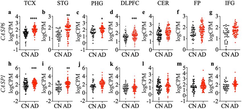
www.nature.com/scientificreports/
whose neurons project their axons into the ERC10. Therefore, Casp6 activity is an early spatial–temporal event
in AD pathogenesis.
Casp6 activity is associated with age-dependent cognitive impairment. In aged NCI individuals, high levels
of Casp6 correlate with global cognitive scores and predict lower episodic and semantic memory performance,
the first types of memories impaired in A D1,7,11. Casp6 activity in the anterior olfactory bulb is associated with
lower global cognitive scores, mini mental state exam (MMSE) scores, and episodic and semantic m emories10.
Cerebrospinal fluid Tau∆Casp6 levels reflect human brain Tau∆Casp6 levels, and correlate with AD severity,
and global cognitive, MMSE, episodic, semantic and working memory s cores12. Furthermore, expression of
self-activated human Casp6 in mouse CA1 neurons is sufficient to induce age-dependent episodic and spatial
memory impairment11.
Active Casp6 is associated with selective neuronal degeneration, but not necessarily cell death. Casp6 activa-
tion in HEK293T cells does not induce cell d eath13,14. Microinjection of recombinant active Casp6 in human
neurons in primary cultures induces a dose-dependent and protracted type of cell death15. Similarly, overexpres-
sion of wild type or mutant forms of amyloid precursor protein (APP) associated with familial AD induces Casp6-
dependent, but amyloid beta peptide (Aβ) independent, neurodegeneration in human n eurons16. This eventually
leads to neuronal cell death that is Aβ dependent. Nerve growth factor (NGF)-deprived mouse primary dorsal
root ganglia and sympathetic neuron cultures leads to compartmentalized Casp6-dependent axonal degeneration
and Caspase-3 (Casp3)-dependent cell d eath17–19. In AD brains, active Casp6-immunopositive neurons do not
show apoptotic features and active Casp3 immunoreactivity is s parse6,20,21. Furthermore, transgenic expression of
a self-activated human Casp6 in mouse brains causes hippocampal CA1 neurodegeneration, defined here as the
loss of neuronal structure or function, in the absence of significant neuronal loss11,22. Consistently, microinjected
active Casp6 causes neuronal degeneration and impairs synaptic transmission in CA1 pyramidal neurons, but
not in medium spiny n eurons22.
Casp6 cleaves many protein substrates such as α-Tubulin, Tau, α-Actinin-4, Drebrin, Glial fibrillary acidic
protein, Spinophilin, and Vimentin which are critical for neuronal structure and function and synaptic
plasticity2,5,23,24. Casp6-cleaved valosin containing protein p97 impairs ubiquitin proteasome system-mediated
protein degradation3. Ubiquitin ligases of mouse double minute 2 homolog, ubiquitin fusion degradation 2,
and developmentally down-regulated protein 4, proteins involved in cellular protein turnover are also Casp6
substrates25,26. Furthermore, Casp6 cleaves APP, Presenilin 1 and 2, and Huntingtin, proteins involved in neu-
rodegenerative diseases27–29. These neuronal substrates reflect Casp6 potential function in neurodegeneration.
Casp6 is activated by overexpression and proteolytic processing. Human Casp6 is expressed as an inactive
zymogen from the CASP6 gene30 and the level of Casp6 protein is relatively low in normal human brain31.
Prokaryotic and eukaryotic overexpression of Casp6 is sufficient for self-proteolytic a ctivation14,32,33. CASP6
transcriptional regulation has not been extensively investigated, but p53 is a CASP6 transcription regulator
in neurons and astrocytes34–36. p53 levels are enhanced in familial AD neurons37, indicating a mechanism for
increased CASP6 gene expression. The Casp6 active site consists of catalytic cysteine 163 (C163) surrounded by
four binding pockets for substrate selectivity38. Casp6 maturation requires three cleavage events at D23, D179,
and D193 to remove the pro-domain and the inter-subunit linker between a large subunit (LS) and a small
subunit (SS), so a small β-sheet structure 190TEVD193 in the inter-subunit linker occupying the active site is
removed and catalytic C163 is exposed33. Proteolytic processing allows the correct conformation of exosites and
allosteric sites important for the regulation of catalytic specificity and efficiency39,40. Casp6 can be processed and
activated by itself at high c oncentration14,32, by Casp3 in apoptotic c ells41, or by Caspase-1 in stressed primary
human neurons42,43. Interestingly, p53 also transcriptionally regulates CASP1 gene expression44, which can also
self-activate, indicating possible co-regulated expression of CASP1 and CASP6 genes.
Many genetic risk factors have been identified for AD, but no genetic association between CASP6 and AD
has been reported. Here, we used SKAT-O analysis45 to identify whether rare functional exonic CASP6 variants
associate with human brain volumes, determined by MRI scans in aged individuals. A rare variant, Casp6N73T,
associated with hippocampal subfield CA1 volumes in ADNI. Recombinant Casp6N73T exhibited altered pro-
teolysis of natural protein substrates compared to wild type Casp6 (Casp6WT), limited enzymatic activity and
stability in cell culture, and eliminated Casp6-mediated dysfunction and degeneration in hippocampal CA1
pyramidal neurons. These results suggest that Casp6N73T may provide a protective effect against hippocampal
atrophy compared to Casp6WT.
Results
Genetic association between the Caspase‑6 gene (CASP6) and hippocampal CA1 volume in
AD. From the Alzheimer’s Disease Neuroimaging Initiative (ADNI) whole genome sequencing (WGS) data
(n = 757), only one functional exonic missense SNP (rs141550898) with a minor allele frequency < 0.05 was iden-
tified within CASP6. We performed SKAT-O of rs141550898 with CA1 volumes with age, sex, year of education,
MRI field strength, and intracranial volume as covariates. One individual (male, early MCI, APOE ε3/ε3) was
heterozygous for this variant. The optimized sequence kernel association test (SKAT-O) yielded a significant
association of rs141550898 in CASP6 with hippocampal CA1 subfield volume (p = 0.0146). The rs141550898
SNP in which AAT is replaced by ACT causes a substitution of asparagine (N) for threonine (T) at amino acid 73
of Casp6, thus generating Casp6N73T. The participant carrying Casp6N73T has maintained its early MCI status
for at least 5 year based on his last visit, raising the possibility that Casp6N73T may slow down hippocampal
CA1 atrophy and AD progression.
Gene expression of CASP6 and CASP1. To confirm the implication of CASP1 and CASP6 in the brains
of individuals assessed in the ADNI cohort, CASP1 and CASP6 mRNA levels were evaluated. The number, age,
Scientific Reports | (2021) 11:12695 | https://doi.org/10.1038/s41598-021-91367-0 2
Vol:.(1234567890)www.nature.com/scientificreports/
Brain FP
region TCX (Mayo) PHG (MSBB) IFG (MSBB) STG (MSBB) (MSBB) DLPFC (ROSMAP) CER (Mayo)
Diagnosis CN AD CN AD CN AD CN AD CN AD CN AD CN AD
Number 71 80 16 78 18 102 21 98 22 111 86 155 72 79
Sex (F/M) 35/36 49/31 11/5 53/25 12/6 69/33 16/5 65/33 16/6 75/36 47/39 109/46 35/37 47/32
Age at
death (SD), 82.7 (8.5) 82.6 (7.7) 83.5 (8.9) 85.5 (6.1) 83.2 (8.6) 85.4 (6.0) 83.8 (8.1) 84.5 (6.7) 83.1 (7.7) 85.4 (5.9) 83.4 (5.9) 88.2 (3.1) 82.3 (8.3) 82.5 (7.7)
years
RIN (SD) 7.7 (1.0) 8.6 (0.6) 7.1 (0.9) 6.5 (0.9) 8.7 (1.4) 8.0 (1.8) 6.4 (1.0) 6.3 (0.9) 6.8 (0.9) 6.8 (0.9) 7.3 (1.0) 6.9 (0.9) 7.7 (1.0) 8.4 (0.7)
APOE
genotype 62/9 38/42 14/2 57/21 16/2 72/30 17/4 65/33 19/3 70/41 77/9 91/64 62/10 38/41
(ε4 + /ε4-)
Table 1. Demographic and clinical characteristics of cognitively normal (CN) and Alzheimer disease (AD)
participants for different brain region analysed. TCX temporal cortex, PHG para-hippocampal gyrus, IFG
inferior frontal gyrus, STG superior temporal gyrus, FP frontal pole, DLPFC dorsolateral prefrontal cortex,
CER cerebellum, RIN RNA integrity number.
Figure 1. Differential expression of CASP6 and CASP1 in Alzheimer disease (AD) and cognitively normal
(CN) brains. The logarithm of read counts per million total reads (logCPM) values for CASP6 mRNA (a–g)
and CASP1 mRNA (h–n) generated from RNA-Seq data for AD and CN temporal cortex (TCX; a,h), superior
temporal gyrus (STG; b,i), para-hippocampal gyrus (PHG; c,j), dorsolateral prefrontal cortex (DLPFC; d,k),
cerebellum (CER; e,l), frontal pole (FP; f,m) and inferior frontal gyrus (IFG; g,n). Each dot represents data from
one individual and the horizontal bar denotes the mean. Statistical evaluations were done with the R package
limma between AD and CN. *P < 0.05, **p < 0.01, ***P < 0.001, ****p < 0.0001.
sex, mean age at death, and apolipoprotein APO E genotype of the participants used for differential gene expres-
sion analysis in each brain region are summarized in Table 1. The RNA integrity number (RIN) where 10 repre-
sents intact RNA and 1 completely degraded RNA ranged from 6.3 to 8.7. Gene expression association analysis
using RNA-Seq data generated from temporal cortex (TCX), para-hippocampal gyrus (PHG), inferior frontal
gyrus (IFG), superior temporal gyrus (STG), frontal pole (FP), dorsolateral prefrontal cortex (DLPFC), and
cerebellum (CER) identified that CASP6 mRNA was up-regulated in the TCX (Fig. 1a), STG (Fig. 1b), PHG
(Fig. 1c), and DLPFC (Fig. 1d) of AD compared to cognitively normal older adults. Levels in the CER (Fig. 1e),
FP (Fig. 1f), and IFG (Fig. 1g) were not significantly different. Casp6 is activated by Caspase-1 (Casp1), there-
fore CASP1 mRNA levels were also evaluated. CASP1 mRNA levels were significantly up-regulated in AD TCX
(Fig. 1h) and STG (Fig. 1i), but not in PHG (Fig. 1j), DLPFC (Fig. 1k), CER (Fig. 1l), FP (Fig. 1m), or IFG
(Fig. 1n), compared to cognitively normal older adults.
Recombinant Caspase‑6 N73T (Casp6N73T) zymogen showed similar self‑processing as Cas‑
pase‑6 wild type (Casp6WT) zymogen. To observe the self-processing of Caspase-6 N73T (Casp6N73T)
zymogen, recombinant protein from three independent clones expressing Casp6N73T zymogen were compared
with Caspase-6 wild type (Casp6WT) zymogen and catalytically inactive triple mutant Casp6D(23,179,193)
A. The Casp6WT zymogen undergoes self-processing at D23, D179, and D193 to remove the pro-domain and
the inter-subunit linker, which allows the formation of active Casp6 consisting of two large subunits (LS) and
two small subunits (SS) (Fig. 2a). Purified recombinant Casp6N73T and Casp6WT zymogens both generated
the expected small subunit (SS) and large subunit with (LSL) or without (LS) inter-subunit linker. Casp6WT
Scientific Reports | (2021) 11:12695 | https://doi.org/10.1038/s41598-021-91367-0 3
Vol.:(0123456789)www.nature.com/scientificreports/
Figure 2. Self-processing of recombinant Casp6N73T zymogen. (a) Schematic diagram of recombinant human
Casp6 zymogen showing the subunits and epitopes recognized by antibodies. The epitope for the SC-81635
antibody is unknown but located in the large subunit (LS), neoepitope for 10630 antisera detects the C-terminus
of the LS of Casp6, and epitope for the Pharmingen antibody is G271-K285. Pro pro-domain, LS large subunit,
L linker, SS small subunit. (b) Coomassie blue stain of 2 µg purified recombinant Casp6WT and Casp6N73T
zymogens processed into LSL, LS and SS subunits. The unprocessed Casp6D(23,179,193)A was used as a control
for the FL form. LSL large subunit with the linker, FL full length. * indicates unspecific band. (c) Quantification
of the density of LSL, LS or SS normalized to density levels of Casp6WT from Coomassie blue stained gels.
(d,f,h) Western blot of Casp6WT, Casp6N73T, and Casp6D(23,179,193)A with SC-81635 antibody anti-LS (d),
with neoepitope antisera 10630 anti cleaved LS (f), and with Pharmingen antibody anti-SS (h). Quantification of
(e) LSL and LS from (d), (g) LS from (f), and (i) SS from (h). Data represents mean and s.e.m of 3 independent
experiments. No statistical differences were found by unpaired student’s test.
and Casp6N73T zymogens generated more LSL than LS (Fig. 2b) indicating preferred self-cleavage at D193, as
previously observed with Casp6WT14,33,46. The catalytically inactive Casp6D(23,179,193)A was not processed, as
expected, and migrated as full-length (FL) Casp6 at 34 kDa. The Casp6N73T zymogen generated LSL, LS, and SS
at levels comparable to those of the Casp6WT zymogen (Fig. 2b,c). FL, LSL, LS, and SS of Casp6 were confirmed
with western blotting against anti-FL and anti-LSL Casp6 antiserum (Fig. 2d,e), neoepitope antiserum recog-
nizing Casp6 cleaved at D179 (Fig. 2f,g), and anti-SS antibodies (Fig. 2h,i). No difference in the levels of each
subunit between Casp6WT and Casp6N73T was detected (Fig. 2e,g,i). These results indicate that Casp6N73T
zymogen self-processing is similar to that of Casp6WT zymogen.
Scientific Reports | (2021) 11:12695 | https://doi.org/10.1038/s41598-021-91367-0 4
Vol:.(1234567890)www.nature.com/scientificreports/
Figure 3. Casp6WT and Casp6N73T enzymatic processing of Ac-VEID-AFC. (a) VEIDase activity of
Casp6WT or Casp6N73T at 2, 10, 20, 50, 100, or 400 nM on 20 µM Ac-VEID-AFC. (b) Cleaved Ac-VEID-AFC
generated with time by Casp6WT or Casp6N73T at 2, 10, or 50 nM on 20 µM Ac-VEID-AFC. Data represents
mean and s.e.m. from 3 independent experiments. No statistical differences were found by two-way ANOVA (a)
or repeated measures two-way ANOVA (b). (c) VMAX, Km, kcat, and kcat/Km values for Casp6WT and Casp6N73T.
Data represents mean and SD from 3 independent experiments.
Recombinant Casp6N73T showed comparable VEIDase activity as Casp6WT on the tetrapep‑
tide Ac‑VEID‑AFC substrate. To determine whether Casp6N73T affects Casp6 catalysis, the catalytic effi-
ciency of Casp6N73T on fluorescent tetrapeptide Ac-VEID-AFC was compared to Casp6WT. Before the activity
assay, the number of active sites was established for each enzyme preparation by active site titration against de-
esterified irreversible pan-caspase inhibitor, zVAD-FMK, to ensure comparison of equal amounts of Casp6WT
and Casp6N73T active sites (Supplementary Fig. S1a-f). Two to four hundred nM active Casp6N73T exhibited
comparable VEIDase activity (defined as cleaved pmol AFC per minute) than similar concentrations of active
Casp6WT (Fig. 3a). The velocities of product formation by 2, 10, and 50 nM Casp6N73T with 20 µM Ac-VEID-
AFC substrate were similar to those of 2, 10, or 50 nM Casp6WT (Fig. 3b). Furthermore, to better understand
the kinetics of Casp6N73T, the Km, Vmax, kcat and kcat/Km were determined based on Michaelis–Menten kinet-
ics measurements (Supplementary Fig. S2). No differences were found in the Km, Vmax, kcat or kcat/Km between
Casp6N73T and Casp6WT (Fig. 3c).
Recombinant Casp6N73T showed increased proteolytic processing of natural Casp6 protein
substrate, Lamin A/C. To assess if Casp6N73T affects Casp6 activity on natural protein substrates, the
cleavage of Lamin A/C by Casp6N73T and Casp6WT was assessed. Lamin A/C was extracted from Casp6 KO
mouse tissue, and was incubated with varying concentrations of active site titrated Casp6N73T or Casp6WT.
Both Casp6N73T and Casp6WT processed Lamin A/C and generated the expected 28 kDa fragment (Fig. 4a)47.
The % FL Lamin A/C and cleaved Lamin A/C generated by Casp6N73T at concentrations from 50 to 500 nM
were similar to that of Casp6 WT after 1 h incubation (Fig. 4b). In order to calculate the initial reaction velocity,
cleaved Lamin A/C was measured every 5 min for the first 15 min of reaction, followed by every 15 min until
60 min. The amount of cleaved Lamin A/C increased with incubation time with both Casp6N73T and Casp6WT
(Fig. 4c). Initially, at the 5 and 10 min time points, 200–500 nM Casp6N73T consistently generated slightly
higher amounts of cleaved Lamin A/C than Casp6WT (Fig. 4d). The slope of the linear phase of the curve in
Fig. 4d determined the initial velocity of the enzymes on Lamin A/C. The initial velocity of Casp6N73T was sig-
nificantly higher than that of Casp6WT (Fig. 4e). These results suggest that the N73T substitution may modify
Casp6N73T interaction with Lamin A/C.
Recombinant Casp6N73T showed decreased catalytic efficiency on α‑Tubulin. To further
understand the effect of Casp6N73T on different protein substrates, the catalytic activity of Casp6N73T and
Casp6WT on α-Tubulin was compared. Both Casp6N73T and Casp6WT processed α-Tubulin and generated
a fragment 2 kDa smaller than the FL (Fig. 5a top panel), consistent with Casp6 cleavage at VGVD438 in α
-Tubulin. Cleavage of α-tubulin at D438 was confirmed with the neoepitope antibody GN20622 (Fig. 5a bot-
tom panel). The % Tub∆Casp6 by Casp6N73T at concentration 15.6–250 nM was significantly lower than that
of Casp6WT after 4 h incubation (Fig. 5b). Kinetically, 15.6–62.5 nM Casp6N73T consistently generated less
Tub∆Casp6 compared to Casp6WT during the first 4 h of incubation (Fig. 5c&d). Analysis of the initial velocity
determined by the slope of the linear phase of the cleavage curve in Fig. 5d, showed a significant 50% reduction
Scientific Reports | (2021) 11:12695 | https://doi.org/10.1038/s41598-021-91367-0 5
Vol.:(0123456789)www.nature.com/scientificreports/
Figure 4. Casp6WT and Casp6N73T enzymatic processing of Lamin A/C. (a) Western blot of full-length
(#2032 Cell Signaling Technology; top) or cleaved Lamin A/C at VEID (neoepitope antibody #2035 Cell
Signaling Technology; middle) and β-Actin (bottom) from 50–500 nM recombinant Casp6WT or Casp6N73T
incubated with Casp6 knockout mouse tissue nuclear extracts. (b) Quantification of full-length Lamin A/C
and cleaved Lamin A/C by Casp6WT or Casp6N73T expressed as % of the control with no added recombinant
active Casp6. (c) Western blots of time-dependent cleaved Lamin A/C by 200, 300, 400, or 500 nM Casp6WT
or Casp6N73T incubated with Casp6 knockout tissue extracts. (d) Quantification of time-dependent generated
cleaved Lamin A/C by Casp6WT or Casp6N73T shown in (c) expressed as % of the control with no added
recombinant active Casp6. Insets: The initial velocity phase of the cleavage curve. The linear regression was
performed on % cleaved Lamin A/C at 5, 10, and 15 min by 200 nM Casp6 (WT: R 2 = 0.82, N73T: R2 = 0.82), or
on % cleaved Lamin A/C at 5 and 10 min by Casp6 with 300 nM (WT: R2 = 0.88, N73T: R2 = 0.80), 400 nM (WT:
R2 = 0.71, N73T: R2 = 0.66), or 500 nM (WT: R 2 = 0.90, N73T: R2 = 0.90). (e) The initial velocity of Casp6WT and
Casp6N73T reacting on Lamin A/C measured from (d). Data were shown as mean ± s.e.m from 3 independent
experiments. Statistical evaluations were done with two-way ANOVA (variant: p = 0.0001, concentration:
p < 0.0001, interaction: p = 0.1329). Post-hoc analyses were done with Tukey’s test. ***p < 0.001 N73T vs WT.
Full-length images of blots/gels are presented in Supplementary Information.
Scientific Reports | (2021) 11:12695 | https://doi.org/10.1038/s41598-021-91367-0 6
Vol:.(1234567890)www.nature.com/scientificreports/
Figure 5. Casp6WT and Casp6N73T enzymatic processing of α-Tubulin. (a) Western blot of full-length
(11H10 Cell Signaling Technology; top) or cleaved α-Tubulin at VGVD438 (GN20622; bottom) in the reactions
of 15.6–250 nM recombinant Casp6WT or Casp6N73T with 242 nM of purified α- and β-Tubulin (121 nM of
α-Tubulin). Tub∆Casp6, α-Tubulin cleaved by Casp6. (b) Quantification of Tub∆Casp6 shown in (A) expressed
as % of FL Tubulin. Statistical evaluations were done with two-way ANOVA (variant: p = 0.0002, concentration:
p < 0.0001, interaction: p = 0.4555). Post-hoc analyses were done with Tukey’s test. ***p < 0.001 N73T vs WT. (c)
Western blot of time-dependent cleaved 121 nM α-Tubulin by 15.6, 31.2, or 62.5 nM Casp6WT or Casp6N73T.
(d) Quantification of Tub∆Casp6 shown in (c) expressed as % of FL Tubulin. Insets: The initial velocity phase of
the cleavage curve. The linear regression was performed on % Tub∆Casp6 at 1, 2, 3, and 4 h by 15.6 nM Casp6
(WT: R2 = 1.00, N73T: R2 = 0.99), on % Tub∆Casp6 at 1, 2, and 3 h by Casp6WT with 31.2 nM ( R2 = 0.99) or
62.5 nM Casp6WT ( R2 = 1.00), or on % Tub∆Casp6 at 1 and 2 h by Casp6N73T with 31.2 nM (R2 = 0.99) or
62.5 nM (R2 = 1.00). (e) Initial velocity of Casp6WT and Casp6N73T reacting on α-Tubulin measured from
(d). Statistical evaluations were done with two-way ANOVA (variant: p < 0.0001, concentration: p < 0.0001,
interaction: p = 0.0015). Post-hoc analyses were done with Tukey’s test. ****p < 0.0001 N73T vs WT. Data
represents mean ± s.e.m. from 3 independent experiments. Full-length images of blots/gels are presented in
Supplementary Information.
Scientific Reports | (2021) 11:12695 | https://doi.org/10.1038/s41598-021-91367-0 7
Vol.:(0123456789)www.nature.com/scientificreports/
Figure 6. Steady levels of Casp6N73T and Casp6WT proteins in transfected HEK293T cells. (a) RedSafe-
stained agarose gel of human Casp6 and Hprt1 mRNA amplicons in non-transfected (NT), mock, pCEP4β,
pCEP4β-CASP6C163A-His, pCEP4β-CASP6WT∆Pro-His, or pCEP4β-CASP6N73T∆Pro-His-transfected
cells. (b) Quantification of Casp6 mRNA levels normalized to Hprt1 shown in (a). (c,e) Western blot and
(d,f) quantification of Casp6 in transfected cells against Pharmingen (c,d) or 10630 neoepitope antibodies
(e,f). ReWT recombinant Casp6WT, ReC163A recombinant Casp6C163A, FL full length, ∆Pro Casp6 without
pro-domain, SS small subunit, LS large subunit. Statistical evaluations were done with one-way ANOVA (d,f:
p < 0.0001) followed by post-hoc Tukey’s test (**p < 0.01, ****p < 0.0001). (g) VEIDase activity in protein extract
from transfected cells. Statistical evaluations were done with one-way ANOVA (p < 0.0001) followed by post-
hoc Tukey’s test. ****p < 0.0001 WT vs NT. (h) Western blot and (i) quantification of Casp6 in transfected cells
with the Pharmingen antibody. The levels of ∆ProCasp6 normalized to the 0 h were fit to a one-phase decay to
calculate the decay rate of Casp6WT or Casp6N73T. * indicates p = 0.012 done with unpaired t-test. Data shown
represents mean ± s.e.m. from 3 independent experiments. Full-length images of blots/gels are presented in
Supplementary Information.
of Casp6N73T catalytic efficiency (Fig. 5e). These results show that Casp6N73T catalytic efficiency on α-Tubulin
is lower than that of Casp6WT. As observed with the Lamin A/C substrate, the results suggest that Casp6N73T
may interact differently than Casp6WT with α-Tubulin.
The steady state level of active Casp6N73T subunits is considerably lower than those of
Casp6WT in transfected human embryonic kidney 293 T (HEK293T) cells. To assess if eukary-
otically expressed Casp6N73T behaves like the prokaryotically expressed recombinant Casp6N73T, CASP6WT
and CASP6N73T cDNAs lacking their pro-domain (∆Pro) to promote self-processing of Casp6 were cloned in
pCep4β and the constructs were transfected in human embryonic kidney 293 T (HEK293T) cells. Catalytically
inactive, CASP6C163A with its pro-domain sequence, was transfected as a control for full length Casp6. Casp6
mRNA levels were equivalent in CASP6WT, CASP6N73T, or CASP6C163A cDNA transfected cells (Fig. 6a,b). In
transfected cells, the ∆ProCasp6 expressed in CASP6WT- and CASP6N73T-transfected cells migrated approxi-
mately 2 kDa below the full length Casp6 in CASP6C163A-transfected cells (Fig. 6c). Unexpectedly, CASP6N73T-
transfected HEK293T cells contained consistently lower levels of ∆Pro-Casp6 than CASP6WT-transfected cells
(Fig. 6c,d). Compared to CASP6C163A-transfected cells, ∆ProCasp6 levels were lower in CASP6WT-transfected
cells, as expected since Casp6WT can be processed into its subunits (Fig. 6d). The levels of LS in CASP6N73T-
transfected cells were significantly lower than those of CASP6WT-transfected cells (Fig. 6e,f). Consistently,
CASP6N73T-transfected HEK293T protein extracts did not show significant Casp6 VEIDase activity compared
to non-, mock-, or empty vector-transfected cell extracts, while CASP6WT-transfected cells exhibited high VEI-
Dase activity (Fig. 6g). Because transfected cells contained similar Casp6WT and Casp6N73T mRNA levels,
decreased Casp6N73T full length protein level and VEIDase activity may be the result by enhanced turnover.
Indeed, in the presence of translational inhibitor cycloheximide (CHD), the degradation rate of ∆ProCasp6N73T
Scientific Reports | (2021) 11:12695 | https://doi.org/10.1038/s41598-021-91367-0 8
Vol:.(1234567890)www.nature.com/scientificreports/
was faster than that of ∆ProCasp6WT levels (Fig. 6h,i), suggesting a higher cellular turnover of Casp6 due to
the N73T substitution. Since Casp6 LS has previously been reported to be degraded by the proteasome13,48, we
verified if proteasomal activity may be responsible for FL or LS Casp6N73T lower levels. The epoxomicin pro-
teasomal inhibitor did not significantly alter levels of either Casp6N73T FL or LS in HEK293T cells, excluding
proteasomal degradation as an explanation (Supplementary Fig. S3). Nevertheless, these results indicate that
Casp6N73T is unstable and degraded by an alternate non-proteasomal dependent mechanism, thereby limiting
the amount of Casp6N73T LS and Casp6N73T activity produced in mammalian cells.
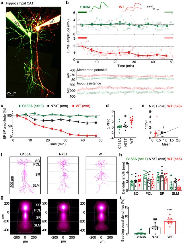
Recombinant Casp6N73T caused less neurofunctional damage and neuronal degeneration
than Casp6WT in hippocampal CA1 pyramidal neurons. The effect of Casp6N73T on neuronal func-
tion was compared with recombinant Casp6WT and catalytically inactive Casp6C163A proteins by patching
CA1 pyramidal neurons in hippocampal organ slices with these proteins (Fig. 7a) and measuring the amplitude
of excitatory postsynaptic potential (EPSP) (Fig. 7b). EPSP amplitudes from recorded neurons were analyzed
when the membrane potential and input resistance were stable (Fig. 7b bottom panel). Activated Casp6WT and
Casp6N73T were stable for 5 h after their preparation in internal solution (Supplementary Fig. S4), within which
the recording data was acquired. Ten pg Casp6WT induced a decrease of EPSP amplitude 30 min after patch-
ing, and a continually decreasing EPSP amplitude during the 50 min of recording (Fig. 7c). In contrast 10 pg
catalytically inactive Casp6C163A did not alter EPSP amplitude within 50 min. Interestingly, 10 pg Casp6N73T
decreased the EPSP amplitude less than Casp6WT in pyramidal neurons for approximately 10 min after patching,
and EPSP amplitude was maintained at 65% of the original levels for 50 min (Fig. 7c). Paired pulse ratio (∆PPR)
of the first and second EPSP wave increased in Casp6WT patched neurons compared to Casp6C163A, suggest-
ing that the decaying EPSP amplitude was at least in part due to a reduced probability of release presynaptically49.
In contrast, Casp6N73T ∆PPR was indistinguishable from that of Casp6C163A (Fig. 7d), possibly hinting at
an absence of perceivable effects on presynaptic release. Although the change in PPR due to Casp6WT suggested
PSP50 indicated a predomi-
a presynaptic locus of action, the coefficient of variation (CV) analysis of the first E
nantly postsynaptic effect, although with variable outcome (Fig. 7e). Taken together, the combined outcome of
PPR and CV analyses is consistent with a coordinated downregulation on both pre- and post-synaptic sides due
to Casp6WT.
The morphology of Casp6WT, Casp6N73T, or Casp6C163A patched CA1 pyramidal neurons was analyzed
by manual 3D reconstruction (Fig. 7f). Dendritic map density and convex hulls indicated the average distribu-
tion and the maximum extent of dendrites, respectively (Fig. 7g). Neurons patched with Casp6WT exhibited
a lower length of apical dendrites in the SLM region compared to Casp6C163A-patched neurons, but the data
did not reach statistical significance (Fig. 7h). The apical dendrites length of Casp6N73T-patched neurons was
longer than those of Casp6WT-patched neurons. In addition, Casp6WT-patched neurons displayed beading
along basal neurites in the stratum oriens region indicative of neuronal degeneration (Fig. 7i), while beading
was rarely seen in neurons patched with Casp6C163A, indicating that this beading is linked to Casp6 activity.
The beading dendrites of Casp6N73T-patched neurons was approximately 50% lower than in neurons patched
with Casp6WT. These results indicate that Casp6N73T is less detrimental to neuron function and causes less
neurodegeneration than Casp6WT.
Discussion
Our study demonstrates that a rare human CASP6 variant encoding Casp6N73T, genetically associated with
preserved hippocampal CA1 volume in an AD cohort, exhibits altered catalyses on natural protein substrates,
lamin A and α-Tubulin. In addition, our study showed that Casp6N73T has less negative impact on neuronal
function and neurodegeneration than the Casp6WT in mice CA1 hippocampal pyramidal neurons. These results
provide initial evidence for a neuroprotective effect by Casp6N73T as reflected by hippocampal subfield volume
preservation but this needs to be validated in independent data sets.
The significant association between Casp6N73T and hippocampal CA1 volume by SKAT-O provides an
advantage in identifying associations between low frequency functional exonic SNPs and pathological features
of AD compared to genome-wide association studies (GWAS)45,51. Different from the single regression model
used in GWASs, SKAT uses a multiple regression model to regress the phenotypes of different SNPs in the same
genomic region and allows different orientations of the phenotype, in order to assess the cumulative effects of
one gene on disease. Another advantage of our approach is the use of a quantitative measure of hippocampal CA1
volume, instead of the numerous variables of clinically diagnosed AD. Hippocampal CA1 volume was chosen
rogression52,53. Large scale longitudinal measures indicate that
because atrophy in this region occurs early in AD p
hippocampal CA1 atrophy is a robust MRI biomarker to distinguish MCI that further develop to AD from stable
MCI54. Furthermore, rare SNPs also mean lower number of individuals carrying the SNP, therefore, our finding
will require testing additional cases to confirm the role of Casp6N73T in MCI and AD. On the other hand, the
ADNI study provides precise cognitive and neuroimaging information on the individual carrying CASP6N73T.
The Casp6N73T variant was identified in a heterozygous individual at the early MCI stage with unexpected
preserved hippocampal CA1 volume compared to those carrying the wild type CASP6.
Here, Casp6N73T was shown to be less damaging to synaptic transmission and plasticity than Casp6WT.
In contrast to catalytically inactive Casp6C163A, Casp6WT rapidly decreased induced EPSP amplitude in CA1
hippocampal neurons, whereas Casp6N73T showed only a minor decrease. In addition, the paired pulse ratio
was increased in Casp6WT, but not in Casp6N73T. Furthermore, Casp6WT, but not Casp6C163A, induced
basal dendritic beading indicating Casp6 activity-mediated neurodegeneration CA1 neurons, a feature that
was reduced with Casp6N73T. Based on the morphological analysis of reconstructed neurons, Casp6WT, but
not Casp6N73T, caused a decrease in the total apical dendritic length. These data are consistent with transgenic
Scientific Reports | (2021) 11:12695 | https://doi.org/10.1038/s41598-021-91367-0 9
Vol.:(0123456789)www.nature.com/scientificreports/
Figure 7. Casp6N73T is less damaging to neuronal function and neurodegeneration than Casp6WT. (a) Representative two-photon images
of 10 pg Casp6C163A- (Alexa 488, green) and 10 pg Casp6WT- or Casp6N73T- (Alexa 594, red) patched hippocampal CA1 pyramidal
neurons. White arrows indicate basal dendrite degeneration. Scale bar: 25 µm. Maximum-intensity projection of two-photon stacks was
compiled using ImageJ, and imaging montage of entire neurons was performed using Affinity Designer 1.7. (b) Sample EPSP time course
plots from Casp6C163A- (green) and Casp6WT- (red) patched neurons in (a), showing reduction of neurotransmission for Casp6WT
(1.53 ± 0.32 mV, n = 8 vs. 0.14 ± 0.07 mV, n = 8, p < 0.01) but not for Casp6C163A (1.94 ± 0.32 mV, n = 10 vs. 1.91 ± 0.29 mV, n = 10, p = 0.76)
when comparing the last 10 traces (light thick line) to the first 10 traces (dark thick line). Open circles: EPSP amplitude recorded every 30 s.
Closed circles: EPSP amplitude binned and averaged across 10 traces. Inset: representative EPSP traces highlight the paired-pulse ratio (PPR).
Scale bars: 2 mV, 25 ms. Resting membrane potential and input resistance remained stable throughout experiment. (c) EPSP time courses for
Casp6C163A- (n = 10), Casp6N73T- (n = 8), or Casp6WT- (n = 8) patched neurons. (d) PPR from Casp6WT-, Casp6C163A- or Casp6N73T-
patched neurons. One-way ANOVA (p = 0.0043), followed by Tukey’s post-hoc test (**p < 0.01 vs. C163A; #p < 0.05 vs. WT). (e) CV
analysis of Casp6N73T- and Casp6WT-patched neurons. Casp6C163A was unaltered. (f) Representative reconstructions of Casp6C163A-,
Casp6N73T-, or Casp6WT-patched neurons. Image stacks were used for manual reconstruction of 3D morphologies using the Neuromantic
freeware (http://www.reading.ac.uk/neuromantic/body_index.php). (g) Dendritic density maps of Casp6C163A- (n = 11), Casp6N73T-
(n = 8), or Casp6WT- (n = 8) patched neurons generated using custom software running in Igor Pro 8 v8.04 (https://www.wavemetrics.com,
https://github.com/pj-sjostrom/qMorph). Dotted lines show the convex hull of the maximum extent. (h) Cumulative dendritic length of
reconstructed neurons in layers SO, PCL, SR, and SLM. (i) Casp6C163A-, Casp6N73T- and Casp6WT-patched CA1 pyramidal neurons
beading basal dendrites. One-way ANOVA (p = 0.0001), followed by Tukey’s test (****p < 0.0001 vs Casp6C163A, ##p < 0.01 vs Casp6WT).
Scientific Reports | (2021) 11:12695 | https://doi.org/10.1038/s41598-021-91367-0 10
Vol:.(1234567890)www.nature.com/scientificreports/
expression of a self-activated form of Casp6WT in CA1 neurons causing age-dependent cognitive impairment11,
small amounts of active Casp6 causing a rapid depression of CA1 neuronal t ransmission22 and impairing long-
term potentiation in hippocampal CA1 circuits in vivo55. Given that the presence of active Casp6 detected in
the entorhinal cortex of aged individuals correlates with decreased cognitive p erformance1,7,12,42 and that early
56,57
cognitive decline is strongly correlated with synaptic loss , these data raise the possibility that the active
Casp6 observed in neuropil threads, neuritic plaques and neurofibrillary tangles4,5 causes synaptic transmission
problems. Most importantly, the data suggest that Casp6N73T variant would not be as detrimental as Casp6WT
for neuronal structure and function and this may protect aged individuals from cognitive decline. In support
of this conclusion, Casp6 deficient mice have been shown to be protected against excitotoxicity, nerve growth
factor deprivation and myelin-induced axonal degeneration, and showed an age-dependent increase in cortical
and striatal v olume58.
The molecular reason for the protective action of Casp6N73T might stem from its altered proteolysis of natu-
ral protein substrates compared to Casp6WT. Casp6 can cleave numerous protein substrates, some of which are
specific to neurons and s ynapses2,59 and involved in neurodegenerative diseases, such as Tau, Vimentin, Drebrin,
Spinophilin, α-Actinin2,5, valosin containing protein p 973, APP60, Presenilin 1 and 2 28, and H
untingtin61, that
may be responsible for structural damage, synaptic loss, and protein aggregation. Here, we show that compared
to Casp6WT, Casp6N73T had increased proteolytic processing of Lamin A/C, decreased proteolysis of α-tubulin
and equivalent processing of the Ac-VEID-AFC peptide substrate. These results indicate that either the N73T
alters the structure of the Casp6 enzyme thereby affecting the catalytic site, or the N73T alters the interaction of
Casp6 with substrates. The altered catalysis of natural protein substrates α-Tubulin and Lamin A/C, but not of
the small peptide substrate, suggests that the active site of the Casp6N73T is relatively normal. Therefore, it is
likely that Casp6N73T interacts differently with α-Tubulin and Lamin A/C than Casp6WT, thus suggesting the
presence of an exosite either within N73, its surroundings, or in a structure that is altered by N73T. Exosites are
defined as sites outside the substrate-binding catalytic site which influence structure or function of an enzyme.
The N73T substitution is located in the middle of Casp6’s helix B with a shorter side chain pointing outward.
The nearby negatively charged D72 is conserved among Casp6 orthologues and unique to Casp6 among the
caspase protein family, which may also contribute to this exosite. Casp6 exosite 42RRR44, located at the hinge
between the core structure and the N-terminus of the large subunit, has been confirmed62,63. Furthermore, two
rare human variants, G66R and R65W completely eliminate or significantly reduce Casp6 activity through
impaired substrate binding, alter the catalytic site activity, and have dominant negative effects on Casp6 W T46.
The alternatively spliced Casp6b isoform lacking amino acids 13–104 while retaining the catalytic site also has
no activity, and inhibits Casp6WT a ctivation64. The N73T data presented here further highlights the importance
of the N-terminus of the large subunit of Casp6 in substrate recognition. Casp6N73T may change the proteoly-
sis of different neuronal substrates in addition to α-Tubulin, which together display a reduced damage due to
Casp6 and favors a neuroprotective phenotype. Future investigation in characterizing the cleavage efficiencies
of Casp6N73T on these substrates could expand our understanding of the new exosite as well as the protective
mechanism of Casp6N73T. The identification of an additional exosite for Casp6 could be useful in specific drug
design against Casp6 activity, which has been shown to cause age-dependent cognitive impairment in m ice11.
The protective action of Casp6N73T might also stem from its instability in mammalian cells. Casp6N73T full
length levels were decreased significantly relative to Casp6WT, despite similar mRNA levels for Casp6N73T and
Casp6WT. This lower level of full length Casp6N73T could be explained by either a lower mRNA translational
rate, increased protein processing, or increased degradation by alternate cellular proteolytic activities. A more
rapid turnover for full length Casp6N73T compared to FL Casp6WT was observed indicating either increased
protein processing into its active subunits or increased degradation by alternate cellular proteolytic activities. The
fact that the level of Casp6N73T processed large subunit (LS) was reduced almost tenfold relative to Casp6WT
indicated that Casp6N73T was processed less efficiently than Casp6WT or that the Casp6N73T LS was degraded
more rapidly than the Casp6WT LS. Proteasomal activity was excluded since proteasomal inhibition did not
significantly alter either FL or LS Casp6N73T levels. The fact that self-processing of prokaryotically expressed
recombinant Casp6N73T is equivalent to that of Casp6WT eliminates the possibility that the Casp6N73T muta-
tion alters processing. Therefore, the most likely explanation for these findings is that the FL Casp6N73T is
unstable and degraded by a non-proteasomal cellular mechanism, thereby limiting the amount of Casp6N73T
LS and Casp6N73T activity produced in mammalian cells.
Lastly, we confirm here that CASP1 and CASP6 mRNA levels are significantly increased in specific regions
of the AD brains in the ADNI cohort compared to cognitively normal control brains from large sample sizes
in the AMP-AD Consortium. We investigated CASP1 expression because it activates Casp6 in human primary
neurons42. Increased CASP1 mRNA levels have been reported previously in AD cortex and entorhinal cortex
(ERC)65,66. Our results additionally show increased CASP1 mRNA levels in AD temporal cortex and superior tem-
poral gyrus. CASP6 expression in AD is more controversial. One early study reports low level of CASP6 mRNAs
in AD b rains65. Others indicate increased CASP6 mRNA levels in cortex and cerebellum of AD brains43,66. Here,
we show increased CASP6 mRNA levels in AD temporal cortex, superior temporal gyrus, para-hippocampal
gyrus, and dorsolateral prefrontal cortex. These results support the implication of CASP6 in AD.
This study highlights the importance of assessing the role of the amazing genetic diversity of humans in
disease by combining human genetic information associated with well-ascertained neuroimaging and cognitive
measures with biochemical and electrophysiological approaches to investigate the potential influence of rare
variants on AD-related pathologies and cognition.
Scientific Reports | (2021) 11:12695 | https://doi.org/10.1038/s41598-021-91367-0 11
Vol.:(0123456789)www.nature.com/scientificreports/
Methods
Alzheimer’s Disease Neuroimaging Initiative (ADNI). Individuals used in the analysis were ADNI
participants o67,68. Inclusion and exclusion criteria, clinical and neuroimaging protocols, and other information
about ADNI can be found at www.adni-info.org and http://www.loni.usc.edu/ADNI/. Written informed con-
sent was obtained at the time of enrollment for imaging and genetic sample collection and protocols of consent
forms were approved by each participating sites’ Institutional Review Board. Human subject ethical approval
was obtained by ADNI and can be found at http://www.loni.usc.edu/ADNI/. All methods and experiments were
performed in accordance with relevant guidelines.
Whole genome sequencing (WGS) analysis. WGS on the Illumina HiSeq2000 platform with paired-
end reads was performed on blood-derived genomic DNA samples obtained from 817 ADNI p articipants69.
Briefly, short-read sequences were mapped to the human genome assembly (GRCh build 37.72) using BWA70.
During the alignment, we use only bases with Phred Quality > 15 in each read to include soft clipping of low-
quality bases, retain only uniquely mapped pair-end reads, and remove potential PCR duplicates. After complet-
ing initial alignment, the alignment was further refined by locally realigning any suspicious reads. The reported
base calling quality scores obtained from the sequencer were re-calibrated to account for covariates of base
errors. All variants with statistical evidence for an alternate allele present among individuals were identified
using GATK HaplotypeCaller for multi-sample variant callings.
Neuroimaging analysis. Baseline T1-weighted brain MRI scans were downloaded from the ADNI data-
base. FreeSurfer software was used to process T1-weighted brain MRI scans71 and extract region of interest
(ROI)-based imaging phenotypes72,73.
Gene‑based association analysis. Since population stratification is known to cause spurious associa-
tion in disease studies, we restricted our analyses to only subjects that clustered with CEU (Utah residents with
Northern and Western European ancestry from the CEPH collection) + TSI (Tuscany in Italy) populations using
HapMap 3 genotype data and the multidimensional scaling analysis (www.hapmap.org)74–76. A total of 757
ADNI participants (259 CN, 219 early MCI, 232 late MCI, and 47 AD) were used for analysis, where late and
early MCI were defined as the cognitive performance below 1.5 standard deviations of the normative mean on
a standard test and at the range of 1 to 1.5 standard deviations, r espectively77. After extracting WGS-identified
functional exonic SNPs within CASP6, we performed a gene-based association analysis of rare variants (minor
allele frequency < 0.05) using SKAT-O software78. For hippocampal CA1 volumes, age, sex, year of education,
MRI field strength, and total intracranial volume were used as covariates.
RNA‑Seq analysis. RNA-Seq data (n = 1,966 individuals for the seven brain regions) reprocessed and rea-
ligned in the AMP-AD Consortium using a RNA-Seq pipeline79, were downloaded from the Sage Bionetworks
(www.synapse.org). Human postmortem brain RNA-Seq data were obtained in three independent studies (Reli-
gious Orders Study and the Memory and Aging Project (ROS/MAP), Mount Sinai School of Medicine (MSSM),
and Mayo Clinic) from seven distinct brain regions. Differential gene expression of CASP6 and CASP1 between
AD and cognitively normal controls were analysed with R package limma80.
Mutagenesis and sub‑cloning of Casp6N73T. The pET23b( +)-Casp6WT-His plasmid (Addgene
#11,823 https://www.addgene.org/11823/, gift from Dr. Guy Salvesen, Burnham Institute, La Jolla, CA,
USA) encodes a human Casp6WT zymogen fused with a C-terminal 6 × His tag. The mammalian pCEP4β-
Casp6∆ProWT-His plasmid encodes a human Casp6WT lacking its pro-domain14. Both plasmids were mutated
using QuikChange Site-Directed Mutagenesis Kit (Stratagene, San Diego, CA, USA) with Casp6N73T primers
5’- GCG CAG ATA GAG ACA CTC TTA CCC GCA GG -3’ and 5’- CCT GCG GGT AAG AGT GTC TCT ATC
TGC GC -3’ to introduce the mutation for the N73T substitution. All constructs were verified by sequencing at
McGill University Genome Quebec Innovation Center.
Protein expression and purification. The pET23b( +)-Casp6WT-His or pET23b( +)-Casp6N73T-His
plasmids were expressed in BL21(DE3)pLysS competent cells (Promega, Madison, WI, USA). Casp6 was puri-
fied using Ni Sepharose Fast Flow 6 (GE Healthcare, Chicago, IL, USA) and Macro Prep High Q Resin (Bio-Rad
Laboratories, Hercules, CA, USA) as described32,46. Pure protein was aliquoted, fast frozen in EtOH/dry ice bath,
and stored at − 80 °C freezer.
Active site titration. Casp6 active site concentration was determined using an irreversible inhibitor
N-benzyloxycarbonyl-Val-Ala-Asp-(O-methyl)-fluoromethylketone (zVAD-FMK; MP Biomedicals, Irvine, CA,
USA) as described46.
Caspase activity assays on Ac‑VEID‑AFC. The release of AFC from 20 µM Ac-VEID-AFC by active
escribed46. The slope of the linear phase of
site titrated Casp6 (2, 10, 20, 50, 100, or 400 nM) was measured as d
cleaved AFC plotted against time was calculated as VEIDase activity.
Determination of Km and kcat. Release of AFC from 1–200 µM Ac-VEID-AFC by 20 nM Casp6 at 37 °C
escribed46. VEIDase activity versus Ac-VEID-AFC concentration was fit-
in Stennicke buffer was measured as d
ted to a Michaelis–Menten equation v = (Vmax × [S])/(Km + [S]) using Prism 7 software (GraphPad Software, CA,
Scientific Reports | (2021) 11:12695 | https://doi.org/10.1038/s41598-021-91367-0 12
Vol:.(1234567890)www.nature.com/scientificreports/
USA) and maximal velocity Vmax, Michaelis–Menten constant Km , and kcat = Vmax/[active site concentration of
Casp6] were calculated.
Casp6 activity assay on protein substrates. To assess Lamin A/C cleavage, nuclear proteins were
extracted from Casp6 knockout mice colon tissue81 as described46. Casp6 (50–500 nM) were incubated with 3 µg
lysate in Stennicke buffer at 37 °C for 5–60 min.
To assess α-Tubulin cleavage, purified porcine Tubulin (Cytoskeleton, Inc., Denver, CO, USA) was used.
Tubulin powder was reconstituted in General Tubulin Buffer (80 mM PIPES pH 6.9, 2 mM M gCl2, 0.5 mM
EGTA) supplemented with 10 mg/mL GTP. Casp6 (15.6–250 nM) was incubated with 252 nM Tubulin in Sten-
nicke buffer at 37 °C for 1–12 h.
After the incubation period, samples were prepared in Laemmli buffer and analyzed by Western blot. The
initial velocity of Casp6 on Lamin A/C or α-Tubulin was calculated based on the linear portion of the cleavage
curve. The number of data points in the linear portion of each cleavage curve used for linear regression depended
on the goodness-of-fit when R 2 was closest to 1.00.
Western blots. Fifty ng recombinant Casp6, 6 µg nuclear or 20 µg HEK293T protein extracts, 0.05–0.2 µg
Tubulin, were submitted to western blot analyses with 1:1000 mouse anti-Casp6 against human Casp6 amino
acids 24–293 (SC-81653, Santa Cruz Biotechnology, Dallas, TX, USA), 1:10,000 rabbit neoepitope 10630
antiserum5, or 1:250 mouse anti-Casp6 small subunit (BD Pharmingen clone B93-4, BD Biosciences, San Jose,
CA, USA), 1:1000 anti-Lamin A/C (Cell Signaling Technology #2032, Danvers, MA, USA), 1;1000 anti-cleaved
Lamin A/C at VEID230 (Cell Signaling Technology #2035), 1;1000 anti- α-Tubulin (Cell Signaling Technology
#11H10), 1:5000 anti-cleaved α-Tubulin at VGVD43814, or 1:5000 anti-β-Actin ( Sigma-Aldrich Co # A5441,
Oakville, ON, Canada). Immunoreactivity was detected with 1:5000 HRP-conjugated anti-rabbit (DAKO P0217,
Agilent Technologies Burlington, ON, Canada) or anti-mouse (Jackson ImmunoResearch Labs #133499, West
Grove, PA, USA) secondary antibody followed by ECL (GE Healthcare), or with alkaline phosphatase-conju-
gated anti-mouse secondary antibody (1:5000; Jackson) followed by NBT/BCIP (Promega). Immunoreactive
bands were scanned, and densitometry was performed using ImageJ software (NIH, Bethesda, MD, USA) by
measuring band intensity values beyond the background.
Transfection, treatments, and protein extraction of HEK293T cells. The transfections were car-
ried out with Lipofectamine 2000 (Invitrogen, Burlington, ON, Canada) and proteins extracted after 24 h in
escribed14. Transfection efficiency of Lipo-
cell lysis buffer for Casp6 activity or in RIPA for western blots as d
fectamine 2000 was optimized to obtain more than 90% transfected cells by calculating the number of fluores-
cent cells transfected with pBud-EGFP plasmid (Addgene #23027) over the number of total cells stained with
Hoechst (Thermo Fisher Scientific). For protein degradation analysis, cells were treated 24 h after transfection
with 75 µg/ml cycloheximide (CHD, Sigma-Aldrich) for the indicated times.
Treatment of transfected HEK293T cells with epoxomicin. Cells were treated with 50 nM of epox-
omicin (Enzo) 24 h after transfection for the indicated times and proteins extracted in cell lysis buffer. Proteaso-
mal inhibition was confirmed in a proteasome activity assay as reported p reviously48 using 50 µM Succinyl-Leu-
Leu-Val-Tyr-7-amino-4-methylcoumarin (Suc-LLVY-AMC; Enzo).
RT‑PCR. Total RNA was extracted using TRIzol (Invitrogen) and converted to cDNA with avian myeloblas-
tosis reverse transcriptase (Roche Diagnostics, Laval, QC, Canada). Casp6 cDNA was amplified with Taq DNA
polymerase (New England Biolabs, Ipswich, MA, USA) as described46.
Electrophysiology. All animal experimentation was approved by the McGill University Animal Care Com-
mittee and performed under guidelines and regulations in accordance with the ARRIVE guidelines. Hippocam-
pal slices (300 µm) from 11–16 days old C57BL/6 mice were prepared in ice-cold A CSF22.
Recombinant Casp6 were activated in Stennicke buffer at 37 °C for 15 min, and diluted to 10 pg/10 µL in
internal solution82 with 30–60 µM Alexa 594 or 40–80 µM Alexa 488 (Invitrogen), filtered through 0.22 µm
hydrophilic polyethersulfone filters (Millipore Sigma, Oakville, ON, Canada), and osmolarity double checked
to be ~ 310 mOsm.
Multiple whole-cell patches were performed as d escribed82. The stimulating electrode was placed in the CA1
stratum orients and delivered five 0.1-µs-long biphasic pulses (10–50 V) at 30 Hz every 30 s to elicit EPSPs in
the range 1–3 mV. The recording was done for 1 h unless the data failed to meet the stability criteria defined as
potential (± 4 mV), input resistance (± 15%) and temperature (32–34 °C). Offline data were analysed with Igor
Pro (WaveMetrics Inc.). EPSP peak amplitudes were measured and averaged every 2.5 min (5 traces). Ensemble
time courses were normalized to the first 2.5 min EPSP.
Two‑photon imaging and neuron reconstruction. Two-photon excitation was achieved using a Cha-
meleon Ultra II femtosecond laser (Coherent, Santa Clara, CA, USA) tuned to 780 nm for both Alexa 594 and
488. Two-photon microscopes were custom-built83. Imaging data were acquired using customized versions of
ScanImage 2018 (Vidrio Technologies, Leesburg VA USA)84 running in Matlab (The MathWorks, Natick, MA,
USA) via a PCI-6110 or a PCIe-6374 data acquisition board (National Instruments, Austin, TX, USA). When
neurons had been well loaded with dye (> 1 h after break-in), the neuronal morphology was acquired as stacks of
512-by-512-pixels slices at 2 ms/line spaced by 2 µm. Maximum-intensity two-photon-imaging stacks compiled
Scientific Reports | (2021) 11:12695 | https://doi.org/10.1038/s41598-021-91367-0 13
Vol.:(0123456789)You can also read