JBC Papers in Press. Published on May 16, 2019 as Manuscript RA118.006074 The latest version is
←
→
Page content transcription
If your browser does not render page correctly, please read the page content below
JBC Papers in Press. Published on May 16, 2019 as Manuscript RA118.006074
        The latest version is at http://www.jbc.org/cgi/doi/10.1074/jbc.RA118.006074

              Extensive metabolic remodeling after limiting mitochondrial lipid burden
                       is consistent with an improved metabolic health profile.

Sujoy Ghosh1,2, Shawna E. Wicks3,4, Bolormaa Vandanmagsar4, Tamra M. Mendoza4, David S. Bayless4,
J. Michael Salbaum5, Stephen P. Dearth6, Shawn R. Campagna6, Randall L. Mynatt4,7, and Robert C.
Noland8
1
 Laboratory of Computational Biology, Pennington Biomedical Research Center, Baton Rouge, LA,
70808; 2Program in Cardiovascular and Metabolic Disorders and Center for Computational Biology,
Duke-National University of Singapore Medical School, Singapore, 169857; 3Talaria Antibodies, Inc.,
Pennington Biomedical Research Center, Baton Rouge, LA, 70808; 4Gene Nutrient Interactions
Laboratory, Pennington Biomedical Research Center, Baton Rouge, LA, 70808; 5Genomics Core
Facility, Pennington Biomedical Research Center, Baton Rouge, LA, 70808; 6Department of Chemistry,
University of Tennessee, Knoxville, TN, 37996; 7Transgenic Core Facility, Pennington Biomedical
Research Center, Baton Rouge, LA, 70808; 8Skeletal Muscle Metabolism Laboratory, Pennington
Biomedical Research Center, Baton Rouge, LA, 70808

                                                                                                               Downloaded from http://www.jbc.org/ by guest on November 8, 2019
                           Running title: Limiting fat oxidation improves health

*To whom correspondence should be addressed: Robert C. Noland: 6400 Perkins Road, Baton Rouge,
LA, 70808; Email: robert.noland@pbrc.edu; Tel: 225-763-2788; Fax: 225-763-0273

Keywords: fatty acid oxidation, mitochondria, peroxisome, genomics, metabolomics, proteomics

ABSTRACT                                                  precursors to meet energy demand; however,
     Mitochondrial lipid overload in skeletal             this doesn’t translate to enhance energy status as
muscle contributes to insulin resistance and              Cpt1bM-/- mice have low ATP and high AMP
strategies limiting this lipid pressure improve           levels, signifying energy deficit. Comparative
glucose homeostasis; however, comprehensive               analysis of transcriptomic data with disease-
cellular adaptations that occur in response to            associated gene-sets not only predicted reduced
such an intervention have not been reported.              risk of glucose metabolism disorders, but were
Herein mice with skeletal muscle-specific                 also consistent with lower risk for hepatic
deletion of carnitine palmitoyltransferase 1b             steatosis, cardiac hypertrophy, and premature
(Cpt1bM-/-), which limits mitochondrial lipid             death. Collectively these results suggest
entry, were fed a moderate fat (25%) diet and             induction of metabolic inefficiency under
samples were subjected to a multi-modal                   conditions of energy surfeit likely contribute to
analysis merging transcriptomics, proteomics,             improvements in metabolic health when
and non-targeted metabolomics to characterize             mitochondrial lipid burden is mitigated.
the coordinated multi-level cellular responses            Moreover, the breadth of disease states to which
that occur when mitochondrial lipid burden is             mechanisms induced by muscle-specific Cpt1b
mitigated. Limiting mitochondrial fat entry               inhibition may mediate health benefits could be
predictably improves glucose homeostasis;                 more extensive than previously predicted.
however, remodeling of glucose metabolism
pathways pale in comparison to adaptations in             INTRODUCTION
amino acid and lipid metabolism pathways,                     In the absence of increased energy demand,
shifts in nucleotide metabolites, and biogenesis          heightened lipid pressure in a hyperlipidemic
of mitochondria and peroxisomes. Despite                  environment can overwhelm mitochondrial β-
impaired fat utilization, Cpt1bM-/- mice have             oxidative demand, resulting in greater
increased acetyl-CoA (14-fold) and NADH (2-               incomplete fatty acid oxidation (1-3). This
fold), indicating metabolic shifts yield sufficient       excess mitochondrial lipid burden appears to be

                                                      1
a key contributor towards insulin resistance,              biology approach to assess global molecular
whereas strategies that limit mitochondrial fatty          differences in skeletal muscle from Cpt1bfl/fl vs.
acid entry improve glucose homeostasis and                 Cpt1bM-/- mice. Specifically, we analyzed
mitigate lipid-induced insulin resistance (3-9).           skeletal muscle (mixed gastrocnemius) samples
Comprehensive examination of models that limit             through datasets derived from transcriptomic,
skeletal muscle mitochondrial lipid overload is            proteomic and metabolomic platforms and
likely to facilitate identification of novel               interrogated the unique and common responses
mechanisms through which alleviating the                   among the different methodologies. This
mitochondrial lipid burden improves whole body             integrative approach revealed substantial
glucose homeostasis. One such model that links             remodeling of substrate metabolism pathways
improved glucose homeostasis with reduced                  which are consistent with our previous findings
mitochondrial fatty acid entry is the skeletal             and provides insight into potential mechanisms
muscle-specific, Cpt1b-deficient (Cpt1bM-/-)               that could contribute to the beneficial phenotype
mouse (8,9).                                               that result when mitochondrial lipid entry is
    Carnitine palmitoyltransferase 1 (Cpt1) is             limited specifically in skeletal muscle.
localized to the outer mitochondrial membrane
and is the rate-limiting enzyme controlling                RESULTS

                                                                                                                Downloaded from http://www.jbc.org/ by guest on November 8, 2019
mitochondrial lipid entry. Cpt1 facilitates                Global Changes in Gene, Protein, and
mitochondrial lipid entry by converting acyl-              Metabolite Levels
CoAs to acyl-carnitines, which then enter the                   Transcriptome analysis via SAGE detected
mitochondrion by passing through the                       26,639 genes, of which 13,602 were deemed as
transmembrane-spanning carnitine-acylcarnitine             reliably identified (gene count >2 in at least 1
translocase (CACT). Once within the                        sample). 539 genes (~4% of all detected genes)
mitochondrial matrix, acylcarnitines are                   displayed an absolute fold-change of ≥1.5
converted back into acyl-CoAs, which then enter            between genotypes, with 229 genes up-regulated
the β-oxidative pathway for use as metabolic               and 310 genes down-regulated in Cpt1bM-/- mice
fuel. Given its critical role in this process, it is       (Figure 1A). Proteomics analysis yielded
not surprising that inhibition of Cpt1 activity            identifications for 3,036 peptides and 591
greatly limits mitochondrial fatty acid oxidation          proteins across all samples. After curation for
(9-12). Three isoforms of Cpt1 have been                   high quality peptides, the final quantitative
discovered (Cpt1a, Cpt1b, and Cpt1c) with                  dataset was based on 2,955 peptides and 581
Cpt1b being predominant in skeletal muscle.                proteins, with 408 proteins containing at least 2
Studies by Mynatt et al. showed Cpt1bM-/- mice             unique peptides. 62 proteins (~11% of the
have reduced mitochondrial lipid oxidation and             identified proteome) were >1.5-fold different
excess intramuscular lipids (triglycerides,                between genotypes with 60/62 being
diglycerides, and ceramides); however, these               significantly higher in Cpt1bM-/- mice (Figure
mice exhibit improved glucose homeostasis and              1B). Non-targeted metabolomics analysis
were resistant to diet-induced obesity (8,9).              annotated 9374 m/z features displaying
These results support the concept that                     chromatographic peak-like qualities. Currently,
mitochondrial lipid overload contributes to                85 of these features have been identified in our
glucose intolerance and strategies that limit              library. The remaining 9289 features likely
mitochondrial fat entry in skeletal muscle can             correspond to other unidentified polar, water-
limit lipid-induced insulin resistance. As such,           soluble metabolites, but may also contain
Cpt1bM-/- mice provide a valuable experimental             isotope and adduct variant metabolites. Using a
model that can be used to investigate adaptations          p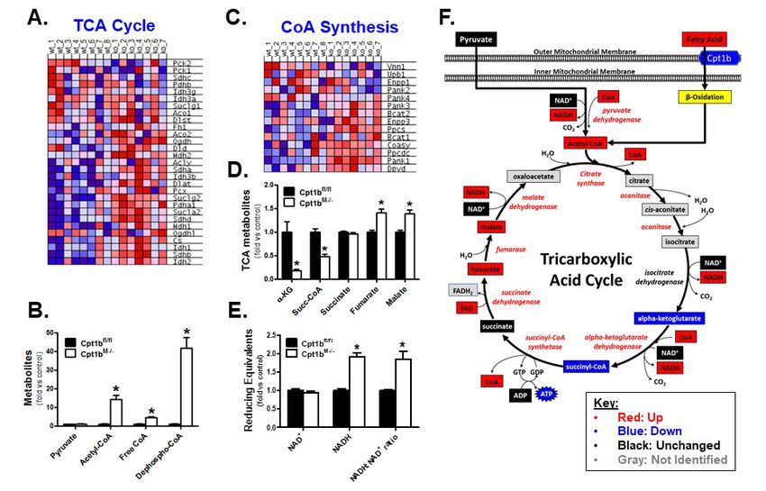
whereas 1637 metabolites were lower compared              metabolites. Alternatively, nucleotides are also
to Cpt1bfl/fl littermates (Figure 1D).                    critical to the synthesis of key factors involved
     Principal component analysis (PCA) was               in energy metabolism pathways including
performed on SAGE, proteomics, and                        nucleoside triphosphates (ATP, GTP, CTP, and
metabolomics datasets to detect potential                 UTP), reducing equivalents (NAD and FAD),
outliers and to further investigate how global            and coenzyme A (CoA). As a result, much of the
gene, protein and metabolite expression could             analyses performed using Cpt1bM-/- mice
differentiate between experimental groups (inset          indicate significant remodeling of energy
within Figures 1A-D). Whereas a moderate level            metabolism pathways and many of these
of group separation was noted for transcriptomic          alterations are discussed in greater detail in the
and proteomic data, PCA results from the                  context of relevant metabolic pathways below.
metabolomics dataset (both known and unknown
metabolites) revealed a clear separation.                 Gene Set Enrichment Analysis (GSEA) and
Additionally, 41 of the 50 identified metabolites         Ingenuity Pathway Analysis (IPA)
that were significantly different between Cpt1fl/fl           To gain understanding of the possible effects
vs Cpt1bM-/- mice were also significantly                 of transcriptomic changes on biological
correlated (p ≤0.0251 and FDR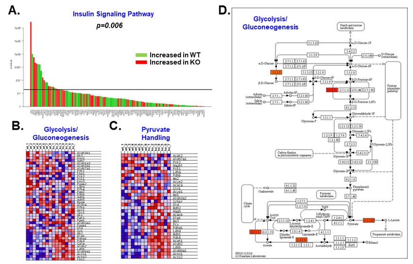
changes, we constructed a regulator/effector            notable differences in insulin signaling (8,9). In
network based on curated information available          the present study, globaltest analysis of 118
from the IPA Knowledge Base. The network                genes linked to the insulin signaling cascade
consists of three layers with the top layer being       indicated significant alterations in this pathway
transcription regulators, the middle layer being        (p=0.006) within the basal (i.e. not insulin-
observed gene expression changes, and the               stimulated) state (Figure 3A). It is, however,
bottom layer being predicted phenotypic effects         worth noting that roughly half of the
(Figure 2A). This network map indicated                 differentially regulated genes were increased,
changes in genes that regulate substrate                while the other half were decreased. We next
metabolism and metabolic disease phenotypes             investigated gene enrichment within glycolysis
were likely mediated by induction of                    (Figure 3B) and pyruvate handling (Figure 3C)
overlapping transcription factor networks               and observed significant upregulation of genes
involving peroxisome proliferator-activated             in both pathways. Mapping changes to a KEGG
receptor (Ppar) pathways, sterol regulatory             pathway map suggests remodeling of the
element-binding transcription factor 1 (Srebf1),        glycolytic pathway was modest and most likely
RAR-related orphan receptor alpha (Rora),               driven primarily by adaptations in pyruvate
Krüppel-like factor 15 (Klf15), estrogen-related        handling (Figure 3D). This coincides well with

                                                                                                             Downloaded from http://www.jbc.org/ by guest on November 8, 2019
receptor alpha (Esrra), and PPAR gamma                  the KEGG map for the pyruvate handling
coactivator 1-alpha (Pgc-1α). Analysis of the           pathway (Figure S4D) and provides mechanistic
portion of the network directly linked to Cpt1b         insight    helping     explain    our     previous
(Figure S4A) revealed induction of genes                observations showing increased pyruvate
involved in fatty acid metabolism (Figure 2B),          dehydrogenase (PDH) activity in Cpt1bM-/-
glycerolipid metabolism (Figure 2C) and                 skeletal muscle (8,9).
peroxisomal metabolism (Figure 2D), which
were some of the most robustly upregulated              Integrative Analysis: TCA Cycle
pathways noted in the GSEA (Table S1).                      GSEA revealed genes linked to TCA cycle
Peroxisomal adaptations showed particular               function were increased in Cpt1bM-/- skeletal
enrichment of genes involved in fatty acid              muscle (Figure 4A), which supports the
oxidation (Figure S4B). Additionally, while             observed heightened pyruvate oxidation in these
individual PPAR isoforms did not appear to be           mice (8,9). Analysis of the identified metabolites
regulated, the PPAR signaling network was               indicated that while pyruvate levels were not
identified as strongly enhanced via GSEA                different between genotypes, acetyl-CoA levels
(Figure 2E). Mapping the PPAR network                   were nearly 14-fold higher in Cpt1bM-/- skeletal
indicated that the genes regulated in Cpt1bM-/-         muscle (Figure 4B). Heightened acetyl-CoA
mice were primarily involved in lipid transport         production appears to be at least partially
and oxidation (Figure S4C). Proteomic analysis          facilitated by a 4.5-fold increase in free CoA
further supported this finding through the              (Figure 4B). Interestingly, enrichment of the
observed induction of several proteins involved         CoA biosynthesis pathway was observed in
in lipid metabolism in Cpt1bM-/- skeletal muscle        Cpt1bM-/- mice (Figure 4C), which included a
(Figure 2F).                                            significant increase in expression of the gene
                                                        encoding the rate-limiting enzyme in CoA
Integrative Analysis: Insulin Signaling                 biosynthesis, pantothenate kinase 1 (Pank1).
Pathway, Pyruvate Handling, and Glycolysis              This was confirmed by RT-PCR (Figure S5A)
    Figure S1B shows Cpt1bM-/- mice have                and CT values (22-24) indicate Pank1 is a fairly
improved glucose homeostasis as they have               abundant transcript in muscle. Additionally,
lower baseline glucose levels and maintain              perhaps the most compelling evidence for
lower blood glucose throughout a glucose                enhanced CoA biosynthesis is that Cpt1bM-/-
tolerance test than Cpt1bfl/fl controls, which is       mice exhibit 41-fold greater levels of the
consistent with previous findings (8,9,13).             metabolite precursor for CoA synthesis,
However, using Akt phosphorylation as a                 dephospho-CoA (Figure 4B).
measure of insulin sensitivity, we did not detect

                                                    4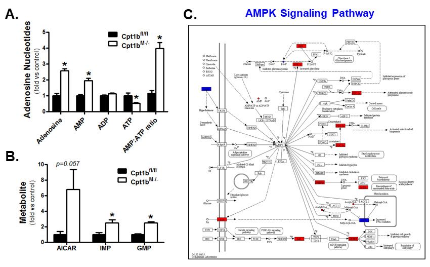
Differences in CoA and acetyl-CoA could              visual summary depicting changes in genes and
also be impacted by a variety of other factors            metabolites related to the TCA cycle and
including, but not limited to: changes in TCA             reducing equivalents is shown in Figure 4F.
flux rates, acyl-CoA synthetase activity (Acss1-
3, Acsf1-4, Acsm1-5, Acsl1-6, Acsbg1/2, Fatp1-            Integrative Analysis: Amino Acid Metabolism
6), carnitine acyltransferase activity (CrAT,                   Leucine oxidation is increased in Cpt1bM-/-
CrOT, Cpt1a, Cpt1b, Cpt1c, Cpt2), activity of             mice (8,9), suggesting that when mitochondrial
dehydrogenase enzymes with a lipoic acid                  lipid entry is impaired, they become more reliant
moiety (PDH, a-KGDH, BCKDH), acetyl-CoA                   on amino acids as fuel. Results in Table S1
acyltransferase activity (Acat1/2, Acaa1/2),              confirm substantial remodeling at the gene level
acyl-CoA thioesterase activity (Acot1-13,                 of several pathways involved in amino acid
Them4/5), acetoacetyl-CoA synthetase activity             metabolism and heatmaps for each identified
(Aacs), acetyl-CoA consuming reactions (CS,               pathway can be found in Figure S6. Using genes
ACC, HS2, etc), as well as activity of various            that contributed to core enrichment in all of
acetyltransferase and acyltransferase enzymes             these amino acid metabolism pathways, a
that regulate post-translational modification.            heatmap was made showing commonality of all
Differential expression of several genes involved         regulated genes amongst the amino acid

                                                                                                              Downloaded from http://www.jbc.org/ by guest on November 8, 2019
in these pathways was observed in the SAGE                metabolism pathways (Figure 5A). As shown,
dataset, so we attempted to validate these with           little overlap between regulated genes is
RT-PCR. We previously reported CrAT, CrOT,                apparent for most of these pathways (Figure
Cpt2, CS, Pdha1, Sdhb, Bckdh, and Fatp1 are               5A), indicating the adaptations identified by
significantly higher in Cpt1bM-/- skeletal muscle         GSEA (Table S1) is due to expansive
(8,9), so we didn’t repeat these analyses. Newly          remodeling of amino acid metabolism, rather
identified genes involved in CoA/acetyl-CoA               than a result of changes within a small set of
metabolism that were differentially regulated in          genes common to all amino acid metabolism
the SAGE dataset included: Acss1, Acss2, Acsl1,           pathways. Since genes involved in numerous
Acot2, Acot8, and Acot13. Of these, only Acot2,           amino acid metabolism pathways were
Acot8, and Acot13 were significantly different            regulated, the metabolomics dataset was probed
(Figure S5B).                                             to identify amino acids in skeletal muscle.
     Elevated acetyl-CoA provides more carbons            Results show decreases in L-arginine, L-
that can enter the TCA cycle in Cpt1bM-/- mice.           glutamine, L-leucine, and L-valine levels in
While our metabolomics platform did not                   Cpt1bM-/- mice, whereas increased levels of L-
resolve citrate or isocitrate, earlier results show       asparagine, L-aspartate, L-homoserine, L-lysine,
Cpt1bM-/- mice have increased complete                    L-ornithine, and L-threonine were identified
oxidation (i.e. CO2 production) of non-lipid              (Figure 5B). While results from this non-
substrates (pyruvate and leucine) that provide            targeted metabolomics screen did not provide a
acetyl-CoA precursors (8,9), suggesting greater           comprehensive amino acid panel, the amino
potential for substrate entry into the TCA cycle.         acids that were identified show very similar
Since Cpt1bM-/- mice likely have greater entry of         patterns to those found in our previous targeted
carbons into the citrate pool, it is interesting to       metabolomics analysis (9). Collectively, the
note that α-ketoglutarate and succinyl-CoA                metabolomics        platforms      from     both
                                                                                        M-/-
levels are substantially lower in these mice vs.          investigations show Cpt1b          mice have 13
littermate controls (Figure 4D). Alternatively,           amino acids increased in skeletal muscle, 4
downstream of these points in the TCA cycle,              amino acids are decreased, and 3 amino acids
succinate is similar between genotypes, while             remain similar when compared to Cpt1bfl/fl
fumarate and malate are both significantly                controls. Since amino acids can be used as
elevated in Cpt1bM-/- mice (Figure 4D). Also              metabolic fuel, results from these datasets were
noteworthy is the fact that NAD+ levels were              used to develop an integrative map showing
similar between genotypes and NADH levels                 points of entry of carbons derived from amino
were higher in Cpt1bM-/- mice, resulting in an            acid substrates into the TCA cycle (Figure 5C).
increased NADH:NAD+ ratio (Figure 4E). A                  As shown, amino acids that can be metabolized

                                                      5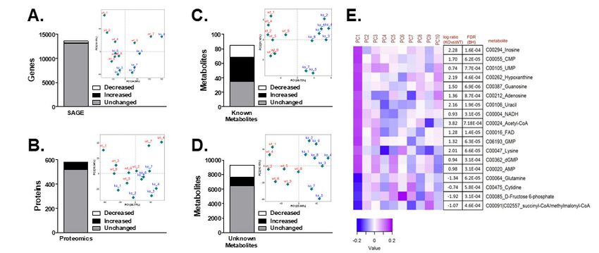
to pyruvate, fumarate or oxaloacetate are almost         concept of enhanced mitochondrial biogenesis in
universally increased. Likewise, amino acids             Cpt1bM-/- skeletal muscle. This is likely driven
that can be catabolized to produce acetyl-CoA            by activation of Pgc-1α and transcription factor
are primarily increased, except leucine.                 A, mitochondrial (Tfam; Figure 6E). It is also
Alternatively, amino acid-derived metabolites            worth noting that changes in the ETC appear to
that enter the TCA cycle as either α-                    be almost exclusively due to induction of
ketoglutarate or succinyl-CoA are not as                 nuclear-encoded subunits as none of the genes
consistently present at higher concentrations.           identified by the SAGE analysis were encoded
Notably, the amino acids most commonly                   by mitochondrial DNA (mtDNA), while only
considered to be important in generating α-              one protein encoded by mtDNA (ATP6) was
ketoglutarate are glutamate and glutamine,               significantly altered.
which are unaltered and decreased, respectively,
in Cpt1bM-/- mouse muscle. Of additional                 AMPK Signaling
interest, the most robustly upregulated pathway               Since flux through the TCA cycle generates
identified by GSEA was leucine, isoleucine, and          reducing equivalents that support energy
valine degradation (Table S1) and two of these           production, the increased NADH:NAD+ ratio
amino acids (leucine and valine) are reduced in          (Figure 4F) suggests there are ample reducing

                                                                                                              Downloaded from http://www.jbc.org/ by guest on November 8, 2019
Cpt1bM-/- mice, which likely represents greater          equivalents to support ATP synthesis in Cpt1bM-
                                                         /-
branched chain amino acid utilization.                      mice. With this in mind, it is interesting that
                                                         analysis of adenosine nucleotides found ATP
Integrative          Analysis:          Oxidative        content is actually lower in Cpt1bM-/- mouse
Phosphorylation                                          skeletal muscle, while AMP levels and free
     With wide-spread alterations in substrate           adenosine are increased (Figure 7A). The
metabolism pathways in Cpt1bM-/- skeletal                elevated AMP:ATP ratio in Cpt1bM-/- mice
muscle, it was important to determine if                 suggests they are in an energy-insufficient state.
alterations in electron transport chain (ETC)            5-aminoimidazole-4-carboxamide ribonucleotide
components were also present. The heatmap in             (AICAR) is a naturally occurring intermediate in
Figure 6A shows genes encoding proteins                  the synthesis of inosine monophosphate (IMP),
involved in oxidative phosphorylation were               which can be used to produce AMP and
enriched in Cpt1bM-/- mice. Additionally,                guanosine monophosphate (GMP). As shown in
analysis of the proteomics dataset for Gene              Figure 7B, IMP and GMP levels are also
Ontology-based      enrichment      of    cellular       significantly higher in Cpt1bM-/- mice, while a
compartments (Figure S7A) demonstrated clear             >6-fold increase in the metabolic precursor,
enrichment for mitochondrial respiratory chain           AICAR, approached statistical significance
components (Figure S7B). A graphical                     (p=0.057). Since both AMP and AICAR are
representation of specific subunits of each ETC          known activators of AMP-activated protein
complex showing significant differences                  kinase (AMPK), the SAGE results were
between genotypes for SAGE (Figure 6B) and               examined for evidence of activation of this
proteomics (Figure 6C) datasets reveal                   pathway. Figure 7C provides a visualization
significant remodeling of the ETC in Cpt1bM-/-           showing changes in several genes downstream
mice. The SAGE dataset identified 55 genes in            from AMPK, which is consistent with activation
the oxidative phosphorylation pathway to be              of this energy-sensing pathway. These findings
regulated, while the proteomics screen identified        provide a mechanism that helps explain the
24 proteins that were altered in the ETC                 increase in AMPK phosphorylation observed
complexes (despite limited coverage of the               previously in Cpt1bM-/- mouse skeletal muscle
proteome). Importantly, there appears to be              (9).
reasonable agreement between SAGE and
proteomics results as 18 of the regulated proteins       DISCUSSION
were also increased at the gene level (Figure                The purpose of this investigation was to
6D). All differentially expressed genes and              integrate information yielded from three distinct
proteins were upregulated, which supports the            ‘omics technologies (SAGE, proteomics, and

                                                     6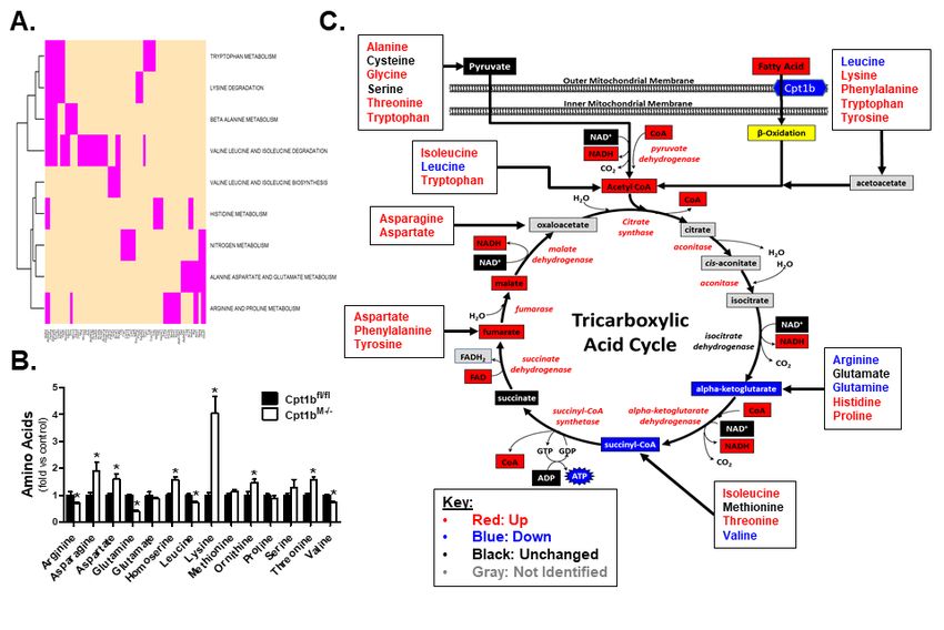
non-targeted metabolomics) to identify regulated         catabolism pathways. Of interest, genes
metabolic systems in skeletal muscle from                encoding peroxisomal proteins were identified
Cpt1bM-/- mice. We previously reported Cpt1bM-/-         as the second most robustly enriched metabolic
mice exhibit changes in substrate oxidation              pathway via GSEA and our previous data
(8,9); therefore, initial analyses of our ‘omics         confirm that peroxisomal function is heightened
datasets were targeted toward pathways involved          in Cpt1bM-/- skeletal muscle (8,9). Notably,
in carbohydrate, amino acid, and lipid                   genes involved in peroxisomal fat oxidation
metabolism. Results confirmed extensive                  were particularly enriched. This is potentially
remodeling of these pathways, with the most              important as unesterified fatty acids can enter
pronounced adaptations being observed in amino           peroxisomes; therefore, peroxisomal lipid entry
acid and lipid metabolism pathways, while shifts         does not require a carnitine shuttle system.
in glucose metabolism were modest. Strikingly,           Within their matrix, peroxisomes contain
a biofunction analysis, based on a large body of         carnitine acetyltransferase (CrAT) and carnitine
published gene-disease associations, indicated           octanoyltransferase (CrOT), which convert
the adaptations were consistent with decreased           chain-shortened moieties of peroxisomal
predicted risk of glucose metabolism disorders,          oxidation into acylcarnitines. These products can
hepatic steatosis, fatigue, cardiac hypertrophy,         then be exported into the cytosol, where they can

                                                                                                                Downloaded from http://www.jbc.org/ by guest on November 8, 2019
and premature mortality; thus signifying that the        enter the mitochondrial matrix independent of
breadth of disease states to which muscle-               Cpt1. As a result, peroxisomes are capable of at
specific Cpt1b inhibition may mediate                    least partially rescuing LCFA oxidation when
improvements in health may exceed that which             Cpt1 is inhibited (7). With this in mind, defining
has previously been believed.                            whether recruitment of peroxisomes in skeletal
     Cpt1b has a critical role in facilitating           muscle of Cpt1bM-/- mice is a critical adaptation
mitochondrial long chain fatty acid (LCFA)               that helps combat a potentially lipotoxic
entry; thus, inhibiting this enzyme markedly             environment and/or contributes to the beneficial
reduces mitochondrial LCFA oxidation. A                  metabolic      phenotype     is    an important
potentially underappreciated role of Cpt1b lies          consideration that warrants further investigation.
in the fact that the acylcarnitine metabolites                Cpt1b deletion limits the ability to use lipids
produced via Cpt1b are also readily exported             to meet energy demand; therefore, Cpt1bM-/-
from the cell, which is particularly relevant in         mice must adapt to rely more heavily on other
conditions of lipid excess (1-3). Limitations in         substrates. In this regard, earlier studies show
the ability to both use lipids as fuel, as well as       Cpt1 inhibition increases reliance on
export excess lipid from the cell in the form of         carbohydrates (4,5,7,14-20). Similarly, we found
acylcarnitines, leads to increased intramuscular         Cpt1bM-/- mice show enhanced whole body
lipid accrual in Cpt1bM-/- mice as verified by           glucose oxidation (indirect calorimetry),
elevated triglycerides, diglycerides, and                improved glucose tolerance, as well as increased
ceramide metabolites (9). Accordingly, it is not         pyruvate dehydrogenase activity and pyruvate
surprising that a primary molecular response is          oxidation in skeletal muscle homogenates (8,9).
to remodel lipid metabolism pathways.                    It is also important to note that Cpt1 inhibition
Regulator/effector network analysis of the               mitigates lipid-induced insulin resistance in
SAGE data predicts this might occur through              skeletal muscle, despite the fact that such
activation of Srebf1, Rora, Klf15, Esrra, Pgc-1α,        interventions promote a potentially lipotoxic
and/or Ppar regulated pathways. Of note, some            environment (3,8,9). Results from the present
adaptations induced by these transcriptional             analysis support these concepts and suggest that
regulators may actually exacerbate ectopic lipid         at least a portion of the adaptive increase in
buildup as evidence herein shows Cpt1bM-/- mice          carbohydrate utilization in skeletal muscle of
have increased expression of genes and proteins          Cpt1bM-/- mice is encoded at the molecular level,
that drive cellular lipid uptake, transport, and         as GSEA revealed enrichment of pathways
storage. Conversely, despite the inability of            involved in glycolysis, pyruvate handling and
mitochondria to directly utilize LCFAs, our              insulin signaling. It is, however, worth
results show significant upregulation of lipid           mentioning that skeletal muscle is predicted to

                                                     7
account for ~20% of the basal metabolic rate upregulation of amino acid metabolism in this
(21) and this can increase greatly during exercise study likely contributes to lower lean mass in
(22), emphasizing skeletal muscle has Cpt1bM-/- mice as they age.
significant substrate requirements. Skeletal SAGE and proteomics datasets both
muscle of Cpt1bM-/- has limited ability to utilize predicted enhanced mitochondrial biogenesis in
the most prevalent substrate available (lipid); skeletal muscle from Cpt1bM-/- mice. AMPK and
therefore, the adaptations in glucose handling Pgc-1α are activated under conditions of energy
pathways actually seem relatively modest deficit and promote mitochondrial biogenesis.
compared to what we expected. In this regard, it Cpt1bM-/- exhibit an increased AMP:ATP ratio
is important to recall that glucose is an essential (Figure 7A), as well as activation of pAMPK
substrate for brain and blood cells and the body and upregulation of Pgc-1α (9); therefore, it is
goes to great lengths to protect against likely that activation of these pathways
hypoglycemia. As such, it is tempting to contributes to the observed mitochondrial
speculate that the relatively modest remodeling biogenesis. Skeletal muscle from traditional
of glucose handling pathways in skeletal muscle models of energy-deficit (caloric restriction,
of Cpt1bM-/- mice is necessary to protect against prolonged fasting, acute exercise, etc) exhibit an
the possibility of hypoglycemia. If true, this increased AMP:ATP ratio, as well as decreases
Downloaded from http://www.jbc.org/ by guest on November 8, 2019
would also require adaptations in tissues other in NADH and FADH2 reducing equivalents.
than skeletal muscle, which provides an Since mitochondrial ATP production is
interesting model to identify novel mechanisms supported by delivery of reducing equivalents to
of organ systems crosstalk that regulate whole the electron transport chain, it is surprising to
body metabolic health that is worth future study. note that lower ATP levels in Cpt1bM-/- mouse
Cpt1bM-/- mice also exhibit remarkable muscle occurs despite greater mitochondrial
remodeling of amino acid metabolism pathways. content and ample reducing equivalents
Earlier we reported Cpt1bM-/- mice have (increased NADH levels and greater
heightened leucine oxidation (8,9), suggesting NADH:NAD+ ratio). Roughly 95% of the total
adaptations occur that allow for greater protein cellular NADH pool is estimated to reside within
catabolism. Findings herein strongly support this mitochondria (23); thus, an apparent mismatch
as 1) pathways involved in amino acid between NADH availability and ATP content is
metabolism represented 9 of the 21 significantly seemingly paradoxical. One possibility is that
enriched pathways identified via GSEA, and 2) despite increased mitochondrial biogenesis, the
the metabolomics screen revealed the majority relative lack of induction of genes and proteins
of amino acids are significantly different encoded by mitochondrial DNA may have
between Cpt1bfl/fl vs. Cpt1bM-/- mice. In fact, created mitochondria with an impaired ability to
based upon results of the current study, it can be utilize reducing equivalents to generate the
argued that adaptations in amino acid proton motive force required to support ATP
metabolism pathways actually exceed those synthesis. While potentially compelling, the
directing carbohydrate metabolism. In this prospect that the newly formed mitochondria are
regard, futile protein cycling has been linked to inherently dysfunctional seems unlikely since
increased energy expenditure (11) and could isolated mitochondria from Cpt1bM-/- mice
potentially contribute to the metabolically exhibited similar functionality (oxygen
advantageous phenotype observed in Cpt1bM-/- consumption, respiratory control ratio, and
mice; however, the importance of adaptations in uncoupled respiration) as littermate controls (9).
amino acid metabolism pathways in this rodent This does not, however, rule out the possibility
model remain to be more fully understood. It is that other cellular alterations occur in Cpt1bM-/-
also worth noting that we previously reported mice that either 1) limit the ability to produce
that Cpt1bM-/- mice begin to exhibit lower lean ATP, and/or 2) increase ATP utilization. Finally,
mass at ~17-20 weeks of age (8,9). Mice in this reducing equivalents are involved in a myriad of
study were 14-16 weeks of age at the time of other redox reactions throughout the cell, so
harvest, which likely precedes significant another possibility is that the increased NADH
differences in lean mass; however, the detected levels are linked to regulation of cellular redox
8balance; however, identifying specific pathways diminishes ATP production (24,27,28), which
requires further study. resembles the energy-deficit phenotype observed
Cpt1b converts long-chain fatty acyl-CoAs in Cpt1bM-/- mice. Second, at the cellular level,
(LCFA-CoA) into long-chain acylcarnitines, acetylation of histone residues is a critical
resulting in the liberation of coenzyme A (CoA). posttranslational modification that regulates
As such, manipulation of Cpt1b activity has the gene transcription within the nucleus. It has been
potential to affect the cellular CoA pool. Since postulated that heightened acetyl-CoA drives
Cpt1b acts directly on LCFA-CoA, it stands to acetylation of histone residues to promote cell
reason that manipulating Cpt1 activity would growth and survival even under states of energy
affect the LCFA-CoA pool; however, previous deficit (26,29), which also resembles results
studies that have measured LCFA-CoA levels in from Cpt1bM-/- mice. Ultimately, however, the
models of variable Cpt1 activity have yielded potential role that enhanced CoA biosynthesis
inconsistent findings (13,14,19,24). At this time &/or acetylation play in the phenotype observed
we cannot offer a resolution to this debate as in Cpt1bM-/- mice remains speculative and
targeted acyl-CoA metabolic profiling in warrants further study.
Cpt1bM-/- mice has not been performed; In summary, despite variable coverage of
however, our results clearly indicate significant the proteome and identified metabolites, the
Downloaded from http://www.jbc.org/ by guest on November 8, 2019
differences in CoA metabolism. Substantial combined use of SAGE, proteomics and non-
increases in free CoA (4.5-fold), dephospho- targeted metabolomics identified regulation of
CoA (41-fold), enrichment of genes involved in several pathways that were consistent across
CoA biosynthesis, and upregulation of acyl-CoA multiple platforms. Furthermore, the fact that
thioesterases point toward a system that is robust differences in numerous metabolites were
attempting to increase CoA availability. Recent observed increases confidence that changes at
work suggests increased CoA biosynthesis the gene and protein levels represent functional
occurs in systems attempting to drive LCFA adaptations. Importantly, findings herein
oxidation (25), which is consistent with the revealed that the extensive remodeling of
extensive remodeling of lipid metabolism substrate metabolism pathways in Cpt1bM-/- mice
pathways in Cpt1bM-/- mice. The rise in CoA are consistent with metabolically healthy
availability almost certainly contributes to the phenotypes. With this in mind, the large
14-fold increase in acetyl-CoA levels in Cpt1bM- publically available datasets developed from this
/-
mice. Heightened acetyl-CoA levels are comprehensive systems biology approach are
usually found in states of energy surplus (26); expected to help investigators define specific
thus, the robust increase in acetyl-CoA in mechanisms that contribute to the notable
Cpt1bM-/- mice signifies yet another seemingly improvements in metabolic health when the
paradoxical metabolite signature in a mouse mitochondrial lipid burden is mitigated in
model that otherwise exhibits a phenotype skeletal muscle.
similar to models of energy deficit. With this in
mind, it is important to note that while acetyl- EXPERIMENTAL PROCEDURES
CoA can enter the TCA cycle for use as EXPERIMENTAL MODEL
metabolic fuel, it can also be used as a substrate Animals: Skeletal muscle-specific Cpt1b-
in other reactions within the cell. Of particular deficient mice (Cpt1bM-/-) and littermate controls
relevance, surplus acetyl-CoA and NADH are (Cpt1bfl/fl) were developed as described (8,9).
known to promote protein acetylation (26). Briefly, floxed Cpt1b mice were bred to mice
While analysis of the SAGE data did not reveal expressing Cre recombinase under the control of
enrichment of pathways involved in protein the Mlc1f promoter (Jackson Laboratories,
acetylation or deacetylation, there are Stock # 024713) to delete Cpt1b in skeletal
observations from this study that support this muscle. As validated in our previous studies
possibility. First, numerous proteins in the TCA (8,9), this deletes Cpt1b expression and function
cycle and oxidative phosphorylation system are specifically in skeletal muscle with no effect on
acetylated and it has been shown that cardiac muscle. Mice were group-housed at
hyperacetylation of mitochondrial proteins room temperature (RT) under a 12:12 h
9light:dark cycle and allowed ad libitum access to 0.2 mL of chloroform was added. The samples
food and water. In an effort to be able to best were shaken vigorously for 15 sec and allowed
compare results from this systems biology study to sit at RT for 2-3 min before they were
to previous findings (8,9,30), mice were fed the centrifuged (12,000 x g; 15 min; 4°C) to induce
same rodent breeder chow (Purina Rodent Chow phase separation. Roughly 600 µL of the upper
#5015, Purina Mills, St. Louis, MO), which aqueous supernatant containing RNA was
provides 20% of calories from protein, 26% transferred to a new microcentrifuge tube
from fat, and 54% from carbohydrate. At 12-14 whereupon 600 µL of 70% ethanol was added
weeks of age body weight was measured (Figure and the samples were vortexed. RNA was then
S1A) and a glucose tolerance test was performed isolated using an RNeasy kit (Qiagen, Valencia,
as described below to confirm the Cpt1bM-/- mice CA) with DNAse treatment per manufacturer’s
used in this study exhibited the established instructions. RNA content and quality (260/280
improvement in glucose homeostasis (Figure ratio range 1.9 - 2.1) were assessed using a
S1B). Tissue harvest occurred at 14-16 weeks of Nanodrop 1000 and used for downstream Serial
age, which is a time point at which fat mass has Analysis of Gene Expression (SAGE) studies.
consistently been shown to be lower in Cpt1bM-/- The lower organic phase developed following
mice, yet lean mass is typically not yet different Trizol extraction contained protein samples that
Downloaded from http://www.jbc.org/ by guest on November 8, 2019
(8,9). Following a 4h food pull, Cpt1bfl/fl (n=8) were shipped to the Duke Proteomics Core
and Cpt1bM-/- (n=8) male mice were euthanized Facility (Durham, NC). Following
by cervical dislocation and tissues were centrifugation, phase separation was performed
collected, snap-frozen in liquid nitrogen, and by adding chloroform, and the aqueous layer
stored at -80°C until subsequent analyses could was discarded and protein precipitated from the
be performed. Mixed gastrocnemius (MG) organic layer by methanol precipitation. After
skeletal muscle was powdered and used for all washing and sonication of the protein pellet, the
assays presented. The Pennington Biomedical precipitate was reconstituted in 50 µL of 0.25%
Research Center has an AALAC-approved Rapigest SF (Waters Corporation, Milford, MA)
Comparative Biology Core facility and in 50 mM ammonium bicarbonate, pH=8.0.
veterinary staff that continuously monitor the Bradford assays were performed to determine
health of the animals via a sentinel program and protein quantity and were used for subsequent
daily inspection. All animal studies performed proteomics analysis.
were approved by the Pennington Biomedical Serial Analysis of Gene Expression
Research Center Institutional Animal Care and (SAGE): A primary goal of this study was to
Use Committee. detect comprehensive changes in gene
expression in skeletal muscle from Cpt1bfl/fl vs.
Cpt1bM-/- mice, thus a serial analysis of gene
METHOD DETAILS expression (SAGE) was performed by the
Glucose Tolerance Test: Glucose tolerance Genomics Core at the Pennington Biomedical
tests were performed after a 4h fast as described Research Center (Baton Rouge, LA). The SAGE
(9,13). Briefly, after measuring baseline blood analysis was performed as reported earlier
glucose levels via tail vein, mice received a (31,32). Briefly, gene expression profiling was
0.2mL intraperitoneal injection of 20% D- performed by expression tag sequencing
glucose (40mg glucose per mouse) and blood (SAGE) on an AB SOLiD 5500XL next-
glucose levels were subsequently monitored at generation sequencing instrument using reagent
20min, 40min, and 60min post-injection. kits from the manufacturer (Applied Biosystems,
RNA and Protein Isolation: RNA and Foster City, CA). Sequence reads were aligned
protein were extracted from 20-30mg of to mouse reference RefSeq transcripts (version
powdered mixed gastrocnemius (MG) skeletal mm9), via SOLiDSAGE (Applied Biosystems).
muscle using Trizol (ThermoFisher Scientific, Only uniquely mapped sequence reads were
Waltham, MA) as previously described (8,9). counted to generate the expression count level
Briefly, samples were homogenized in 1 ml for each respective RefSeq gene. Changes in
Trizol, allowed to sit at RT for 5 min, and then certain genes detected via SAGE were
10confirmed by RT-PCR using described methods after which the analytical separation was
(8,9) and primers shown in Table S3. performed using a 1.7 µm Acquity BEH130 C18
Proteomics: Another goal of this study was 75 mm × 250 mm column (Waters) using a 90-
to detect broad-scale adaptations in proteins min gradient of 5-40% acetonitrile with 0.1%
involved in metabolism in Cpt1bM-/- mice, with a formic acid at a flow rate of 300 nL/min and a
particular interest in mitochondrial adaptations. column temperature of 45°C. Data collection on
As such, we had a proteomics analysis the Synapt G2 mass spectrometer was performed
performed by the Duke Proteomics Core in ion-mobility assisted data-independent
(Durham, NC). Trizol-extracted protein (20 µg) acquisition (HDMSE) mode, using 0.6 sec
from each sample (concentration range 1-7.5 alternating cycle time between low (6 V) and
µg/µL) was normalized to an equal volume (20 high (27-50 V) collision energy (CE). Scans
µL) to yield a 1 µg/µL sample. This sample was performed at low CE measured peptide accurate
reduced and denatured with 10 mM DTT and mass and intensity (abundance), while scans at
heating at 80°C for 10 min. Samples were elevated CE allowed for qualitative
subsequently alkylated with 20 mM identification of the resulting peptide fragments
iodoacetamide at room temperature in the dark via database searching. The total analysis cycle
for 30 min and then digested with 1:50 (w/w) time for each sample injection was
Downloaded from http://www.jbc.org/ by guest on November 8, 2019
trypsin:protein overnight at 37°C. After approximately 2 h. Following the 21 total
digestion, 25 fmol/µL trypsinized yeast alcohol analyses, data was imported into Rosetta
dehydrogenase 1 (MassPrep, Waters Elucidator v3.3 (Rosetta Biosoftware, Inc), and
Corporation, Milford, MA) was added as a all LC-MS runs were aligned based on the
surrogate standard to each sample, and samples accurate mass and retention time of detected
were acidified to 1% trifluoroacetic acid and ions (“features”) using the PeakTeller algorithm
heated to 60°C for 2 h to hydrolyze Rapigest. in Elucidator. Relative peptide abundance was
After centrifugation, samples were transferred to calculated based on area-under-the-curve (AUC)
liquid chromatography (LC) vials and a quality of aligned features across all runs. The overall
control (QC) pool was prepared by mixing equal dataset had 168,700 quantified features, and
volumes of each sample. Quantitative one- 108,834 high collision energy (peptide
dimensional liquid chromatography/tandem fragment) spectra that were subjected to
mass spectrometry (LC/MS/MS) was performed database searching. This MS/MS data was
on 1 µg of the peptide digests (described in searched against a custom RefSeq database with
Supplemental Methods). Individual peptides Mus musculus taxonomy which contained a
were scored using PeptideProphet algorithm reversed-sequence “decoy” database for false
(Elucidator), and the data was annotated at a discovery rate determination. ProteinLynx(Phenomenex, Torrance, CA) held at 25˚C. The combined supernatants were dried under
mass spectrometer was run in negative nitrogen gas and subsequently resuspended in
ionization mode similar to a previously reported 300 µL of sterile water. The samples were
method (33). The chromatographic elution was transferred to autosampler vials and immediately
introduced to the Exactive Plus Orbitrap mass placed in an Ultimate 3000 RS autosampler
spectrometer (Thermo Fisher Scientific, (Dionex, Sunnyvale, CA) cooled to 4˚C.
Waltham, MA) via an electrospray ionization Samples were analyzed using Ultraperformance
source through a 0.1 mm internal diameter fused Liquid Chromotography-High Resolution Mass
silica capillary tube. Instrumental parameters Spectrometry (UPLC-HRMS) run in negative
were as follows: 3 kV spray voltage, 10 nitrogen ionization mode similar to a previously reported
sheath gas flow rate (unitless), 320˚C capillary method (33). Metabolite identification was
temperature, 3e6 AGC target, 140,000 performed by first converting raw spectrum files
resolution, and a scan window of 85 to 800 m/z to the open-source mzML format using the
from 0 to 9 min and 100 to 1000 m/z from 9 to Proteowizard package (35). Total ion
25 min. Solvent A was composed of 97:3 chromatograms were automatically corrected
water:methanol, 10 mM tributylamine, and 15 based on retention times for each sample using
mM acetic acid. Solvent B was methanol. The MAVEN software (Princeton University)
Downloaded from http://www.jbc.org/ by guest on November 8, 2019
gradient was as follows: 0 to 5 minutes, 0% B; 5 (36,37). Metabolites were identified based on
to 13 minutes, 20% B; 13 to 15.5 minutes, 55% retention times and m/z (± 5 ppm) and
B; 15.5 to 19 minutes, 95% B; and 19 to 25 chromatographic intensities were integrated for
minutes, 0% B. The flow rate was maintained each sample. Unidentified spectral features were
200 µL/min for the duration. annotated using MAVEN’s automated peak
Metabolomics: A third goal of this study detection feature, selecting m/z features
was to get a comprehensive view of global exhibiting chromatographic peak-like qualities.
adaptations in metabolism in Cpt1bM-/- skeletal Peak detection settings were as follows: 10 ppm
muscle, while also providing detail on mass domain resolution, 10 scan time domain
metabolites involved in glucose and amino acid resolution, 11 scan EIC smoothing, 0.50 min
metabolism, as well as energy status and redox peak grouping, 5 scan baseline smoothing, 80%
balance. To achieve this goal, non-targeted chromatograph intensity dropped baseline, 1
metabolomics was performed by the Biological minimum peak per group, 3 signal to baseline
and Small Molecule Mass Spec Core at the and signal to blank ratios, 5 scan minimum peak
University of Tennessee (Knoxville, TN). width, and 10,000 ion minimum peak intensity,
Metabolite extraction was performed at 4˚C using default peak classifier model.
unless stated otherwise. To 1.5 mL
microcentrifuge tubes containing 10-15 mg of QUANTIFICATION AND STATISTICAL
powdered mixed gastrocnemius skeletal muscle, ANALYSIS
1.3 mL of extraction solvent was added Gene expression data analysis: Differential
consisting of a 40:40:20 HPLC grade methanol, analysis of RNA read count data was performed
acetonitrile, and water with 0.1 M formic acid using DESeq2 software (38), which models read
(34). Samples were thoroughly mixed by counts as a negative binomial distribution and
vortexing before extraction was allowed to uses an empirical Bayes shrinkage-based method
proceed for 20 min at –20˚C. Following to estimate signal dispersion and fold-changes.
extraction, samples were centrifuged (5 min at Gene expression signals were logarithmically
16.1 rcf) and supernatants were transferred to transformed (to base 2) for all downstream
new vials. Pellets were resuspended in an analyses (the lowest expression value being set
additional 50 µL of extractions solvent and to 1 for this purpose).
extraction was again allowed to proceed for 20 Pathway enrichment analysis: Pathway
min at –20˚C before being centrifuged. enrichment was conducted via three separate
Supernatants were again transferred to vials and approaches - (a) competitive gene-scoring based
the process was repeated once more with an on gene set enrichment analysis (GSEA) (39),
additional 50 µL of extraction solvent. The (b) over-representation analysis (ORA) based on
12a pre-filtered list of differentially expressed In addition to the above 2 ‘competitive test’
genes (Ingenuity Pathway Analysis (IPA), methods (44), we also queried gene expression
Qiagen, Redwood City, differences via a ‘self-contained’ test for gene-
www.qiagen.com/ingenuity), and (c) enrichment set enrichment known as the globaltest. The
analysis of pre-selected pathways via the globaltest method is based on regression
globaltest package in R (http://bioconductor.org) analysis of the association of a gene-set’s
(40). GSEA was performed by first ranking the component gene expression to a phenotype, and
expression of all genes in the Cpt1bfl/fl (WT) and tests the null hypothesis that no gene in the
Cpt1bM-/- (KO) groups via the signal-to-noise gene-set is associated with the phenotype (with
ratio (SNR) metric, and then employing a no additional consideration for genes in other
weighted Kolmogorov-Smirnov test to gene-sets). We applied globaltest to the same
determine if the gene SNRs deviate significantly gene expression data as used for GSEA analysis,
from a uniform distribution in a priori defined and queried KEGG specific gene-sets obtained
gene-sets (pathways) derived from the Kyoto from the package kegg.db (Carlson M.
Encyclopedia of Genes and Genomes (KEGG) KEGG.db: A set of annotation maps for KEGG.
database (41) via the Molecular Signatures R package version 3.0.0.). Gene annotations
Database repository (MSigDb, were provided as Entrez IDs to match the gene-
Downloaded from http://www.jbc.org/ by guest on November 8, 2019
http://software.broadinstitute.org/gsea/msigdb/, set annotations utilized by globaltest from
(42)). Statistical significance for the observed org.Mm.eg.db (release 3.3) (Carlson M.
enrichment was ascertained by permutation org.Mm.eg.db: Genome wide annotation for
testing over size-matched gene-sets. Significant Mouse. R package version 3.2.3).
gene-sets were selected by control of the false For visual representations of gene and
discovery rate (FDR) at 25% (43). The per- metabolite expression pattern differences in
sample expression profiles of genes contributing pathways, we used the Search and Color
to core enrichment of the significant pathways Pathway tool from the KEGG Mapper suite of
were visualized via row-normalized blue-red tools (www.genome.jp/kegg/mapper.html). A
heatmaps with blue representing lower, and red total of 389 genes with nominal p-value 1.5-fold between the
IPA analysis was carried out on Cpt1bfl/fl and Cpt1bM-/- groups were selected for
differentially expressed genes (with absolute representation on the KEGG pathway maps.
fold-change >1.5-fold and FDRwere uploaded excluded an outlier (KO8) that as an outlier from the metabolomics dataset.
was identified in both, while WT8 was removed
ACKNOWLEDGEMENTS
We thank Jaycob Warfel, Susan Newman, Claudia Kruger, and Richard Carmouche for critical advice,
reagents and/or technical assistance. We recognize Drs. Matthew Foster, J. Will Thompson and Arthur
Moseley from the Duke Proteomics Core Facility (Duke University School of Medicine) for performing
the proteomic analysis. This work utilized PBRC core facilities (Genomics, and Transgenic and Animal
Phenotyping) that are supported in part by COBRE (NIH 3 P30-GM118430) and NORC (NIH 2P30-
DK072476) center grants from the National Institutes of Health. This research was supported by NIH
3P30-GM118430 (R.C.N.), NIH 1R01DK103860 (R.C.N.), ADA grant # 1-10-BS-129 (R.L.M.), and
NIH 1R01DK089641 (R.L.M.). S.E.W. was supported by a T32 AT004094 fellowship and received a
pilot and feasibility grant from NORC (NIH 2P30-DK072476). J.M.S. is funded in part by a Botanicals
and Dietary Supplements Research Center grant (P50 AT002776). Dr. Sujoy Ghosh was supported in part
by 2 U54 GM104940 from the National Institute of General Medical Sciences of the National Institutes of
Health, which funds the Louisiana Clinical and Translational Science Center.
Downloaded from http://www.jbc.org/ by guest on November 8, 2019
DECLARATION OF INTERESTS
The authors have no conflicts of interest to report.
REFERENCES
1. Muoio, D. M., Noland, R. C., Kovalik, J. P., Seiler, S. E., Davies, M. N., DeBalsi, K. L., Ilkayeva, O. R.,
Stevens, R. D., Kheterpal, I., Zhang, J., Covington, J. D., Bajpeyi, S., Ravussin, E., Kraus, W., Koves,
T. R., and Mynatt, R. L. (2012) Muscle-specific deletion of carnitine acetyltransferase
compromises glucose tolerance and metabolic flexibility. Cell Metab 15, 764-777
2. Noland, R. C., Koves, T. R., Seiler, S. E., Lum, H., Lust, R. M., Ilkayeva, O., Stevens, R. D., Hegardt,
F. G., and Muoio, D. M. (2009) Carnitine insufficiency caused by aging and overnutrition
compromises mitochondrial performance and metabolic control. J Biol Chem 284, 22840-22852
3. Koves, T. R., Ussher, J. R., Noland, R. C., Slentz, D., Mosedale, M., Ilkayeva, O., Bain, J., Stevens,
R., Dyck, J. R., Newgard, C. B., Lopaschuk, G. D., and Muoio, D. M. (2008) Mitochondrial overload
and incomplete fatty acid oxidation contribute to skeletal muscle insulin resistance. Cell Metab
7, 45-56
4. Ganesh, S. K., Sharma, Y., Dayhoff, J., Fales, H. M., Van Eyk, J., Kickler, T. S., Billings, E. M., and
Nabel, E. G. (2007) Detection of venous thromboembolism by proteomic serum biomarkers.
PLoS One 2, e544
5. Breton, J., Lavergne, J., Wakeham, M. C., Nabedryk, E., and Jones, M. R. (2007) The unusually
strong hydrogen bond between the carbonyl of Q(A) and His M219 in the Rhodobacter
sphaeroides reaction center is not essential for efficient electron transfer from Q(A)(-) to Q(B).
Biochemistry 46, 6468-6476
6. Nabedryk, E., Paddock, M. L., Okamura, M. Y., and Breton, J. (2007) Monitoring the pH
dependence of IR carboxylic acid signals upon Q(B)- formation in the Glu-L212 --> Asp/Asp-L213
--> Glu swap mutant reaction center from Rhodobacter sphaeroides. Biochemistry 46, 1176-
1182
7. Noland, R. C., Woodlief, T. L., Whitfield, B. R., Manning, S. M., Evans, J. R., Dudek, R. W., Lust, R.
M., and Cortright, R. N. (2007) Peroxisomal-mitochondrial oxidation in a rodent model of
obesity-associated insulin resistance. Am J Physiol Endocrinol Metab 293, E986-E1001
148. Vandanmagsar, B., Warfel, J. D., Wicks, S. E., Ghosh, S., Salbaum, J. M., Burk, D., Dubuisson, O.
S., Mendoza, T. M., Zhang, J., Noland, R. C., and Mynatt, R. L. (2016) Impaired Mitochondrial Fat
Oxidation Induces FGF21 in Muscle. Cell Rep 15, 1686-1699
9. Wicks, S. E., Vandanmagsar, B., Haynie, K. R., Fuller, S. E., Warfel, J. D., Stephens, J. M., Wang,
M., Han, X., Zhang, J., Noland, R. C., and Mynatt, R. L. (2015) Impaired mitochondrial fat
oxidation induces adaptive remodeling of muscle metabolism. Proc Natl Acad Sci U S A 112,
E3300-3309
10. Chase, J. F., and Tubbs, P. K. (1972) Specific inhibition of mitochondrial fatty acid oxidation by 2-
bromopalmitate and its coenzyme A and carnitine esters. Biochem J 129, 55-65
11. She, P., Reid, T. M., Bronson, S. K., Vary, T. C., Hajnal, A., Lynch, C. J., and Hutson, S. M. (2007)
Disruption of BCATm in mice leads to increased energy expenditure associated with the
activation of a futile protein turnover cycle. Cell Metab 6, 181-194
12. Koves, T. R., Noland, R. C., Bates, A. L., Henes, S. T., Muoio, D. M., and Cortright, R. N. (2005)
Subsarcolemmal and intermyofibrillar mitochondria play distinct roles in regulating skeletal
muscle fatty acid metabolism. Am J Physiol Cell Physiol 288, C1074-1082
13. Lopaschuk, G. D., Wall, S. R., Olley, P. M., and Davies, N. J. (1988) Etomoxir, a carnitine
Downloaded from http://www.jbc.org/ by guest on November 8, 2019
palmitoyltransferase I inhibitor, protects hearts from fatty acid-induced ischemic injury
independent of changes in long chain acylcarnitine. Circ Res 63, 1036-1043
14. Keung, W., Ussher, J. R., Jaswal, J. S., Raubenheimer, M., Lam, V. H., Wagg, C. S., and Lopaschuk,
G. D. (2013) Inhibition of carnitine palmitoyltransferase-1 activity alleviates insulin resistance in
diet-induced obese mice. Diabetes 62, 711-720
15. Hubinger, A., Knode, O., Susanto, F., Reinauer, H., and Gries, F. A. (1997) Effects of the carnitine-
acyltransferase inhibitor etomoxir on insulin sensitivity, energy expenditure and substrate
oxidation in NIDDM. Horm Metab Res 29, 436-439
16. Lopaschuk, G. D., McNeil, G. F., and McVeigh, J. J. (1989) Glucose oxidation is stimulated in
reperfused ischemic hearts with the carnitine palmitoyltransferase 1 inhibitor, Etomoxir. Mol
Cell Biochem 88, 175-179
17. Martin, C., Odeon, M., Cohen, R., and Beylot, M. (1991) Mechanisms of the glucose lowering
effect of a carnitine palmitoyl transferase inhibitor in normal and diabetic rats. Metabolism 40,
420-427
18. Ratheiser, K., Schneeweiss, B., Waldhausl, W., Fasching, P., Korn, A., Nowotny, P., Rohac, M.,
and Wolf, H. P. (1991) Inhibition by etomoxir of carnitine palmitoyltransferase I reduces hepatic
glucose production and plasma lipids in non-insulin-dependent diabetes mellitus. Metabolism
40, 1185-1190
19. Obici, S., Feng, Z., Arduini, A., Conti, R., and Rossetti, L. (2003) Inhibition of hypothalamic
carnitine palmitoyltransferase-1 decreases food intake and glucose production. Nat Med 9, 756-
761
20. Wolf, H. P., and Engel, D. W. (1985) Decrease of fatty acid oxidation, ketogenesis and
gluconeogenesis in isolated perfused rat liver by phenylalkyl oxirane carboxylate (B 807-27) due
to inhibition of CPT I (EC 2.3.1.21). Eur J Biochem 146, 359-363
21. Wang, Z., Heshka, S., Gallagher, D., Boozer, C. N., Kotler, D. P., and Heymsfield, S. B. (2000)
Resting energy expenditure-fat-free mass relationship: new insights provided by body
composition modeling. Am J Physiol Endocrinol Metab 279, E539-545
22. Noland, R. C. (2015) Exercise and Regulation of Lipid Metabolism. Prog Mol Biol Transl Sci 135,
39-74
23. White, A. T., and Schenk, S. (2012) NAD(+)/NADH and skeletal muscle mitochondrial adaptations
to exercise. Am J Physiol Endocrinol Metab 303, E308-321
15You can also read